Embed Size (px)
Citation preview

Effect of cavity configuration and aging on thebonding effectiveness of six adhesives to dentin
Kenichi Shiraia,b, Jan De Muncka, Yasuhiro Yoshidac, Satoshi Inoued,Paul Lambrechtsa, Kazuomi Suzukic, Hideaki Shintanib,Bart Van Meerbeeka,*
aLeuven BIOMAT Research Cluster, Department of Conservative Dentistry, School of Dentistry,Oral Pathology and Maxillo-Facial Surgery, Catholic University of Leuven, Kapucijnenvoer 7,3000 Leuven, BelgiumbDepartment of Operative Dentistry, Hiroshima University Graduate School of Dentistry, Hiroshima, JapancDepartment of Biomaterials, Okayama University Graduate School of Medicine and Dentistry,Okayama, JapandDivision for General Dentistry, Hokkaido University Dental Hospital, Sapporo, Japan
Received 31 July 2003; received in revised form 10 December 2003; accepted 30 January 2004
KEYWORDSAdhesion; Etch-and-
rinse; Self-etch; Micro-
tensile bond strength;
Polymerization
shrinkage; Durability;
Aging
Summary Objectives: The purpose of this study was to determine the effectpolymerization contraction stress may have on bond durability.
Methods: Bonding effectiveness was assessed by micro-tensile bond strengthtesting (mTBS) and electron microscopy. The mTBS to flat dentin surfaces and instandardized cavities was determined (this after 1 day as well as 1 year waterstorage). Six adhesives representing all current classes were applied: two etch-and-rinse (OptiBond FL, Kerr; Scotchbond 1, 3M ESPE), two self-etch (Clearfil SE Bond,Kuraray; Adper Prompt, 3M ESPE) and two glass-ionomer (Fuji Bond LC, GC;Reactmer, Shofu) adhesives.
Results: The conventional 3-step etch-and-rinse adhesive OptiBond FL bondedmost effectively to dentin, and appeared insensitive to polymerization shrinkagestress and water degradation. The 2-step self-etch adhesive Clearfil SE Bond mostclosely approached this superior bonding effectiveness and only slightly lost bondstrength after 1-year water exposure. The 2-step etch-and-rinse adhesiveScotchbond 1 and the ‘strong’ 1-step self-etch adhesive Adper Prompt appearedvery sensitive to cavity configuration and water-aging effects. The 2-step resin-modified glass-ionomer adhesive Fuji Bond LC only suffered from shrinkage stress,but not from 1-year water-exposure. Remarkable also is the apparent repairabilityof the ‘mild’ 1-step glass-ionomer adhesive Reactmer when stored for 1 year inwater, in spite of the very low 1-day mTBS.
Significance: Simplified bonding procedures do not necessarily imply improvedbonding performance, especially in the long term.Q 2004 Academy of Dental Materials. Published by Elsevier Ltd. All rights reserved.
Dental Materials (2005) 21, 110–124
www.intl.elsevierhealth.com/journals/dema
0109-5641/$ - see front matter Q 2004 Academy of Dental Materials. Published by Elsevier Ltd. All rights reserved.doi:10.1016/j.dental.2004.01.003
*Corresponding author. Tel.: þ32-16-33-75-87; fax: þ32-16-33-27-52.E-mail address: [email protected]

Introduction
Restoring teeth with minimal sacrifice of soundtooth structure forms the basis of today’s restora-tive practice.1 Essential in achievement of thisconcept are adhesives that provide strong anddurable bonding to the remaining sound enameland dentin. Many laboratory reports have proventhat modern adhesives do effectively bond to toothtissue, at least in the short term.2,3 Clinically,marginal deterioration of composite restorationsremains however problematic in the long term andstill forms the major reason to replace adhesiverestorations.4,5
Consequently, the long-term stability of bond-ing, in particular to dentin, remains question-able.6,7 A factor known to promote bonddegradation is long-term water exposure.8–11 Bonddeterioration by water storage might be caused bydegradation of interface components, such as dena-turation of collagen and/or elution of degraded orinsufficiently cured resin.6,12 Otherwise, bonding toenamel is known to be more stable over time,especially using etch-and-rinse adhesives.13 Bond-ing to etched enamel has recently even been shownto seal the more vulnerable resin–dentin bond andso to protect it against water degradation.7,13
Most studies currently investigating the dura-bility of adhesion do, however, not take intoaccount polymerization contraction stresses.These stresses put resin–tooth interfaces undersevere tension, in particular when restoring cavitieswith high C-factor, and thus yield less chance forrelaxation of shrinkage stress.14 Such pre-stressedinterfaces may be more susceptible to degradation.They may for instance explain the relatively fast invivo degradation noted for high C-factor Class-Irestorations,15 even when adhesives are used thatin vitro predicted durable bonding.16
The main objective of this study was therefore todetermine the effect polymerization contractionstress may have on bond durability. A micro-tensilebond strength (mTBS) protocol was used to deter-mine the bonding effectiveness of six adhesivesrepresenting the three classes of today’s adhesiveapproaches (‘etch-and-rinse’, ‘self-etch’ or ‘glass-ionomer’ approach following the classification byVan Meerbeek et al.17,18). The hypothesis testedwas that adhesives bonded equally well to dentin atthe bottom of Class-I cavities as to flat ‘laboratory’dentin surfaces. In addition, the degradationresistance of resin–dentin bonds formed at thebottom of Class-I cavities was tested by mTBSdetermination after the restored teeth were agedin water for 1 year.
Materials and methods
mTBS-testing
Sixty-five non-carious human third molars (gatheredfollowing informed consent approved by the Com-mission for Medical Ethics of the Catholic Universityof Leuven) were stored in 0.5% chloramine solutionat 4 8C and used within 1 month after extraction.First, all teeth were mounted in gypsum blocks inorder to ease manipulation. A standard box-typeClass-I cavity (4.5 £ 4.5 mm2) was then prepared atthe occlusal crown center with the pulpal floorending at mid-coronal dentin, using a high-speedhand piece with a cylindrical medium-grit (100 mm)diamond bur (842; Komet, Lemgo, Germany)mounted in a MicroSpecimen Former (University ofIowa, Iowa City, IA, USA). Next, all specimens wererandomly divided into six groups, and subjected to abonding treatment strictly according to the manu-facturer’s instructions using one of the sixadhesives listed in Table 1. The cavity was filled inthree horizontal layers with Z100 (3M ESPE, St Paul,MN, USA). Light-curing was done using an Optilux500 (Demetron/Kerr, Danbury, CT, USA) devicewith a light output not less than 550 mW/cm2. Theteeth were stored either for 24 h or for 1 year at37 8C in 0.5% chloramine in water, to preventbacterial growth.9 After storage, the teeth weresectioned perpendicular to the adhesive–toothinterface using an Isomet diamond saw (Isomet1000, Buehler Ltd, Lake Bluff, IL, USA) to obtainrectangular sticks (1.8 £ 1.8 mm2 wide; 8–9 mmlong). Out of each tooth, four sticks were sectionedfrom the central cavity floor. They were mounted inthe pin-chuck of the MicroSpecimen Former andtrimmed at the biomaterial –tooth interface to
Table 1 List of adhesives investigated.
Product name Manufacturer Class of adhesivea
OptiBond FL Kerr, Orange, CA,USA
3-Step etch-and-rinseadhesive
Scotchbond 1b 3M ESPE, Seefeld,Germany
2-Step etch-and-rinseadhesive
Clearfil SE Bond Kuraray, Osaka,Japan
2-Step self-etchadhesive
Adper Prompt 3M ESPE 1-Step self-etchadhesive
FujiBond LC GC, Tokyo, Japan 2-Step glass-ionomeradhesive
Reactmer Shofu, Kyoto, Japan 1-Step glass-ionomeradhesive
a According to Van Meerbeek et al.18
b Marketed in US as ‘Adper Single Bond’.
Effect of cavity configuration and aging on the bonding effectiveness of six adhesives to dentin 111

a cylindrical hour-glass shape with a bondingsurface of about 1 mm2 using a fine cylindricaldiamond bur (835KREF, Komet, Lemgo, Germany) ina high-speed handpiece under air/water spraycoolant. Specimens were then fixed to Ciucchi’sjig with cyanoacrylate glue (Model Repair II Blue,Sankin Kogyo, Tochigi, Japan) and stressed at acrosshead speed of 1 mm/min until failure in a LRXtesting device (LRX, Lloyd, Hampshire, UK) using aload cell of 100 N. The mTBS was expressed in MPa,as derived from dividing the imposed force (N) atthe time of fracture by the bond area (mm2). Whenspecimens failed before actual testing, a bondstrength of 0 MPa was included in the calculation ofthe mean mTBS. The actual number of pre-testingfailures (ptf) was explicitly noted as well. The modeof failure was determined light-microscopically at amagnification of 50 £ using a stereomicroscope,and recorded as either ‘cohesive failure in dentin’,‘adhesive failure’ or ‘mixed adhesive and/orcohesive resin failure’.
Following an identical specimen preparationprotocol, the mTBS of the six adhesives to flat‘laboratory’ mid-coronal dentin surfaces was deter-mined (control). Again, four mTBS sticks weresectioned from the mid/mid-coronal dentin area,trimmed at the biomaterial–tooth interface to acylindrical hour-glass shape, and eventually pulledapart in the LRX material tester. A detaileddescription of specimen processing has previouslybeen described by De Munck et al.3
Statistical analysis
Kruskal –Wallis analysis and Dwass–Steel –Chrit-chlow–Fligner multiple comparisons were used todetermine statistical differences in mTBS betweenadhesives applied to either flat ‘laboratory’ dentinor to Class-I cavity bottom dentin, respectively,after 1-day as well as after 1-year water storage, at
a significance level of 0.05. Similar statistics werecarried out to assess the effect of cavity configur-ation and aging on the bonding effectiveness ofeach adhesive separately.
Failure analysis using Fe-SEM and TEM
From each group, representative mTBS-specimenswere processed for field-emission scanning electronmicroscopy (Fe-SEM, Philips XL30, Eindhoven, TheNetherlands) using common specimen processingincluding fixation, dehydration, chemical drying,and gold-sputter coating.19 Some selected Fe-SEMspecimens with particular ultra-structuralfeatures were further processed for transmissionelectron microscopy (TEM). They were immersedfor 12 h in epoxy resin prior to embedding inmolds.20 Non-demineralized 70–90 nm sectionsthrough the fracture plane were cut using adiamond knife (Diatome, Bienne, Switzerland) inan ultramicrotome (Ultracut UCT, Leica, Vienna,Austria). For evaluation of collagen, TEM sectionswere positively stained with 5% uranyl acetate (UA)for 20 min and saturated lead citrate (LC) for 3 minprior to TEM examination (Philips CM10, Eindhoven,The Netherlands).
Results
The mean mTBS, standard deviations and the ratio’sof the number of pre-testing failures over the totalnumber of specimens (n) are summarized peradhesive and experimental condition in Table 2,and graphically presented in box-whisker plots inFig. 1. The results from light-microscopy failureanalysis are presented in Table 3. The results frommultiple comparisons statistical analysis (p-values)are mentioned in Table 4 for bonding to flat‘laboratory’ dentin (control), and in Table 5 for
Table 2 mTBS to flat ‘laboratory’ dentin, and to Class-I cavity bottom dentin after 1-day and 1-year water storage.
1 Day, flat ‘laboratory’dentin
1 Day, Class-1 cavitybottom dentin
1 Year, Class-1 cavity bottomdentin
mTBS ptf=n mTBS ptf=n mTBS ptf=n
OptiBond FL 47.3 (13.1)a 0/12 51.5 (10.3)a 0/12 40.7 (11.9)a 0/9Scotchbond 1 42.0 (11.4)a,b 0/12 11.9 (6.0)c 15/20 0c 11/11
Clearfil SE Bond 48.1 (11.5)a 0/12 41.3 (8.4)a 0/16 26.8 (10.6)a,b 0/8Adper Prompt 14.8 (8.4)d 2/12 7.2 (9.8)c 12/23 3.2 (6.2)c 9/12
Fuji Bond LC 31.3 (8.0)b,c 0/18 19.9 (6.2)b 0/10 19.4 (5.8)b 0/10Reactmer 28.4 (9.9)c 0/18 4.0 (7.6)c 17/24 27.5 (6.1)b 0/12
ptf, pre-testing failure; n; total number of specimens; (SD). Means with the same superscript are not significantly different withintheir group (column).
K. Shirai et al.112

bonding to Class-I cavity bottom dentin after 1-dayand 1-year water storage. Finally, the statisticalresults of the effect of cavity configuration andaging on bonding effectiveness are mentioned peradhesive in Table 6.
When bonded to flat laboratory ‘dentin’ (1-daywater storage; Fig. 1, Tables 2 and 4), no significantdifference in bonding effectiveness was recordedbetween, respectively, the 3- and 2-step etch-and-rinse adhesive, Optibond FL and Scotchbond 1, andthe 2-step self-etch adhesive, Clearfil SE Bond.Their bonding effectiveness was significantly higherthan that of the 1-step self-etch adhesive, AdperPrompt, and that of the 2- and 1-step glass-ionomeradhesives, Fuji Bond LC and Reactmer. The only
exception is the mTBS of Scotchbond 1 that is notsignificantly different from that of Fuji Bond LC andonly nearly significantly different from that ofReactmer. There is no significant difference inbonding performance between both glass-ionomeradhesives, Fuji Bond LC and Reactmer. Significantlythe lowest mTBS was recorded for the 1-step self-etch adhesive Adper Prompt.
When bonded to Class-I cavity bottom dentin(1-day water storage; Fig. 1, Tables 2 and 5 topright), the mTBS of the 3-step etch-and-rinseadhesive OptiBond FL and of the 2-step self-etchadhesive Clearfil SE Bond were significantly higherthan that of all other adhesives. The glass-ionomeradhesive Fuji Bond LC bonded relatively well to
Figure 1 mTBS to dentin. The boxes represent non-parametric statistics (lower quartile, median, upper quartile) withthe whiskers extending to minimum and maximum value. The central vertical line represents the median value. Thecolor of the box refers to the experimental group: white, mTBS to flat dentin after 1-day water storage; gray, mTBS tocavity bottom dentin after 1-day water storage; dark gray, mTBS to cavity bottom dentin after 1-year water storage.
Effect of cavity configuration and aging on the bonding effectiveness of six adhesives to dentin 113

Class-I cavity bottom dentin and significantly betterthan Scotchbond 1, Adper Prompt, and Reactmer.Basically due to the high number of pre-testingfailures, recorded as 0 MPa, the bonding effective-ness of the 2-step etch-and-rinse adhesive Scotch-bond 1 was as low as that of Adper Prompt andReactmer.
When bonded to Class-I cavity bottom dentin andafter 1-year water storage (Fig. 1, Tables 2 and 5bottom left), again the highest mTBS was recordedfor the 3-step etch-and-rinse adhesive OptiBond FL,which also again was only not significantly differentfrom the mTBS recorded for the 2-step self-etchadhesive Clearfil SE Bond. In contrast with the 1-day
results, no significant difference could be found inbonding effectiveness between Clearfil SE Bond andboth glass-ionomer adhesives, Fuji Bond LC andReactmer. Again the significantly lowest mTBS toClass-I cavity bottom dentin after 1-year of waterstorage was recorded for the 2-step etch-and-rinseadhesive Scotchbond 1 and the 1-step self-etchadhesive Adper Prompt.
Comparing the bonding effectiveness to Class-Icavity bottom dentin with that to flat ‘laboratory’dentin (1-day water storage; Table 6 first column),the factor cavity configuration did not affect thebonding effectiveness of the 3-step etch-and-rinseadhesive OptiBond FL and of the 2-step self-etch
Table 3 Failure mode analysis.
Cohesive failurein dentin
Adhesive failure(þpre-testing failure)
Mixed adhesiveand/or cohesiveresin failure
Total specimennumber
1 Day, flat ‘laboratory’ dentinOptiBond FL 5 0 þ 0 7 12Scotchbond 1 3 0 þ 0 9 12
Clearfil SE Bond 6 0 þ 0 6 12Adper Prompt 0 0 þ 2 10 12
Fuji Bond LC 0 0 þ 0 18 18Reactmer 0 8 þ 0 10 18
1 day, Class-1 cavity bottom dentinOptiBond FL 6 0 þ 0 6 12Scotchbond 1 0 4 þ 15 1 20
Clearfil SE Bond 4 4 þ 0 8 16Adper Prompt 0 4 þ 11 8 23
Fuji Bond LC 0 2 þ 0 8 10Reactmer 1 6 þ 17 0 24
1 year, Class-1 cavity bottom dentinOptiBond FL 3 2 þ 0 4 9Scotchbond 1 0 0 þ 11 0 11
Clearfil SE Bond 2 0 þ 0 6 8Adper prompt 0 0 þ 0 3 þ 9 12
Fuji Bond LC 0 1 þ 0 9 10Reactmer 0 0 þ 0 12 12
Table 4 p-Values of all pairwise Kruskal–Wallis comparisons (Dwass–Steel–Chritchlow–Fligne) for mTBS to flat ‘laboratory’ dentin(1-day water storage).
1 Day surface OptiBond FL Scotchbond1 Clearfil SE Bond Adper Prompt Fuji Bond LC Reactmer
OptiBond FL *** 0.7696 .0.9999 0.0006 0.0092 0.0043Scotchbond 1 *** 0.8014 0.001 0.1015 0.0519Clearfil SE Bond *** 0.0006 0.0037 0.0012Adper Prompt *** 0.0008 0.0317Fuji Bond LC *** 0.9886Reactmer ***
p-Values in italics are smaller than 0.05 and thus indicate significant difference; p-value in bold indicates nearly significantdifference.
K. Shirai et al.114

adhesive Clearfil SE Bond. A significant bond-redu-cing effect was recorded for the 2-step etch-and-rinse adhesive Scotchbond 1 and the 2- and 1-stepglass-ionomer adhesives, Fuji Bond LC and React-mer. A nearly significantly lower (borderline)bonding performance was recorded for the 1-stepself-etch adhesive Adper Prompt.
Evaluating the effect of aging (Class-I cavitydentin after 1-year water storage versus after 1-daywater storage; Table 6 second column), a signifi-cant bond-reducing effect was only recorded for the2-step self-etch adhesive Clearfil SE Bond, while aremarkable significant bond-enhancing effect wasrecorded for the 1-step glass-ionomer adhesiveReactmer. The aging factor was not significant forall other adhesives (OptiBond FL, Scotchbond 1,Adper Prompt, and Fuji Bond LC).
With regard to failure patterns, typical failurepatterns are shown in Figs. 2–6 for the six adhesivestested following the three experimental conditions.Most failures were recorded as ‘mixed adhesiveand/or cohesive resin failure’, irrespective ofadhesive and experimental condition (Table 3).Only in case a rather high number of pre-testingfailures were recorded (Scotchbond 1 bonded toClass-1 cavity bottom dentin after 1-day and 1-yearwater storage, and Reactmer bonded to Class-1cavity bottom dentin after 1-day water storage),the most frequent failure pattern was recorded as‘adhesive’, and was clearly associated with arelatively low bonding effectiveness. An exceptionto this observation are the failure patternsrecorded for Adper Prompt, which failed predomi-nantly mixed adhesively and cohesively in resin(Table 3, Fig. 4). When a relatively high mTBS wasmeasured (in particular for Optibond FL and ClearfilSE Bond), the specimens tended to fail morecohesively in dentin. Noteworthy is also that after1-day as well as after 1-year water storage, a largepart of the Fuji Bond LC specimens failed (at leastpartially) cohesively within the glass-ionomer
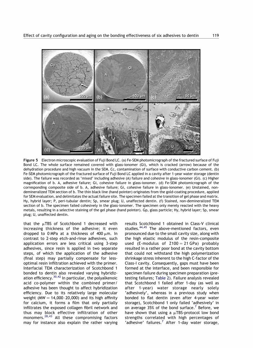
adhesive (Fig. 5). This indicates that the actualbonding effectiveness of Fuji Bond LC was notassessed because the cohesive strength of the glass-ionomer material itself was lower than, or at leastas low as, the interfacial bond strength. The latterwas partially corroborated by Fe-SEM analysis of thefractured surfaces, which revealed that actuallysome failures were located at the glass-ionomer/composite interface (Fig. 5).
Adhesive failure patterns were typically recog-nized by exposure of similarly curved scratches(circles) with a diameter corresponding to thediameter (1.4 mm) of the diamond bur used forcavity preparation (Figs. 2–6). The 3-step etch-and-rinse adhesive OptiBond FL hardly ever failed‘adhesively’. When this occurred after 1-yearwater storage, the adhesively failed parts neverexhibited poorly resin-impregnated collagen fibrils(image not shown), confirming the high hybridiz-ation efficacy of this 3-step etch-and-rinseapproach. On the contrary, the rather low bondingeffectiveness recorded for the 2-step etch-and-rinse adhesive Scotchbond 1 was associated with ahigh number of adhesive failures (Table 3), oftenexhibiting poorly resin-impregnated collagen(Fig. 2).
Table 5 p-Values of all pairwise Kruskal–Wallis comparisons (Dwass–Steel–Chritchlow–Fligne) for mTBS to Class-1 cavity bottomdentin after 1-day water storage (top right) and after 1-year water storage (bottom left).
1 Year 1 Day
OptiBond FL Scotchbond1 Clearfil SE Bond Adper Prompt Fuji Bond LC Reactmer
OptiBond FL *** , 0.0001 0.0972 , 0.0001 0.0011 , 0.0001Scotchbond 1 0.0005 *** , 0.0001 0.8837 0.0093 .0.9999Clearfil SE Bond 0.2763 0.0007 *** , 0.0001 0.0004 , 0.0001Adper prompt 0.0009 0.5089 0.0014 *** 0.0166 0.8291Fuji Bond LC 0.008 0.0004 0.5973 0.0018 *** 0.0003Reactmer 0.0415 0.0002 0.9825 0.0004 0.0623 ***
p-Values in italics are smaller than 0.05 and thus indicate significant difference; p-value in bold indicates nearly significantdifference.
Table 6 p-Values for significancy of effect of cavityconfiguration and aging (Kruskal–Wallis, Dwass–Steel–Chrit-chlow–Fligner comparisons).
Cavity configuration Aging
OptiBond FL 0.6618 0.0706Scotchbond 1 , 0.0001 0.1789
Clearfil SE Bond 0.0921 0.0272Adper prompt 0.0667 0.4204
Fuji Bond LC 0.0044 .0.9999Reactmer , 0.0001 , 0.0001
p-Values in italics are smaller than 0.05 and thus indicatesignificant difference; p-value in bold indicates nearlysignificant difference.
Effect of cavity configuration and aging on the bonding effectiveness of six adhesives to dentin 115

Discussion
Numerous studies reported on bonding perform-ance of adhesives as measured following amTBS-protocol. However, most were performed onflat ‘laboratory’ dentin surfaces that have a lowC-factor of 1/5.16,21,22 In a tooth cavity, shrinkagestress is, however, generated during polymerizationof the composite, pulling the adhesive from thecavity wall.14,23 This phenomenon is especiallypronounced in a Class-I cavity (used in this study)with five bonded walls and only one free surface,revealing a C-factor of 5/1. High shrinkage stressesmay induce gaps between the restoration and thecavity wall/floor that must result in micro-leakage,post-operative pain and other related clinicalproblems.5,24
Six adhesives representing all different classes ofadhesives were tested (Table 1).17,18 To insure
minimal variation in polymerization shrinkagestress, standardized cavities were made using atemplate and the MicroSpecimen Former. Inaddition, the same resin composite (Z100, 3MESPE), known for its relatively high E-modulus,25
was used for all adhesives. Because of the highC-factor of the experimental cavities (5/1) and thehigh E-modulus of the resin composite used, weassumed that this in vitro generated shrinkage stressapproaches the highest stress generated clinically.Qualitative Fe-SEM and TEM examination of thefracture planes combined with fractographic anal-ysis was used to substantiate the bond strength data.
Previous studies have shown that with increasingC-factor the mTBS decreases.11,26 In this study, thefactor ‘cavity configuration’ did not weaken thebond produced by the 3-step etch-and-rinseadhesive OptiBond FL and the 2-step self-etchadhesive Clearfil SE Bond. Among the different
Figure 2 Electron microscopic evaluation of Scotchbond 1. (a) Fe-SEM photomicrograph of the fractured surface ofScotchbond 1 applied in a cavity after 1-day water storage (dentin side). In contrast to when Scotchbond 1 was applied toa flat dentin surface, the failure occurred completely adhesively (A). (b) Magnification of a (box). On some sites, thedentin surface remained covered with a thin layer of resin (Cr); other areas failed at the bottom of the hybrid layer (Hb).Tubules are occluded by resin tags (arrow). (c) Composite counterpart at a similar area as in b. At some sites, the hybridlayer (Hy) remained attached to the resin composite (C). (d) Magnification of the edge of the composite side of themTBS-specimen, viewed at an angle of 608. The hybrid layer (Hy) remained attached to the composite (C). Loosecollagen fibrils incompletely enveloped by resin were disclosed. Consequently, the main failure site occurred within orat the bottom of the hybrid layer. Rt, resin tag. (e) Fe-SEM photomicrograph of the fractured surface of Scotchbond 1applied in a cavity after 1-year water storage (dentin side). Again, the specimen failed mainly adhesively at the bottomof the hybrid layer, exposing unaffected dentin (D), as well as it failed within the hybrid layer (Hy), exposing non-resin-enveloped collagen fibrils. Some areas failed near the top of the hybrid layer (Ht). Arrow, resin tag. (f) Compositecounterpart of e. The same failure sites can be detected; they occurred at the top of hybrid layer (Hy), uncovering thebonding layer (B) and within or at the bottom of the hybrid layer, exposing non-resin-enveloped collagen fibrils.
K. Shirai et al.116
classes of adhesives,18 the 3-step etch-and-rinseadhesive OptiBond FL often presented with thehighest bond strength values.2,3,18,27 This superiorbonding effectiveness must probably, to a largeextent, be attributed to optimal dentinhybridization, as was demonstrated in severalultra-morphologic interface analyses.17,18,28 – 31
The conventional 3-step application procedurethat guarantees a low technique-sensitive appli-cation procedure, the specific monomer/solventcocktail of the primer solution (containing glycer-ophosphoric acid dimethacrylate or GPDM, 2-hydro-xyethyl methacrylate or HEMA, and phthalic acidmono ethyl methacrylate or PAMM in an ethanol–water solution), the viscous solvent-free and glass-filled adhesive, and other ingredients such as anadequate polymerization initiator, may allhave contributed to this in vitro and in vivovery successfully and reliably performingadhesive.2,7,17,30,32 Also in this study, this 3-stepapproach resulted in the highest mTBS in Class-Icavities, after 1-day as well as after 1-year water
storage. Because of this high bond strength, thespecimens nearly never failed solely ‘adhesively’,as some of the less performing adhesives do(Table 3, Fig. 2).
Among the self-etch adhesives, the 2-stepadhesive Clearfil SE Bond has also consistentlybeen associated with favorable laboratory results.In particular when bonded to dentin, Clearfil SE Bonddid not underscore OptiBond FL, despite its beingapplied following a self-etch approach with oneapplication step less.16,17,27 Recently, a randomizedcontrolled clinical trial did not reveal any differencein clinical behavior, when Clearfil SE Bond wasapplied following manufacturer’s instructions orwhen enamel was selectively acid-etched with 40%phosphoric acid prior to the application of Clearfil SEBond.18,33 The specific molecular composition with10-methacryloxydecyl dihydrogen phosphate(10-MDP) as functional monomer has recently beenproven to be capable of interacting intimately withresidual hydroxyapatite that remained within theshallow 1-mm hybrid layer18,34 From the three
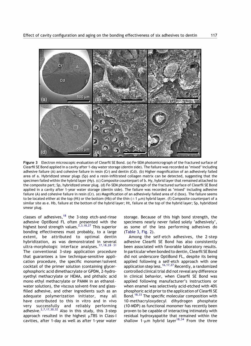
Figure 3 Electron microscopic evaluation of Clearfil SE Bond. (a) Fe-SEM photomicrograph of the fractured surface ofClearfil SE Bond applied in a cavity after 1-day water storage (dentin side). The failure was recorded as ‘mixed’ includingadhesive failure (A) and cohesive failure in resin (Cr) and dentin (Cd). (b) Higher magnification of an adhesively failedarea of a. Hybridized smear plugs (Sp) and a resin-infiltrated collagen matrix can be detected, suggesting that thespecimen failed within the hybrid layer (Hy). (c) Composite counterpart of b. Hy, hybrid layer that remained attached tothe composite part; Sp, hybridized smear plug. (d) Fe-SEM photomicrograph of the fractured surface of Clearfil SE Bondapplied in a cavity after 1-year water storage (dentin side). The failure was recorded as ‘mixed’ including adhesivefailure (A) and cohesive failure in resin (Cr). (e) Magnification of an adhesively failed area of d (box). The failure seemsto be located either at the top (Ht) or the bottom (Hb) of the thin (^1 mm) hybrid layer. (f) Composite counterpart of asimilar site as e. Hb, failure at the bottom of the hybrid layer; Ht, failure at the top of the hybrid layer; Sp, hybridizedsmear plug.
Effect of cavity configuration and aging on the bonding effectiveness of six adhesives to dentin 117

monomers investigated (besides 10-MDP, also4-methacryloxyethyl trimellitic acid or 4-MET, and2-methacryloxyethyl phenyl hydrogen phosphate orphenyl-P), the chemical bonding potential of10-MDP with hydroxyapatite was significantly thehighest and the most hydrolytically stable.34
Another reason contributing to the superiorbonding effectiveness of OptiBond FL and ClearfilSE Bond, might be the particle-filled adhesive resinthat is typically applied in a relatively thick layer. Ithas been hypothesized before that a relatively thickadhesive layer may act as an intermediary stressreliever to compensate for the shrinkage stressimposed during polymerization of the composite tothe resin-cavity wall/bottom bond.18,35 –40 Finiteelement analysis also revealed that with increasingthickness or decreasing elastic modulus of theadhesive resin, the shrinkage stresses can beconsiderably decreased.41 Consequently, this elas-tic bonding concept may, to a large extent, explainthe good resistance OptiBond FL and Clearfil SE
Bond have against polymerization shrinkage, whenapplied in a high C-factor cavity (Table 6).
The cavity configuration significantly affectedthe bonding performance of the 2-step etch-and-rinse adhesive Scotchbond 1. When bonded to flat‘laboratory’ dentin, Scotchbond 1 presented withbond strengths as high as that measured for theconventional 3-step system OptiBond FL. Identicalresults were obtained before.2,7 When Scotchbond1 was applied in a cavity, however, different factorsmay have compromised its bonding capacity. Therisk on improper drying of etched dentin increasedbecause of the narrow dimensions of the Class-Icavities prepared in this study. Likewise, it mustalso have been more difficult to adequately removeresidual solvents (water and ethanol) from thecombined primer/adhesive resin. Pooling of theadhesive was especially inevitable in the cavitycorners, having lead to thicker adhesive layers, butalso more difficult solvent removal in such regions.In this regard, a study by Zheng et al.22 reported
Figure 4 Electron microscopic evaluation of Adper Prompt. (a) Fe-SEM photomicrograph of the fractured surface ofAdper Prompt applied in a cavity after 1-day water storage (dentin side). The failure was recorded as ‘mixed’ includingadhesive failure (A) and cohesive in resin (Cr). (b) Magnification of an adhesively failed area. Tubules are widely openedby the strong self-etch adhesive and filled with resin tags. A reticular pattern is formed by a resin rim that surrounds thetubuli (arrow). (c) Fe-SEM photomicrograph of the corresponding composite counterpart of b. The heads of the resin tagsare still attached to the adhesive resin layer. Consequently, the specimen failed mainly at the top of the hybrid layer. (d)Fe-SEM photomicrograph of the fractured surface of Adper Prompt applied in a cavity after 1-year water storage (dentinside). The failure was recorded as ‘mixed’ including adhesive failure (A) and cohesive in resin (Cr). (e) Magnification ofthe transition zone between adhesive and cohesive failure, viewed at an angle of 458 (box in d). The three componentsthat constitute the resin–dentin interaction zone can easily be distinguished: bonding layer (B), hybrid layer (Hy) andunaffected dentin (D). (f) Composite side of a similar site as e. B, bonding; Hb, bottom of the hybrid layer; Ht, top of thehybrid layer; Rt, resin tag.
K. Shirai et al.118
that the mTBS of Scotchbond 1 decreased withincreasing thickness of the adhesive; it evendropped to 0 MPa at a thickness of 400 mm. Incontrast to 2-step etch-and-rinse adhesives, suchapplication errors are less critical using 3-stepadhesives, since resin is applied in two separatesteps, of which the application of the adhesive(final step) may partially compensate for less-optimal resin infiltration achieved with the primer.Interfacial TEM characterization of Scotchbond 1bonded to dentin also revealed varying hybridiz-ation efficiency.30,42 In particular, the polyalkenoicacid co-polymer within the combined primer/adhesive has been thought to affect hybridizationefficiency. Due to its relatively large molecularweight (MW ¼ 14,000–20,000) and its high affinityfor calcium, it forms a film that only partiallyinfiltrates the exposed collagen fibril network andthus may block effective infiltration of othermonomers.28,43 All these compromising factorsmay for instance also explain the rather varying
results Scotchbond 1 obtained in Class-V clinicalstudies.44,45 The above-mentioned factors, evenpronounced due to the small cavity size, along withthe high elastic modulus of the resin-compositeused (E-modulus of Z100 ¼ 21 GPa) probablyresulted in a rather poor bond at the cavity bottomthat could not withstand the high polymerizationshrinkage stress inherent to the high C-factor of theClass-I cavity. Consequently, gaps must have beenformed at the interface, and been responsible forspecimen failure during specimen preparation (pre-testing failures; Table 2). Failure analysis revealedthat Scotchbond 1 failed after 1-day (as well asafter 1-year) water storage nearly solely‘adhesively’, whereas in a previous study whenbonded to flat dentin (even after 4-year waterstorage), Scotchbond 1 only failed ‘adhesively’ inon average 35% of the bond surface.7 Before, wehave shown that using a mTBS-protocol low bondstrengths correlated with high percentages of‘adhesive’ failures.7 After 1-day water storage,
Figure 5 Electron microscopic evaluation of Fuji Bond LC. (a) Fe-SEM photomicrograph of the fractured surface of FujiBond LC. The whole surface remained covered with glass-ionomer (Gi), which is cracked (arrow) because of thedehydration procedure and high vacuum in the SEM. Cc, contamination of surface with conductive carbon cement. (b)Fe-SEM photomicrograph of the fractured surface of Fuji Bond LC applied in a cavity after 1-year water storage (dentinside). The failure was recorded as ‘mixed’ including adhesive (A) failure and cohesive in glass-ionomer (Gi). (c) Highermagnification of b. A, adhesive failure; Gi, cohesive failure in glass-ionomer. (d) Fe-SEM photomicrograph of thecorresponding composite side of b. A, adhesive failure; Gi, cohesive failure in glass-ionomer. (e) Unstained, non-demineralized TEM section of b. The thin black line (hand pointer) originates from the gold-coating procedure, appliedfor SEM evaluation, and delimitates the actual failure site. The specimen failed at the transition of gel phase and matrix.Hy, hybrid layer; P, peri-tubular dentin; Sp, smear plug; U, unaffected dentin. (f) Stained, non-demineralized TEMsection of b. The specimen failed cohesively in the glass-ionomer. The specimen only merely reacted with the heavymetals, resulting in a selective staining of the gel phase (hand pointer). Gp, glass particle; Hy, hybrid layer; Sp, smearplug; U, unaffected dentin.
Effect of cavity configuration and aging on the bonding effectiveness of six adhesives to dentin 119
most of the failures were located at the base of thehybrid layer, where SEM revealed many loosecollagen fibrils that did not appear enveloped byresin (Fig. 2). This confirms the poorer hybridizationefficiency mentioned above. A weak layer ofexposed collagen that was insufficiently impreg-nated by resin may have been present in betweenan incompletely formed hybrid layer and theunderlying unaffected dentin. Similar rather dis-appointing results, related to technique-sensitivity,were reported in a study, in which the long-termbonding effectiveness of Scotchbond 1 was assessedfollowing a mTBS-protocol after cyclic thermal andmechanical loading.46
Currently, glass-ionomers are the only self-adhesive restorative materials,17,47,48 though alsotheir bonding effectiveness gains from beforehandconditioning with a low-concentrated polyalkenoicacid (10–20%) conditioner.31 Their bonding mech-anism is twofold. The polyalkenoic acid conditionersuperficially and partially demineralizes dentin,leaving HAp around exposed collagen fibrils. As aresult, a submicron hybrid layer is formed that
provides micro-mechanical retention. In addition,the residual HAp within the hybrid layer serves as areceptor for chemical interaction with the carboxylgroups of the polyalkenoic acid.48 By addingmethacrylate monomers, resin-modified glass-iono-mers can be used to bond resin composites to toothsubstrates. Even though this bonding strategy invitro commonly underscores that of conventionaletch-and-rinse adhesives,17,18,49 the resin-modifiedglass-ionomer Fuji Bond LC has been highly success-ful in vivo in Class-V studies for periods up to fiveyears.17,18,33,50 Nevertheless, in this study Fuji BondLC appeared sensitive to the cavity configuration,since its mTBS slightly, but significantly decreasedin Class-I cavities.
Reactmer should be regarded as a one-step resin-based adhesive. However, due to the incorporationof fillers that are produced from the reaction of ion-leachable glass with polyalkenoic acid, it wasintroduced as a one-step glass-ionomer or so-called‘giomer’ on the dental market. After 24 h, a verylow mTBS was recorded and nearly all failures weresolely ‘adhesive’, with only few Reactmer remnants
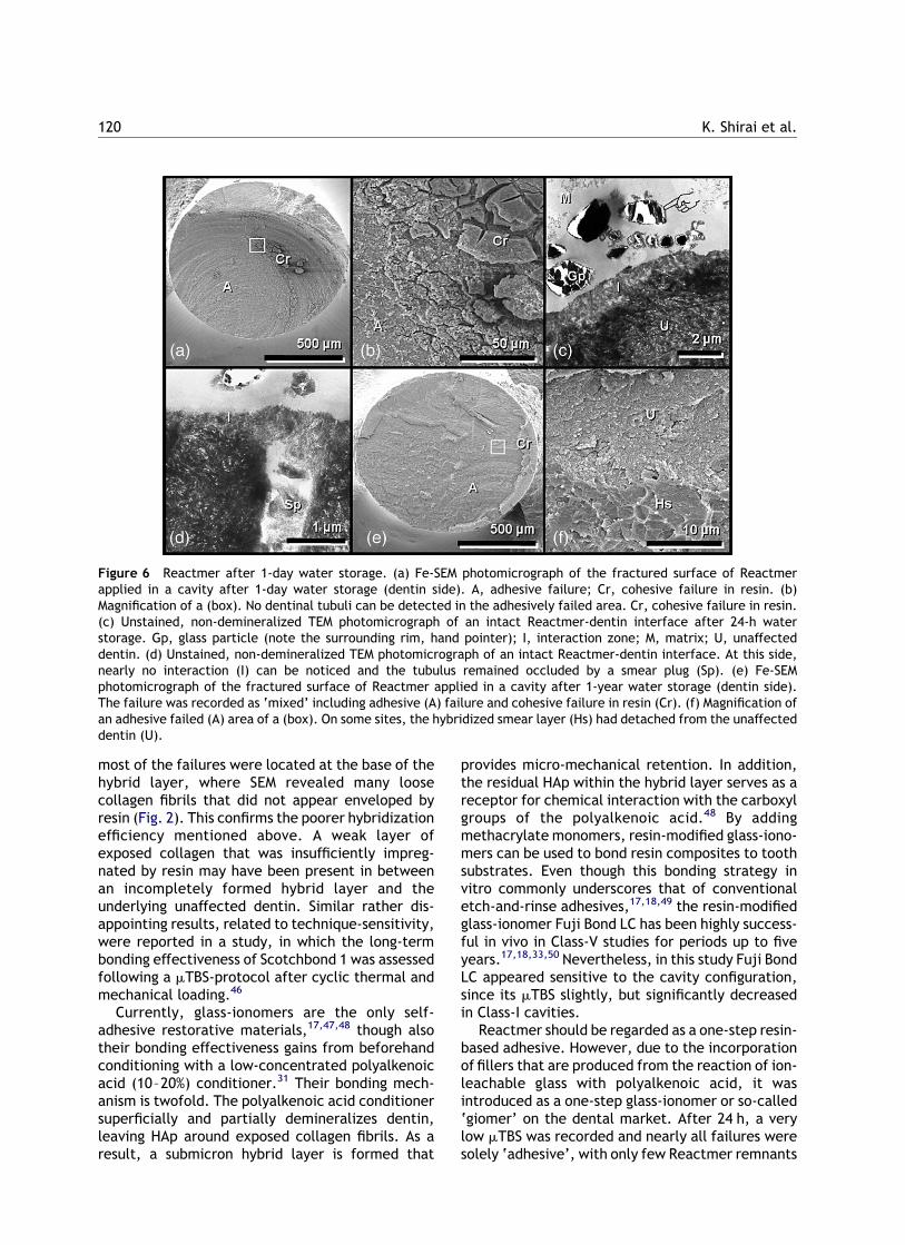
Figure 6 Reactmer after 1-day water storage. (a) Fe-SEM photomicrograph of the fractured surface of Reactmerapplied in a cavity after 1-day water storage (dentin side). A, adhesive failure; Cr, cohesive failure in resin. (b)Magnification of a (box). No dentinal tubuli can be detected in the adhesively failed area. Cr, cohesive failure in resin.(c) Unstained, non-demineralized TEM photomicrograph of an intact Reactmer-dentin interface after 24-h waterstorage. Gp, glass particle (note the surrounding rim, hand pointer); I, interaction zone; M, matrix; U, unaffecteddentin. (d) Unstained, non-demineralized TEM photomicrograph of an intact Reactmer-dentin interface. At this side,nearly no interaction (I) can be noticed and the tubulus remained occluded by a smear plug (Sp). (e) Fe-SEMphotomicrograph of the fractured surface of Reactmer applied in a cavity after 1-year water storage (dentin side).The failure was recorded as ‘mixed’ including adhesive (A) failure and cohesive failure in resin (Cr). (f) Magnification ofan adhesive failed (A) area of a (box). On some sites, the hybridized smear layer (Hs) had detached from the unaffecteddentin (U).
K. Shirai et al.120

left on the dentin surface (Table 3, Fig. 6). Thisrather low bonding effectiveness must be attribu-ted to the Class-I configuration, as this adhesive inconjunction with the same composite performedremarkably better to a flat surface after 24 h ofwater storage.3 Additional TEM analysis revealedthat Reactmer only superficially interacts with thedentin surface. Hence, the smear layer may nothave been fully dissolved, but only infiltrated andconsequently stabilized, as can be concluded fromthe following observations: (1) the interaction layerbetween dentin and Reactmer was very irregular.The thickness of the interaction zone varied from1 mm to nearly no interaction (Fig. 6); (2) thedentinal tubules appeared still filled with smear and(3) the primer has a relatively high pH of 3.2.3 Fromthese data, one could speculatively expect that the24-h micro-mechanical bonding effectiveness mighthave been relatively low and especially sensitive toshrinkage stress induced by polymerization of thecomposite in the high C-factor Class-I cavity, asshown by the low mTBS after 24 h (Table 2).
The effect of cavity configuration on the bondingeffectiveness of the ‘strong’ one-step self-etchadhesive Adper Prompt was only nearly significant.However, this must be regarded as statisticallysignificant, taking into account the significantlyhigher number of pre-testing failures when placedin a Class-I cavity. Like in this study when applied toflat or to Class-I cavity bottom dentin, it was shownbefore that this one-step self-etch adhesive and itspre-decessor Prompt L-pop produced mTBSs thatwere among the lowest ones recorded for adhesivesfrom the diverse classes of contemporaryadhesives.2,18 This lower bonding performance isalso reflected in the varying clinical resultsreported from Class-V studies. Relatively favorableshort-time retention rates of 100% at 6 months and96% at 1 year were recorded, respectively, byMunoz et al.51 and by Boghosian.52 However,relatively high loss rates of 21% at 2 years andeven 35% at 1 year were reported by, respectively,van Dijken45 and Brackett et al.53 Several expla-nations such as inhibition of polymerization of therestorative composite on top due to its highacidity,54 incomplete wetting and insufficientlythick adhesive layer,55 and phase separationbetween hydrophilic and hydrophobic ingredientsand resultant sensitivity to hydrolysis,17,18,56 havebeen advanced to explain this lower bondingperformance to dentin as compared to moreconventional etch-and-rinse and self-etchadhesives.
Besides reduced bond strength in Class-I cavities,pre-stressed interfaces may also be more suscep-tible to degradation, for example by gaps and
micro-voids that facilitate fluid exchange along theinterface. In this study, the long-term degradationof resin–dentin bonds formed in Class-I cavities wasstudied by exposure to water for 1 year at 37 8C.When sensitive to aging, the mTBS of the adhesiveshould have been reduced after 1-year waterstorage. Nevertheless, as the restored teeth werestored intact, the occlusal seal produced by bond-ing of the adhesive to the outer enamel margin ofthe Class-I cavities may have protected the bond ofthe adhesive to the Class-I bottom dentin againstdegradation. This beneficial effect was demon-strated before when four etch-and-rinse adhesiveswere applied to dentin disks surrounded by anenamel rim.7 Again, the bonding performance ofthe 3-step etch-and-rinse adhesive Optibond FLappeared stable despite the 1-year water storage,though the p-value nearly reached the level ofsignificance (Table 6). This confirms our previousresults when OptiBond FL was bonded to flat dentinand even after exposure to water for 4 years did notappear to have lost bond strength.7
The mTBS of the ‘mild’ 2-step self-etch adhesiveClearfil SE Bond decreased significantly after 1 yearof water storage. Although the failure modes didnot change noteworthy over time, SEM analysispointed out that adhesive failures after 1 dayoccured pre-dominantly more within the hybridlayer, whereas after 1 year the failures werelocated more at the transition of the hybrid layerto unaffected dentin (Fig. 3).
Using the 2-step etch-and-rinse adhesive Scotch-bond 1, all specimens failed prior to being tested sothat no mTBS could be recorded when it was bondedto Class-I cavity bottom dentin and exposed towater for 1 year. Also in our previous study,Scotchbond 1 appeared sensitive to 4-year wateraging, except when the Scotchbond-1-dentin bondwas all-around sealed by a resin–enamel bond.7 Inthat study, Scotchbond 1 was applied to flat dentindisks. Also in this study, some protection must havebeen provided by bonding of Scotchbond 1 to theocclusal enamel margins of the Class-I restorations.However, this appeared insufficient already after 1year. This may indicate that the additional polym-erization stress in Class-I cavities rendered thebonding performance even more vulnerable towater degradation.
When using the 2-step glass-ionomer adhesiveFuji Bond LC, no difference was found between1-day and 1-year mTBS. This is in contrast with asimilarly conducted study where Fuji Bond LC wasbonded to flat dentin surfaces and stored for 4 yearsin water.58 In that study, the mTBS dramaticallydecreased, mainly because of degradation of theglass-ionomer matrix itself. By storing the entirely
Effect of cavity configuration and aging on the bonding effectiveness of six adhesives to dentin 121
restored tooth in water in this study, for only 1year, it can be assumed that the water exposure ofthe glass-ionomer matrix at the pulpal floor of thecavity and the resultant damage to it must havebeen limited, thereby explaining the differencewith the former results. This is also confirmed by anon-changing failure analysis over the 1-year period(Table 3). Also SEM analysis did not reveal anystructural changes over time (Fig. 5). TEM analysison the other hand showed that sites rated as‘adhesive’ failure, actually failed at the transitionof the gel phase to the glass-ionomer matrix,leaving the hybrid layer intact (Fig. 5). This effectshould be further investigated in depth.
Remarkably, the ‘mild’ 1-step self-etch adhesiveReactmer was the only adhesive, of which the mTBSto Class-I cavity bottom dentin did not decreaseafter one year of water storage. It even increasedconsiderably (and highly significantly). No pre-testing failures were recorded after 1-year waterstorage, whereas about 70% of the specimens failedprior to being tested for the 1-day water-storedClass-I specimens (Fig. 1 and Table 2). Also the mainfailure mode changed from exclusively ‘adhesive’after 1 day to ‘mixed’ after 1 year (Table 3), andthus sustains the hypothesis of improved bondingeffectiveness over time. The most plausible expla-nation for this remarkable effect is that the glass-ionomer phase within Reactmer may by a kind ofion-exchange mechanism have additionally chemi-cally interacted with dentin. These water-depen-dent reactions may have taken a few weeks toestablish57, especially at the pulpal floor of theClass-I restoration, a site relatively remote fromthe water source. Alternatively, maturation of theglass-ionomer adhesive with time may haveenforced its cohesive strength and subsequentlyalso its mTBS. This ongoing reaction may then alsohave induced expansion of the adhesive, and sorelieved polymerization stress and avoided gapformation57. This expansion effect in combinationwith chemical interaction with dentin may beindicative of a kind of ‘repair’ effect, whencompared with the poor 1-day bond performance.
No further significant reduction in mTBS wasrecorded for the 1-step self-etch adhesive AdperPrompt after 1-year of water storage, thoughan already low bonding performance was achievedat 1 day.
In conclusion, the conventional 3-step etch-and-rinse adhesive still remained most effective inbonding to dentin, and appeared insensitive toeffects of increased polymerization shrinkage stressand water degradation. Most closely approachingthis superior bonding effectiveness of OptiBond FL,the ‘mild’ 2-step self-etch adhesive Clearfil SE Bond
only slightly lost bond strength after 1-yearexposure to water. The 2-step etch-and-rinseadhesive Scotchbond 1 and the ‘strong’ 1-stepself-etch adhesive Adper Prompt appeared verysensitive to cavity configuration and water-agingeffects, whereas the 2-step resin-modified glass-ionomer adhesive Fuji Bond LC only suffered fromhigher shrinkage stress, but not from the 1-yearwater-exposure. Remarkable is the apparentrepairability of the ‘mild’ 1-step self-etch orglass-ionomer adhesive Reactmer when stored for1 year in water. In general, simplified bondingprocedures do not necessarily imply improvedbonding performance, especially on the longterm. The application of technique-sensitiveadhesives such as the 2-step etch-and-rinse adhesive ScotchBond 1 and the 1-step self-etch adhesive Adper Prompt in more complexconfigurations leads to dramatic bond deterio-ration, on a short as well as long-term basis.
Acknowledgements
This study was supported in part by a ResearchGrant of the Fund for Scientific Research—Flanders(F.W.O.-grant ‘Krediet aan Navorsers’ 1.5.054.99)and by a fund of the Toshio Nakao Chair forAdhesive Dentistry inaugurated at the CatholicUniversity of Leuven with B. Van Meerbeek and P.Lambrechts awarded as Chairholders. We thank therespective manufacturers for the generousdonation of materials.
References
1. Degrange H, Roulet JF. Minimally invasive dentistry withbonding. Chicago: Quintessence Publishing; 1997.
2. Inoue S, Vargas MA, Van Meerbeek B, Abe Y, Yoshida Y,Lambrechts P, Vanherle G, Sano H. Micro-tensile bondstrength of eleven modern adhesives to dentin. J AdhesDent 2001;3:237—46.
3. De Munck J, Van Meerbeek B, Inoue S, Vargas M, Yoshida Y,Armstrong S, Lambrechts P, Vanherle G. Micro-tensile bondstrengths of one- and two-step self-etch adhesives to bur-cutenamel and dentin. Am J Dent 2003;16:414—20.
4. Van Meerbeek B, Perdigao J, Lambrechts P, Vanherle G. Theclinical performance of adhesives. J Dent 1998;26:1—20.
5. Roulet JF. Marginal integrity: clinical significance. J Dent1994;22:S9—S22.
6. Hashimoto M, Ohno H, Sano H, Tay FR, Kaga M, Kudoi Y,Oguchi H, Araki Y, Kuboto M. Micromorphological changes inresin—dentin bonds after 1 year of water storage. J BiomedMater Res 2002;63:306—11.
7. De Munck J, Van Meerbeek B, Lambrechts P, Vanherle G.Four-year water degradation of total-etch adhesives bondedto dentin. J Dent Res 2003;82:136—40.
K. Shirai et al.122

8. Gwinnett AJ, Yu S. Effect of long-term water storage ondentin bonding. Am J Dent 1994;7:109—11.
9. Burrow MF, Satoh M, Tagami J. Dentin bond durability afterthree years using a dentin bonding agent with and withoutpriming. Dent Mater 1996;12:302—7.
10. Sano H, Yoshikawa T, Pereira PN, Kanemura N, Morigami M,Tagami J, Pashley DH. Long-term durability of dentin bondsmade with a self-etching primer, in vivo. J Dent Res 1999;78:906—11.
11. Armstrong SR, Keller JC, Boyer DB. The influence of waterstorage and C-factor on the dentin-resin compositemicrotensile bond strength and debond pathway utilizing afilled and unfilled adhesive resin. Dent Mater 2001;17:268—76.
12. Santerre JP, Shajii L, Leung BW. Relation of dentalcomposite formulations to their degradation and the releaseof hydrolyzed polymeric-resin-derived products. Crit RevOral Biol Med 2001;12:136—51.
13. Frankenberger R, Kramer N, Petschelt A. Long-term effect ofdentin primers on enamel bond strength and marginaladaptation. Oper Dent 2000;25:11—19.
14. Feilzer AJ, De Gee AJ, Davidson CL. Setting stress incomposite resin in relation to configuration of the restaura-tion. J Dent Res 1987;66:1636—9.
15. Hashimoto M, Ohno H, Kaga M, Endo K, Sano H, Oguchi H. Invivo degradation of resin—dentin bonds over 1 to 3 years.J Dent Res 2000;79:1385—91.
16. De Munck J, Van Meerbeek B, Yudhira R, Lambrechts P,Vanherle G. Micro-tensile bond strength of two adhesives toEr:YAG-lased vs. bur-cut enamel and dentin. Eur J Oral Sci2002;110:322—9.
17. Van Meerbeek B, Vargas M, Inoue S, Yoshida Y, Peumans M,Lambrechts P, Vanherle G. Adhesives and cements topromote preservation dentistry. Oper Dent 2001;26(Suppl6):S119—44.
18. Van Meerbeek B, De Munck J, Yoshida Y, Inoue S, Vargas M,Vijay P, Van Landuyt K, Lambrechts P, Vanherle G.Buonocore memorial lecture: adhesion to enamel anddentin: current status and future challenges. Oper Dent2003;28:215—35.
19. Perdigao J, Lambrechts P, Van Meerbeek B, Vanherle G,Lopes ALB. Field emission SEM comparison of four postfixa-tion drying techniques for human dentin. J Biomed Mater Res1995;29:1111—20.
20. Robinson G, Gray T. Electron microscopy 2: practicalprocedures. In: Bancroft JD, Stevens A, editors. Theoryand practice of histological techniques. New York: ChurchillLivingstone; 1996. p. 585—626.
21. Armstrong SR, Keller JC, Boyer DB. Mode of failure in thedentin—adhesive resin—resin composite bonded jointas determined by strength-based (mTBS) and fracture-based(CNSB) mechanical testing. Dent Mater 2001;17:201—10.
22. Zheng L, Pereira PNR, Nakajima M, Sano H, Tagami J.Relationship between adhesive thickness and microtensilebond strength. Oper Dent 2001;26:97—104.
23. Versluis A, Douglas WH, Cross M, Sakaguchi RL. Does anincremental filling technique reduce polymerization shrink-age stresses? J Dent Res 1996;75:871—8.
24. Roulet JF, Reich T, Blunk U, Noack M. Quantitative marginalanalysis in the scanning electron microscope. ScanningMicrosc 1989;3:147—58.
25. Abe Y, Lambrechts P, Inoue S, Braem MJA, Takeuchi M,Vanherle G, Van Meerbeek B. Dynamic elastic modulus of‘packable’ composites. Dent Mater 2001;17:520—5.
26. Yoshikawa T, Sano H, Burrow MF, Tagami J, Pashley DH.Effects of dentin depth and cavity configuration on bondstrength. J Dent Res 1999;78:898—905.
27. De Munck J, Van Meerbeek B, Vargas M, Iracki J, Van LandugtK, Poitevin A, Lambrechts P. One day bonding effectivenessof new self-etch adhesives to bur-cut enamel and dentin.Oper Dent 2004 (in press).
28. Van Meerbeek B, Conn Jr LJ, Duke ES, Eick JD, Robinson SJ,Guerrero D. Correlative transmission electron microscopyexamination of nondemineralized and demineralized resin—dentin interfaces formed by two dentin adhesive systems.J Dent Res 1996;75:879—88.
29. Van Meerbeek B, Yoshida Y, Lambrechts P, Vanherle G, DukeES, Eick JD, Robinson SJ. A TEM study of two water-basedadhesive systems bonded to dry and wet dentin. J Dent Res1998;77:50—9.
30. Van Meerbeek B, Yoshida Y, Snauwaert J, Hellemans L,Lambrechts P, Vanherle G, Wakasa K, Pashley DH. Hybrid-ization effectiveness of a two-step versus a three-step smearlayer removing adhesive system examined correlatively byTEM and AFM. J Adhes Dent 1999;1:7—23.
31. Inoue S, Van Meerbeek B, Vargas M, Yoshida Y, Lambrechts P,Vanherle G. Adhesion mechanism of self-etching adhesives.In: Tagami J, Toledano M, Prati C, editors. Proceedings of3rd International Kuraray Symposium on Advanced AdhesiveDentistry. Como: Grafiche Erredue; 2000. p. 131—48.
32. Boghosian A. Clinical evaluation of a filled adhesive system inClass 5 restorations. Compend Contin Educ Dent 1996;7:750—4.
33. Peumans M, Van Meerbeek B, Lambrechts P, Vanherle G.Two-year clinical effectiveness of a resin-modified glass-ionomer adhesive. Am J Dent 2003;16:363—8.
34. Yoshida Y, Nagakane K, Fukuda R, Nakayama Y, Okazaki M,Shintani H, Inoue S, Tagawa Y, Suzuki K, De Munck J, VanMeerbeek B. Comparative study on adhesive performance offunctional monomers. J Dent Res 2004 (in press).
35. Kemp-Scholte CM, Davidson CL. Complete marginal seal ofClass V resin composite restorations effected by increasedflexibility. J Dent Res 1990;69:1240—3.
36. Kemp-Scholte CM, Davidson CL. Marginal integrity related tobond strength and strain capacity of composite resinrestorative systems. J Prosthet Dent 1990;64:658—64.
37. Van Meerbeek B, Willems G, Celis JP, Roos JR, Braem M,Lambrechts P, Vanherle G. Assessment by nano-indentationof the hardness and elasticity of the resin—dentin bondingarea. J Dent Res 1993;72:1434—42.
38. Perdigao J, Lambrechts P, Van Meerbeek B, Braem M, YildizE, Yucel T, Vanherle G. The interaction of adhesive systemswith human dentin. Am J Dent 1996;9:167—73.
39. Choi KK, Condon JR, Ferracane JL. The effects of adhesivethickness on polymerization contraction stress of composite.J Dent Res 2000;79:812—7.
40. Lee S-Y, Chiang HC, Lin CT, Huang HM, Dong DR. Finiteelement analysis of thermo-debonding mechanism in dentalcomposites. Biomaterials 2000;21:1315—26.
41. Ausiello P, Apicella A, Davidson CL. Effect of adhesive layerproperties on stress distribution in composite restorations—a3D finite element analysis. Dent Mater 2002;18:295—303.
42. Spencer P, Wang Y, Walker MP, Wieliczka DM, Swafford JR.Interfacial chemistry of the dentin/adhesive bond. J DentRes 2000;79:1458—63.
43. Eliades G, Vougiouklakis G, Palaghias G. Heterogeneousdistribution of single-bottle adhesive monomers in theresin—dentin interdiffusion zone. Dent Mater 2001;17:277—83.
44. Brackett MG, Dib A, Brackett WW, Estrada BE, Reyes AA.One-year clinical performance of a resin-modifiedglass ionomer and a resin composite restorative materialin unprepared class V restorations. Oper Dent 2002;27:112—6.
Effect of cavity configuration and aging on the bonding effectiveness of six adhesives to dentin 123

45. van Dijken JWV. Simplified adhesive systems in class V non-carious cervical dentin lesions. In: Programme and abstractsof the European Festival of Oral Science, Cardiff, Wales,September 25—28; 2002 (Abstr. 8).
46. Nikaido T, Kunzelmann KH, Ogata M, Harada N, Yamaguchi S,Cox CF, Hickel R, Tagami J. The in vitro dentin bondstrengths of two adhesive systems in class I cavities of humanmolars. J Adhes Dent 2002;4:31—9.
47. Wilson AD, Prosser HJ, Powis DM. Mechanism of adhesion ofpolyelectrolyte cements to hydroxyapatite. J Dent Res 1983;62:590—2.
48. Yoshida Y, Van Meerbeek B, Nakayama Y, Snauwaert J,Hellemans L, Lambrechts P, Vanherle G, Wakasa K. Evidenceof chemical bonding at biomaterial—hard tissue interfaces.J Dent Res 2000;79:709—14.
49. Belli S, Unlu N, Ozer F. Bonding strength to two differentsurfaces of dentin under simulated pulpal pressure. J AdhesDent 2001;3:145—52.
50. Tyas MJ, Burrow MF. Clinical evaluation of a resin-modifiedglass ionomer adhesive system: results at five years. OperDent 2002;27:438—41.
51. Munoz CA, Dunn JR, Bernal G, Torres J, Wilson A. Clinicalevaluation of Prompt L-Pop at 6 months. J Dent Res 2001;80:65. Abstr. No. 237.
52. Boghosian A. Clinical evaluation of a self-etchingadhesive: 1 year results. J Dent Res 2002;81:A-52.Abstr. No. 0192.
53. Brackett WW, Covey DA, St Germain Jr HA. One-year clinicalperformance of a self-etching adhesive in Class V resincomposites cured by two methods. Oper Dent 2002;27:218—22.
54. Tay FR, King NM, Suh BI, Pashley DH. Effect of delayedactivation of light-cured resin composites on bonding ofall-in-one adhesives. J Adhes Dent 2001;3:207—25.
55. Pashley EL, Agee KA, Pashley DH, Tay FR. Effects of oneversus two applications of an unfilled, all-in-one adhesive ondentine bonding. J Dent 2002;30:83—90.
56. Tay FR, Pashley DH, Suh BI, Carvalho RM, Itthagarun A.Single-step adhesives are permeable membranes. J Dent2002;30:371—82.
57. Huang C, Kei LH, Wei SH, Cheung GS, Tay FR, Pashley DH.The influence of hygroscopic expansion of resin-basedrestorative materials on artificial gap reduction. J AdhesDent 2002;4:61—71.
58. De Munck J, Van Meerbeek B, Yoshida Y, Inoue S, Suzuki K,Lambrechts P. Four-year water degradation of a glass-ionomer adhesive bonded to dentin. Eur J Oral Sci 2004;112:73—83.
K. Shirai et al.124