Embed Size (px)
Citation preview

_____________________________________________________________________________________________________ *Corresponding author: E-mail: [email protected];
Journal of Scientific Research & Reports 26(1): 80-92, 2020; Article no.JSRR.54350 ISSN: 2320-0227
Effect of Cardisoma guanhumi (Land Crab) Extract on Haematology and Lungs Histology in Swiss Mice
Infected with Bordetella pertussis
I. O. Enyi1*, A. I. Hart1 and I. M. Siminialayi2
1Department of Animal and Environmental Biology, Faculty of Science, University of Port Harcourt,
Nigeria. 2Department of Pharmacology, Faculty of Basic Medical Science, University of Port Harcourt, Nigeria.
Authors’ contributions
This work was carried out in collaboration among all authors. Author IOE designed the study,
performed the statistical analysis, wrote the protocol and wrote the first draft of the manuscript. Authors AIH and IMS managed the analyses of the study. Author IMS managed the literature
searches. All authors read and approved the final manuscript.
Article Information
DOI: 10.9734/JSRR/2020/v26i130216 Editor(s):
(1) Dr. Farzaneh Mohamadpour, Department of Organic Chemistry, University of Sistan and Baluchestan, Iran. Reviewers:
(1) Itamar Souza de Oliveira Junior, Federal University of São Paulo, Brazil. (2) Heba Gamal Abd El-Aziz Nasr, Al-Azhar University, Egypt.
Complete Peer review History: http://www.sdiarticle4.com/review-history/54350
Received 05 December 2019 Accepted 10 February 2020 Published 21 February 2020
ABSTRACT
Pertussis is an acute human respiratory tract disease caused by Bordetella pertussis, a known international pathogen that affects infants, children and adults. This study is aimed at investigating the changes in haematological parameters and histopathological changes of the lungs connected with Bordetella pertussis infection in swiss mice and to evaluate the potential of Cardisoma guanhumi extract to reverse these adverse changes about erythromycin treatment. The animals were divided into five groups: Group 1 was normal to control, group 2 was infected with Bordetella pertussis without treatment (negative control), group 3 and 4 were Bordetella pertussis infected and treated with 300 mg/kg and 600 mg/kg of the extract respectively and group 5 were infected and treated with 4000 mg/70 kg of erythromycin in divided doses. The animals were inoculated with a single infectious dose of Bordetella pertussis bacteria and were consequently treated with the graded doses of the extract and erythromycin for eighteen days after the animals were confirmed infected. The mice were humanely sacrificed using diethyl ether anaesthesia and blood samples
Original Research Article

Enyi et al.; JSRR, 26(1): 80-92, 2020; Article no.JSRR.54350
81
were taken for liver function investigation and liver tissue harvested and processed for histological examination. The result showed that Cardisoma guanhumi extract reversed the changes in the haematological parameters and pathological changes in the lungs of mice infected with Bordetella pertussis in a dose and time-dependent manner which suggests prophylactic and curative potentials of Cardisoma guanhumi extract against B. pertussis.
Keywords: Cardisoma guanhumi; land crab; lungs histology; Bordetella pertussis.
1. INTRODUCTION
Since the identification of pertussis as a clinical disease more than 1,600 years ago, Bordetella pertussis remains a major global pathogen that affects infants, children and adults [1]. B. pertussis is a complicated bacterium that expresses numerous bacterial factors with immune-modulating functions and produces different bacterial factors responsible for the symptoms seen during the disease [2]. Pertussis is mostly a toxin-mediated disease. The bacteria close to the cilia of the respiratory epithelial cells, produce toxins that paralyze the cilia and cause inflammation of the respiratory tract, which interferes with the clearing of pulmonary secretions [3]. Pertussis antigens appear to allow the organism invade host defences by enhancing lymphocytosis but impairing chemotaxis (NCDC, 2011). The understanding of this leaves us with the question of why has pertussis remained a major challenge to conquer internationally and what are the imminent dangers if nothing is done to curtail the prevalence of this disease. Numerous studies have progressively proven that pertussis toxin (PT) is the leukocytosis enhancing factor that is produced by Bordetella pertussis. Furthermore, other experimental animals, including rats (Samore and Siber 1992), pigs [4] and macaques [5] (Pauza et al., 1997), develop leukocytosis (an increase in the number of white blood cell especially during infection) when administered purified pertussis toxin (PT). Mice and baboons inoculated with B. pertussis have lowered levels of leukocytosis when induced with PT-specific monoclonal antibodies [6]. In a study by Temple et al., (2009) to determine the influence of living Bordetella pertussis on the induction and duration of pathophysiological reactions in mice infected intranasally with graded doses of culture, reported that fatally infected mice showed loss of body weight, spleen atrophy, obvious hypothermia and hypoglycemia (low blood sugar), and highly increased levels of leukocytes and serum immune-reactive insulin while non-fatal infected mice showed normal weight gain, almost normal temperature, spleen enlargement, not too pronounced hypoglycemia, lower but
obvious increased levels of leukocytes and serum immune-reactive insulin and histamine sensitization. Leukocytosis (an increase in the number of white blood cell especially during infection) and lymphocytosis (an increase in the number of lymphocytes in the blood) are clear indications of pertussis infection [7]. Similarly, Momoh et al., [8] noted that infection with S. typhi, similar bacteria to the one under study produced a reduction in pack cell volume (PCV), Red blood cell (RBC) and Hemoglobin (Hb) while there was an increase in monocyte, neutrophil and WBC but there was no significant difference. Crabs are decapod crustaceans which belong to the infra order Brachyura. They are mainly covered with the thick exoskeleton. Their lower region is completely hidden under the thoracic cavity. They can be found in most tropical and subtropical regions of the world as reported by Sammy et al., [9]. Cardisoma guanhumi is a species of land crab that is found in tropical and subtropical estuaries and other maritime areas of land along the Atlantic coast of the Americas [10]. They are known to be good sources of essential macro and micro minerals such as potassium, phosphorus, calcium, magnesium, copper, iron, manganese and zinc. Bae [11] and Sujeetha et al., [12] reported the biomedical and nutritional properties of crabs to include Omega3 (a polyunsaturated acid) contained in crab meat which helps in protecting against heart diseases and inhibit the aggressive behaviour. Mahae et al., [13] noted that the selenium contained in crab meat plays an important role in human's antioxidant defence system by preventing cells and tissues from damage and helps in proper functioning of the immune system and metabolism of thyroid hormone while riboflavin present in them helps in the production of steroids and red blood cells, maintenance of the skin, promote normal growth and iron absorption from the digestive tract and support antioxidant activity. Garry [14] explained that copper and phosphate content in crab helps in the absorption, storage and metabolism of iron and is involved in the formation of red blood cells. Ming et al., [15] and Suneeta [16] in their separate studies reported that crabs lower blood pressure, protect against heart diseases and

Enyi et al.; JSRR, 26(1): 80-92, 2020; Article no.JSRR.54350
82
possess anti-inflammatory properties. Chitosan derived from crab shell have several properties including anti-microbial and antibacterial properties due to its peculiar characteristics [13]. Chitosan fights against numerous pathogenic organisms like fungi, spoilage microorganisms, gram-positive and gram-negative bacteria [13]. However, this is the first attempt at establishing the antibacterial effects of crab extract which was prejudiced by unreliable evidence of the healing potential of crabs in whooping cough among the bonny people of Rivers State, Nigeria. This study is aimed at determining the effect of Cardisoma guanhumi (land crab) extract on haematology and lungs histology in swiss mice infected with Bordetella pertussis.
2. MATERIALS AND METHODS 2.1 Sample Collection and Identification Cardisoma guanhumi was caught using a trap in the buguma creek, Rivers State, Nigeria. The samples collected were transferred into perforated plastic containers to allow for air during transportation and was transported to the Pharmacognosy research laboratory, Department of Pharmacognosy, University of Port Harcourt, Nigeria. The samples were identified Mr. Otufu Paciya using Food and Agriculture Organization species identification sheets for freshwater and marine crab species.
2.2 Method of Extraction
Using the Shahidi and Synowiecki [17] extraction method, 60 of the freshly collected crabs were sacrificed and the shell separated from the meat and washed with tap water to remove all impurities. The crab shells and meat were then transferred to the oven and dried at 70
oC until
they were completely dry. Using a laboratory mortar and pestle, the dried crab shells and meat were ground and sieved into the size of 500 µm.
40 g of the sieved crab was measured using WANT precision electric weighing balance made by WANT balance instrument company limited, China. into a beaker and 200 ml of cord liver oil was added and stirred with a magnetic stirrer until it was completely mixed for 20 minutes. The beaker was then transferred into a water bath at a temperature of 60
oC and allowed for 30 mins.
The mixture was then filtered with a white handkerchief to drain off the oil and the residue transferred into a beaker. The residue was treated with 2% potassium hydroxide (KOH) at a
ratio of 1:20 w/v and was stirred continuously for 2 hours at a temperature of 90°C to remove protein from the crab. The sample was filtered under vacuum and the residues were continuously washed until the pH became neutral i.e pH=7. This was done to ensure that all the salt had been removed after removing the protein. The deproteinized crab was transferred into an oven and dried at 60°C until it was completely dry [17]. Two-point five per cent w/v of hydrochloric acid (HCl) was used at room temperature (20°C) for 6 hours to remove the mineral content of the deproteinized crab at a ratio of 1:20 w/v. The samples were filtered under vacuum and washed with tap water until the pH was neutral. The demineralized crab was then transferred to the oven and dried at a temperature 60°C until completely dried [17]. The demineralized crab was treated with 300 ml acetone for 10 mins and dried for 2hrs at ambient temperature and the residues were removed to achieve decolourization. The decolourized sample was washed in running water, filtered and dried at 60°C until it was completely dried to obtain crab chitin [17].
Deacetylation of chitin was
carried out using the method of Yen et al., (2009). The obtained chitin was treated with 40% w/v aqueous sodium hydroxide (NaOH) in the ratio of chitin to the solution 1:15 w/v at 105°C in a water bath for 2hrs. Thereafter, the chitin was filtered with a filter pump and washed with deionized water until the pH was neutral to obtain the extract. The obtained extract was then dried at 60°C for 2hrs in the oven. The dried extract was preserved in a well-labelled bottle and kept for the experiment.
2.3 Isolation of Test Organism The test organism Bordetella pertussis (ATCC
®9340
TM) was gotten from the American
Type Culture Collection (ATCC), USA. The culture media used for isolation according to ATCC is medium 35: Bordet Gengou/Broth medium from a human clinical specimen at a growth temperature of 37°C in the aerobic atmosphere. The product was received freeze-dried at 2°C-8°C
and stored at -80°C. The
bacterium was reconstituted using Regan-Lowe agar (Charcoal blood Agar) in the Department of Microbiology laboratory, University of Port Harcourt.
2.4 Experimental Design A total of one hundred and twenty-two (122) animals (swiss mice) were divided into five
Enyi et al.; JSRR, 26(1): 80-92, 2020; Article no.JSRR.54350
83
groups for the curative treatment study. Group 1 (normal) had 10 animals, group 2 (negative control) had 28 animals; group 3 and 4 consisted of 28 swiss mice each. Group 1 served as the normal control without treatment but was fed with the normal animal feed and water. Group 2 (negative control group) consisted of B. pertussis inoculated mice without treatment. Group 3 consisted of B. pertussis infected mice exposed to low dose (300 mg/kg) of Cardisoma guanhumi extract while group 4 consisted of B. pertussis infected mice exposed to high dose (600 mg/kg) of Cardisoma guanhumi extract and group 5 consisted of B. pertussis infected mice exposed to 4000 mg/70 kg of erythromycin [18]. On day 0, at day 6days interval and on day 18, seven animals were sacrificed using diethyl ether anaesthesia; samples of blood were collected and the liver removed for assessment of liver function status and histopathological examination respectively.
2.5 Challenging Healthy Animals with Bordetella pertussis Infective Dose
One hundred and twenty-two animals were intraperitoneally challenged with the infective dose of Bordetella pertussis which was calculated to be 5 x 10
5 cfu/ml. After infection
had set in (through physical observation of signs like weakness, non-productive cough, anorexia and the isolation of the organism from the blood of the infected animals on day 0) seven animals were sacrificed and blood samples and liver tissue was collected for preliminary investigation and the rest animals from the other treatment groups were given two times daily of the various doses of the extract and the standard antibiotics (erythromycin) for 18days.
2.6 Antibiotic and Extract Concentration Preparation
The extract solution for the study was prepared by dissolving 0.5 g of the extract in 1 ml of di-methyl-sulfoxide (DMSO) solvent to have a stock concentration of 500 mg/ml. Since 70 kg (70000 g) takes 4000 mg of erythromycin daily at the severe case of whooping cough, then 25 g (average weight of test animal) will take 25 g × 4000 mg/70000 g = 1.429 mg. This means that 25 g will take 1.429 mg/ml or 2.858 mg/0.5ml (1.429×2) or 5.716 mg/0.25 ml (1.429×4). 5.716 mg / 0.25 ml was prepared from 500 mg tablet of erythromycin tablet thus 500 mg/Xml = 5.716
mg/ml. therefore, X = 500 mg x ml/5.716 mg = 87.47 ml. Hence, 500 mg tablet of erythromycin was dissolved in 87.47 ml distilled water to prepare the erythromycin solution for the study.
2.7 Blood Collection Each animal was anaesthetized with diethyl ether in a desiccator and blood was collected by cardiac puncture method and transferred into a well-labelled sample bottle containing anti-coagulant.
2.8 Haematological Analysis The haematological analysis was carried out using the method as described by Randox Laboratories Limited, United Kingdom.
2.9 Histopathology Studies The study animals (swiss mice) were subjected to diethyl ether anaesthesia in a desiccator and dissected aseptically to collect the liver for histopathological studies. The collected tissues were kept in 10% chloroform for preservation and were subsequently trimmed to the size of 3-4 mm thickness for fixation. These tissues were fixed, dehydrated, cleared, impregnated, embedded, sectioned and stained with hematoxylin and eosin before mounting according to the method described by Baker (1945).
2.10 Statistical Analysis
The results of the measurements are shown as Mean Standard Deviation of Mean. The mean differences were obtained by ANOVA and post hoc with least significant difference (LSD) [19].
3. RESULTS 3.1 Effect of Cardisoma guanhumi Extract
on Post-inoculation Treatment on Hematological Indices in B. pertussis Infected Mouse
From the result, inoculation of mice with an infective dose of B. pertussis reveals a gradual decrease in PCV, Hemoglobin, Platelet, RBC and Eosinophil levels when compared to the normal control but these alterations were reversed when treated with Cardisoma
Enyi et al.; JSRR, 26(1): 80-92, 2020; Article no.JSRR.54350
84
guanhumi extract. Treatment with Cardisoma guanhumi extract reveals a steady increase in PCV (Table 1) Hemoglobin (Table 2) platelet (Table 3) RBC (Table 4) as the day progresses. Contrarily, the negative control shows a constant decrease in all the parameters. Statistical ANOVA comparison shows a significant difference (p<0.05) between normal control, negative control, standard drug and the treated groups on 6
th, 12
th and 18
th day. Similarly,
inoculation with an infective dose of B. pertussis shows an increase in WBC, neutrophils, lymphocytes and monocytes when compared with the normal control. However, treatment with Cardisoma guanhumi extract reversed the variations causing a gradual decrease in the haematological parameters (neutrophils, lymphocytes, WBC and monocytes). ANOVA comparison between the normal control, negative control, standard drug and the treatment group shows a significant difference in WBC on day 6, 12 and 18. Also, neutrophils showed no significant (p>0.05) difference on day 12 and 18 when compared to the standard drug. Similarly, there was a significant difference (P<0.05) in lymphocytes on day 6 and 12 but showed no significant (P>0.05) on day 18 when compared to the normal control and standard drug. Finally, monocytes count showed no significant difference in the standard drug on day 6 and 12 but a significant difference (P<0.05) when compared to negative control on day 18.
3.2 Effect of Cardisoma guanhumi Extract on Lungs Histo-architecture in B. pertussis Infected Mice
The lungs histopathological examination of the control animals reveals a normal structure with clear alveolar spaces, epithelial cells and blood vessels. There was no histologic alteration in the lungs. However, mice lungs tissue infected with B. pertussis with no treatment for four days shows various distortions in the lungs tissue such as widened interstitial, interstitial inflammation and bullae formation. The lungs tissues in mice administered with (300 mg/kg) of Cardisoma guanhumi extract for 12 days shows infected tissues with bullae formation while those treated for 18 days shows no interstitial inflammation. The lungs tissues not treated (negative control) for 18 days shows widened interstitial, interstitial inflammation, haemorrhage and bullae formation. However, Lungs tissues in mice treated with (600 mg/kg) of Cardisoma guanhumi extract for 6 days and 12 days shows mile interstitial Flammarion, bullae formation and widened interstitial while treatment for 18 days shows no obvious histologic change while for those treated with erythromycin for 6 days showed interstitial inflammation and bullae formation. However, lungs tissues infected with B. pertussis and treated with erythromycin for 18 days shows no obvious histologic change and appeared normal as the control. This is shown in Plates 1-14.
Table 1. Effect of post – IT on Cardisoma guanhumi extract on PCV (g/dl)] in B. pertussis infected mice
Day 0 Day 6 Day 12 Day 18
Control 37.00±0.000 37.00±0.000 37.00±0.000 37.00±0.000 Negative control 24.67±1.155 22.33±.577 18.67±1.155 15.33±1.155 Erythromycin 24.67±1.155 28.33±.577 34.33±.577 36.67±.577 Low Dose 24.67±1.155 26.67±.577
abc 29.67±.577
abc 31.33±1.155
abc
High Dose 24.67±1.155 26.67±.577abc
31.67±1.155abc
34.67±1.528abc
a = Significant (p<0.05) between test groups and control; b = Significant (p<0.05) between test groups and negative control; c = Significant (p<0.05) between test groups and erythromycin; Control = Animal fed with
normal feed and water; Negative control = Animal infected with Bordetella pertussis without treatment; Low dose = 300 mg/kg; High dose = 600 mg/kg; Erythromycin = standard antibiotics drug
Table 2. Effect of post - IT on Cardisoma guanhumi extract on Hgb (g/dl)] in B. pertussis
infected mice
Day 0 Day 6 Day 12 Day 18
Control 12.30±0.000 12.30±0.000 12.30±0.000 12.30±0.000 Negative control 8.40±.200 7.47±.115 7.00±.173 6.47±.153 Erythromycin 8.40±.200 9.13±.208 11.10±.173 12.20±.100 Low Dose 8.40±.200 8.37±.058
abc 9.07±.058
abc 9.53±.153
abc
High Dose 8.40±.200 8.53±.058abc
10.33±.115abc
11.60±.100abc

Enyi et al.; JSRR, 26(1): 80-92, 2020; Article no.JSRR.54350
85
Table 3. Effect of post - IT on Cardisoma guanhumi extract on platelets count (x103/µl)] in
B. pertussis infected mice
Day 0 Day 6 Day 12 Day 18
Control 100.00±0.000 100.00±0.000 100.00±0.000 100.00±0.000 Negative control 56.00±0.000 53.33±1.155 50.33±.577 48.33±.577 Erythromycin 56.00±0.000 73.67±3.512 85.00±2.000 99.33±.577 Low Dose 56.00±0.000 63.67±1.528
abc 70.33±2.517
abc 76.00±2.000
abc
High Dose 56.00±0.000 66.67±1.155abc
76.33±.577abc
91.67±.577abc
Table 4. Effect of post - IT on Cardisoma guanhumi extract on RBC (x103/µl) in B. pertussis
infected mice
Day 0 Day 6 Day 12 Day 18
Control 9.29±0.000 9.29±0.000 9.29±0.000 9.29±0.000 Negative control 4.90±.300 4.50±.200 3.77±.115 3.07±.153 Erythromycin 4.90±.300 6.33±.058 7.90±.200 9.28±.017 Low Dose 4.90±.300 5.50±.173
abc 6.00±.265
abc 6.80±.200
abc
High Dose 4.90±.300 5.83±.058abc
6.80±.300abc
8.83±.115abc
Table 5. Effect of post - IT on Cardisoma guanhumi extract on WBC (x103/µl) in B. pertussis
infected mice
Day 0 Day 6 Day 12 Day 18
Control 5.10±0.000 5.10±0.000 5.10±0.000 5.10±0.000 Negative control 5.77±.058 6.43±.153 7.70±.173 9.27±.153 Erythromycin 5.77±.058 5.33±.058 5.20±0.000 5.13±.058 Low Dose 5.77±.058 5.73±.058
abc 5.47±.058
abc 5.27±.058
b
High Dose 5.77±.058 5.63±.058abc
5.43±.058abc
5.17±.058b
Table 6. Effect of post - IT on Cardisoma guanhumi extract on neutrophil count (x103/µl) in
B. pertussis infected mice
Day 0 Day 6 Day 12 Day 18
Control 20.00±0.000 20.00±0.000 20.00±0.000 20.00±0.000 Negative control 24.67±1.155 27.33±.577 30.00±1.000 42.00±1.732 Erythromycin 24.67±1.155 21.67±.577 21.00±0.000 20.00±0.000 Low Dose 24.67±1.155 24.00±0.000
abc 23.00±0.000
abc 23.00±0.000
abc
High Dose 24.67±1.155 23.00±0.000abc
22.33±.577ab
20.67±.577b
Table 7. Effect of post - IT on Cardisoma guanhumi extract on lymphocyte count (x103/µl) in B.
pertussis infected mice
Day 0 Day 6 Day 12 Day 18
Control 82.00±0.000 82.00±0.000 82.00±0.000 82.00±0.000 Negative control 88.00±1.000 91.33±2.082 94.00±1.000 101.67±2.517 Erythromycin 88.00±1.000 84.33±.577 83.67±.577 82.00±0.000 Low Dose 88.00±1.000 87.67±.577
abc 85.67±.577
abc 84.67±.577
b
High Dose 88.00±1.000 85.33±1.155ab
84.33±.577ab
82.67±.577b
Table 8. Effect of post - IT on Cardisoma guanhumi extract on monocyte count (x103/µl) in
B. pertussis infected mice
Day 0 Day 6 Day 12 Day 18
Control 2.00±0.000 2.00±0.000 2.00±0.000 2.00±0.000 Negative control 4.67±.577 6.67±.577 9.00±1.000 11.00±1.000 Erythromycin 4.67±.577 3.33±.577 2.67±.577 2.00±0.000 Low Dose 4.67±.577 4.00±0.000
ab 3.67±.577
ab 3.33±.577
b
High Dose 4.67±.577 4.00±0.000ab
3.33±.577ab
2.67±.577b

Enyi et al.; JSRR, 26(1): 80-92, 2020; Article no.JSRR.54350
86
Histological Plates
Plate 1. Photomicrograph of lungs tissue of normal mice showing no histologic alteration
Plate 2. Photomicrograph of lungs of mice infected with B. pertussis showing interstitial inflammation and bullae formation (Day 0)
Interstitial inflammation Bullae formation
Plate 3. Photomicrograph of lungs of mice infected with B. pertussis without treatment for 6 days showing interstitial inflammation and bullae formation
Enyi et al.; JSRR, 26(1): 80-92, 2020; Article no.JSRR.54350
87
Interstitial inflammation Bullae formation
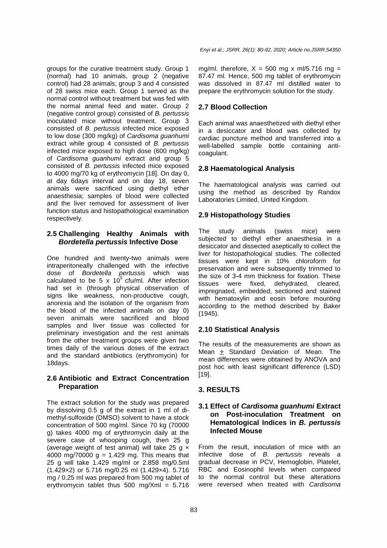
Plate 4. Photomicrograph of lungs of mice infected with B. pertussis treated with 4000 mg/
70 kg of erythromycin for 6 days showing interstitial inflammation and bullae formation
Interstitial inflammation Bullae formation
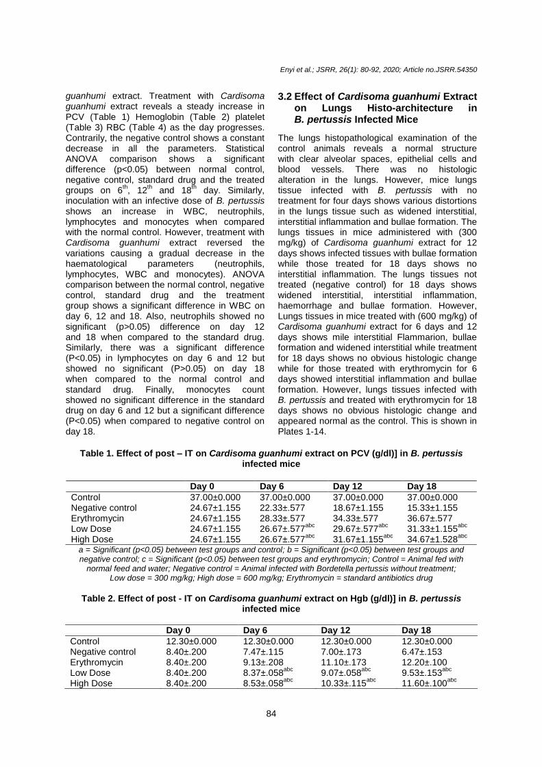
Plate 5. Photomicrograph of lungs of mice infected with B. pertussis and treated with 300 mg/kg of Cardisoma guanhumi extract for 6 days showing interstitial inflammation and
bullae formation
Interstitial inflammation Bullae formation
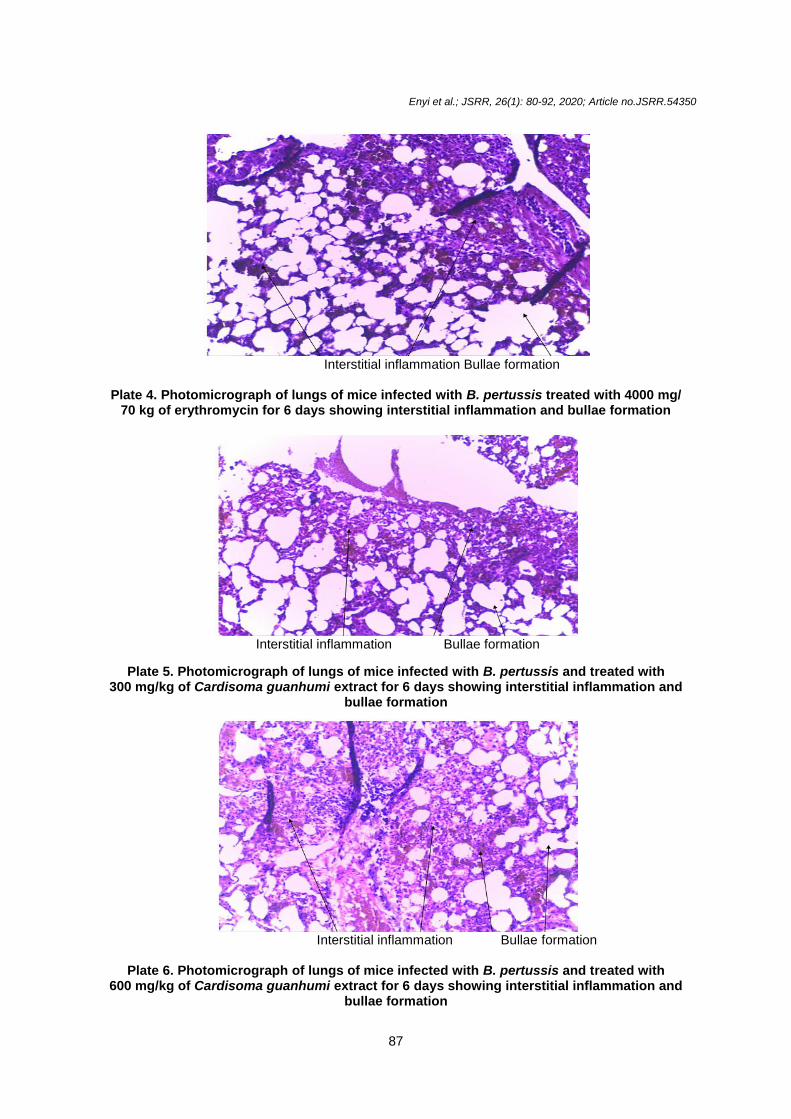
Plate 6. Photomicrograph of lungs of mice infected with B. pertussis and treated with
600 mg/kg of Cardisoma guanhumi extract for 6 days showing interstitial inflammation and bullae formation

Enyi et al.; JSRR, 26(1): 80-92, 2020; Article no.JSRR.54350
88
Interstitial inflammation Bullae formation
Plate 7. Photomicrograph of lungs of mice infected with B. pertussis without treatment for 12 days showing interstitial inflammation and bullae formation
Alveolar spaces Pulmonary vessels
Plate 8. Photomicrograph of lungs of mice infected with B. pertussis treated with 4000 mg/ 70 kg of erythromycin for 12 days showing no obvious histologic change with clear alveolar
spaces and pulmonary vessels
Mild interstitial inflammation Bullae formation
Plate 9. Photomicrograph of lungs of mice infected with B. pertussis and treated with
300 mg/kg of Cardisoma guanhumi extract for 12 days showing mild interstitial inflammation and bullae formation

Enyi et al.; JSRR, 26(1): 80-92, 2020; Article no.JSRR.54350
89
Interstitial inflammation Bullae formation
Plate 10. Photomicrograph of lungs of mice infected with B. pertussis and treated with 600 mg/kg of Cardisoma guanhumi extract for 12 days showing interstitial inflammation and
bullae formation
Interstitial inflammation Bullae formation
Plate 11. Photomicrograph of lungs of mice infected with B. pertussis without treatment for
18 days showing interstitial inflammation and bullae formation
Alveolar spaces Pulmonary vessels
Plate 12. Photomicrograph of lungs of mice infected with B. pertussis and treated with 4000 mg/70 kg of erythromycin for 18days showing no obvious histologic change

Enyi et al.; JSRR, 26(1): 80-92, 2020; Article no.JSRR.54350
90
Plate 13. Photomicrograph of lungs of mice infected with B. pertussis and treated with 300 mg/kg of Cardisoma guanhumi extract for 18 days showing no histologic alteration
Plate 14. Photomicrograph of lungs of mice infected with B. pertussis and treated with
600 mg/kg of Cardisoma guanhumi extract for 18 days showing normal lungs tissue
4. DISCUSSION Several studies have shown that during B. pertussis infection, the common haematological changes include leukocytosis (an increase in the number of white blood cell (WBC)) lymphocytosis (an increase in the number of lymphocytes in the blood) and monocytosis (an increase in the number of monocytes in the blood [7], Temple et al. [6]. Significant decrease in mean levels reduced PCV (Pack Cell Volume), Red Blood Cell (RBC) a Hemoglobin while there is an increased neutrophil [8]. The result of this study agrees with the reports from the other researchers. The increase in neutrophil and white blood cell are associated with the invasion of the hemopoietic organ (bone marrow) by
B. pertussis while monocytosis and lymphocytosis are attributed to the increased release of these cells from the myeloid/ lymphoid tissues in response to the infection [20] (Anusuya and Sumathi, 2015). However, the decrease in RBC, PCV and haemoglobin could be as a result of the destruction of Red Blood Cell by the infection [21] hemophagocytosis (a potentially fatal disease of normal but overactive histiocytes and lymphocytes that are common in infants) and bone marrow suppression (Khosla et al., 1995). The result further reveals that the post-inoculation treatment with Cardisoma guanhumi extracts reversed the usual trend of significant increase (P<0.05) in WBC, lymphocytes, monocytes, neutrophils and decrease in PCV, RBC, platelets and haemoglobin usually

Enyi et al.; JSRR, 26(1): 80-92, 2020; Article no.JSRR.54350
91
associated with B. pertussis infection. Therefore, the post-inoculation treatment with Cardisoma guanhumi extract reversed B. pertussis invasion of the hemopoietic organs, bone marrow suppression, destruction of RBC, hemophagocytosis and the invasion of the macrophages although the level of reversal is time and suggests that prophylactic and curative potentials of Cardisoma guanhumi extract against B. pertussis when combined with other therapeutic agents. The result of this study agrees with the report of Andreasen and Carbonetti, [22], Karen et al., [23] who said infection of B. pertussis causes lung tissue inflammation, necrosis and widened interstation. However, treatment with Cardisoma guanhumi slowly reversed the trend with an increase in time and dosage. The result confirms the anti-Bordetella pertussis property of the extract.
5. CONCLUSION Inoculation of mice with an infective dose of B. pertussis produces haematological changes such as leukocytosis (an increase in the number of white blood cell (WBC)) lymphocytosis (an increase in the number of lymphocytes in the blood) and monocytosis (an increase in the number of monocytes in the blood) Significant decrease in mean levels of PCV (Pack Cell Volume), Red Blood Cell (RBC) and Hemoglobin with an increase in neutrophil but treatment with Cardisoma guanhumi extract reversed the changes in a dose and time-dependent manner. Similar, infection with B. pertussis caused distortions in the lungs tissue such as widened interstitial inflammation and bullae formation but treatment with Cardisoma guanhumi extract reversed the changes in a dose and time-dependent manner.
ETHICAL APPROVAL Animal ethic Committee approval has been collected and preserved by the authors.
COMPETING INTERESTS Authors have declared that no competing interests exist.
REFERENCES 1. Dorji D, Mooi F, Yantorno O, Deora R,
Graham RM, Mukkur TK. Bordetella pertussis virulence factors in the continuing
evolution of whooping cough vaccines for improved performance. Medical Microbiolog and Immunology. 2017;10:17-52.
2. Mattoo S, Cherry JD. Molecular pathogenesis, epidemiology, and clinical manifestations of respiratory infections due to Bordetella pertussis and other Bordetella subspecies. Clinical Microbiology Review. 2005;18:326–382.
3. Sheridan SL, Ware RS, Grimwood K, Lambert SB. Number and order of whole cell pertussis vaccines in infancy and disease protection. JAMA. 2012;308:454–456.
4. Elahi S, Brownlie R, Korzeniowski J. Infection of newborn piglets with Bordetella pertussis: A new model for pertussis. Infection and Immunology. 2005; 73:3636–45.
5. Hinds PW, Yin C, Salvato MS. Pertussis toxin induces lymphocytosis in rhesus macaques. Journal of Medical Primatol. 1996;25:375–81.
6. Nguyen AW, Wagner EK, Laber JR. A cocktail of humanized anti-pertussis toxin antibodies limits disease in murine and baboon models of whooping cough. Sci Transl Med. 2015;7:316-95.
7. Beck TC, Gomes AC, Cyster JG. CXCR4 and a cell-extrinsic mechanism control immature B lymphocyte egress from bone marrow. Journal of Experimental Medicine. 2014;211:2567–81.
8. Momoh AO, Adebolu TT, Ogundare AO. Evaluation of beniseed extract and fermented liquors in treatment of diarrhea in albino rats infected with Salmonella typhi. European Journal of Biology and Medical Science Research. 2013;1(2):16-23.
9. Sammy D, Grave N, Dean P, Pentcheff N. A classification of living a fossil genera of decapod crustaceans. Raffles bulletin of zoology. 2009;21:1-109.
10. Renata AS, Jose RF, Fábio HV. Development of male reproductive system of the blue land crab Cardisoma guanhumi Latreille, 1828 (Decapoda: Gecarcinidae). Acta Zoologica. 2012;93(4):390–399.
11. Bae Keun Park. Application of chitin and its derivatives in biological medicine. International Food Research Journal. 2010;11(12):5152-5164
12. Sujeetha M, Sharmila S, Jayanthi J, Ragunathan G. Antioxidant property of some extracts derived from the mud crab,

Enyi et al.; JSRR, 26(1): 80-92, 2020; Article no.JSRR.54350
92
scylla serrate. International Journal of Phytopharmacology. 2015;6(2):111-113.
13. Mahae N, Chalat C, Muhamud P. Antioxidant and antimicrobial properties of chitosan sugar complex. International Food Research Journal. 2011;18(4):1543-1551.
14. Garry Kerch. The potential of chitosan and its derivatives in prevention and treatment of age related disease. International Food Research Journal. 2015;13:2158-2182.
15. Ming Kong, Ke Xing, Hyun Kin Park. Antimicrobial properties of chitosan and mode of action: A state of art review. International Journal of Food Microbiology. 2010;144(1):50-63.
16. Suneeta kumara (2014) Extraction and characterization of chitin and chitosan from (Labeo rohit) fish scales. Raffles bulletin of zoology, 6:482-489
17. Shahidi F, Synowiecki J. Isolation and characterization of nutrients and value- added products from snow crab (Chionoecetes opilio) and shrimp (Pandalus borealis) processing discards. Journal of Agricultural Food Chemistry. 1991;39(8):1527–1532.
18. Enyi IO, Hart AI, Siminialayi IM. Effect of Cardisoma guanhumi (Land crab) extract on liver function and liver histology of Swiss mice infected with Bordetella pertussis. International Journal of
Contemporary Research and Review. 2020;11(01):20201-20211.
19. Mead R, Curnow RN. A simple statistical method in Agriculture and Experimental Biology. Charpman Hall, London, UK. 1982;33-46.
20. Das BK, Mukherjee SC. Toxicity of cypernethrin in Labeo rohita fingerlings: Biochemical enzymatic and heamatological consequence. Journal of Comparative Biochemistry, Physiology, Toxicology and Pharmacology. 2003;134: 109-121.
21. Dangana A, Ajobiewe J, Nuhu A. Hematological changes associated with Salmonella typhi and Salmonella paratyphi in humans. International Journal of Biomedical Health Science. 2010;6:219-222.
22. Andreasen C, Carbonetti NH. Pertussis toxin inhibits early chemokine production to delay neutrophil recruitment in response to Bordetella pertussis respiratory tract infection in mice. Infections and Immunology. 2008;76:5139–5148.
23. Karen M, Scanlon Y, Snyder G, Nicolas H. Fatal pertussis in the neonatal mouse model is associated with pertussis toxin-mediated pathology beyond the airways. Infectious Immunology. 2017;85(11):355-17.
_______________________________________________________________________________ © 2020 Enyi et al.; This is an Open Access article distributed under the terms of the Creative Commons Attribution License (http://creativecommons.org/licenses/by/4.0), which permits unrestricted use, distribution, and reproduction in any medium, provided the original work is properly cited.
Peer-review history: The peer review history for this paper can be accessed here:
http://www.sdiarticle4.com/review-history/54350