Embed Size (px)
Citation preview

RESEARCH Open Access
Effect of 150 kHz electromagnetic radiationon the development of polycystic ovariesinduced by estradiol Valerate in SpragueDawley ratsStephanie Mohammed1* , Venkatesan Sundaram2 and Nikolay Zyuzikov1
Abstract
Background: Polycystic ovary syndrome (PCOS) is the most common complex endocrine disorder affectingapproximately 2–20% of reproductive aged females. Tumour Treating Fields (100–300 kHz) is a recent innovative,non-invasive therapeutic approach to cancer therapy. This frequency as an alternative therapy for the managementof polycystic ovaries has not yet been explored.
Objectives: To explore the effect of full-body exposure of 150 kHz Electromagnetic Radiation (EMR), on thedevelopment of polycystic ovaries in an estradiol valerate-induced PCO rat model.
Method: Twenty-one female adult rats were divided into three groups (n = 7 each): control, Estradiol Valerate (EV)and EV + EMR groups. The EV + EMR group was subjected to full body exposure at 150 kHz EMR continuously foreight consecutive weeks. Estradiol valerate was administered orally to induce polycystic ovaries in EV and EV + EMRgroups. Body and ovarian weights were recorded and analysed. The regularity of the estrous cycle was assessed inall three groups. The histological study of ovarian tissue was carried out by haematoxylin and eosin staining. Theserum concentration levels of Luteinizing Hormone (LH), Follicle-Stimulating Hormone (FSH) and testosterone weremeasured using the ELISA method.
Results: The body and ovary weights did not differ significantly between the EV and EV + EMR groups. The estrouscycle was found to be irregular in both the EV and EV + EMR groups. Ovarian histology revealed near normalmorphology with little or no degenerative and morphological changes in developing follicles in the exposedgroup. Histometrical analysis showed an increased number of developing follicles and a significant reduction in thenumber and size of follicular cysts (p < 0.05) in the EV + EMR group. Hormonal analysis revealed no significantdifference in the testosterone and FSH levels between the EV + EMR and EV groups. However, the LH, LH/FSH ratiodecreased significantly in the EV + EMR group compares to the EV group.
Conclusion: The 150 kHz EMR appear to have little or no degenerative and morphological changes in thedeveloping follicles, an increased number of typical developing follicles and a significant reduction in the numberand size of the follicular cysts (p < 0.05).
Keywords: 150 kHz electromagnetic radiation, Estradiol Valerate, Polycystic ovary model
© The Author(s). 2021 Open Access This article is licensed under a Creative Commons Attribution 4.0 International License,which permits use, sharing, adaptation, distribution and reproduction in any medium or format, as long as you giveappropriate credit to the original author(s) and the source, provide a link to the Creative Commons licence, and indicate ifchanges were made. The images or other third party material in this article are included in the article's Creative Commonslicence, unless indicated otherwise in a credit line to the material. If material is not included in the article's Creative Commonslicence and your intended use is not permitted by statutory regulation or exceeds the permitted use, you will need to obtainpermission directly from the copyright holder. To view a copy of this licence, visit http://creativecommons.org/licenses/by/4.0/.The Creative Commons Public Domain Dedication waiver (http://creativecommons.org/publicdomain/zero/1.0/) applies to thedata made available in this article, unless otherwise stated in a credit line to the data.
* Correspondence: [email protected] of Physics, Faculty of Science and Technology, The Universityof the West Indies, St. Augustine, Trinidad and TobagoFull list of author information is available at the end of the article
Mohammed et al. Journal of Ovarian Research (2021) 14:26 https://doi.org/10.1186/s13048-021-00774-4

Subject classification codes Electromagnetic Radiation,Polycystic Ovary.
IntroductionPolycystic ovary syndrome (PCOS) is recognized as themost common complex endocrine disorder affecting ap-proximately 2–20% of reproductive aged females [8].This condition manifests polycystic ovaries, hyperandro-genism, androgenic alopecia, hirsutism, acne, menstrualirregularity, anovulation or oligo-amenorrhea, miscar-riage, and infertility [28]. It presents symptoms such asunwanted hair growth and hormonal changes which cannegatively affect the emotional character of womenwhich may subsequently may result in depression andanxiety [2, 15]. Women with PCOS are more susceptibleto several chronic conditions including obesity, dyslipi-daemia, hypertension, heart disease, and type 2 diabetesmellitus (T2DM) [20]. The definite aetiology of PCOSremains largely unknown. However, complex interac-tions between genetic, behavioural, and environmentalfactors play critical roles in the development of PCOSand subsequent therapeutic options [11]. Present treat-ment options focus on controlling the associated signsand symptoms. Therefore, the search for moreefficacious, affordable treatment options attracts interestfor the management of PCO and its subsequentcomplications.Electromagnetic radiation (EMR) consists of electro-
magnetic waves, which are synchronized oscillations ofboth electric and magnetic fields that travel through avacuum at the speed of light. These waves which areconstantly emitted from the natural environment, as wellas from everyday appliances, frequently influence the hu-man body. The effect of this type of energy waves on liv-ing tissues may exert various effects on their ability tofunction, although the mechanisms conditioning thisphenomenon have not been fully understood. The ef-fects of the EMR on the reproductive system is catego-rized as hazardous, neutral or beneficial [34]. The resultsof reproductive studies confirming a beneficial effect ofelectromagnetic waves evoke hope for the need of theseinventions in the treatment of PCO.Currently, the Intermediate Frequency (300 Hz to 10
MHz) range has offered controversial outlook on thetherapeutic use of this range of frequency. Confirminglythe range of 100 kHz – 300 kHz known as Tumor Treat-ing Fields (TTF) has provided substantial evidence for amore positive advancement in the field of oncology.Tumor Treating Fields is an innovative, non-invasiveand advanced therapeutic approach to various cancertherapies. This particular frequency disrupts mitosis andselectively kills rapidly dividing cancer cells by deliveringcontinuous (over 18 h per day) low intensity, intermedi-ate frequency, alternating electric fields (100 kHz – 300
kHz) to the tumor site [30]. Tumor Treating Fields havebeen found very effective in the treatment of several can-cers including Glioblastoma multiforme and ovariancancers [12, 17] in a preclinical setting.It is postulated that the follicular disruption of PCOS is
2-fold [7]. First, the early follicular growth is extreme andsecond, the selection of one follicle from this increasedpool of growing follicles for further maturation to a dom-inant follicle is arrested. It remains unknown whether theprimary defects lie within the theca, granulosa or oocytebut it is presumed a consequence of intra-ovarian hyper-androgenism. Previous reports of TTFs on the action ofabnormally proliferating cells, therefore evokes interest forexploring the effect of this frequency during the folliculardevelopment of polycystic ovaries.The optimal frequency of TTFs for antimitotic effect
varies by cancer type, and can be adjusted for maximalanticancer effect. In a preclinical setting, 150 kHz TTFswas found to be effective for pancreatic cancer, Non-Small-Cell lung carcinoma (NSCLC), brain metastasisfrom NSCLC and mesothelioma treatment when com-bined with chemotherapy [17]. Currently, 200 kHz is be-ing explored for ovarian cancers in the same setting [33].Therefore, this current study is designed to test the ef-fect of 150 kHz Electromagnetic radiation (EMR) duringthe development of estradiaol valverate induced polycys-tic ovaries in rats.
Materials and methodsAnimals and experimental designA total of twenty-one (21) adult female Sprague Dawley(SD) rats (12–15 weeks-old) weighing 250-300 g wereprocured from the Lab Animal Facility at the School ofVeterinary Medicine for the study. The animals wereplaced in ventilated metal cages with the dimensions of40 × 24 × 14 cm (2 rats per cage) with paper bedding ma-terial in a pathogen free room at a temperature of 24 ±2 °C, humidity of 50–60% and 12-h light/dark cycle. Therats were fed with a standard pellet diet and water adlibitum. The rats were allowed to acclimatize to labora-tory condition for 7 days.The animals were divided randomly into three groups
(n = 7 each): control group, Estradiol Valerate Group(EV) and EV + EMR group. The EV + EMR group wassubjected to full body exposure of EMR at 150 kHz con-tinuously (except for about one hour per week that wasneeded for changing the cages) for eight consecutiveweeks [1]. Polycystic ovarian condition were induced inthe EV and EV + EMR groups of animals by administer-ing commercially available estradiol valerate tablets at asingle oral dosage of 4 mg per animal on the first and14th day of experiment as reported by Brawer et al. [3]to ensure the EV activity was maintained for the devel-opment of PCO. The control and EV group was kept
Mohammed et al. Journal of Ovarian Research (2021) 14:26 Page 2 of 10

under similar conditions without EMR. The Campus Re-search Ethics Committee of the University of the WestIndies approved the protocols for animal experimenta-tion (CEC 310/09/17).
Exposure deviceThe animals were kept in a uniform electromagneticfield with a frequency of 150 kHz and Amplitude voltageof 12 V. The electric signal was produced by KenwoodAG-203A oscillator 10 Hz-1MHz with maximum out-come intensity. The field was generated by two parallelelectrodes made of cardboard covered by aluminium foil.The electrodes were placed at opposite cage walls. Thedistance between electrodes of 40 cm was determined bythe cage size, so, the amplitude field strength was 0.3 V/cm. The intensity of the field in the cages was measuredby broadband (100 kHz-6 GHz) radiation meter AirmedNarda NBM-550. The control group of animals were inthe same room and to reduce leaking radiation, the con-trol and EV group cages were surrounded by foil oncardboards from all 4 sides. The intensity of field was50–80 μW/cm2 inside experimental cages and 20–50nW/cm2 in the control and EV cages. The overall roomhad an exposure of 0–100 nW/cm2. Thus the intensityof electromagnetic field in the irradiation cages wasmore than 1000 times higher in comparison to the con-trol/sham exposed cages which was due to the gener-ation of an EM field by the oscillator. During theelectromagnetic field intensity measurements, all cellulardevices were placed away. The device was the onlysource for emitting the desired EMR frequency. Geom-etry and positions of cages, electrodes and oscillatorwere not changed during the experiment. The EMR levelwas monitored weekly to ensure consistent levels of ex-posure to each cage and to each group.
Assessment of estrous cycleAll animals were assessed for regularity of the estrouscycle by exfoliative vaginal cytology before and through-out the experiment. The animals with three consecutivenormal estrous cycles alone were used for the study. Theoestrus cycle was assessed by vaginal swab method. Thevaginal smears were taken early in the morning daily atthe same time to reduce variability and to ensure evalua-tors were aware of inherent variations. Cotton tippedswabs moistened with phosphate buffered saline wereinserted into the vaginal cavity to obtain exfoliative cells.The cells were directly smeared onto clear microscopeglass slides with pre-labelled identification numbers. Theslides were immediately stained with Methylene blueand left to air dry [16]. After 10 min, vaginal cytologywas analysed to determine the stage of the estrous cyclewith the aid of an Olympus BX51 system microscope.The different stages of the estrous cycle were identified
by exfoliative cytology as seen in (Fig. 1). These con-sisted of: Proestrous - predominance of small nucleatedcells; Estrous - predominance of irregularly shaped epi-thelial cells with invisible nucleus; Metestrous - mixtureof nucleated, cornified and neutrophils and Diestrous -predominance of neutrophils. The persistent vaginal cor-nification is a sign of PCO development and animalswith these cytology is confirmed as PCO(S) animals.
Hormonal analysisAt the end of the exposure period, the animals wereweighed and sedated with ketamine hydrochloride at adosage rate of 80 mg/kg intraperitoneally. Once the ratswere sedated, they were put under deep anaesthesia byadministering pentobarbital sodium at a dosage rate of40 mg/kg intraperitoneally. Once the anaesthetic hadtaken effect, 5 ml of blood was collected using a standardterminal cardiac puncture protocol. Immediately aftercollection of blood, the animals were euthanized byoverdosing with pentobarbital sodium at a dosage rate of120 mg/kg intraperitoneally. The blood samples werecentrifuged 1500 rpm for 10 mins at 4 °C and serum wasseparated. The serum samples were then stored at −80 °C until testing. A testosterone ELISA kit (ab 108,666,Abcam), sensitive to 0.07 ng/ml, was used to measurethe levels of testosterone in the serum. ELISA Assays forLuteinising Hormone (LH) (cat no. ENZKIT 107, EnzoLife Sciences) and Follicle Stimulating Hormone (FSH)(cat no. LS-F38636, Lifespan Biosciences, NC) were usedto estimate levels of LH and FSH.
Histological analysisThe ovaries were dissected out, weighed and fixed in10% buffered neutral formalin and processed further byroutine histological procedure. Sections were cut at 3–5 μm thickness using a rotary microtome (ThermoShandon Finesse ME). The slides were stained withHaematoxylin and Eosin (H&E) using standard protocoland analysed with aid of the Olympus BX51 systemmicroscope. All follicles were classified as either normalor atretic. Follicles with intact oocytes surrounded bylayers of complete granulosa cells were considered asnormal. While, atretic follicles presented with vacuoliza-tion and pyknotic nuclei within the granulosa cells andalso some occasional shrinkage of oocytes. Photomicro-graphs were then taken with the help of an OlympusDP71 microscope digital camera.
Histomorphometric analysisThe ovarian tissues that were stained with haematoxylinand eosin (H&E) were used for histomorphometry. Folli-cles were assigned four groups based on their develop-mental stage: (1) primordial follicles (oocytes of folliclessurrounded by a layer of squamous or flattened
Mohammed et al. Journal of Ovarian Research (2021) 14:26 Page 3 of 10

granulosa cells); (2) primary follicles (oocytes sur-rounded by a single layer of cuboidal granulosa cells); (3)preantral/secondary follicles (oocytes surrounded bymore than one layer of cuboidal granulosa cells with noantrum); and (4) antral follicles (oocytes surrounded bymore than one layer of cuboidal granulosa cells with avisible antrum). A quantitative assessment was made bycounting the number of follicles in each section of theovary. Follicles with visible oocytes in the sections werecounted three times and averaged [31]. The number ofcorpora lutea (CL) were also counted.
Statistical analysisData was analysed with the use of IBM SPSS StatisticsV21 (Armonk, New York, USA) software. Descriptivestatistics were calculated for all animals used in the ex-periment. The mean and standard deviation were calcu-lated among the categorical groups using ANOVA.Statistical significance was set at p < 0.05.
ResultsEffect on body and ovary weightThe body weights measured at the end of the experi-ment revealed that the mean and standard deviation hadreduced significantly when animals were given EV re-gardless of exposure. However, there was no significantdifference among the EV group and the exposure group
with regards to body weight. Additionally the weight ofthe left and right ovary did not vary significantly amongall three groups (Table 1).
Effect on estrous cycleAll three groups showed normal estrous cycle prior tothe experiment. During the experiment, the normal es-trous cycle of 4–5 days with all four phases was observedin the control group, whereas it was disrupted in EV in-duced group with a dominant estrous stage (many corni-fied cells). The EV+ EMR showed less cornificationstages with improved estrous cycle than the EV group(Fig. 2).
Effect on histological structure of ovaryThe ovarian follicles at different stages of developmentwere normal and intact for the control group. The pre-antral and antral follicles revealed signs of degeneration,including cell pyknosis, thin granulosa cells layer, nu-merous cystic follicles, thickened theca layer, distortedzona pellucida and cumulus oophorous and blood con-gestion and reduced number of CL in EV group rats.The EV + EMR group showed little signs of distortionfrom the antral follicle to the mature follicle. Follicles atvarious stages were observed for this group (almost simi-lar to the control), with a smaller number of cystspresent (Fig. 3).
Fig. 1 Exfoliative cytology during the estrous cycle. (a) Proestrous stage shows small nucleated cells. (b) Estrous stage shows cornified cells. (c)Metestrous stage shows nucleated, cornified and neutrophil cells. (d) Diestrous stage shows neutrophils
Mohammed et al. Journal of Ovarian Research (2021) 14:26 Page 4 of 10

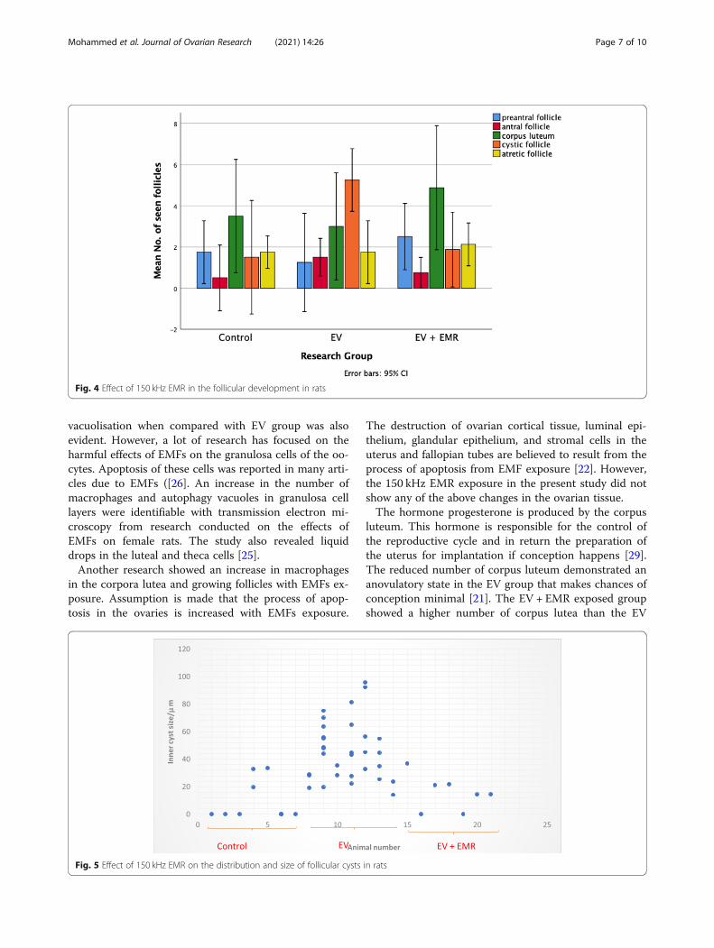
Effect on histomorphometry of ovaryThe histomorphometric analysis of ovarian follicles inthe control, EV and EV + EMR groups are presented in(Fig. 4). In the EV group, a significant decline was ob-served in the number of preandral follicles whereas, thenumber of antral follicle and cystic follicle increased innumber compared with control and EV + EMR groups.The number of atretic follicles did not show any signifi-cant difference among the groups. In EV + EMR group,the number of ovarian follicles at the different stages ofdevelopment were closely similar to the control group.The number of corpus luteum was lower in the EVgroup and highest in the EV + EMR among all threegroups. The mode number of follicular cysts per ovaryin the EV group was higher than all groups as each ratspresented with at least 2 cysts with inner diameter >40 μm. Two rats in the control group were observed tohave cysts with inner diameter < 40 μm as seen in (Fig. 5).The EV + EMR group had an average of 1 follicular cystper animal with inner diameter < 30 μm and some hadno visible follicular cysts.
Effect on gonadotrophic hormonesThe serum concentrations of gonadotropic and sex hor-mones in the control, EV and EV + EMR are presentedin (Fig. 6). There was a significant difference in the LHlevels between control (22.37 ± 9.10 ng/ml), EV (34.66 ±
7.19 ng/ml) and EV + EMR (27.92 ± 8.82 ng/ml) with p =0.04 with an increase in EV group. The LH/FSH ratiowas also significantly (p = 0.05) different among thegroups (control – times 1; EV – times 3 and EV +EV&EMR – times 2). The control group (18.19 ± 10.90ng/ml) showed the highest level of FSH when comparedto the EV group (12.16 ± 5.77 ng/ml) and the EV + EMRgroup (13.30 ± 5.65 ng/ml). There was no significant dif-ference in the testosterone levels among the threegroups as p = 0.66.
DiscussionThe present study successfully induced the pathophysio-logical development of PCOS as identified in humans[5]. Estradial valerate chemically designated as estra-1, 3,5(10)-triene-3,17β-diol 17β-pentanoate, a synthetic ster-ane steroid, long- acting oestrogen on administrationcaused hypothalamic–pituitary dysregulation of GnRH,resulting in improper release and storage of LH [3, 4].The young cyclic adult rats treated with single dose of 4mg of EV per animal on 1st and 14th day of [3] experi-ment in the present study developed a successful poly-cystic ovary which was confirmed by the presence ofnumerous large follicular cysts without an oocyte, re-duced granulosa cells, theca layer hyperplasia and re-duced number of corpus lutea, very much similar towomen PCOS [27]. The vaginal exfoliative cytology, a
Table 1 The effect of 150 kHz EMR on the body and ovarian weights (n = 7)
Parameter Control EV EV + EMR p
Body weight/g 391.19 ± 51.28 283.27 ± 33.33 281.80 ± 23.46 0.14
Weight of left ovary/g 0.07 ± 0.02 0.08 ± 0.01 0.07 ± 0.03 0.74
Weight of right ovary/g 0.06 ± 0.02 0.07 ± 0.01 0.06 ± 0.02 0.40
*The mean difference is significant at the p = 0.05 confidence interval
Fig. 2 Representation of estrous cycle of EV and EV + EMR group for 46 days
Mohammed et al. Journal of Ovarian Research (2021) 14:26 Page 5 of 10

key indicator of ovarian physiology, confirmed that EVtreated rats in the present study were almost acyclic,specifying the presence of cysts contrary to the controlgroup similar to that reported by [10].The EV + EMR exposed groups in the present study
exhibited several positive effects such as, slightly lowerbody weight but not by a significant amount, improvedreproductive cycle, usual morphology of developing folli-cles, increased number of typical developing follicles, re-duction in the mean number and diameter of thefollicular cysts than the PCOS rats (EV group) andclosely similar to the control group. The most important
finding in the study is the reduction in the number offollicular cysts present per animal. The highest numberof cysts in the EV + EMR group was only two cysts peranimal and some had none. This revealed that the EMRmight have reduced the formation of the cysts.The present study revealed that overall follicular dy-
namics was less disrupted in the group exposed to EMRand the observations were very close to the controlgroup. This is because the follicular developmental sta-ging from primordial to secondary was observed to bepresent. The follicular cell differentiation into granulosacells and thecal cells with little to no distortion and
Fig. 3 Photomicrographs of different ovarian follicles in control, EV and EV + EMR groups. The photomicrograph showing the nests of primordialfollicles in the Control(A), EV(b) and EV + EMR (c) groups. The unilaminar primary follicles in the Control (d), EV (e) and EV + EMR (f) groups. Themultilaminar primary follicles in the Control (g), EV (h) and EV + EMR (i) groups. The antral follicles in the Control (j), EV (k) with marked distortedgranulosa and theca layer cells greater than the EV + EMR (l) groups. The matured follicle in the control (m) and EV + EMR (o) groups with lessdistortion of the granulosa cells. The cystic follicle (n) in the EV group with thin layer of granulosa cells. The cross section of the ovary of theControl (p), EV (q) and EV + EMR (r) groups. o- oocytes; f: follicular cells; g granulosa cells; t-thecal cells; a; a-antrum; c-cysts cl; corpus luteum gf;Graffian follicle
Mohammed et al. Journal of Ovarian Research (2021) 14:26 Page 6 of 10
vacuolisation when compared with EV group was alsoevident. However, a lot of research has focused on theharmful effects of EMFs on the granulosa cells of the oo-cytes. Apoptosis of these cells was reported in many arti-cles due to EMFs ([26]. An increase in the number ofmacrophages and autophagy vacuoles in granulosa celllayers were identifiable with transmission electron mi-croscopy from research conducted on the effects ofEMFs on female rats. The study also revealed liquiddrops in the luteal and theca cells [25].Another research showed an increase in macrophages
in the corpora lutea and growing follicles with EMFs ex-posure. Assumption is made that the process of apop-tosis in the ovaries is increased with EMFs exposure.
The destruction of ovarian cortical tissue, luminal epi-thelium, glandular epithelium, and stromal cells in theuterus and fallopian tubes are believed to result from theprocess of apoptosis from EMF exposure [22]. However,the 150 kHz EMR exposure in the present study did notshow any of the above changes in the ovarian tissue.The hormone progesterone is produced by the corpus
luteum. This hormone is responsible for the control ofthe reproductive cycle and in return the preparation ofthe uterus for implantation if conception happens [29].The reduced number of corpus luteum demonstrated ananovulatory state in the EV group that makes chances ofconception minimal [21]. The EV + EMR exposed groupshowed a higher number of corpus lutea than the EV
Fig. 4 Effect of 150 kHz EMR in the follicular development in rats
Fig. 5 Effect of 150 kHz EMR on the distribution and size of follicular cysts in rats
Mohammed et al. Journal of Ovarian Research (2021) 14:26 Page 7 of 10

group and the reproductive cycle was an improved onein this group than the EV group. Morphological atresiawas evident in all three groups. No major comparisonwas observed as follicular atresia is considered an activecellular process. Yet, the susceptibility to programmedcell death at various stages during follicular developmentremains undefined [26].A key factor in hormone function changes and
causes of infertility symptoms in females are the re-sult of neuroendocrine changes caused by the impactof EMFs on females [19]. The decrease in numberpreandral follicles in the PCOS ovaries cause theoverproduction of androgens that impedes with nor-mal follicular maturation process [24]. But the presentstudy did not show significant elevation in the testos-terone level among the all groups. In the presentstudy, the FSH concentrations did not alter but LHconcentrations increased in rats with PCOS, thus thematuration of follicles was impaired and multi-sizedcystic follicles were formed. The LH/FSH ratio in EVgroup and EV + EMR group were also significantlyhigher than the control group. The group exposed toEMR showed an increase in the LH and LH/FSH ra-tio, which is contrary to the reports in a DHEA-induced PCOS rat model [9, 23]. Generally, a highfrequency in gonadotropin-releasing hormone (GnRH)pulses in the hypothalamus leads to LH secretionfrom the pituitary. In this case, increased levels canalso be from accelerated GnRH activity, increased re-sponsiveness to GnRH or decreased sensitivity of thehypothalamus received via negative feedback from sexsteroids [32].
Overall, the reduction in cystic formation from expos-ure to NIR can be a possible avenue for further research.The possible mechanism on which this 150 kHz workscan be linked to some of the Bio-Electromagnetic Princi-ples. One major principle being the sensitivity of recep-tor efficiency on the surface of target cells to signaltransduction [18] but cannot be confirmed in thisexperiment.As alternating electric fields of intermediate frequency
and low intensity, the TTFields have been reported toslow down the growth of tumor cells while having noobvious bioeffects on normal cells [6, 13, 30]. However,this frequency has never been examined during the de-velopment of non-cancerous conditions such as PCO.Hence, the consistency of the present results cannot beconfirmed without results of the mechanistic studies in-volved in cancer cell lines in this frequency.The effect of EMR on cells can be direct as shown by
previous experiments on glioma cell lines [14] but it isunlikely because the same changes in all different layersof different types of cells in ovaries were observed in thepresent study. It can be speculated that there is an indir-ect effect either by the influence of EMR to cell recep-tors or the effect on the hypothalamus and signalling viacertain hormones. Since the present study is focussed onthe effect of 150 kHz on estrous cycle, ovarian histologyand serum levels of gonadotrophic hormones, which isnot strong enough to come to a solid conclusion. So,further investigations are required to assess the effect ofEMR during follicuogenesis of PCO development by in-vestigating the follicular ultrastructure and immunohis-tochemical characterisation of surface receptors of the
Fig. 6 Effect of 150 kHz EMR on serum levels of gonadotrophic hormones in the rats
Mohammed et al. Journal of Ovarian Research (2021) 14:26 Page 8 of 10

granulosa and thecal cells and a detailed study on theHypothalamo-hypopysio-gonal axis which forms thelimitation of this study. Additionally, the study shouldbe explored using various PCO inducing models toevaluate a more definite conclusion on cystic develop-ment. The authors are currently repeating this experi-ment to fully understand the effect of 150 kHz EMR onthe HPG axis.
ConclusionThe 150 kHz EMR appears to have a positive effect likeimproved reproductive cycle, reversal to usual morph-ology of developing follicles, increased number of typicaldeveloping follicles, reduction in the mean number anddiameter of the follicular cysts. However, a more detailedstudy, which includes the limitations as highlighted.
AbbreviationsCL: Corpora lutea; DHEA: Dehydroepiandrosterone; ELISA: Enzyme-linkedImmunosorbent Assay; EM: Electromagnetic; EMF: ElectromagneticFrequency; EMR: Electromagnetic Radiation; EV: Estradiol Valverate;FSH: Follicle Stimulating Hormone; GnRH: Gonadotropin-releasing hormone;H & E: Haematoxylin and Eosin; LH: Luteinizing Hormone; NIR: Non-ionizingRadiation; PCO: Polycystic ovary; PCOS: Polycystic Ovary Syndrome;SD: Sprague Dawley
AcknowledgmentsThe authors wish to express sincere thanks to Department of Physics, andSchool of Veterinary Medicine, The University of the West Indies for thesupport rendered throughout the project. The authors also acknowledge theservices offered by Dr. Jenelle Johnson, in-charge of Lab Animal Facility atSchool of Veterinary Medicine for this study. Authors also acknowledge thelaboratory help from Mr. Lester Gilkes, Dr. Samuel Rampersad and Mr. GeraldChadoo.
Authors’ contributionsSM led the design, conceived the study, performed the experimental work,vaginal cytology, morphometrical analysis and hormone estimation.Compiled and analysed all the results and wrote the original manuscript. VSperformed histology, supervised and edited the manuscript. NZ conceivedthe study, performed induction of EMR system and overall supervision of theexperiment and edited the manuscript. All authors read and approved thefinal manuscript.
Author’s informationStatistical Analysis: Stephanie Mohammed. Email: [email protected] Sharing Statement: All data generated and analysed during this studyare included in this published article.
FundingThe authors thanks to School of Graduate Studies and Research, TheUniversity of the West Indies, St. Augustine, Trinidad and Tobago for thefinancial support through Campus Research and Publication Fund(CRP.5.NOV19.60).
Availability of data and materialsAll data is available for this experiment. It will not be released because thereare other phases of this experiment in progress.
Ethics approval and consent to participateThe Campus Research Ethics Committee of the University of the West Indiesapproved the protocols for animal experimentation (CEC 310/09/17).
Consent for publicationAll author consent for publication of this manuscript.
Competing interestsAll authors declare no conflict of interest.
Author details1Department of Physics, Faculty of Science and Technology, The Universityof the West Indies, St. Augustine, Trinidad and Tobago. 2Department of BasicVeterinary Sciences, School of Veterinary Medicine, Faculty of MedicalSciences, The University of the West Indies, St. Augustine, Trinidad andTobago.
Received: 8 December 2020 Accepted: 25 January 2021
References1. Ahmadi SS, Khaki AA, Ainehci N, Alihemmatic A, Khatooni AA, Khaki A, et al.
Effect of non-ionizing electromagnetic field on the alteration of ovarianfollicles in rats. Electron Physician. 2016;8(3):2168.
2. Blay S, Aguiar JVA, Passos IC. Polycystic ovary syndrome and mentaldisorders: a systematic review and explatory meta-analysis. NeuropsychiatrDis Treat. 2016;12:2895.
3. Brawer JR, Munoz M, Farookhi R. Development of the polycystic ovariancondition (PCO) in the estradiol Valverate-treated rat. Biol Reprod. 1986;35:647–55.
4. Carriere PD, Brawer JR, Farookhi R. Pituitary gonadrtropin-releasing hormonereceptor content in rats with polycystic ovaries. Biol Reprod. 1988;38:562–7.
5. Chaudhari NK, Nampoothiri LP. Neurotransmitter in a testosteronepropionate-induced polycystic ovarian. Horm Mol Biol Clin Investig. 2017;29(2):71–7.
6. Davies AM, Winberg U, Palti Y. Tumor treating fields: a new frontier incancer therapy. Annals of the New York Academy of Science. 2013;1291(1):86–95.
7. Dewailly D, Cortet-Rudelli C, Decanter C. The polycystic ovary syndrome:reproductive aspects. In: Wass JAH, Shalet SM, editors. The Oxford textbookof endocrinology. Oxford UK: Oxford University Press; 2003. p. 1135–43.
8. Ding T, Hardiman PJ, Petersen I, Wang F-F, Qu F, Baio G. The prevalence ofpolycystic ovary syndrome in reproductive-aged women of differentethnicity: a systematic review and meta-analysis. Oncotarget. 2017;8:9635.
9. Francou MM, Durdos M, Salvetli RN, Bravalle C, Rey F, Ortega HH.Characterization of pituitary cell populations in rats with induced polycysticovaries. Cell Tissues Organs. 2008;188:310–9.
10. Kafali H, Iriadam M, Ozardali I, Demir N. Letrozole-induced polycystic ovariesin the rat: a new model for cystic ovarian disease. Arch Med Res. 2004;35(2):103–8.
11. Kakoly AS, Earnest A, Teede HJ, Moran LJ, Joham AE. The impact ofobesity on the incidence of type 2 diabetes among women withpolycystic ovary syndrome. Diabetes Care 2019. https://doi.org/https://doi.org/10.2337/dc18-1738.
12. Kinzel A, Ambrogi M, Varshaver M, Kirson ED. Tumor treating fields forGlioblastoma treatment: patient satisfaction and compliance with thesecondary-generation Optune system. Clinical Medicine Insights Oncology.2019;13.
13. Kirson ED, Dbaly V, Tovarys F, Vymazal J, Soustiel JF, Itzhaki A, et al.Alternating electric fields arrest cell proliferation in animal tumor modelsand human brain tumors. Proc Acad Sci USA. 2007;104(24):10152–7.
14. Lukas B, Almke B, Meshksar S, Dierks A, Majernik GH, Krauss JK, et al.Tumor treating fields (TTFields):investigations on the mechanism ofaction by electromagnet exposure of cells in telophase/cytokinesis. SciRep. 2019;9:7362.
15. Mohammed SB, Nayak BS. Polycystic ovarian syndrome trend in a nutshell.International Journal of Women’s Health and Reproduction Sciences. 2017;5(3):153–7.
16. Mohammed SB, Sundaram V. Comparative study of metachromatic stainingmethods in assessing the Exfoliative cell types during Oestrous cycle inSprague-Dawley laboratory rats. Int J Morphol. 2018;36(3):962–8.
17. Mun EJ, Babiker HM, Weinberg U, Kirson ED, Van Hoff DD. Tumor-treating fields: a fourth modality in cancer treatment. Clin Cancer Res.2017;24:266–75.
18. Neil C. Criticism of health assessment in the ICNIRP guidelines forradiofrequency and microwave radiation (100kHz – 300kHz). Lincoln: NewZeeland Lincoln University 2002.
Mohammed et al. Journal of Ovarian Research (2021) 14:26 Page 9 of 10

19. Nelson JF, Karelus K, Bergman MD, Felicio LS. Neuroendocrine involvementin aging: evidence from studies of reproductive ageing and caloricrestriction. Neurobiol Aging. 1995;16(5):837–43.
20. Orio F, Muscogiuri G, Nese C, Palomba S, Savastano S, Tafuri D, et al.Obesity, type 2 diabetes mellitus and cardiovascular disease risk: an updatein the management of polycystic ovary syndrome. Eur J Obstet GynecolReprod Biol. 2016;207:214.
21. Palomba S, Daolio J, La Sala BG. Oocyte competence in women withpolycystic ovary. Trends Endocrinol Metab. 2017;28(3):186–98.
22. Rad SJ, Rowshangar L, Karimi K. The effect of Electromagnetic field onFallopian Tube. IFFS 2001 Selected free communication. Internationalproceedings division; Melbourne. November 2001:25–30.
23. Rencher FS, Ozbek KS, Eraldemir C, Zehra S, Tugba K, Ceylan S, et al. Effectof resveratrol and metformin on ovarian ultrastructure in PCOS: anexperimental study. Journal of Ovarian Research. 2018;11:55.
24. Rezvanfar M, Ahmadi A, Saadi HS, Baeeri M, Abdollahi M. Mechanistic linksbetween oxidative/nitrosative stress and tumor necrosis factor alpha inletrozole-induced murine polycystic ovary: biochemical and pathologicalevidence for beneficial effect of pioglitazone. Hum Exp toxicol. 2012;31(9):887–97.
25. Roshangar L, Rad SJ. Electron microscopic study of folliculogenesis afterelectromagnetic field exposure. Journal of Reproduction and Infertility. 2004;5(4):299–307.
26. Roshanhar L, Hamdi BA, Khaki AA, Soleimani Rad J, Soleimani RS. Effect oflow frequency electromagnetic field exposure on oocyte differentiation andfollicular development. Adv Biomed Res. 2014;3:76.
27. Schulster A, Farookhi R, Brawer JR. Polycystic ovarian condition in estradiolvalverate-treated rats: spontaneous changes in characteristic endocrinefeatures. Biol Reprod. 1984;31:587–93.
28. Sirmans SM, Pate KA. Epidemiology, diagnosis and management ofpolycystic ovary syndrome. Clin Epidemiol. 2014;6:1.
29. Stocco C, Telleria C, Gibori G. The molecular control of Corpus luteumformation, function and regression. Endocr Rev. 2007;28(1):117–49.
30. Stupp R, Taphoorn M, Driven L, Taillibert S, Chen HT, Paek SSH, et al. TumorTreating Fields (TTfields)- A Novel Cancer Treatment Modality: TranslatingPreclinical Evidence and Engineering into a Survial Benefit with DelayedDecline in Quality of Life. International Journal of Radiation Oncology.Biology. Physics. 2017:99(5).
31. Tayefi NH, Gavami M, Aknarzadeh A, Beheshti R, Mohammednejad D,Abedelahi A. Preservation of mouse ovarian tissue follicle morphology andultra-structure after vitrifying in biotechnological protocols. J Ovarian R.2015;8:7.
32. Teharni FR, Noroozzadeti M, Zahediasl S, Piryaei A, Azizi F. Introducing a ratmodel of prenatal androgen-induced polycystic ovary syndrome inadulthood. Exp Physiol. 2014;99(5):792–801.
33. Vergote I, Copeland L, Monk B, Coleman R, Cibula D, Sehouli J, et al.P154 tumor treating fields (200kHz) concomitant with weekly paclitaxelfor platinum-resistant ovarian cancer: phase 3 INNOVATIVE/ENGOT-OV50study 2019.
34. Wdowiak A, Mazurek P, Wdowiak A, Bojar I. Effect of electromagnetic waveson human reproduction. Ann Agric Environ Med. 2017;24(1):13–8.
Publisher’s NoteSpringer Nature remains neutral with regard to jurisdictional claims inpublished maps and institutional affiliations.
Mohammed et al. Journal of Ovarian Research (2021) 14:26 Page 10 of 10