Embed Size (px)
Citation preview

EEG

These are neurons. Your brain has hundreds of billions of them!
Diagram of a neuron. A group of real neurons.

Brainwaves (EEGs) reflect the brain’s electrical activity. A neuron at rest is like a little battery. Whenever a neuron is active, its voltage briefly changes.
When a neuron is active, its voltage may change by 100 mV or more.
Electrical activity in a single neuron.

When millions of neurons fire at the same time, they may produce electrical activity detectable to an electrode placed on the head.
Two illustrations of the brain producing electricity.
Two real human brains. The left image is an MRI.

For example, if you hear a tone, many different groups of neurons fire as your brain processes that tone. EEGs can tell us when and where these groups of neurons fire. Doctors often use this technique to diagnose hearing disabilities, since EEGs can reveal which groups of neurons are damaged.

This figure shows some of the EEGs evoked by a tone. Early responses (within 0.1 seconds of the tone) are very consistent. Later EEG components may vary depending on whether you ignored the tone, if it was meaningful to you, if you expected it, and other factors.

Most EEG studies use an electrode cap. This is a special cap that contains electrodes at certain locations over different areas of the head.
On rare occasions, doctors may need to use surgery to implant an electrode inside the skull to get better recordings. This is only done when medically necessary. For example, doctors may need to know exactly what area of a patient’s brain is creating seizures.
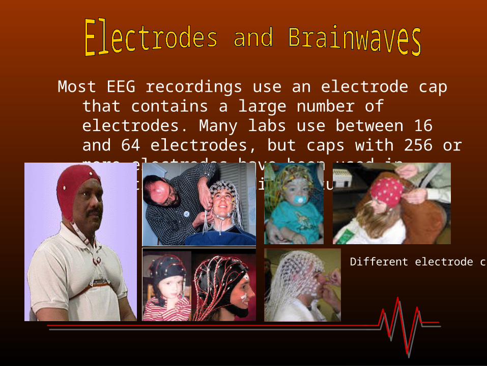
Most EEG recordings use an electrode cap that contains a large number of electrodes. Many labs use between 16 and 64 electrodes, but caps with 256 or more electrodes have been used in scientific and medical studies.
Different electrode caps.

Most electrode caps are designed with electrodes over specific areas of the skull (and thus specific areas of the brain). Otherwise, you would be recording from different brain areas each time you use a cap.
These are standardized electrode locations, called the International 10-20 system.

Scientists run subjects all the time in EEG experiments. Subjects may be paid, they may volunteer, or they may receive class credit for participating.
Before preparing a subject for EEG recording, s/he is shown the lab and the equipment, and is asked to sign a consent form agreeing to be in the study. This is very important. Scientists are required to get “informed consent” from subjects. After this, the preparation begins….

1. It is often necessary to place an electrode on or behind the ear before donning the electrode cap. Scientists often clean the area behind the ear with rubbing alcohol. Some people put electrodes near the eye to detect blinking and other eye movements.
2. The scientist measures the subject’s head and then places the correct sized cap on his head.

3. Electrode gel is then placed between each electrode and the scalp to get a good connection. Everyone agrees that electrode gel in your hair is a wonderful experience.
Two types of electrode gel. Squirting gel under an electrode.

4. The scientist checks the cap to make sure there is a good connection between each electrode and the brain.
5. The subject is now ready for recording! A typical recording session lasts about an hour. It takes roughly 30 minutes to prepare a subject for recording, depending on the number of electrodes, the subject’s hair, the scientist’s skill, type of electrode cap, and other factors.

6. After recording, the cap is removed. Electrode gel washes out easily with water, so many subjects rinse or wash their hair after a recording session. Of course, smart people know that electrode gel in your hair makes you cool.
7. That’s it! The subject is done, but the scientist now has data to analyze.

Most people have heard of free-running EEGs. These are naturally produced, rhythmic brainwaves that do not require outside activity.
Well known free running EEGs include:Delta (1-4 Hz), found in deep sleepTheta (4-8 Hz), found in sleep, meditation, hypnosisAlpha (8-14 Hz), indicate relaxation and closed eyesMu (8-14 Hz), largest when individual is not moving Beta (non specific higher frequencies), indicate alertness

This graph shows about four seconds of EEG from a human subject. Each of the 15 lines represents a different electrode site. This has a lot of alpha activity (about 10 waves per second), meaning the subject was probably awake but drowsy with eyes closed. Again, alpha waves are a type of free running EEG

However, people sometimes are interested in the brain’s response to a certain event. For example, if someone touches your hand or plays a tone, your EEG will change as your brain processes that event.
The technical term for EEG activity based on a specific event is an event related potential (ERP). One common bump is called the P300, named because it starts about 300 milliseconds after an event.

These two figures show responses to flashes. Specifically, they show how the brain responds differently to flashes people notice compared to flashes they choose to ignore. In each graph, the relatively flat lines (red or dashed) show the brain’s response to ignored flashes. Notice the large bump starting around 300 milliseconds in the other lines (blue or solid), showing the response to flashes people counted. These are examples of the P300, a type of Event Related Potential (ERP).
Top figure: from Allison thesis (Allison 2003)
Bottom figure: from Bayliss thesis (Bayliss 2001)

There are other technologies for studying brain activity. Two well known techniques are PET and fMRI. These approaches provide different information than EEGs. EEGs are very good at telling when a brain area was active, but are poor at finding exactly where in the brain the signal came from. EEG recording equipment is also much cheaper, easier to use, and more portable that the tools needed for PET or fMRI.
Some people use EEGs in combination with fMRI. This can be a very powerful tool for finding exactly when and where something occurs.

Richard Caton was the first person to record electrical activity from the brain in 1875.
Dr. Caton placed electrodes on the surface of rabbit and monkey brains. He found that this activity changed in response to flashing lights.

The first person to record brainwaves from humans without surgery was Hans Berger.
Dr. Berger began studying the EEG in 1924, and first published his results in 1929.
Doctor Hans Berger

Though Berger lacked modern electronics, he made many important discoveries. He showed that attention affects alpha and beta waves.
Dr. Berger’s EEG recording apparatus Dr. Berger’s electrode cap
Pure research: EEGs help us learn more about when, where, and how different brain areas work together when thinking, speaking, responding to tones, etc.

Medical: Isolate areas or processes that respond slowly, improperly, or not at all. Detect onset of seizures, strokes, psychotic episodes, or other problems. Enable communication for severely disabled with brain computer interface systems. Train kids with ADD to pay attention longer. Study the effects of drugs over time.

Entertainment/relaxation: Some people have used EEG systems designed for alpha biofeedback. This means that you train yourself to have more alpha waves in your EEG, which helps some people relax. EEGs can be used to play simple games or make music.

Passive monitoring: There has been a lot of progress recently in alertness monitors based on the EEG. These might warn people they are dozing off. This has been proposed for people in “attention-critical” situations like nuclear plant technicians, sonar operators, security guards, and truck drivers.

Any questions ?