Embed Size (px)
Citation preview
EEG and Clinical Neurophysfatogy Proceedings of the 2nd European Congress of EEG and Clinical Neurophysiology, Salzburg, Austria, September 16-19, 1979
E d i t o r s : H. Lechner and A. Aranibar
EXCERPTA MEDICA, Amsterdam-Oxford-Princeton

© Excerpta Medica 1980 All rights reserved. No part of this publication may be repro-duced, stored in a retrieval System or transmitted, in any form or by any means, electronic, mechanical, photocopying, recording or otherwise, without permission in writing from the publisher.
International Congress Series No. 526 ISBN Excerpta Medica 90 219 0457 8 ISBN Elsevier North-Holland 0 444 90172 8
P u b l i s h e r : Excerpta Medica 305 Keizersgracht 1000 BC Amsterdam P.O. Box 1126
S o l e D i s t r i b u t o r s f o r t h e U S A a n d C a n a d a : Elsevier North-Holland Inc. 52 Vanderbilt Avenue New York, N.Y. 10017
Printed in The Netherlands by Casparie-Amsterdam
2nd European Congress of EEG and Clinical Neurophysiology
Organized by: The Austrian Society of EEG and Clinical Neurophysiology Presidents: Prof. Dr. H. Lechner
Prof. Dr. K. Pateisky Prof. Dr. E. Deisenhammer
Secretaries: Prof. Dr. E. Scherzer OA. Dr. S. Enge
Treasurer: Dr. H. Feldner-Bustin
Organizing Committee of the Ist European Congress of EEG and Clinical Neurophysiology President: Prof. Dr. E. Lugaresi Secretary: Prof. Dr. R. Muntani
ORGANIZING COMMITTEE Board: Prof. Dr. H. Lechner, Graz Prof. Dr. K. Pateisky, Vienna Prof. Dr. E. Scherzer, Vienna Secretarial Committee: Dr. A. Aranibar, Graz Mrs. Ch. Sulzer, Graz
v

Contents
I. Position of EEG in diagnosis and treatment of epilepsy with consideration of findings of computerized axial tomography
The value of CT and EEG in the diagnosis of epilepsy G. L a d u r n e r , R. M a r t i s c h n i g , A . Ära n i b a r , M . J . M o h s e n , W. D . Sager a n d H . L e c h n e r 3
EEG and computerized transaxial tomography in patients with temporal lobe epilepsy
G. S c o l l o - L a v i z z a r i a n d Ch. B a l m e r 11 Correlative EEG and CT findings in epilepsies of early and late onset
M . S c h u m a c h e r , F. Schümm a n d H . D . L a n g o h r 16 Correlations between computerized tomography, EEG and clinical findings in patients with seizures
F. L . Glötzner, R. Geiser a n d M . Ratzka 32 Value of EEG and CT in post-traumatic epilepsy
P. Wesse/y, Th. Reisner a n d K. Zeiler 37 Post-traumatic early onset epilepsy with consideration of Computer tomography
W.l. S t e u d e l , J. Krüger, Th. Göller a n d H . C h . Grau 42 EEG and Computer tomography in post-traumatic epilepsy
G. B e r t h a , G . L a d u r n e r , H . L e c h ner a n d W . D . S a g er 50
II. Value of EEG in the Classification, treatment and follow-up of epilepsies
Value of ictal EEG in diagnosis and Classification of epileptic patients L . Oller-Daurella a n d L . Oller 57
Ictal "psychical phenomena" and stereo-electroencephalographic findings
H . G. Wieser 62 Diagnostic value of EEG in spontaneous sleep following sleep deprivation in epilepsy
B . Clemens a n d I. M e z e y 1 1 Long-term EEG and video monitoring in epilepsy
C D . B i n n i e , J. O v e r w e g a n d A . J . R o w a n 83 Electroneurophysiological evaluation of clonazepam as antiepileptic
A . C . Dec/erck 89 EEG diagnosis and incidence of epilepsy in children with cerebral palsy
H . Bauer 100 CEEB (CT, EEG, ECHO-EG, brain scan) in epilepsies related to etiologic macrofactors
F. Hajnsek, N . G u b a r e v a n d Z C e m a l o v i c - B o k o 106
ix

Paroxysmal development in a case of malformation of hypothalamus J . H a n n 116
IM. EEG in the study of cerebrovascular disorders
Quantitative EEG in cerebral ischemia. A . Parameters for the detection of abnormalities in "normal" EEGs in patients with acute unilateral cerebral ischemia (A.U.C.I.)
A . C . van H u f f e i e n , D . C . J . P o o r t v l i e t , C . J . M . van der Wu/p a n d 0. M a g n u s 125
Quantitative EEG in cerebral ischemia. B. Parameters valuable for follow-up of patients with acute unilateral cerebral ischemia (A.U.C.I.)
A . C . van H u f f e i e n , D . C . J . P o o r t v l i e t , C . J . M . van der Wu/p a n d 0. M a g n u s 131
Quantitative EEG in cerebral ischemia. C. The significance of photic Stimulation (PS) in patients with acute unilateral cerebral ischemia (A.U.C.I.)
A . C . van H u f f e i e n , D . C . J . P o o r t v l i e t , C . J . M . van der Wu/p a n d 0. M a g n u s 138
Correlation of EEG and CT findings with cerebral blood flow measurements in patients with cerebrovascular disorders
E. O t t , H . L e c h n e r , K. M a r g u c , G. B e r t h a , A . A r a n i b a r , G. L a d u r n e r a n d W . D . Sager 143
Correlation between computerized tomography and EEG findings in acute cerebrovascular disorders
G . A . O t t o n e l l o , G. R e g e s t a a n d P. T a n g a n e l l i 148 Correlations and discrepancies between clinical aspects, EEG and CT brainscan data in ischaemic cerebral disease
J . H . A . van der D r i f t , S . L . Visser, E . J . J o n k m a n a n d A . v . d . S t e e n 163 Clinical value of EEG in transient ischemic attacks
S. Enge, H . L e c h n e r , Ch. L o g a r a n d G. L a d u r n e r 173 Cerebral refractory period of somatosensory System. EEG and clinical findings before and after vascular surgery in cerebrovascular disease
J. Jörg, H . M . M e h d o r n a n d R. P o d e m s k i 181 Value of homonymous Visual and somatosensory evoked potentials compared to EEG in ischemic hemispheric Syndromes
R. P o d e m s k i , J. Jörg a n d H . J . L e h m a n n 191 Computer EEG analysis of sensorimotor functions: Results on hemiplegic patients
G. Harrer, G. P f u r t s c h e l l e r , H . Harrer a n d W. P r i b y l 201 EEG study in 51 cases of intracerebral hematoma
M . Va/uet a n d D . S a m s o n - D o l l f u s 209 Auditory evoked potentials to verbal Stimulation in focal cerebrovascular disorders
x
S. P o p o v a n d D . T s c h a v d a r o v 212 Clinical value of computerized driving analysis (CDA) of photic following in cerebrovascular accident
B . Barac, V. Brinar a n d V. I l g u m 219 Value of EEG and computerized tomography in ischemic cerebrovascular disorders
A . M o g l i a , A . Tartara, M . M o l a , R. M a n n / , F. R o g n o n e a n d A . M a r t e l l i 226
EEG and cerebrospinal fluid lactate in recent ischemic stroke G. Prüll a n d 0. B u s s e 231
Analysis of spontaneous sleep in ischemic cerebrovascular disease and brain stem lesions
0. Zsadänyi a n d Gy. B e r e c z 241 Frontal intermittent rhythmic delta activity in ischemic brainstem disorder
H . Walser a n d H . /s/er 247 Electroclinical Symptoms in patients with epilepsy of cerebrovascular origin
P. Tariska, P. Rajna a n d G. Gereby 254 EEG findings in migraine accompagnee
K. Christian/, B . Volker a n d D . S o y k a 259 Serial EEG records during migrainous attacks
H . M a t t h i s , P. P e r r i a u d , M . J e k i e l a n d A . B e a u m a n o i r 267
IV. EEG in children and developmental disorders
Electrophysiological characteristics of attention as an index of functional maturation of the brain in children
N . V. D u b r o v i n s k a y a 275 Age dependent differences in EEG power spectra of dyslexic children in relation to normals
E. C o l o n , S. N o t e r m a n s a n d J. d e Weerd 283 Auditory evoked brainstem responses in the evaluation of children with severe speech impairment
R . J . M c C l e l l a n d , I. L y n e s s a n d R . S . M c C r e a 289 EEG, Visual evoked potentials, somatosensory evoked potentials and nerve conduction velocity in a family with adrenoleukodystrophy
B. M a m o l i , M . Graf, K. Toifl a n d P. D a l - B i a n c o 299 Propagation defects in somatosensory evoked responses in children
£ C o l o n , W. Renier a n d F. Gabreels 306 Occipital reflex spikes and their evolution in healthy children
P. D e M a r c o 314 EEG in 3 to 4 year-old children exposed to risk of asphyxia at birth
H . K l e p e l 321
xi
Multivariate analysis indicating relations between cerebral lateralization (as measured by EEG, EP, and CNV) and IQ scores, for normal and MCD children
L . J . R o g e r s , A . K . M c B u r n e y , H . G . D u n n , J . U . C r i c h t o n a n d M . D . L o w 329
The EEG profile J . L . B l o m a n d K. M e c h e l s e 338
EEG correlates of System Organization of brain integrative activity in ontogenesis and possibilities to use them as age and individual characteristics of children
D . A . Farber 346 Specific manifestation of genotypic factors in parameters of Visual evoked responses
T . M . M a r y u t i n a 354 Clinical use of auditory brainstem responses in premature and newborn infants
P . A . Desp/and 360
V. EEG monitoring in head trauma The prognostic significance of the EEG in the initial phase after severe cranial trauma
M . Egli a n d F. K u n z 369 Combined EEG-CT examinations in early phase of traumatic brain stem lesions
J. Krüger, W.l. S t e u d e l a n d H . C h . Grau 377 Clinical and EEG findings in the first ten days of traumatic coma: 24-hour recording studies
P. M a n g i n , J. Krieger, J. K o w a l s k i , J . P . D u p e y r o n , T. P o t t e c h e r a n d D . K u r t z 392
Heart rate studies in association with EEG as a means of following the progress of head injuries
B . M . Evans 403 Frequency analysis of EEG background activity in children with head injury
H . F i c h s e l a n d H . C. Söding 409 EEG in cases of post-traumatic headache
A . Tartara, A . M o g l i a , M . M o l a a n d F. S a v o l d i 417 Patients with severe brain injury committed to a Psychiatric hospital
R. D e S m e d t , E. R o d r i g u s , R. D e b a n d t , J. S e r v a i s a n d L . Van Eyken 423
xii

VI. Evoked responses
Maps of visually evoked multichannel EEG potential fields D . L e h m a n n a n d W. S k r a n d i e s 427
Effects of intravenous Clonazepam on cortical somatosensory evoked responses (SER) in dyssynergia cerebellaris myoclonica (Ramsay-Hunt Syndrome)
F. M a u g u i e r e a n d J. C o u r j o n 433 Computer EEG analysis of sensorimotor functions: Methodological background
G. P f u r t s c h e l l e r , H . M a r e s c h a n d W. P r i b y l 445 Flash and pattern-reversal Visual evoked responses in retrobulbar-neuritis and controls: A comparison of conventional and TV Stimulation techniques
K. L o w i t z s c h , H . D . R u d o l p h , D . Trincker a n d E. Müller 451 Components of the visually evoked subcortical potential (VESP) to flash Stimulation in man
G . F . A . H a r d i n g a n d M . P . R u b i n s t e i n 464 Correlations between evoked potentials during different stages of perception of electrical Stimuli
V . B . S t r e l e t s A l b Visual evoked potential research of emotional reaction mechanisms typical of people with increased level of anxiety
A . A . I b a t u l l i n a 480 Evoked instantaneous coherency increase in the electrical activity of the cat brain upon sensory Stimulation
P. U n g a n a n d E. B a s a r 485
VII. Special neurophysiological studies
The relation of certain EEG phenomena with age and sex 0. M a g n u s a n d L . P o n s e n 507
Quantification of the EEG for clinical application, based on spectral analysis of the ongoing activity
0. M a g n u s , A . C . van H u f f e i e n , D . C . J . P o o r t v l i e t a n d C . J . M . van der Wu/p 518
Clinical, EEG and Computer tomographic findings in herpes simplex encephalitis
W. H a c k e , H . Z e u m e r a n d G. F r e u n d 531 The EEG in a group of centenarians
D2. K a n t a r d z i c a n d M . G a v r a n o v i c 545 Electroencephalographic and neuropathologic correlations in dementias
G. Gereby 553
Computer EEG analysis in assessment of effect of nootropic drugs G. D o l c e , V. C e c c o n i , G. C r u c c u , P. Pola, F. Vigevano a n d A , Z a m p o n i 560
Quantitative determination of respiratory cardiac arrhythmia in neurological diagnosis
G. R a b e n d i n g , G. R e i c h e l , H . Klöckner a n d E. Grimmberger 565 Diazepam in clinical electroencephalography: Effect on abnormal slow activity
Z M a r t i n o v i c 570 Computerized EEG analysis in the management of comatose patients: An operational proposal
W. G. S a n n i t a a n d G. R o s a d i n i 583 Prevalence of EEG abnormalities on the left side: A Statistical analysis
G. F i l l i g o i , E. B e r t i n i , A . B i a n c h i , A . B o l l e a , N . D a g n i n o , P. Gabriele, M . M a n f r e d i , G. S i d e r i a n d M . T o n d i 592
EEG changes during adaptation to high temperature Z T. T u r s u n o v a n d N . Tahirova 595
Computerized EEG and prediction of psychopathology after open heart surgery
W. S p e h r a n d P. Götze 603 Dynamics of spatial phasic relationship of bioelectrical potentials in normal subjects and Schizophrenie patients
N . E . Sviderskaja 611 A study of inter and intra individual variability of the EEG of 16 normal subjects by means of segmentation
B . H . J a n s e n , A . H a s m a n , R. L e n t e n a n d S . L . Visser 617 Neurophysiological mechanisms of amblyopia
L . A . N o v i k o v a , N . N . Z i s l i n a , L G . K u m a n , V . A . Tolstova a n d L . l . F i l c h i k o v a 629
Sonomotor reflex as a descending control mechanism in the auditory System in man
D . F i a l k o w s k a , G. J a n c z e w s k i , A . K u k w a , K. K o c h a n e k a n d P. D o b r z y h s k i 637
Electrophysiological studies of patients with focal lesion of sensorimotor area during real and imagined hand movement
L . M . P u c h i n s k a y a 644 Pattern sensitivity: The role of movement
C D . B i n n i e , A . J . Wi/kins a n d C E . Darby 650 Nervous and Visual effects of occupational n-hexane exposure
A . M . Seppäläinen, C. R a i t t a a n d M . S . H u u s k o n e n 656 Fluctuations of electronystamogram parameters throughout the day
B . H o f f e r b e r t h a n d M . Dessauer 662 Peripheral neuropathy at onset of insulin dependent diabetes
G. C o m i , M . R o d o c a n a c h i , L . L o z z a , L . Beccaria, R. Vanini a n d V. S a i b e n e 666
xiv

Electromyographic, electroneurographic and electron microscopic observations in predominantly juvenile collagenosis
G. S i t z e r a n d G. G. B r u n e 670 Value of Single fiber myography in ocular forms of juvenile myasthenia gravis
G. Sitzer a n d G. G. B r u n e 681 Role of EEG rhythms in the generation of neuronal Signals of Visual perception
V . A . L u t s k y 688 Relationship of EEG rhythms to Short term memory mechanisms
A . N . L e b e d e v 691 Components of evoked hyperpolarization in the spinal motoneuron of the cat
B . S u t o r a n d J . Vieth 694 Excitatory and inhibitory interneurons in the cortex of the cat
J. Vieth a n d J. Faust 703 Analysis of spread of acetylcholine-induced seizures
Ff. Vollmer, I. Szirmai a n d P. R a p p e l s b e r g e r 715
VIII. Brain electrical potentials and human higher nervous activity
Evoked potentials and mental processes A . M . I v a n i t s k y 727
Late slow waves and their relationship to decision and action W. C. M c C a l l u m a n d S . H . Curry 733
Hemispheric asymmetry of cortical visual evoked potentials to neutral and emotional Stimuli
E . A . K o s t a n d o v 740 Stereoelectroencephalographic studies on event related slow potentials in the human brain
E. G r o l l - K n a p p , J. Gang/berger, M . H a i d e r a n d H . S c h m i d 746 Activating mechanisms of reticular influence on cerebral cortex
M . N . L i v a n o v 763 Anticipatory unit responses of human deep brain structures during initiation of voluntary movements and Performance of mental tests
S . N . Rayeva 770
List of contributors 777 Index of authors 789
xv

COMPONENTS OF EVOKED HYPERPOLARIZATION IN THE SPINAL MOTONEURON OF THE CAT
B. Sutor and J . Vieth
Department of Experimental Neuropsychiatry, University Neurology C l i n i c , Erlangen, F.R.G.
By means of in t racel lu lar recordings from pyramidal ce l ls of the cat 's sensorimotor cortex, we have described two kinds of evoked hyperpolarization (1,2). Depending on the Stimulus intensity , a short last ing hyperpolarization, up to 100 msec, and a long last ing one,longer than 100 msec,can be evoked. By a r t i f i c i a l a l terat ion of the mem-brane potential and combination of dif ferent Stimulus conditions, these two kinds of evoked hyperpolarization have been shown to depend on dif ferent mechanisms. The question arose, i f the spinal motoneurons of the cat behave s imi lar ly to cor t ica l pyramidal c e l l s .
MATERIAL AND METH0DS
The experiments were carried out under l ight pento-barbital anaesthesia, immobilization with gallamin and a r t i f i c i a l respirat ion. After a laminectomy from L2 to S1, the spinal cord was covered with paraff in o i l held con-stantly at 37°C. Then the dura mater was opened. Stimulat ion to evoke a response in motoneurons was done at the transsected ventral and dorsal roots of the lumbar Segments L6 and L7. A l l recordings were made in the ventral horn of the lumbar segments L6 or L7.
RESULTS
Figure 1 shows an in t race l lu lar recording of a moto-neuron in lumbar segment 7 during the Stimulation of the dorsal root L6. With a Stimulus intensity of 0.4 mA no response could be seen. A Stimulus intensity of 0.5 mA, however, evoked an EPSP followed by a 75 msec last ing hyperpolarization. Further increase of the Stimulus intens i ty also increased the duration of the hyperpolarization. With 0.9 mA a threshold-l ike Prolongation of the hyperpola r i za t ion , up to 148 msec, appeared. Further increase of the Stimulus intensity did not have any effect on the duration. Thus, also in spinal motoneurons we were able to dist inguish two kinds of evoked hyperpolarization: F i r s t l y , a short las t ing , Stimulus dependent hyperpolarization of a duration up to 100 msec and a threshold between 0.4 and 0.5 mA. Secondly, a long last ing evoked hyperpolarization with a duration of more than 100 msec
694

which was not stimulus-controlled and with a threshold between 0.8 and 0.9 mA.
Figure 1. Intracel lular recordings of a motoneuron of spinal cord segment L7 of cat. Short and long last ing hyperpolarization as response to Single e lec t r ic Stimulus of dorsal root L7. Stimulus intensi t ies are indicated in mA. The Stimulation threshold of the short last ing hyperpolarizat ion is between 0.4 and 0.5 mA. Further increasing intensity causes also an increase of duration of hyperpolar izat ion. Between 0.8 and 0.9 mA a l imit is reached, when further increase of intensity has no effect on the duration. The duration remains constant.
To analyze the components of the different kinds of evoked hyperpolarization, the membrane potential was a l tered a r t i f i c i a l l y by means of current in ject ion.
Figure 2 shows recordings of short last ing evoked hyperpolarization in a lumbar motoneuron under normal conditions and under a r t i f i c i a l current in ject ion. The
695

60 mV
Figure 2. Intracel lular recordings of a motoneuron of spinal cord segment L7 of cat. Short last ing hyperpolarization, e l i c i t e d by an e lect r ic Single Stimulus of dorsal root L7. Change of membrane potential by a r t i f i c i a l a l te r -at ion. co = control recording; de = recording with a r t i f i c i a l depolarization; hy = recording with a r t i f i c i a l hyperpolarization; Eipsp = IPSP equilibrium potent ia l . In trace hy the Eipsp has been surpassed and the hyper-polariz ing IPSP potential changed to a depolarizing one, and the s l ight ly depolarized potential of the d i s i n h i b i -t ion changed to a hyperpolarizing one. The recordings have been arranged without reference to absolute potential values.
696
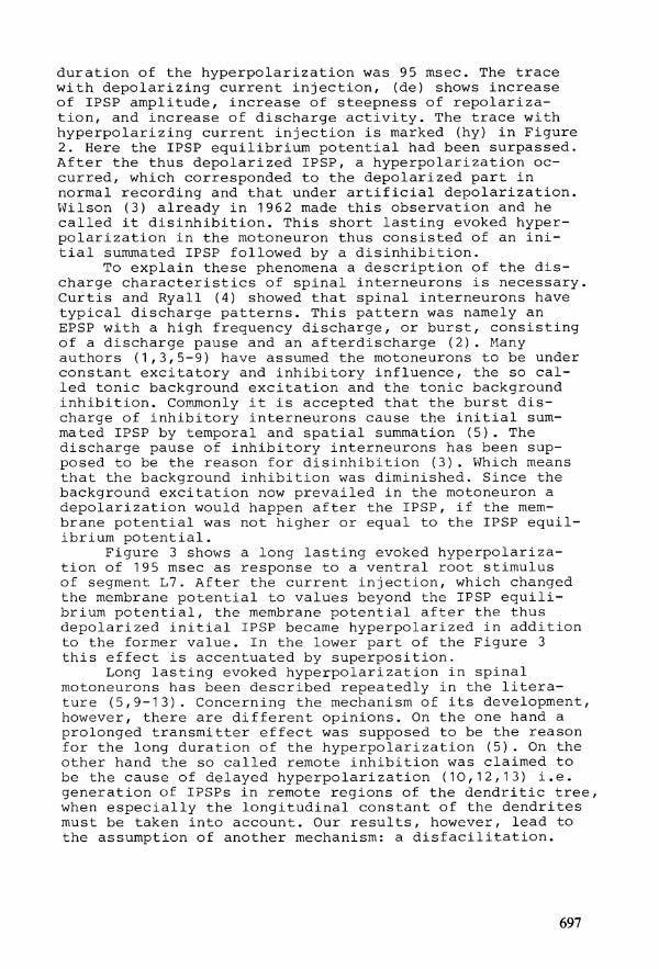
duration of the hyperpolarization was 95 msec. The trace with depolarizing current in ject ion, (de) shows increase of IPSP amplitude, increase of steepness of repolar iza-t ion , and increase of discharge ac t iv i ty . The trace with hyperpolarizing current inject ion is marked (hy) in Figure 2. Here the IPSP equilibrium potential had been surpassed. After the thus depolarized IPSP, a hyperpolarization oc-curred, which corresponded to the depolarized part in normal recording and that under a r t i f i c i a l depolarization. Wilson (3) already in 1962 made this Observation and he cal led i t d is inh ib i t ion . This short last ing evoked hyperpolar izat ion in the motoneuron thus consisted of an i n i t i a l summated IPSP followed by a d is inh ib i t ion .
To explain these phenomena a description of the d i s charge character ist ics of spinal interneurons is necessary. Curtis and Ryall (4) showed that spinal interneurons have typical discharge patterns. This pattern was namely an EPSP with a high frequency discharge, or burst, consisting of a discharge pause and an afterdischarge (2). Many authors (1,3,5-9) have assumed the motoneurons to be under constant excitatory and inhibitory influence, the so c a l led tonic background excitat ion and the tonic background inh ib i t ion . Commonly i t is accepted that the burst d i s charge of inhibitory interneurons cause the i n i t i a l summated IPSP by temporal and spat ial summation (5). The discharge pause of inhibitory interneurons has been sup-posed to be the reason for d is inh ib i t ion (3). Which means that the background inhib i t ion was diminished. Since the background excitation now prevailed in the motoneuron a depolarization would happen after the IPSP, i f the mem-brane potential was not higher or equal to the IPSP equ i l ibrium potent ia l .
Figure 3 shows a long last ing evoked hyperpolarizat ion of 195 msec as response to a ventral root Stimulus of segment L7. After the current in ject ion, which changed the membrane potential to values beyond the IPSP e q u i l i brium potent ia l , the membrane potential after the thus depolarized i n i t i a l IPSP became hyperpolarized in addition to the former value. In the lower part of the Figure 3 this effect is accentuated by superposition.
Long last ing evoked hyperpolarization in spinal motoneurons has been described repeatedly in the l i t e r a -ture (5,9-13). Concerning the mechanism of i ts development, however, there are di f ferent opinions. On the one hand a prolonged transmitter effect was supposed to be the reason for the long duration of the hyperpolarization (5). On the other hand the so cal led remote inhib i t ion was claimed to be the cause of delayed hyperpolarization (10,12,13) i . e . generation of IPSPs in remote regions of the dendrit ic tree, when especial ly the longitudinal constant of the dendrites must be taken into account. Our resul ts , however, lead to the assumption of another mechanism: a d i s f a c i l i t a t i o n .
697

80 mV
80 mV
Figure 3. Intracel lular recordings of a motoneuron of the spinal cord segment L7 of cat. The long last ing hyperpolar izat ion, e l i c i t e d by an e lec t r ic Single Stimulus, of the ventral root L7. Change of the membrane potential by a r t i f i c i a l a l terat ion. Abbreviations as in Figure 2. The lower trace (co/hy) is a protection of the upper traces one upon another to c la r i f y the ef fect . In trace hy the Eipsp has been surpassed and the hyperpolarizing IPSP potential changed to a depolarizing one and the hypolar i -zing part after the IPSP is hyperpolarized in addit ion. The recordings have been arranged without reference to absolute potential values.
698
This assumption is possible , since injected hyperpolar-iz ing current caused an additional hyperpolarization of the evoked hyperpolarization following the IPSP, which i t s e l f was changed to a depolarizing potent ia l . Conduc-tance measurements of the membrane only showed meaningful increases during the i n i t i a l IPSP, but not during the late part of the evoked hyperpolarization. If one assumes a prolonged transmitter e f fect , also during the late part then the conductance should be increased.
Against the assumption of a prolonged transmitter e f fec t , also the addit ional hyperpolarization must be mentioned, which does not f i t for an inh ib i t ion . If one assumes the late part of the long last ing evoked hyperpolar izat ion to be originated by remote inhibitory synap-ses, this hyperpolarization should decrease. It should even be reversed just as the i n i t i a l IPSP. Therefore we conclude that this hyperpolarization was a d i s f a c i l i t a -t ion .
Tonic background excitat ion as well as tonic background inhib i t ion are probably due to the act iv i ty of spinal interneurons (6-8,14). Since the d i s f a c i l i t a t i o n is an interruption of excitatory synaptic influence on the motoneuron, we may conclude that this interruption would predominantly be due to a discharge pause in exc i tatory interneurons. The threshold of interruption of tonic background excitat ion is higher than that of the tonic background inh ib i t ion , because higher Stimulus intensi t ies must be used to obtain a d i s f a c i l i t a t i o n . At the time we are not able to dist inguish between excitatory and inhibitory interneurons in the spinal cord, but in our recordings of 35 spinal interneurons we always saw, with weak and with strong St imuli , after the burst d i s charge a discharge pause. And after strong Stimuli which caused a d i s f a c i l i t a t i o n in motoneurons, we always ob-served in interneurons a long discharge pause correspond-ing to d i s f a c i l i t a t i o n . Thus we have to assume that during d i s f a c i l i t a t i o n also d is inh ib i t ion s t i l l ex is ts .
DISCUSSION
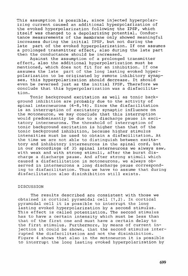
The results described are consistent with those we obtained in cor t ica l pyramidal c e l l (1,2). In cor t ica l pyramidal c e l l i t is possible to interrupt the long last ing evoked hyperpolarization by a second Stimulus. This effect is cal led potentiat ion. The second Stimulus has to have a certain intensity which must be less than that of the f i r s t one and must have a certain delay to the f i r s t Stimulus. Furthermore, by means of current i n jection i t could be shown, that the second Stimulus inter-rupted the d i s f a c i l i t a t i o n and not the d is inh ib i t ion . Figure 4 shows that also in the motoneuron i t is possible to interrupt the long last ing evoked hyperpolarization by
699

100 mV
-Figure 4. Intracel lular recordings of a motoneuron of spinal cord segment L7 of cat. Potentiation of the evoked long last ing hyperpolarization (upper trace; Stimulus of 3.0 mA) during the late part by a second shock of 1.0 mA at the dorsal root L6 (middle trace) . The delay between either Stimuli is 80 msec. The lower trace shows the response to the potentiating Stimulus, when i t i s applied alone.
700

a second Stimulus. The intensity of this Stimulus must be between 0.8 and 1.1 mA, in other words, i t must be weaker than the f i r s t one. The delay to the f i r s t Stimulus must be at least between 70 and 80 msec. This means that poten-t ia t ion only can be obtained, when no IPSP is present anymore. These values are independent of the intensity of the f i r s t Stimulus. It is only necessary that the f i r s t Stimulus has at least intensity to evoke a long last ing hyperpolarization. That means, that a d i s f a c i l i t a t i o n ex is ts . In most cases the second Stimulus applied alone only evoked a short last ing hyperpolarization. Up to now we do not have the evidence, but i t can be assumed -corresponding to our results in the cortex - , that also in the motoneuron the second Stimulus w i l l only interrupt the d i s f a c i l i t a t i o n and not the d is inh ib i t ion .
SUMMARY
In spinal motoneurons there are two kinds of evoked hyperpolarization: a short and a long last ing one. The short last ing hyperpolarization is Stimulus dependent and consists of an i n i t i a l summated IPSP followed by a d i s i n h i b i t i o n . This d is inh ib i t ion apparently is caused by the interruption of the tonic background inh ib i t ion , due to a discharge pause in inhibi tory interneurons. The compon-ents of long last ing evoked hyperpolarization are also i n i t i a l summated IPSPs followed by s t i l l present d i s i n h ib i t ion and additional d i s f a c i l i t a t i o n which probably is caused by the interruption of the tonic background exci tat ion, due to a discharge pause in excitatory inter neurons. It is possible to interrupt the long last ing evoked hyperpolarization by a second but weaker Stimulus. The reported results are consistent with those we obtained in cor t ica l pyramidal c e l l s and interneurons.
ACKNOWLEDGEMENT
Supported by the Deutsche Forschungsgemeinschaft (Vi 36/1-8).
REFERENCES
1. Vieth, J . , Kneise, U. and Kaeferlein, J . (1974): Evozierte Enthemmung und Bahnungsminderung in c o r t i -calen Nervenzellen. Arch.Psychiat.Nervenkr. 218: 271-290.
2. Vieth, J . and Faust, J . : Excitatory and inhibitory interneurons in the cortex of the cat . In this volume.
3. Wilson, V . J . and Burgess, P.R. (1962): Dis inhibi t ion in the cat spinal cord. J.Neurophysiol. 25: 392-404.
4. Cur t is , D.R. and Rya l l , R.W. (1966): The synaptic excitat ion of Renshaw c e l l s . Exp.Brain Res. 2: 81-96.
701

5. Eccles, J . C . (1964): The Physiology of Synapses. Springer, Heidelberg.
6. Hunt, C.C. and Kuno, M. (1959): Background discharge and evoked responses of spinal interneurons. J . Physiol . 147: 364-384.
7. Koizumi, K., Ushiyama, J . and Brooks, C.M.C. (1959): A study of ret icular formation action on spinal interneurons and motoneurons. Jap .J .Phys io l . 9: 282-303.
8. Terzuolo, C A . (1959): Cerebellar inhibitory and excitatory actions upon spinal extensor-motoneurons. A r c h . i t a l . B i o l . 97: 316-339.
9. Sutor, B. and Vieth, J . (1979): Kurz- und langdauernde evozierte Hyperpolarisation in Motoneuronen der Katze. EEG-EMG 10, 50.
10. Cook j r . , W.A. and Cangiano, A. (1972): Presynaptic and postsynaptic inhibi t ion of spinal motoneurons. J . Neurophysiol. 35: 389-403.
11. Davenport, J . , Schwindt, P.C. and C r i l l , W.E. (1978): Presynaptic and long-last ing postsynaptic inhibi t ion during penic i l l in- induced spinal seizures. Neurology, 28: 592-597.
12. Kel ler th , J.0.(1968): Aspects of the relat ive s ign i f -icance of pre and postsynaptic inhib i t ion in the spinal cord. In: Structure and Function of Inhibitory Neuronal Mechanisms. E d . : C. von Euler, S. Skoglund and U. Soederberg, Pergamon, Oxford.
13. Schomburg, E .D. , Meinck, H.M., Haustein, J . and Roesler, J . (1978): Functional Organization of the spinal ref lex pathways from forelimb afferents to hindlimb motoneurons in the cat. Brain Res. 139: 21-33.
14. Frank, K. and Fuortes, M.G.F. (1956): Unitary act iv i ty of spinal interneurons of cats. J . Physiol . 131: 424-435.
702