Embed Size (px)
Citation preview

Molecular characterization of root-knot nematodes (Meloidogyne spp.)
parasitizing potatoes (Solanum tuberosum) in South Africa
By
Edward M Onkendi
A thesis submitted in partial fulfillment of the requirements for the degree
of
Magister Scientiae
MSc. Microbiology
In the
Department of microbiology and plant pathology
Faculty of agricultural sciences
University of Pretoria
Pretoria
South Africa.
November 2012
©© UUnniivveerrssiittyy ooff PPrreettoorriiaa

i
Declaration
I, Onkendi Edward M, declare that this thesis that I hereby submit for the award of the degree
MSc. Microbiology at the University of Pretoria is my own work and has not been submitted
for any other award in this or any other tertiary institution. Studies referenced in this work
have been acknowledged appropriately.
_______________________________
Onkendi Edward M.
Dated this…………………day of……………………..2012.
This thesis has been approved for submission for the award of the degree MSc. Microbiology
at the University of Pretoria by,
Dr. L. N. Moleleki,
Department of Microbiology and Plant Pathology,
University of Pretoria,
Pretoria,
South Africa.
Signature _______________________ Date ____________________ as the University
supervisor.

ii
Acknowledgements
First, I am grateful to the Almighty God for His abundant grace and provision of good health
during the time I spent in South Africa taking up my studies. All this could not have been
possible were it not for His constant care.
Second, my sincere gratitude goes to my supervisor Dr. L. N Moleleki for the invaluable
guidance and support that she gave me throughout the period I was involved in my studies till
the production of this manuscript. Seldom do you get such a supervisor who is caring,
supportive and dedicated in reading an avalanche of her students’ write ups in time. Through
her constant support, South Africa became a home far away from home.
I feel also indebted to thank the National Research Foundation (NRF), Potato South Africa
(PSA) and the University of Pretoria for financial support to undertake my studies. During
this period I gained a wealth of skills which I will find highly relevant in my future
undertakings as a scientist.
I also wish to thank FABI for the immense scientific support that it accorded me during the
studies. The working ethics of other students in this institution always encouraged me to
work and finish my assignments in time. The nights we worked together, shared about our
countries and the music we listened to will always be memorable.
I cannot forget Dr. Marriette Marais (ARC), Prof. Hammes (UP) and C. K Wairuri for their
help on technical aspects. I also appreciate PROKON and my HPI colleagues in lab 9-39;
Lenny Mashavha, Gugulethu Kubheka, Aobakwe Mongae and Rudi Pretorius for their
willingness to help me whenever I needed their help.
Finally my heartfelt appreciation goes to my loving parents, siblings, Dr. G. M Kariuki, Dr. J.
Kamau, Eston Mutitu and all friends who constantly encouraged me in my studies even when
I missed my country. You will always be remembered.

iii
Dedication
To the lovely grandchildren of my dad and mum.

iv
Summary
Molecular characterization of root-knot nematodes (Meloidogyne spp.) parasitizing
potatoes in South Africa
by
Onkendi Edward M
Supervisor: Dr. L. N. Moleleki
Department of Microbiology and Plant Pathology
University of Pretoria
Pretoria
South Africa
for the degree MSc. Microbiology
Potato (Solanum tuberosum) is regarded as one of the single most important vegetable crops
in South Africa, with an average annual production of 2 million metric tons. The potato
industry contributes to an average of $ 0.37b worth of potatoes annually. Over the years,
potato production in South Africa has been affected by, among other factors, diseases and
plant parasitic nematodes particularly root-knot nematodes (Meloidogyne spp.). In infected
potato fields, root-knot nematodes cause great damage to the crop leading to substantial
losses in yield and compromised produce quality. The direct and indirect damage caused by
Meloidogyne species results in revenue loss due to a high number of table and processing
potatoes rejected in markets both locally and internationally. The presence of resistance
breaking Meloidogyne populations, the withdrawal of methyl bromide and lack of
commercially grown resistant cultivars suggests that growers are likely to experience more
losses in the future. Furthermore, distribution of seed tubers harbouring root-knot nematodes,
which may also be asymptomatic, inadvertently facilitates transmission of these parasites to
new areas thus perpetuating the problem. Therefore, for the potato industry to adequately
address the threat of root-knot nematodes, accurate identification and quantification of root-

v
knot nematode levels in the field as well as in seed tubers is of importance. Currently most
methods of identifying Meloidogyne species largely rely on the use of morphological traits.
However, it can be a challenge to accurately differentiate between closely related species
using morphology and other classical methods. To resolve this, recent trends globally have
focused on the development of DNA-based diagnostics to rapidly and accurately identify
different Meloidogyne species. This study therefore sought to; (a) develop a PCR-based
diagnostic tool for accurate detection and identification of various Meloidogyne species
parasitizing potatoes in South Africa; (b) use this tool to map their distribution and; (c)
develop real-time PCR (qPCR) techniques for accurate quantification and characterization of
tropical Meloidogyne species from infected potato tubers. In this study, of the 78 composite
potato tuber samples collected from various potato growing regions across seven provinces,
24% were found infected with M. javanica, 23% with M. incognita, 17% with M. arenaria,
14% with M. enterolobii, 3% M. chitwoodi, 1% M. hapla and 1% as M. artiellia. The identity
of the remaining 17% could not be established. The three tropical species; M. javanica, M.
incognita and M. arenaria were identified as the dominant species, occurring almost in every
region sampled. Meloidogyne hapla and M. enterolobii occurred in Mpumalanga and
KwaZulu–Natal respectively while M. chitwoodi was isolated from two growers located
within the Free State. In the study the use of HRMC and real-time PCR was also developed
for identification and quantification of tropical Meloidogyne species infesting potato tubers.
Using these two techniques, we were able to show that Meloidogyne arenaria populations
produced specific melting peaks (79.3183± 0.0295°C, P < 0.05) thus distinguishing
themselves from M. incognita (79.5025± 0.0224°C, P < 0.05) and M. javanica (79.96 ±
0.0459°C, P < 0.05). Real-time PCR was also able to detect 1.53/100th
of a nematode using
second stage juveniles.

vi
Table of contents
Declaration i
Acknowledgements ii
Dedication iii
Summary iv
List of tables ix
List of figures x
List of appendices xiv
List of abbreviations and acronyms xv
CHAPTER ONE: A general review of root-knot nematodes (Meloidogyne spp.)
1
1.0 Potato production in South Africa 2
1.1 History and nomenclature of Meloidogyne species 3
1.1.1 Distribution of Meloidogyne species 4
1.1.2 Economic significance 5
1.1.3 Meloidogyne disease cycle 6
1.1.4 Movement of Meloidogyne species 10
1.1.5 Reproduction of Meloidogyne species 10
1.1.6 Symptoms of infected potato plants 11
1.1.7 Host range and pathogenicity 12
1.2 Identification of Meloidogyne species 12
1.2.1 Morphological identification 13
1.2.2 Biochemical identification 16
1.2.2.1 Use of monoclonal (Mab) and polyclonal (Pab) antibodies 16
1.2.2.2 Isozyme characteristics 17
1.2.3 Molecular identification 18
1.3 Management of Meloidogyne species 21
1.3.1 Chemical methods 21
1.3.2 Cultural methods 23
1.3.3 Biological methods 24

vii
1.4 Aim of the study 25
1.5 References 26
CHAPTER TWO: Detection, discrimination and phylogenetic analyses of
Meloidogyne species 35
2.0 Introduction 36
2.1 Materials and methods 40
2.2 Sample collection 40
2.3 Root-knot nematode extraction 41
2.4 DNA extraction 42
2.5 PCR amplification 43
2.5.1 Primers 44
2.6 Cloning, sequence analyses and species identification 44
2.7 Phylogenetic analyses 45
2.8 Results 47
2.8.1 Source of Meloidogyne species used in this study 47
2.8.2 Comparison of DNA extraction methods 49
2.8.3 Identification of Meloidogyne samples based on IGS region of the rDNA 51
2.8.3.1 Phylogenetic analysis of Meloidogyne samples based on IGS sequences 55
2.8.4 Identification of Meloidogyne samples based on 28S D2-D3 region of the rDNA 57
2.8.4.1 Phylogenetic analysis of Meloidogyne samples based on 28S D2-D3 sequences 59
2.8.5 Identification of Meloidogyne samples based on COII region within 16S rRNA 61
2.8.5.1 Phylogenetic analysis of Meloidogyne samples based on COII sequences 63
2.8.6 Distribution of Meloidogyne species in South Africa 66
2.9 Discussion 68
2.10 Conclusion 73
2.11 References 75
CHAPTER THREE: Development of a high resolution melting curve (HRMC)
analysis for tropical Meloidogyne species 82
3.0 Introduction 83
3.1 Materials and methods 86

viii
3.1.1 Biological material 86
3.1.2 DNA extraction 87
3.1.3 Quantification of target DNA from second stage juveniles using qPCR 88
3.1.4 IGS PCR amplification 88
3.1.5 Nucleotide sequencing and species identification 89
3.1.6 SCAR PCR amplification 90
3.1.7 Characterization of Meloidogyne species using HRMC analysis 90
3.1.8 Analysis of unknown test sample using HRMC analysis 91
3.1.9 Statistical analysis 92
3.2 Results 93
3.2.1 Quantification of target DNA from J2s using real-time PCR 93
3.2.2 IGS PCR amplification and species identification 95
3.2.3 SCAR PCR amplification 97
3.2.4 Characterization of Meloidogyne species using HRMC analysis 98
3.2.5 Analysis of unknown test sample using HRMC analysis 100
3.3 Discussion 102
3.4 Conclusion 105
3.5 References 106
CHAPTER FOUR: General conclusions and future prospects 111
4.1 References 116
Appendices 119
Appendix I 119
Appendix II 120

ix
List of tables
Table 2.1 Geographic origin of samples used in this study. The number of
samples indicated here is composite; each composite sample
consisting of five infected potato tubers. Some of the growers
submitted more than one composite sample.
Page
41
Table 2.2 List of PCR primers used in this study. 44
Table 2.3 Amount of DNA extracted from four representative samples using
the four methods tried in this study.
50
Table 2.4 Results of isolated Meloidogyne species based on IGS-rDNA. 53
Table 2.5 BLAST results for isolated Meloidogyne species based on IGS-
rDNA sequences.
54
Table 2.6 GenBank, NCBI accession numbers of Meloidogyne IGS reference
sequences used for the construction of phylogenetic relationships.
55
Table 2.7 BLAST results for isolated Meloidogyne species based on D2-D3
28S sequences.
58
Table 2.8 GenBank, NCBI accession numbers of Meloidogyne D2-D3 (28S)
reference sequences used for the construction of phylogenetic
relationships.
59
Table 2.9 BLAST results for isolated Meloidogyne species based on COII 16S
rRNA sequences.
63
Table 2.10 GenBank, NCBI accession numbers of Meloidogyne COII of the
16S rRNA (lRNA) reference sequences used for the construction of
phylogenetic relationships.
64
Table 3.1 Meloidogyne species used in this study. 87
Table 3.2 List of oligonucleotide primers used for real-time PCR studies 91
Table 3.3 Table showing means for One-way ANOVA for logarithm of DNA
amount and the Ct values obtained from juveniles using the JMV
primers (P < 0.05).
94
Table 3.4 Real-time PCR results for the study samples. The average melting
temperatures generated from M. arenaria study samples and
reference samples were obtained from six independent replicates.
99

x
List of figures
Figure 1.1 The disease cycle of root-knot nematodes. The second stage juvenile
is the infective stage that infects the root cells to initiate disease
progression. The characteristic galls are as a result of giant cells
which form as a response to the parasitism proteins injected into the
root cells by the nematode.
Page
7
Figure 1.2 Common morphological features for the identification of
Meloidogyne species. (a); A pear shaped adult female with a twisted
‘neck.’ (b); Perineal patterns of an adult female (c); Vermiform adult
male (d); Stylet of common Meloidogyne species (Mitkowski and
Abawi, 2003; Coyne et al., 2009).
14
Figure 2.1 Organization of rDNA. The 18S, 26S or 28S and 5.8S ribosomal
genes are arranged in tandem repeats that are kept apart by ITS and
ETS regions. Adjacent to them are the IGS regions (Petersen and
Vrain, 1996).
38
Figure 2.2 Pictorial representation of various cultivars found infected in this
study. Cultivar information was provided by PROKON and some of
the growers who submitted their samples to the University of
Pretoria.
47
Figure 2.3 Photo showing brown spots on an infected potato tuber from root-
knot nematodes. A high level of root-knot nematode infection in
tubers is manifested by characteristic galling that is pimple like on the
tuber’s skin. The potato tuber is from the Mondial cultivar and it was
obtained from the Northern Cape. Majority of the tubers with the
characteristic brown spots were found to be infected from root-knot
nematodes.
48
Figure 2.4
Different stages of Meloidogyne genus as observed under the electron
microscope. (a) Meloidogyne eggs, J1 stage and a young developing
female. (b) Second juvenile stage. (c) Male stage. (d) Eggs, egg
masses and a young developing female. (e) Adult female stages. (f)
Adult females with a gelatinous matrix.
49

xi
Figure 2.5 PCR amplification of the IGS region from various Meloidogyne
populations using primers 194/195. The numbers represent different
Meloidogyne populations. Representative samples: 1, 2, 3; L132, L16
and L15 respectively, 5; M. incognita population, 8; M. chitwoodi
population, 9; M. javanica population, 16 and 17; M. hapla
population, 20 and 21; M. enterolobii population. L is 1kb ladder.
52
Figure 2.6 Pie chart showing the percentage of Meloidogyne species identified
based on IGS-rDNA sequence results. The information was obtained
by analyzing 23 sequences; 16 new from this study and seven which
were retrieved from GenBank. The identity of the closely related
tropical Meloidogyne species was further confirmed through the COII
and D2-D3 sequence analysis.
54
Figure 2.7 Maximum likelihood tree after an alignment of consensus sequences
based on IGS rDNA region of various Meloidogyne species identified
in this study. Newly obtained sequences in this study are in bold.
Analysis was done using 1000 bootstrap replicates and support for
each clade is indicated on the nodes.
56
Figure 2.8 PCR amplifications of the 28S region of the rDNA from various
Meloidogyne samples in this study using primers D2A/D3B. The
numbers (1-6) represent different Meloidogyne samples in this study.
58
Figure 2.9 Maximum parsimony tree that has been rooted after an alignment of
consensus sequences based on the 28S D2-D3 rDNA region of
various Meloidogyne species identified in this study. Newly obtained
sequences in this study are in bold. Analysis was done using 1000
bootstrap replicates and support for each clade is indicated on the
nodes.
60
Figure 2.10 PCR amplification of the cytochrome oxidase subunit II (COII)
within the mtDNA. Lanes 1, 2 and 3 represent M. arenaria (L32), M.
javanica (L16) and M. incognita (L15) respectively which were used
as reference samples. The other lanes (4-11) represent various
Meloidogyne samples collected in this study. Primers C2F3/1108
were used.
62
Figure 2.11 Maximum parsimony tree that has been rooted after an alignment of 65

xii
consensus sequences based on the COII of the 16S rRNA (lRNA)
gene of various Meloidogyne species identified in this study. Newly
obtained sequences in this study are in bold. Analysis was done using
1000 bootstrap replicates and support for each clade is indicated on
the nodes.
Figure 2.12 Map of South Africa showing the distribution of various Meloidogyne
species. A total of 78 composite samples were analyzed during this
study. The numbers do not represent the exact location from which
the individual Meloidogyne species were collected.
67
Figure 3.1 Curve illustrating a calibration for logarithm of DNA amount from
individual juvenile nematode against Ct values. Each point represents
the mean of six independent real-time PCR assays with the JMV
primers using M. arenaria samples.
95
Figure 3.2 IGS PCR amplification of Meloidogyne arenaria and reference
samples used in this study. The representative M. arenaria study
sample gave an amplicon size of 1000bp and it was compared to the
reference samples as a preliminary way of authenticating the identity.
Samples 1; M. arenaria, sample SCRI L32, 2; M. incognita sample
SCRI L15, 3; M. javanica sample SCRI L16, 4; M. arenaria study
sample.
96
Figure 3.3 SCAR PCR amplification to confirm presence of M. arenaria.
Samples: 1; M. arenaria SCRI L32, 2; study sample A7, 3; study
sample 68, 4; M. javanica sample SCRI L16, 5; M. incognita sample
SCRI L15. An amplicon of 500bp was obtained for all the three M.
arenaria samples (lane1-3). No PCR product was obtained for the
two negative controls (M. javanica and M. incognita in lanes 4 and 5
respectively).
98
Figure 3.4 Melting peak profiles for M. arenaria, M. incognita, M. javanica and
other study sample. Meloidogyne arenaria sample SCRI L32 gave an
early peak (79.184 ± 0.0742°C) compared to the other two mitotically
parthenogenetic Meloidogyne species; M. incognita and M. javanica
(L15 and L16) respectively. Meloidogyne javanica had the highest Tm
value (79.96 ± 0.0459°C) compared to M. incognita (79.5025 ±
100

xiii
0.0224°C) and M. arenaria with 79.184 ± 0.0742°C (P < 0.05).
Figure 3.5
Electrophoresis of unknown sample after real-time PCR assay with
JMV primers. Sample: 1; unknown test sample 30, 2; M. chitwoodi
sample 51, 3; M. arenaria sample L32. The 100bp molecular ladder
is shown by L.
101

xiv
List of appendices
Appendix I Centrifugal floatation method for root-knot nematode extraction.
Appendix II Publications from this thesis and other work done during MSc studies.
a) Onkendi E. M., & Moleleki, L. N. (2012). Detection of Meloidogyne
enterolobii in potatoes in South Africa and phylogenetic analysis based on
intergenic region and the mitochondrial DNA sequences. European Journal
of Plant Pathology, DOI: 10.1007/s10658-012-0142-y.
b) Onkendi E. M., & Moleleki, L. N. (2012). Distribution and genetic
diversity of root-knot nematodes (Meloidogyne spp.) in potatoes from South
Africa. Plant Pathology, DOI: 10.1111/ppa.12035.
c) Onkendi E. M., Kariuki, G. K., Marais, M., & Moleleki, L. N. (2012).
Threat of invasive root-knot nematodes (Meloidogyne spp.) in Africa. Plant
Pathology, to be submitted.
d) Moleleki L. N., Onkendi, E. M., Mongae, A., & Kubheka, C. G. (2012).
Characterization of Pectobacterium wasabiae causing black leg and soft rot
diseases in South Africa. European Journal of Plant Pathology, DOI
10.1007/s10658-012-0084-4.

xv
List of abbreviations and acronyms
AFLPs amplified fragment length polymorphisms
ANOVA analysis of variance
ARC agricultural research corporation
BLAST basic local alignment search tool
bp base pair
BS bootstrap support
Bt cry Bacillus thuringiensis crystals
COI cytochrome oxidase sub-unit I
COII cytochrome oxidase sub-unit II
Ct threshold cycle
DNA deoxyribonucleic acid
dNTP dinucleotide triphosphate
EDTA ethylenediaminetetraacetic acid
ELISA enzyme linked immunosorbent assay
EPPO European and Mediterranean plant protection organization
ETS external transcribed spacer
FABI forestry and agricultural biotechnology institute
FH formin homology genes
fig figure
g gram(s)
GCN golden cyst nematode
g/l grams per litre
ha-1
per hectare
HGT horizontal gene transfer
HPI host pathogen interaction
hr hour(s)
IEF isoelectric focusing
IGS intergenic spacer region
ITS internal transcribed spacer
ITS1 internal transcribed spacer 1
J2 second stage juvenile or larvae

xvi
J3 third stage juvenile or larvae
J4 fourth stage juvenile or larvae
kb kilobase
kg kilogram(s)
M molar
Mabs monoclonal antibodies
MAFFT multiple alignment with fast fourier transform
Mdh malate dehydrogenase
MEGA molecular evolutionary genetics analysis
min minute(s)
Mi-1 resistance gene in tomatoes against Meloidogyne species
ml millilitre
ML maximum likelihood
MLSA multilocus sequence analyses
mm millimeter
mM millimolar
MOTU molecular organizational taxonomic unit
MP maximum parsimony
mtDNA mitochondrial DNA
MUSCLE multiple sequence comparison by log-expectation
NCBI national center for biotechnology information
ng/µl nanogram per microlitre
NRF national research foundation
Pabs polyclonal antibodies
PCR polymerase chain reaction
PPN plant parasitic nematode
PROKON product control for agriculture
PSA Potato South Africa
qPCR real-time PCR
R rand
RAPD randomly amplified polymorphic DNA
rDNA ribosomal DNA
RKN root-knot nematode
RLFPs restriction length fragment polymorphisms

xvii
RNA ribonucleic acid
RNAi ribonucleic acid interference/silencing
rpm revolutions per minute
s second(s)
SA South Africa
SAPPNS South Africa plant parasitic nematode survey
SCAR sequence characterized amplified region
SDS-CGE sodium doedecyl sulphate capillary-gel electrophoresis
SCRI, UK The Scottish crop research institute, United Kingdom (currently TJHI, UK)
spp species
STS sequence tag site
TAE tris-acetate EDTA
TJHI, UK The James Hutton institute, United Kingdom (previously SCRI, UK)
Tm melting temperature
ton tonne(s)
U taq polymerase enzyme units
UP university of Pretoria
VNTR variable number tandem repeats
v/v volume per volume
WLB worm lysis buffer
w/v weight per volume
2-DGE two-dimensional gel electrophoresis
TM trade mark sign
β beta
°C degrees Celsius
$ dollar (US)
% percent
µl microlitre
µg microgram
µg/ml microgram per milliliter
® registered sign
± plus or minus
µM micro-molar
pH the logarithm of the reciprocal of hydrogen-ion concentration

1
CHAPTER ONE
A general review of root-knot nematodes (Meloidogyne spp.)

2
1.0 Potato production in South Africa
Potato (Solanum tuberosum) together with maize, wheat, banana and sorghum is considered as
one of the major food crops in South Africa. In the vegetable category, it is regarded to date as
one of the single most important vegetable crops in South Africa with an average annual
production of two million metric tons (Potato South Africa, 2009). In 2009, it was estimated that
there are about 639 farming units under commercial potato production, with many more engaged
in small scale production in South Africa (Hammes, pers communication). According to Potato
South Africa (2009), potato production is spread across the whole country in varying production
quantities as follows; Limpopo (18%), Eastern Free State (17%), Sandveld (14%), Western Free
State (12%), KwaZulu-Natal (8%) and the rest which account for 31%.
The potato industry contributes to an average of R 2.8b revenue per year with an average of 60,
000 workers on potato farms, which occupy approximately 50,000 ha. Potato production in
South Africa is subdivided mainly into four broad categories namely export, processing, seed
and table potato. In table production, potato produce is packaged into more than 157 million bags
each weighing approximately 10kg before they are sold out for local consumption. On average, a
total of 7% of the potato produce in South Africa is exported to other countries. Potatoes which
are meant for processing account for 19% of the total potato produce which approximately
translates to 380,000 tons per year. This fresh produce is processed to produce products such as
crisps, chilled potatoes, French fries, frozen vegetables and other products. South Africa is also
involved in production of seed tubers. This is meant to assist growers with ready planting
material whenever they need it at an affordable cost compared to the imported seed potatoes.
Testing of all planting materials is done by accredited laboratories before being released to the
growers.
Potato production in South Africa has been greatly affected over the years by various diseases,
which include phytoparasitic nematodes. Phytoparasitic nematodes, in particular root-knot
nematodes (Meloidogyne spp.) significantly reduce potato yields and quality. In 1989, potato
production losses associated with Meloidogyne species in South Africa were estimated to be
16.7%, accounting for R 55.2 Million (Jones, 2006).

3
Other phytoparasitic nematodes such as the potato cyst nematodes (Globodera spp.) and the root-
lesion nematodes (Pratylenchus spp. Flipijev, 1936) although regarded as serious potato
parasites across the world, they are a lesser threat to potato production in South Africa. Both the
potato cyst nematodes (PCNs) and the lesion nematodes are widely distributed across the world
where they cause significant losses in economic production of potatoes annually (Nowaczyk et
al., 2008; Mahran et al., 2010).
1.1 History and nomenclature of Meloidogyne species
Root-knot nematodes (Meloidogyne spp.) were first discovered parasitizing glasshouse
cucumbers in England in 1855 (Mitkowski and Abawi, 2003; Perry et al., 2009). The name
Meloidogyne was first used by Goeldi in 1887 to describe the current Meloidogyne exigua
species that causes galling in coffee (Perry et al., 2009). It is derived from the Greek word
meaning „apple-shaped females‟. Jobert (1878) confirmed presence of galls (knots) in the roots
of coffee trees in Brazil, but he did not manage to study and find out more about the „worms‟ that
were hatching from the eggs associated with these roots (Perry et al., 2009).
After the investigation of Goeldi (1887) on Meloidogyne species parasitizing coffee trees in
Brazil, root-knot nematodes were assigned Anguillula marioni by Cornu (1879) as the name to
describe these pathogens (Perry et al., 2009). Several names were later given to this genus until
Chitwood (1949) reverted to Meloidogyne as the genus name in describing the four widely
distributed Meloidogyne species; M. incognita, M. javanica, M. hapla and M. arenaria. Since
then, many species names (over 92) have been assigned to this genus (De Waele and Elsen,
2007; Adam et al., 2007; Dhandaydham et al., 2008).
Although the genus is evolving, some basic rules as outlined in the International Rules of
Zoological Nomenclature have been adhered to in an effort to classify various species of this
genus accurately. Presently, Meloidogyne genus is classified as follows (Kleyhans et al., 1996);
Phylum: Nemata
Class: Secernentea

4
Subclass: Diplogasteria
Order: Tylenchida
Suborder: Tylenchina
Super family: Tylenchoidea
Family: Heteroderidae
Subfamily: Meloidogyninae
Genus: Meloidogyne
Classification of Meloidogyne genus is based on the differential North Carolina host range test,
morphological, biochemical, serological and molecular characteristics. Recently, there has been a
shift focusing on classification that is based on morphological, biochemical and molecular traits
only. Examples of studies focusing primarily on molecular characterization of Meloidogyne
species that have been carried in the recent past include Baum et al. (1994), De Ley et al. (2002),
Tigano et al. (2005), Landa et al. (2008), Lunt (2008), Holterman et al. (2009) and Sirias (2011).
Other studies have also been reviewed by Blok (2005) and Blok and Powers, (2009). All of these
studies have demonstrated that the mitotic parthenogenetic species M. javanica and M. arenaria
are most closely related to each other than they are to M. incognita (Castagnone-Sereno et al.,
1993). Both are distantly related to M. hapla which is also a mitotic parthenogenetic species
(Baum et al., 1994). On the other hand, the temperate (automictic) species namely M. fallax and
M. chitwoodi are closely related and slightly distant from the other temperate species such as M.
minor (Landa et al., 2008).
1.1.1 Distribution of Meloidogyne species
Various Meloidogyne species are distributed worldwide, some occurring in the tropics,
subtropics and others in temperate regions where they cause serious problems both to the quality
and quantity of potato and other crop yield (Sasser, 1980). Meloidogyne chitwoodi, M. fallax
and M. hapla are found in cool temperate regions, while M. arenaria, M. incognita and M.
javanica are more common in warm temperate, tropical and subtropical regions of the world
(Perry et al., 2009). Among the dominant tropical Meloidogyne species, M. incognita is
considered to be the most destructive pathogen that highly damages crops. For example in

5
Ecuador, M. incognita has been observed to cause more than 20% damage to horticultural crops
(Trudgill et al., 2000).
The presence and distribution of Meloidogyne species that parasitize potato tubers have been
reported in different parts of the world, including South Africa (Jones, 2006), Belgium
(Waeyenberge and Moens, 2001), Florida, USA (Chitwood, 1949), Malta (Vovlas et al., 2005),
Netherlands (Karssen, 1996) and Saudi Arabia (Al-Hazmi et al., 1993). In 2009, the South
African Plant Parasitic Nematode Survey (SAPPNS) database recorded M. javanica as the most
prevalent Meloidogyne species in South Africa with an incidence of 7.35%. Meloidogyne
incognita was found to be the second most prevalent Meloidogyne species with a 5% incidence.
These two species have also been reported previously as the most prevalent root-knot nematodes
in South African potatoes with M. javanica and M. incognita accounting for 62% and 72%
respectively (Jones, 2006). To our knowledge, percentage incidence resulting from co-infections
has not yet been reported.
1.1.2 Economic Significance
In infected potato fields, root-knot nematodes can cause great damage to the potato crop leading
to substantial losses in yield and poor potato quality, reducing tuber marketability. The direct and
indirect damage caused by Meloidogyne species results in revenue loss due to high rates of
rejection of potatoes both locally and internationally (Powers et al., 2005). It is estimated that
Meloidogyne species cause an annual economic loss of $157 billion globally (Abad et al., 2008).
In 1989, potato production losses associated with Meloidogyne species in South Africa were
estimated to be 16.7%, accounting for R 55.2 Million (Jones, 2006). Recent studies carried out
on soy bean have reported the presence of Meloidogyne species in 16 different localities in South
Africa (Fourie et al., 2001). This poses a threat to potato farmers given the fact that these
pathogens are known to have a wide host range. Farms practicing mixed cropping offer a
platform for alternative hosts to these parasites which tend to encourage Meloidogyne species
build up with time therefore acting as a constant source of infection. Economic losses associated
with Meloidogyne species can be great particularly in areas where infestation, due to species

6
adaptation is high, in sandy soils and where there is lack of awareness. Overall, four of these
Meloidogyne species (M. incognita, M. javanica, M. hapla and M. arenaria) are responsible for
up to 5% of annual yield loss in South Africa (M. Marais, pers. comm.).
1.1.3 Meloidogyne disease cycle
Meloidogyne species are obligate sedentary plant endo-parasites. These nematodes have six
developmental stages; the egg, first juvenile (J1), second juvenile (J2), third juvenile (J3), fourth
juvenile (J4) and the adult stage which can either be female or male depending on environmental
factors during development (Eisenback et al., 1981). The egg stage undergoes developmental
changes to give rise to the J1 stage which briefly remains in the egg until it transforms to the
infective J2 stage that finally hatches (Figure 1.1). The second stage juveniles (J2s) are the
infective stage of Meloidogyne species. The J2s do not carry out feeding until they enter a
suitable host. They use food reserves (lipids) stored in their guts during this time (Bird and
Kaloshian, 2003). They locate their plant hosts and settle in the roots where they undergo major
developments in their life cycle. The J2 larva in the soil gets attracted to the roots by root
exudates and penetrates a suitable root by regularly punching the cells at the root surface using a
stylet (Bird and Kaloshian, 2003). This process (repeated thrusting and entry) appears to be
mediated by mechanical force of the stylet and enzymatic secretions from the J2 (Williamson
and Hussey, 1996).
After entering the root, the second stage juvenile (J2) moves between and through cells to the
still-undifferentiated conductive tissues. Within a period of two or three days, the larva (J2)
becomes settled, embeds its head within the developing vascular bundle, and starts feeding
(Williamson and Hussey, 1996). The larva then increases in girth and loses its ability to move
once it is mature. While the nematode is getting mature, it goes through two more larval stages;
the J3 and J4, differentiated from each other by molts. The only significant change is in diameter,
so the adult female is not much longer than the J2. At this point, her body appears spherical/pear-
shaped, with a diameter of about 2.5mm and a narrow neck. The female‟s motility is also lacking
since the nematode has established a feeding site within the root.

7
Figure 1.1: The disease cycle of root-knot nematodes. The second stage juvenile is the
infective stage that infects the root cells to initiate disease progression. The characteristic
galls are as a result of giant cells which form as a response to the parasitism proteins
injected into the root cells by the nematode (Mitkowski and Abawi, 2003).

8
The males on the other hand, after the final molt, exit the galled tissue and search for females in
the amphimictic species. However, males are not required in many root-knot nematode species
as reproduction occurs by parthenogenesis. They usually occur in high numbers only under harsh
conditions (Taylor and Sasser, 1978). The Meloidogyne disease cycle is completed when the
adult female begins laying eggs. Mature females usually deposit single celled eggs at or near the
surface of the root. Egg masses contain approximately 300 to 500 eggs but may vary greatly
from almost none under harsh conditions to as many as 2,000 under highly conducive conditions.
The eggs are contained in a gelatinous matrix. The matrix is meant to cushion the eggs from
harsh environmental factors, among others, extreme temperatures and high humidity.
Embryonation starts immediately and continues until a second stage nematode (J2) hatches
(Goverse et al., 2000).
The length of the life cycle and the rate of population increase depend on several factors, the
most important of which are soil temperature, host susceptibility and soil type. At 27°C, which is
the average optimum temperature for most Meloidogyne species, one complete cycle on a
conducive host requires approximately 21 to 25 days, whereas at 19°C a minimum of 27 days are
sufficient. The life cycle is usually long on a less-suitable host and this generally applies to all
crops (Taylor and Sasser, 1978). Sandy, organic material and soils with high vegetable matter are
highly suitable for nematode multiplication than are heavier clay soils. Equally, soils with low
clay content and pH between 4.0 and 8.0 are suitable ecosystems for Meloidogyne species to
survive in potato fields (Jones, 2006).
In the plant root cells, within a day of infection, five to seven cells of the root vascular system
around the nematode‟s head are stimulated to multiply (hypertrophy) and enlarge (hyperplasia)
abnormally in response to its secretions (Gheysen and Fenoll, 2002; Huang et al., 2006).
Important microscopic changes such as cell wall invaginations into finger like projections and
significant reductions in plasmodesmatal connections with neighbouring cells begin to occur in
the conductive tissues (Williamson and Hussey, 1996; Bird and Kaloshian, 2003). Walls of cells
surrounding the nematode stylet dissolve and the cell contents are integrated into an ever-
enlarging giant cell that is multinucleate. This may contain up to 100 nuclei of different cells
which have been endoreduplicated and also a dense cytoplasm containing a large number of

9
endoplasmic reticulum and mitochondria compared to other cells (Gheysen et al., 1996; Favery
et al., 1998).
Giant cell formation is associated with changes in certain gene expression and regulation
(Caillaud et al., 2008). Certain genes for example the cofilin family; actin-depolymerizing factor
(ADF) are up-regulated leading to re-arrangement of the plant cytoskeleton (actin and
microtubules) within the parenchymal cells of the infected roots (de Almeida Engler et al.,
2010). Usually ADF genes code for cofilin family proteins such as ADF2 which are responsible
for normal cell growth (Ruzicka et al., 2007). Secretions from Meloidogyne species therefore
trigger abnormal ADF2 expression in the parenchymal root cells shortly after infection (Clement
et al., 2009). Other genes such as the formin genes (fh1, fh6 and fh10) have also been associated
with re-arrangement of the plant cytoskeleton during nematode infection (Favery et al., 2004).
Induction of these formin genes during early nematode infection redirects the cell to undergo
extensive plasma membrane and cell wall establishment during independent growth by
regulating the formation of actin network that serves as channels for vesicle transport needed for
extensive cell membrane and cell wall growth (Favery et al., 2004; de Almeida Engler et al.,
2010). On average, the final size of these giant cells is 400 times larger than that of vascular cells
in the roots.
During nematode feeding, the stylet does not pierce the cell plasma membrane. Instead, it is
inserted through the cell wall as the plasma membrane folds around it. A micro-pore is then
created at the open end of the stylet to allow the nematode in sucking cytoplasmic nutrients from
the giant cells (Williamson and Hussey, 1996; Davis et al., 2004). The nematode feeds upon the
giant cells throughout the rest of its life.
Continued enlargement of these giant cells, rapid multiplication of other root cells and growth of
the nematode contributes to the developing root gall, which protects the growing nematode from
the outside environment. The conductive tissues are highly dysfunctional at this stage.
Translocation of water and food is obstructed and, as a result, top growth is affected adversely.
The heavier the nematode infestation, the more stunting and chlorosis occur above ground.
However, at this stage, although symptoms are showing, most farmers are not able to accurately

10
identify this as a nematode problem as these symptoms are often misdiagnosed as nutrient or
water deficiency. Moreover, at this stage, transmission is high since farmers are not able to
isolate infected plants or prevent nematode dissemination in the soil.
1.1.4 Movement of Meloidogyne species
The majority of Meloidogyne species which are distributed across various potato fields have
been introduced into the fields as a result of movement of infected potato planting materials both
locally and internationally (Wesemael et al., 2011). Infections can also spread across farms when
certain stages of Meloidogyne species such as eggs and the J2s are moved from one place to the
other through adhering to the surfaces of farm implements or soles of animals and human beings
or through running water. Wind has also been found to be an agent in the transmission of the egg
stages of Meloidogyne species (Jones, 2006). The transferred stages finally develop into
subsequent stages therefore facilitating colonization of new niches.
1.1.5 Reproduction of Meloidogyne species
Meloidogyne genus is associated with three forms of reproduction; mitotic parthenogenesis
(apomixis), meiotic parthenogenesis (automixis) and cross fertilization (amphimixis) (Eisenback
et al., 1981). Mitotic parthenogenesis is the most common form of reproduction and it is usually
exhibited by species such as M. arenaria, M. javanica and M. incognita. Meiotic parthenogenesis
is associated with M. graminis, M. chitwoodi and M. fallax while cross fertilization can be found
in species such as M. megatyla, M. microtyla and M. carolinensis. In mitotic parthenogenesis,
eggs produced by the females do not undergo meiosis. They therefore end up with the equivalent
number of chromosomes such as those present in somatic cells after attaining maturity. Males
may mate with females in the population but due to chemicals present in the egg cytoplasm, the
sperm nucleus disintegrates before fusion with the egg nucleus. Meloidogyne hapla is
facultatively parthenogenetic. In cross fertilization, males mate freely with females in a
population to give rise to a zygote that undergoes further development to form larvae. The

11
female reproductive system is well developed with two ovaries that are each associated with a
germinal zone, oviduct, spermatotheca and uterus (Eisenback et al., 1981).
1.1.6 Symptoms of infected potato plants
Potato infection by Meloidogyne species can result in above ground and below ground
symptoms. Visible symptoms (galling) manifest later on the roots after the first hours of
infection by the second stage juvenile (Huang et al., 2006). Infection by Meloidogyne species
initiates the formation of giant cells in potato tubers which leads to the characteristic galling.
This is due to uncontrollable increase in number (hypertrophy) and size (hyperplasia) of giant
and other surrounding cortex cells of the root as a result of the secretions injected into them as
the root-knot nematode feeds (Gheysen and Fenoll, 2002; Vanholme et al., 2004). Unlike
syncytia which are associated with cyst forming nematodes where a number of cells coalesce
together, the giant cells are formed by nuclear multiplication without dissolving the cell-wall
(Goverse et al., 2000).
The small galls on the roots may become large due to multiple and mixed infections both from
the nematode and other soil pathogens such as bacteria, fungi and viruses. Some species such as
M. hapla produce very small galls not only in potato roots but even in other hosts such as rice.
Gall size can also be a function of the individual plant species; plants with soft root tissue tend to
have large galls. Within the gall, there is increased synthesis of protein that is directly related to
the disturbance of the normal transport of nutrients to the shoot from roots. This drastically
reduces the efficiency of roots in carrying out transportation and dislocation of water and
nutrients effectively to the rest of the potato plant.
Stunting of the potato plant, decreased leaf and tuber size, chlorosis of leaves in severe cases,
similar to nutrient deficiency symptoms are some of the key symptoms that are associated with
nematode infection (Heinrich et al., 1998). In water deficient soils nematode infected plants may
undergo early wilting (Jones, 2006). Since the potato plant‟s defense mechanism is compromised
due to infection from these Meloidogyne species, opportunistic infections such as Fusarium spp.,

12
Phytophthora spp., Rhizoctonia solani among others sets in and contributes greatly to the
damage of the potato plant (Taylor and Sasser, 1978).
1.1.7 Host range and pathogenicity
Meloidogyne species are polyphagous plant parasites which parasitize up to 5500 different plant
species (Sasser, 1980; Trudgill and Blok, 2001). These plant species include commercial plants,
ornamentals and even weeds. Although most Meloidogyne species have a wide host range, others
exhibit host specificity limited to particular hosts. Some Meloidogyne species, such as M.
incognita and M. arenaria can be regrouped into races based on host specificity (Taylor and
Sasser, 1978). Meloidogyne incognita and M. arenaria have been found to be highly parasitic
compared to other species (Marais, 2007 unpublished). Meloidogyne enterolobii is also known to
break the Mi-1 resistance gene in crops such as tomato (Kiewnick, 2009). Meloidogyne species
host range/race and pathogenecity can be determined using the North Carolina host range test
(Eisenback et al., 1981). This is done by subjecting each pure species isolate to certain plant
species and then scoring their level of pathogenecity.
1.2 Identification of Meloidogyne species
Accurate identification of Meloidogyne species like any other nematode species has been
difficult due to several factors. These include; limited number of nematology taxonomists,
inadequate funding to carry out research and also training of young scientists, wide host ranges,
sexual dimorphisms, polyploidy and overlapping morphological characters (Oliveira et al.,
2011). Nevertheless, different approaches have been devised for improved accurate identification
of various nematode species (Blok and Powers, 2009). Identification methods for root-knot
nematodes are based on either morphological, biochemical and/or molecular approaches. In the
next section, we will endevour to give a brief review of these methods of identification, their
advantages and their limitations.

13
1.2.1 Morphological Identification
Morphological identification of Meloidogyne species is based on direct observations of various
stages of Meloidogyne species under a stereomicroscope or electron microscope. Distinct
morphological characters that are used to distinguish among different Meloidogyne species
include, the morphology of the adult females, second stage juveniles and males, the stylet shape
(usually stomatostylet), body length, perineal patterns, head and tail, excretory pore, dorsal
esophageal gland opening, phasmids and the spicule (Eisenback et al., 1981). Some of these
features have been illustrated in Figure 1.2.

14
Figure 1.2: Common morphological features for the identification of
Meloidogyne species. (a) A pear shaped adult female with a twisted ‘neck.’ (b)
Perineal patterns of an adult female. (c) Vermiform adult male and (d) Stylet of
common Meloidogyne species (Mitkowski and Abawi, 2003; Coyne et al., 2009).

15
Adult females are approximately 0.44mm to 1.30mm long and 0.32mm to 0.70mm in width.
These females can be easily identified by their distinct pear shape. Furthermore, the body of an
adult female in most Meloidogyne species is symmetrical with a „neck‟ region slightly twisted to
the side and its white body is transparent where the stylet, esophageal bulb and excretory canal
are usually visible (Figure 1.2a). Perineal patterns are distinct features comprising of the dorsal
arch, lateral lines, striae and punctuations which are present on the anal side of the adult female
(Eisenback et al., 1981). Most of the Meloidogyne species have characteristic perineal patterns
on the posterior of the adult female which are used during morphological identification (Figure
1.2b).
Adult males are vermiform with slender bodies tapering interiorly and rounded posteriorly
(Figure 1.2c). They have developed stylets, conspicuous annules on their cuticle and spicules
protruding through the cloaca which combine both functions of the anus and sex opening
(Eisenback et al., 1981). Unlike larval stages and females with well developed esophageal glands
for feeding, males lack a well developed feeding system and therefore they do not feed
(Eisenback et al., 1981).
With regard to the stylet, it is predominantly present in adult males, females and J2s and it
consists of three knobs and a straight shaft with a tapering end. In root-knot nematodes, the stylet
is usually a stomatostylet and it is used to pierce the root tip cells during infection. With the aid
of the muscles attached at the end of the three knobs, the continuous lumen of the esophageal
tube, the nematode is able to deliver food to the intestine after piercing the plant cells (Figure
1.2d).

16
1.2.2 Biochemical identification
For over fifty years, various biochemical methods have been employed for identification of
various phytoparasitic nematodes. These biochemical methods are categorized into two broad
groups; serological and protein separation (isozyme and general proteins) (Abrantes et al., 2004).
Serological methods include use of polyclonal and monoclonal antibodies. On the other hand,
protein characterization is based on unique separations for each species. This includes use of
one-dimensional gel electrophoresis, two-dimensional gel electrophoresis (2-DGE), Sodium
doedecyl sulphate capillary-gel electrophoresis (SDS-CGE) and isoelectric focusing (IEF)
(Abrantes et al., 2004).
1.2.2.1 Use of monoclonal (Mab) and polyclonal (Pab) antibodies
Meloidogyne species can be identified on the basis of antigen antibody reactions using
monoclonal and polyclonal antibodies. This is based on the nematode surface coat, secretions
and the mode of interaction with the host and the nematode‟s environment (Blok and Powers,
2009). Polyclonal or monoclonal antibodies when used in diagnostic immunoassays can provide
sufficient information on the identity and the number of certain stages of Meloidogyne species
present in a given sample. Polyclonal antibodies (Pabs) are usually designed to detect more than
one antigenic determinant (Abrantes et al., 2004). They are usually sensitive though they may
lack the specificity required due to cross reactivity with one another (Blok and Powers, 2009).
To overcome this challenge of cross reactivity, one can either use specific polyclonal antibodies
that can target a given diagnostic protein or monoclonal antibodies which are capable of
detecting very low antigen amounts (50-200µg) and are more specific (Blok and Powers, 2009).
Monoclonal antibodies (Mabs) produced from Meloidogyne species cell lines can give high
specificity and better reproducibility though the cell lines are fraught with instability and
occasionally, it takes a long duration in trying to select suitable Mabs (Abrantes et al., 2004).
However, when dealing with unknown Meloidogyne species, such as in surveys or in quarantine
situations, antibodies are usually not the most appropriate technique to employ due to the fact
that they are time consuming and lack greater sensitivity.

17
Use of antibodies as diagnostic tools for Meloidogyne species has varying levels of success. In a
study carried out by Davies et al. (1996), Mabs were able to differentiate female stages of M.
javanica, M. arenaria and M. incognita using enzyme linked immunosorbent assay (ELISA) and
dot blots. Unfortunately, an attempt to differentiate the three Meloidogyne species using western
blots failed due to cross reactivity (Davies et al., 1996). In another study carried out by Ibrahim
et al. (1996), monoclonal antibodies developed against a purified esterase from M. incognita
were able to discriminate between M. javanica and M. incognita from crude extracts containing
non-denatured proteins. Furthermore, a successful method of estimating population numbers of
various Meloidogyne species in the soil based on Pabs and Mabs has not been developed due to
sensitivity and cross-reactivity challenges (Davies et al., 1996). Therefore certain DNA-based
diagnostic methods, which generally have greater sensitivity and specificity, are employed to
complement this.
1.2.2.2 Isozyme characteristics
Isozymes are variants of a particular enzyme which differ from one another in terms of their
biochemical properties such as their amino acid sequence and substrate requirements. They can
be distinguished from each other using biochemical assays. The change in amino acid sequence
in isozymes contributes to a significant change in the electric charge thus making it easy to
identify them by use of gel electrophoresis. Some of these isozymes include: esterases, α-
glycerophosphate dehydrogenase, malate dehydrogenase (mdh) and glutamate dehydrogenase
(Eisenback et al., 1981).
Isozyme characteristics have been used to identify various Meloidogyne species (Esbenshade and
Triantaphyllou, 1990). The adult female stage is usually the preferred one since it is associated
with the expression of a given gene product (Esbenshade and Triantaphyllou, 1990). However,
the adult stage is not readily isolated from the soil as it generally resides in the host. The
infective second stage juveniles are usually in large numbers therefore overshadowing the adult
female stage. In 1985, Esbenshade and Triantaphyllou used isozyme phenotypes to distinguish
Meloidogyne species. They reported esterase patterns from 16 Meloidogyne species, with the
most common phenotypes being A2 and A3 for M. arenaria, H1 for M. hapla, I1 for M.
incognita and J3 for M. javanica. In 1990, Esbenshade and Triantaphyllou again used isozymes

18
in their survey involving about 300 populations of Meloidogyne species originating from 65
countries and different continents. This was a comprehensive survey to have ever been carried
out to identify Meloidogyne species using isozymes. Later, 18 esterase phenotypes from 111
populations of Meloidogyne species were found in Brazil and in other South American countries
while in 2004, China recorded, five esterase phenotypes (Xu et al., 2004).
Isozymes continue to be widely used for studies of Meloidogyne species despite some of their
limitations (Molinari et al., 2005; Wesemael et al., 2011). Enzyme phenotypes are designated,
indicating the Meloidogyne species that they specify and the number of bands detected.
Phenotypes with the same number of bands are differentiated by small letters (Esbenshade and
Triantaphyllou, 1990; Muturi et al., 2003). Enzyme patterns are usually compared with a known
standard, frequently isozymes from M. javanica. Isozymes are used primarily with the female
egg-laying stage using single individuals (Esbenshade and Triantaphyllou, 1990). Use of single
isozyme phenotypes has been unsuccessful in resolving species identities due to inconsistent size
variations between species. This has led to the use of more than one enzyme to resolve this
problem. The enzyme malate dehydrogenase (mdh) has been found to separate M. hapla from M.
incognita, M. arenaria and M. javanica, whereas glutamate dehydrogenase can separate M.
incognita from M. javanica, M. arenaria and M. hapla (Esbenshade and Triantaphyllou, 1990;
Muturi et al., 2003). In surveys targeting Meloidogyne species, isozymes can be used as a
convenient preliminary stage in species identification. Remarkably many useful esterase patterns
are still being discovered, but to determine their specificity and sensitivity, other additional
identification methods such as morphological and molecular should be employed.
1.2.3 Molecular identification
Various molecular approaches have been designed for accurate identification of various
members of Meloidogyne genus. This is primarily because DNA-based methods are rapid and
reliable compared to morphological or biochemical methods (Powers et al., 2005). The most
popularly used DNA-based methods include: mitochondrial DNA (mtDNA), restriction fragment
length polymorphisms (RFLPs), amplified fragment length polymorphisms (AFLP), random
amplified polymorphism DNA (RAPD), sequence characterized amplified region markers
(SCAR-PCR), ribosomal DNA (rDNA), microsatellite DNA (satDNA), microarrays and real-

19
time PCR (qPCR). All these approaches have different accuracies and sensitivities. A more
comprehensive review of these techniques will be presented in chapter two of the thesis.
Restriction fragment length polymorphisms and AFLP are an easy way of looking at genomic
differences or large scale profiling. In these methods, genomic DNA is subjected to
endonuclease digestion and transferred onto a hybridization membrane with a radio isotope
labelled probe (Fargette et al., 2005). During the early stages of RFLP and AFLP methods,
hybridization probes were used to identify various root-knot nematodes (Curran et al., 1986).
Both RFLP and AFLP methods have challenges associated with radioactivity, low sensitivity,
poor band visibility in some cases, lack of reproducibility between different laboratories and use
of high amounts of DNA to achieve desired results.
The RAPD method can also be adopted as a diagnostic tool to resolve the identity of various
Meloidogyne species. This method employs short sequence RAPD primers to distinguish several
Meloidogyne species on the basis of species characteristic patterns. The disadvantage of this
method, like with RFLP and AFLP, is reproducibility (Adam et al., 2007).
Sequence characterized amplified region (SCAR) PCR is a novel molecular approach that is used
to identify various Meloidogyne species by targeting repetitive sequence regions (Zijlstra, 2000).
Fragments obtained from randomly amplified fragments (RAPD) are cloned and sequenced and
information obtained from these used to synthesize specific primers for those regions (Adam et
al., 2007). This method can be accurate in identifying various Meloidogyne species but
sensitivity and specificity is a function of several factors such as the working conditions (DNA
extraction methods or PCR enzyme) and the number of species under test. This method can also
be multiplexed to identify various Meloidogyne species from mixtures though cases of primer
interference can compromise its accuracy.
Molecular identification has also been based on ribosomal DNA (rDNA) to identify various
Meloidogyne species. The 5.8S, 18S, 26S, 28S, coding genes, the internal transcribed spacer
(ITS), the external transcribed spacer (ETS) and the intergenic spacer (IGS) regions are usually
employed in diagnostics and phylogenetic studies (Adam et al., 2007). The repetitive nature of

20
rDNA provides a better template for PCR work due to more variation among Meloidogyne
species than other regions such as the 28S D2-D3 expansion segments (Powers, 2004). Greater
variations in sequences occur between regions of the rDNA that codes for 18S, 28S (26S) and
5.8S compared to the ITS and ETS regions. These repetitions and sequence variations can be
exploited for identification of Meloidogyne species.
Identification based on ITS region of the rDNA is limited due to lack of sequence
polymorphisms particularly in the mitotically parthenogenetic tropical species (M. javanica, M.
arenaria and M. incognita) (Blok, 2005). These species have ITS sequences which vary from
one another by a single nucleotide or a few base pairs (Blok, 2005). To resolve this, sequence
characterized amplified region, SCAR markers have been devised to specifically differentiate
these Meloidogyne species from one another (Zijlstra, 2000; Zijlstra et al., 2000).
The use of microsatellites (satDNA) to discriminate various Meloidogyne species from the rest
has also been explored by a number of studies (Castagnone-Sereno et al., 1993; Piotte et al.,
1994; Mestrovic et al., 2006). Microsatellites are high tandem repeats of short sequences which
are usually located in the heterochromatin, centromeric and telomeric regions of the
chromosomes. They vary in copy numbers, sequence lengths and polymorphisms (Mestrovic et
al., 2006). Fluorescent labelled primers that are specific to certain sequence repeats are used in a
PCR reaction before the PCR products are checked in agarose gel. Based on their locus, the PCR
products are pooled before the samples are sent for sequencing and result analysis. The
advantage of using satDNA in root-knot nematode diagnostics is that it requires little expertise,
limited molecular equipment to perform, the technique is applicable to all stages of nematode
development and can be applied in screening large samples since, there is no need for DNA
extraction and amplification steps (Blok and Powers, 2009).
Microarrays are also useful in root-knot nematode diagnostics due to their potential to target
various regions simultaneously. They involve isolation of DNA from a tissue sample of
Meloidogyne species, incubation with probe labelled controls for hybridization and finally
scanning the image before analysis is done. One of the challenges of microarrays is that they are

21
costly. Furthermore, for non-established laboratories, cases of detecting unknown targets are
missed and experimental design requires high level of expertise (Blok and Powers, 2009).
Advancement in molecular approaches has revolutionalized PCR techniques into new
approaches such as real time PCR (qPCR) which has significantly improved identification and
quantification of Meloidogyne species, with improved accuracy, reliability and sensitivity. Real-
time PCR includes two major chemistry groups where the reporter molecule can either be
sequence specific probe (hydrolysis and hybridization) or non-specific double stranded DNA
binding dyes such as SYBR® green 1 or Eva Green dye. The choice of chemistry to use in qPCR
is dependent on the level of accuracy, budget, sensitivity, skills of the researcher and the number
of targets which can be single or multiple. Besides detection, qPCR can also be used in
genotyping (Bates et al., 2002). Therefore, remarkable progress has been made in the use of
qPCR to study different Meloidogyne species as indicated by different studies (Berry et al., 2008;
Toyota et al., 2008; De Weerdt et al., 2011; Holterman et al., 2012). All these studies have
registered a high level of specificity of different Meloidogyne species and have equally
demonstrated the capability to distinguish and quantify different species present.
1.3 Management of Meloidogyne species
Management of Meloidogyne in potato fields is aimed at protecting the potato crop from
nematode damage, reduce or eliminate nematode population in the soil and ultimately protect
other potato crops in subsequent seasons. Various methods have been employed in Meloidogyne
management and these include cultural, chemical and biological approaches. The next sections
will briefly discuss these management options.
1.3.1 Chemical methods
Chemical methods encompass the use of different inorganic compounds with different
formulations to kill or interfere with the reproduction cycle of root-knot nematodes in infected

22
potato fields. The most commonly used chemicals are those that act either as fumigants (volatile)
or as contact nematicides (non-volatile) (Lamberti, 1997; Strajnar and Širca, 2011).
Fumigants work on the principle of high volatility in ambient temperatures. They usually occur
as either gases or liquids and therefore their low molecular weight allows them to vaporize once
they are introduced into the soil, thus killing all nematodes living in the spaces within the soil
particles. All fumigants are very effective in warm soils where they easily become volatile. In
South Africa, fumigants are broadly grouped as either true nematicides or soil sterilants. True
nematicides are nematode specific while soil sterilants not only target parasitic nematodes but
also free-living nematodes and other micro-organisms in the soil such as bacteria and fungi (De
Villiers et al., 2002). Common examples of true nematicides include; 1-3 dichloropropene
(Telone/DD-95) and Ethylene dibromide (Dowfume W-85) whereas soil sterilants include
methyl bromide (Dowfume) and chloropicrin. Methyl bromide is highly effective against a wide
range of Meloidogyne and other nematode species. Owing to the fact that it is highly toxic and
volatile, it must therefore be applied in the farm by skilled people and under a plastic tarp.
Unfortunately the majority of farmers cannot afford the high cost of the chemical, skilled people
to apply it or at times they are not able to access it in the market. Overall, the efficacy of most
fumigants is determined by a number of factors, among them, soil porosity, soil temperature,
rainfall amount and the moisture content of the soil. All these factors play a critical role in
disseminating the right concentrations of the fumigants in the soil to their intended target
(Lamberti, 1997).
Contact nematicides, which are non-volatile, can also be used to control populations of various
Meloidogyne species in potato fields. These nematicides are either oximecarbamate or
organophosphate in nature with granular formulations that make them easier to apply. When they
are applied to the soil surface, they release the active ingredient which is then spread in the soil
to the target nematodes through rain water, irrigation or normal soil moisture. Common
examples of oximecarbamates available in South Africa include; Aldicarb (Temik), Carbofuran
(Furadan) and Oxamyl (Vydate). For organophosphates, contact nematicides such as Fenamiphos
(Nemacur), Terbufos, Isozofos (Viral) and Cadusafos (Rugby) form part of this group (De
Villiers et al., 2002). For contact nematicides to work efficiently, breaking of large lumps of the
soil, good soil humidity and removing crop remains of the previous season from the soil is

23
essential. Other factors such as the mode of action and the residual duration (shelf life) of the
contact nematicides in the soil should be put into consideration (Lamberti, 1997).
Although chemicals such as nematicides are expensive and environmentally unfriendly, they can
significantly reduce Meloidogyne species in potato fields within a short period of time (Lamberti,
1997). Unfortunately most chemicals do not completely eliminate nematodes particularly once
the symptoms have started to be noticed (Sirias, 2011). In addition they are expensive to small
scale farmers and their continued use can confer some level of resistance to the target nematode
species (Lamberti, 1997). This resistance can be mainly as a result of mutation given the fact that
the phylum Nematoda is associated with high mutation rates (Gasser and Newton, 2000).
Currently, due to chemical residues in the food chain and environmental concerns, there is
growing pressure to ban the use of nematicides such as methyl bromide and other compounds
with active ingredients in many countries (Wesemael et al., 2011). Therefore to address this,
there is need to investigate alternative methods of pest management (Giannakou et al., 2007).
1.3.2 Cultural methods
Cultural methods include the use of resistant potato cultivars, planting clean potato seed tubers,
intercropping, crop rotation and cleaning farm implements to scale down or get rid of root-knot
nematodes in infected fields. These methods have been used successfully for ages in various
parts of the world such as the Andes Mountains of Peru to scale down the spread of Meloidogyne
species in potato farms (Brown et al., 2006). In most cases, these methods are usually affordable
and easy to apply since most of them can either use locally available materials and they do not
require special skills for application.
The limitation of employing crop rotation as a control strategy in extensive farms is that it is not
economically feasible due to economic losses which may be incurred during fallow periods.
Before practices such as crop rotation are employed, the identity of Meloidogyne species should
be understood, its host range and also the cropping history of the field evaluated. Physical
methods such as heat treatment and solarization of the soil before planting can be combined with
cultural methods for effective control of Meloidogyne species (Ioannou, 2000).

24
1.3.3 Biological methods
Biological methods entail the use of living organisms either alive or in an inactive form to
control root-knot nematodes. Certain biological products such as those developed by Pasteuria
Inc and Koppert Biological Systems against Meloidogyne species have been demonstrated to be
successful in the control of these plant parasitic nematodes. Typical biological control agents
include Pasteuria penestrans, Pasteuria hartismeri, Pochonia chlamydosporia, Bacillus firmus,
Paecillomyces lilacinus and Tricoderma spp which attach to the nematode cuticle and end up
killing them (Kariuki and Dickson, 2007; Bishop et al., 2007).
Soil amendment procedures such as application of organic materials for example farm manure
and extracts from Marigold (Tagetes species), to release toxic compounds that can kill plant
parasitic nematodes have also been explored (Mcsorley and Duncan, 1995). During
decomposition of these organic materials, antagonistic bacteria such as Pseudomonas aeruginosa
have been shown to act as competitors or release metabolic toxins which kill Meloidogyne
species (Putten et al., 2006). Despite the fact that it requires one to apply high rates of organic
material (more than 1 ton ha-1
), the use of organic material improves the efficiency of these
antagonistic bacteria by providing ready nutrients which are essential for their growth and
survival (Putten et al., 2006).

25
1.4 Aim of the study
From literature review, it is clear that root-knot nematodes (Meloidogyne spp.) cause a
considerable loss in potato yield and quality. So far there has been no comprehensive study and
accurate identification of various Meloidogyne species parasitizing potatoes in South Africa. A
reliable control strategy for these parasites, despite their economic importance globally is also
lacking. To achieve sustainable success in managing problems associated with these
phytoparasites, a better understanding of Meloidogyne species present, genetic diversity and the
overall threat posed by these phytoparasites and other factors important for parasitism is
essential.
Thus, based on the above, the overall objectives of this research were;
i. To map all the root-knot nematode species by potato growing regions using PCR
ii. To compare genetic diversity of South African Meloidogyne species populations in this
study with Meloidogyne populations isolated from other parts of the world and
construction of phylogenetic relationships
iii. To develop real-time PCR (qPCR) techniques for quantification and characterization of
root-knot nematodes.

26
1.5 References
Abad, P., Gouzy, J., Aury, J-M., & Castagnone-Sereno, P., et al. (2008). Genome sequence of
the metazoan plant-parasitic nematode Meloidogyne incognita. Nature Biotechnology, 26,
909-915.
Abrantes, I. M. D. O., Vieira dos Santos, C. M., Da Conceicao, L. P. M. I., Da Cunha, J. M. M.,
& Santos de S. M. N. (2004). Biochemical and molecular characterization of plant-parasitic
nematodes. Phytopathologia Mediterranea, 43, 232-258.
Adam, M. A. M., Phillips, M. S., & Blok, V. C. (2007). Molecular diagnostic key for
identification of single juveniles of seven common economically important species of root-
knot nematode (Meloidogyne spp.). Plant Pathology, 56, 190-197.
Al-Hazmi, A. S., Ibrahim, A. M., & Abdul-Raziq, A. T. (1993). Distribution, frequency and
population density of nematodes associated with potato in Saudi Arabia. Afro-Asian Journal
of Nematology, 3, 107-111.
Bates, J. A., Taylor, E. J. A., Gans, P. T., & Thomas, J. E. (2002). Determination of relative
proportions of Globodera species in mixed populations of potato cyst nematodes using PCR
product melting peak analysis. Molecular Plant Pathology, 3, 153-161.
Baum, T. J., Gresshoff, P. M., Lewis, S. A., & Dean, R. A. (1994). Characterization and
phylogenetic analysis of four root knot nematode species using DNA fingerprinting and
automated polyacrylamide gel electrophoresis. Molecular Plant and Microbe Interactions, 7,
39-47.
Berry, S. D., Fargette, M., Spaull, V. M., Morand, S., & Cadet, P. (2008). Detection and
quantification of root-knot nematode (Meloidogyne javanica), lesion nematode (Pratylenchus
zeae) and dagger nematode (Xiphinema elongatum) parasites of sugarcane using real-time
PCR. Molecular and Cellular Probes, 22, 168-76.

27
Bird, D. M., & Kaloshian, I. (2003). Are roots special? Nematodes have their say. Physiological
and Molecular Plant Pathology, 62, 115-123.
Bishop, A. H., Gowen, S. R., Pembroke, B., & Trotter. J. R. (2007). Morphological and
molecular characteristics of a new species of Pasteuria parasitic on Meloidogyne ardenensis.
Journal of invertebrate Pathology, 96, 28-33.
Blok, V. C. (2005). Achievements in and future prospects for molecular diagnostics of plant
parasitic nematodes. Canadian Journal of Plant Pathology, 27, 176-185.
Blok, V. C., & Powers, T O. (2009). Biochemical and molecular identification. In R. N Perry, M.
Moens, & J. L. Starr (eds.), Root-knot nematodes (pp. 98-118). CAB International.
Nosworthy Way. Wallingford, Oxfordshire OX10 8DE, UK.
Brown, C. R., Mojtahedi, H., James, S., Novy, R. G., & Love, S. (2006). Development and
evaluation of potato breeding lines with introgressed resistance to Columbia root-knot
nematode (Meloidogyne chitwoodi). American Journal of Potato Research, 83, 1-8.
Caillaud, M-C., Dubrei, G., Quentin, M., Perfus-Barbeoch, L., Lecomte, P., Engler, J. D. A., et
al. (2008). Root-knot nematodes manipulate plant cell functions during a compatible
interaction. Journal of Plant Physiology, 165, 104-113.
Castagnone-Sereno, P., Piotte, C., Uijthof, J., Abad, P., Wajnberg, E., Vanlerberghe-Masutti, F.,
et al. (1993). Phylogenetic relationships between amphimictic and parthenogenetic
nematodes of the genus Meloidogyne as inferred from repetitive DNA analysis. Heredity, 70,
195-204.
Castagnone-Sereno, P., Semblat, J-Philippe., & Abad, P. (1993). A new AluI satellite DNA in
the root-knot nematode Meloidogyne fallax: relationships with satellites from the sympatric
species M. hapla and M. chitwoodi. Molecular Biology and Evolution, 15, 1115-1122.
Chitwood, B. G. (1949). Root-knot nematodes- Part I. A revision of the genus Meloidogyne,
Goeldi 1887. Proceedings of the Helminthological Society (pp. 90-103).

28
Clement, M., Ketelaar, T., Rodiuc, N., Banora, M. Y., Smertenko, A., Engler, G., et al. (2009).
Actin-depolymerizing factor 2-mediated actin dynamics are essential for root-knot nematode
infection of Arabidopsis. The Plant Cell, 21, 2963-2979.
Curran, J., McClure, M. A., & Webster, J. M. (1986). Genotypic differentiation of Meloidogyne
populations by detection of restriction fragment length difference in total DNA. Journal of
Nematology, 18, 83-86.
Davies, K. G., Curtis, R. H., & Evans, K. (1996). Serologically based diagnostic and
quantification tests for nematodes. Pesticide Science, 47, 81-87.
Davis, E. L., Hussey, R. S., & Baum, T. J. (2004). Getting to the roots of parasitism by
nematodes. Trends in Parasitology, 20, 134-141.
de Almeida Engler, J. De Rodiuc, N., Smertenko, A., & Abad, P. (2010). Plant actin cytoskeleton
re-modeling by plant-parasitic nematodes. Plant signalling and behaviour, 5, 213-217.
De Ley, I. T., De Ley, P., Vierstraete, A., Karssen, G., Moens, M., & Vanfleteren, J. (2002).
Phylogenetic analyses of Meloidogyne small subunit rDNA. Journal of Nematology, 34,
319-327.
De Villiers, V., Nieuwoudt, C. J. L., Stoffberg, L., Fourie, M. P., Louw, J. F., Van Vuuren, G., et
al. (2002). Basic principles of responsible plant protection. (pp. 312-325).
De Waele, D., & Elsen, A. (2007). Challenges in tropical plant nematology. Annual review of
Phytopathology, 45, 457-485.
De Weerdt, M., Kox, L., Waeyenberge, L., Viaene, N., & Zijlstra, C. (2011). A real-time PCR
assay to identify Meloidogyne minor. Journal of Phytopathology, 159, 80-84.
Dhandaydham, M., Charles, L., Zhu, H., Starr, J. L., Huguet, T., Cook, D. R., et al. (2008).
Characterization of root-knot nematode resistance in Medicago truncatula. Journal of
Nematology, 40, 46-54.

29
Eisenback, J. D., Hirschmann, H., Sasser, J. N., & Triantaphyllou, A. C. (1981). A guide to the
four most common species of root-knot nematodes (Meloidogyne spp.) with a pictorial key.
Plant Pathology (pp. 1-51). Department of Plant Pathology and Genetics North Carolina
State University and the United States Agency for International Development.
Esbenshade, P. R., & Triantaphyllou, A. C. (1990). Isozyme phenotypes for the identification of
Meloidogyne species. Journal of Nematology, 22, 10-15.
Fargette, M., Lollier, V., Phillips, M., Blok, V., & Frutos, R. (2005). AFLP analysis of the
genetic diversity of Meloidogyne chitwoodi and M. fallax, major agricultural pests. C. R
Biologies, 328, 455-462.
Favery, B., Chelysheva, L. A., Lebris, M., Jammes, F., Marmagne, A., de Almeida Engler, J. D.,
et al. (2004). Arabidopsis formin AtFH6 is a plasma membrane-associated protein
upregulated in giant cells induced by parasitic nematodes. The Plant Cell, 16, 2529-2540.
Favery, B., Lecomte, P., Gil, N., Bechtold, N., Bouchez, D., Dalmassso, A., et al. (1998). RPE, a
plant gene involved in early developmental steps of nematode feeding cells. The EMBO
Journal, 17, 6799-6811.
Fourie, H., McDonald, A. H., & Loots, G. C. (2001). Plant-parasitic nematodes in field crops in
South Africa. 6. Soybean. Nematology, 3, 447-454.
Gasser, R. B., & Newton, S. E. (2000). Genomic and genetic research on bursate nematodes:
significance, implications and prospects. International Journal for Parasitology, 30, 509-
534.
Gheysen, G., Eycken, W. V. D., Barthels, N., Karimi, M., & Montagu, van M. (1996). The
exploitation of nematode responsive plant genes in novel nematode control methods.
Pesticide Science, 47, 95-101.
Gheysen, G., & Fenoll, C. (2002). Gene expression in nematode feeding sites. Annual Review of
Phytopathology, 40, 191-219.

30
Giannakou, I. O., Anastasiadis, I. A., Gowen, S. R., & Prophetou-Athanasiadou, D. A. (2007).
Effects of a non-chemical nematicide combined with soil solarization for the control of
root-knot nematodes. Crop Protection, 26, 1644-1654.
Goverse, A., Engler, J. D. A., Verhees, J., Krol, S. V. D., & Gheysen, G. (2000). Cell cycle
activation by plant parasitic nematodes. Plant Molecular Biology, 43, 747-761.
Holterman, M., Karssen, G., Elsen, S. V. D., Megen, H. V., Bakker, J., & Helder, J. (2009).
Small subunit rDNA-based phylogeny of the Tylenchida sheds light on relationships among
some high-impact plant-parasitic nematodes and the evolution of plant feeding.
Phytopathology, 99, 227-235.
Holterman, M. M., Oggenfuss, M., Frey, J. E., & Kiewnick, S. (2012). Evaluation of high-
resolution melting curve analysis as a tool for root-knot nematode diagnostics.
Phytopathology, 160, 59-66.
Huang, G., Dong, R., Allen, R., Davis, E. L., Baum, T. J., & Hussey, R. S. (2006). A root-knot
nematode secretory peptide functions as a ligand for a plant transcription factor. Molecular
Plant and Microbe Interactions, 19, 463-470.
Ibrahim, S. K., Davies, K. G., & Perry, R. N. (1996). Identification of the root-knot nematode,
Meloidogyne incognita, using monoclonal antibodies raised to non-specific esterases.
Physiological and Molecular Plant Pathology, 49, 79-88.
Ioannou, N. (2000). Soil solarization as a substitute for methyl bromide fumigation in
greenhouse tomato production in Cyprus. Phytoparasitica, 28, 248-256.
Jones, R. (2006). Understanding root knot nematode and principles affecting its control.
Nematological Myths V, 120, 2-4.
Kariuki, G. M., & Dickson, D. W. (2007). Transfer and development of Pasteuria penestrans.
Journal of Nematology, 39, 55-61.

31
Karssen, G. (1996). Description of Meloidogyne fallax n sp. (Nematoda: Heteroderidae), a root-
knot nematode from the Netherlands. Fundam. Appl. Nematol, 19, 593-599.
Kiewnick, S., Dessimoz, M., & Franck, L. (2009). Effects of the Mi-1 and the N root-knot
nematode-resistance gene on infection and reproduction of Meloidogyne enterolobii on
tomato and pepper cultivars. Journal of Nematology, 41, 134-139.
Kleyhans, K. P. N., Van den Berg, E., Swart, A., Marais, M., & Buckley, N. H. (1996). Plant
nematodes in South Africa. (p. 165). Pretoria, South Africa: Business Print.
Lamberti, F. (1997). Plant nematology in developing countries: Problems and progress. Plant
nematode problems and their control in the Near East region (pp. 1-2). Agriculture and
Consumer Protection, FAO. http://www.fao.org/docrep/V9978E/v9978e05.htm. Accessed
15 March 2011.
Landa, B. B., Palomares-Rius, J. E., Vovlas, N., Carneiro, R. M. D. G., Maleita, C. M. N.,
Abrantes, I. M. D. O., et al. (2008). Molecular characterization of Meloidogye hispanica
(Nematoda, Meloidogynidae) by phylogenetic analysis of genes within the rDNA in
Meloidogyne spp. Plant Disease, 92, 1104-1110.
Lunt, D. H. (2008). Genetic tests of ancient asexuality in root knot nematodes reveal recent
hybrid origins. BMC Evolutionary Biology, 8, 1-16.
Mahran, A., Tenuta, M., Shinners-Carenelly, T., Mundo-Ocampo, M., & Daayf, F. (2010).
Prevalence and species identification of Pratylenchus spp. in Manitoba potato fields and
host suitability of “Russet Burbank.” Canadian Journal of Plant Pathology, 32, 272-282.
Mcsorley, R., & Duncan, L. W. (1995). Economic thresholds and nematode management.
Advances in Plant Pathology, 11, 147-162.
Mestrovic, N., Castagnone-Sereno, P., & Plohl, M. (2006). Interplay of selective pressure and
stochastic events directs evolution of the MEL172 satellite DNA library in root-knot
nematodes. Molecular Biology and Evolution, 23, 2316-2325.

32
Mitkowski, N. A., & Abawi, G. S. (2003). Root-knot nematode. The plant health instructor,
doi:10.1094/PHI-I-2003-0917-01.
Muturi, J., Gichuki, C., Waceke, J. W., & Runo, S. M. (2003). Use of isozyme phenotypes to
characterize the major root knot nematodes (Meloidogyne spp.) parasitizing indigenous leafy
vegetables in Kisii. KARI Biennial (pp. 605-612).
Nowaczyk, K., Dobosz, R., Kornobis, S., & Obrepalska-Steplowska, A. (2008). TaqMan real-
time PCR-based approach for differentiation between Globodera rostochiensis (golden
nematode) and Globodera artemisiae species. Parasitology Research, 103, 577-581.
Oliveira de Gonçalves, C. M., Monteiro, R. A., & Blok, V. C. (2011). Morphological and
molecular diagnostics for plant-parasitic nematodes: working together to get the
identification done. Tropical Plant Pathology, 36, 65-73.
Perry, R. N., Moens, M., & Starr, J. L. (2009). Root knot nematodes. (Maurice Moens, Roland N
Perry, & J. L. Starr, Eds) Current (pp. 1-101). CAB International.
Piotte, C., Castagnone-Sereno, P., Bongiovanni, M., Dalmasso, A., & Abad, P, (1994). Analysis
of satDNA from M. hapla and its use as a diagnostic probe. Phytopathology, 85, 458-462.
Powers, T. O., Mullin, P. G., Harris, T. S., Sutton, L. A., & Higgins, R. S. (2005). Incorporating
molecular identification of Meloidogyne spp. into a large-scale regional nematode survey.
Journal of Nematology, 37, 226-235.
Powers, T. (2004). Nematode molecular diagnostics: from bands to barcodes. Annual Review of
Phytopathology, 42, 367-383.
Putten, W. H. V. D., Cook, R., Costa, S., Davies, K. G., Fargette, M, Freitas, H., et al. (2006).
Nematode interactions in nature: models for sustainable control of nematode pests of crop
plants? Advances in Agronomy, 89, 227-260.

33
Ruzicka, D. R., Kandasamy, M. K., Mckinney, E. C., Burgos-Rivera, B., & Meagher, R. B.
(2007). The ancient subclasses of Arabidopsis actin depolymerizing factor genes exhibit
novel and differential expression. The Plant Journal, 52, 460-472.
Sasser, J. N. (1980). Root-knot nematodes: a global menace to crop production. Plant Disease,
64, 36-45.
Sirias, H. C. I. (2011). Root-knot nematodes and coffee in Nicaragua: management systems,
species identification and genetic diversity. Plant breeding. Swedish University of
Agricultural Sciences, PhD thesis.
Strajnar, P., & Širca, S. (2011). The effect of some insecticides, natural compounds and tomato
cv. Venezia with Mi gene on the nematode Meloidogyne ethiopica (Nematoda) reproduction.
Acta Agriculturae Slovenica, 97, 5-10.
Taylor, A. L., & Sasser, J. N. (1978). Biology, identification and control of root-knot nematodes
(Meloidogyne species) (pp. 1-111). North Carolina State University Graphics, Cooperative
publication of Department of Plant Pathology, North Carolina State University and US
Agency for International Development.
Toyota, K., Shirakashi, T., Sato, E., Wada, S., & Min, Y. Y. (2008). Development of a real time
PCR method for the potato-cyst nematode Globodera rostochiensis and the root-knot
nematode Meloidogyne incognita. Soil Science and Plant Nutrition, 54, 72-76.
Trudgill, D. L., Bala, G., Blok, V. C., Daudi, A., Davies, K. G., Gowen, S. R., et al. (2000). The
importance of tropical root-knot nematodes (Meloidogyne spp.) and factors affecting the
utility of Pasteuria penetrans as a biological agent. Nematology, 2, 823-845.
Trudgill, D. L., & Blok, V.C. (2001). Apomictic, polyphagous root-knot nematodes:
exceptionally successful and damaging biotrophic root pathogens. Annual Review of
Phytopathology, 39, 53-77.
Vanholme, B., Meutter, J. D., Tytgat, T., Montagu, M. V., Coomans, A., & Gheysen, G. (2004).
Secretions of plant-parasitic nematodes: a molecular update. Gene, 332, 13-27.

34
Vovlas, N., Misfud. D., Landa, B. B., & Castillo, P. (2005). Pathogenecity of the root-knot
nematode Meloidogyne javanica on potato. Plant Pathology, 54, 657-664.
Waeyenberge, L., & Moens, M. (2001). Meloidogyne chitwoodi and M. fallax in Belgium.
Nematology Medit, 29, 91-97.
Wesemael, W. M. L., Viaene, N., & Moens, M. (2011). Root-knot nematodes (Meloidogyne
spp.) in Europe. Nematology, 13, 3-16.
Williamson, V. M., & Hussey, R. S. (1996). Nematode pathogenesis and resistance in plants. The
Plant Cell, 8, 1735-1745.
Xu, J., Liu, P., Meng, Q., & Long, H. (2004). Characterization of Meloidogyne species from
China using isozyme phenotypes and amplified mitochondrial DNA restriction fragment
length polymorphisms. European Journal of Plant Pathology, 110, 309-315.
Zijlstra, C. (2000). Identification of Meloidogyne chitwoodi, M. fallax and M. hapla based on
SCAR-PCR: a powerful way of enabling reliable identification of populations or individuals
that share common traits. European Journal of Plant Pathology, 106, 283-290.
Zijlstra, C., Donkers-Venne, D. T. H. M., & Fargette, M. (2000). Identification of Meloidogyne
incognita, M. javanica and M. arenaria using sequences characterised amplified region
(SCAR) based PCR assay. Nematology, 2, 847-853.

35
CHAPTER TWO
Detection, discrimination and phylogenetic analyses of Meloidogyne species

36
2.0 Introduction
One of the major challenges facing root-knot nematode taxonomy today is the need to establish
an accurate, efficient and reliable method of identifying various closely related Meloidogyne
species. Because of this, DNA-based diagnostic methods have gained much acceptance due to
their accuracy, reliability and robust nature (Powers, 2004). Besides this, DNA-based methods
have great advantages over classical methods of Meloidogyne species identification such as use
of morphological characters and esterase phenotypes since they can be applied on various stages
of development, discriminate individual species from mixed populations and also utilize DNA
voucher specimens that have been stored for several years (De Ley et al., 2005).
Up to date, various molecular approaches have been adopted to identify and quantify
Meloidogyne genus at the species level even for races in members which exhibit two or more
races for example M. incognita and M. arenaria (Blok and Powers, 2009). Most of these
methods are employed mainly for diagnostic reasons and they include use of target regions such
as: the mitochondrial DNA (mtDNA) (Tigano et al., 2005); intergenic spacer region (IGS)
(Petersen and Vrain, 1996; Blok et al., 1997; Wishart et al., 2002; Adam et al., 2007); external
transcribed spacer region (ETS) and internal transcribed spacer regions (ITS) (Palomares- Rius et
al., 2007). Others methods include the use of sequence characterized amplified region (SCAR)
(Zijlstra, 2000; Zijlstra et al., 2000; Randig et al., 2002; Tigano et al., 2010); amplified fragment
length polymorphisms (AFLP) (Semblat et al., 1998); loop-mediated isothermal amplification
(LAMP) (Niu et al., 2011); randomly amplified polymorphic DNA (RAPD) (Tigano et al.,
2010); restriction fragment length polymorphisms (RFLP) (Carpenter et al., 1992; Xu et al.,
2004); southern-blot and dot-blot (Garate, 1991); microsatellite and satellite DNA probes (De
Luca et al., 2002; Castagnone-Sereno et al., 1999); polymerase chain reaction (PCR) and
multiplexed PCR (Randig et al., 2002; Hu et al., 2011). Each of these methods has its own merits
and demerits. Most of these methods have been reviewed in Chapter one but we will briefly
review those that were employed in this chapter.
The mitochondrial DNA (mtDNA) is one of the most useful targets that have been used in
identifying various Meloidogyne species (Hyman, 1990; Hugall et al., 1994; Hyman and
Whipple, 1996; Blok et al., 2002). The multiple copies present in the mitochondrial DNA in each

37
cell offer a ready template for PCR assays and other molecular studies (Brown et al., 1979). The
low level of recombination that is associated with the mitochondrial DNA coupled with high
rates of evolution also provides a unique region that has been utilized for phylogenetic studies
and studying species variation in different Meloidogyne species (Blouin, 2002; Blok and Powers,
2009). The cytochrome oxidase subunit I (COI) within the mitochondrial DNA is currently being
viewed as a potential gene that can be used in bar coding all Meloidogyne species and also in
studying evolutionary trends and intra-specific variations within Meloidogyne populations
(Powers, 2004; Blok, 2005). Based on this barcoding concept, other studies have suggested the
clustering of nematodes into molecular operational taxonomic units (MOTUs) which have
individuals with highly similar sequence homology based on a specific gene which may not
necessarily be the COI of the mtDNA (Floyd et al., 2002).
Molecular identification based on the ribosomal DNA (rDNA) has also been used to identify
various Meloidogyne species (Blok et al., 1997; Castillo et al., 2003 ; Skantar et al., 2008; Landa
et al., 2008). Ribosomal DNA genes (18S, 26S or 28S and 5.8S) are arranged in tandem repeats
that are kept apart by ITS and ETS regions and adjacent to them are the IGS regions (Figure 2.1)
(De Luca et al., 2011). The 18S, 26S or 28S and 5.8S coding genes are usually used in nematode
diagnostics or in the construction of phylogenetic relationships since they are greatly conserved
compared to the transcribed and non-transcribed regions (ITS, ETS and IGS) and they are
involved in coding for structural RNAs (Blok and Powers, 2009). These coding genes have also
been used to design non-specific primers which are able to amplify the rDNA of many nematode
genera such as Xiphinema and Pratylenchus in a conventional PCR setup (Gutierrez-Gutierrez et
al., 2010; Mahran et al., 2010). A more detailed discussion of the use of ITS, ETS and IGS
regions will ensue in the proceeding paragraphs.

38
Amplicon products from regions such as IGS within the rDNA can discriminate (preliminarily)
some Meloidogyne species and therefore some studies have used this approach to design efficient
protocols for root-knot nematode identification (Adam et al., 2007). Different studies have also
utilized the IGS region either singly or in conjunction with other methods such as RAPD and
SCAR to resolve the identity of various Meloidogyne species (Petersen and Vrain, 1996; Blok et
al., 1997; Wishart et al., 2002; Handoo et al., 2005; Adam et al., 2007; Tigano et al., 2010).
Preference of the IGS region is attributed to sufficient sequence variation and repeats among
various Meloidogyne species within this region which allow identification and differentiation of
individual Meloidogyne species from others (Blok et al., 1997). The IGS region together with
RAPD also provides a better platform to design species specific primers due to sequence
polymorphisms unlike the ITS region which is less conserved in this genus of nematodes (Blok,
2005). Furthermore, the IGS region is the ideal target in that the amplicon size polymorphisms
obtained during PCR assays based on this region can preliminarily inform the kind of species
specific primers (SCAR) to use in distinguishing various Meloidogyne species rather than going
through all the pairs of SCAR primers in trying to identify Meloidogyne species (Adam et al.,
2007).

39
Apart from IGS region, another target region within the rDNA that has been explored widely to
identify various Meloidogyne species is the ITS region (Zijlstra et al., 1995; Zijlstra, 1997;
Handoo et al., 2005). Typically, different size amplicons of the ITS region are generated and the
amplified PCR products are later subjected to restriction enzyme digestion (ITS-RLFP) or
sequencing for sequence comparison with those deposited in the GenBank database (Zijlstra et
al., 1995; Zijlstra et al., 2004; Handoo et al., 2005; Palomares-Rius et al., 2007). Through the
production of different characteristic patterns of ITS-RFLP profiles, this method can be used to
identify different Meloidogyne species (for example M. fallax, M. hapla, M. chitwoodi and M.
incognita) present in composite or complex samples containing mixtures of different species
(Zijlstra, 1997). The limitation with the use of ITS region for diagnostics is that there is little
sequence divergence in the ITS sequences of the common tropical Meloidogyne species (M.
javanica, M. arenaria and M. incognita) (Blok, 2005). This therefore makes it difficult to resolve
the identity of sympatric species based on the ITS region as it creates a loophole for
misidentification. Some studies have supported these findings by reporting similar ITS sequences
between M. hispanica and M. ethiopica a situation that makes it difficult to accurately identify
the two Meloidogyne species based on the ITS region (Blok, 2005; Landa et al., 2008).
Variation in terms of gene lengths within the rDNA is mainly attributed to the D expansion
segments on the 28S region (Zijlstra, 2000). These segments are considered defunct and
therefore have been tolerated and co-evolved with the rest of the rDNA gene family because they
do not interfere with ribosomal functions (Zijlstra, 2000). The expansion segments are
characterized by bias in base composition which is consistent in any given species and this can
be utilized for species identification (Adam et al., 2007). The aspect of variation within the
rDNA has been utilized in a number of studies to carry out phylogenetic studies in various
Meloidogyne and other nematode species (Skantar et al., 2008; Castillo et al., 2009; Gutierrez-
Gutierrez et al., 2010; De Luca et al., 2011). The rDNA genes are conserved in terms of certain
specific nucleotides and are repetitive in nature therefore allowing easy detection and
discrimination of various Meloidogyne species (De Luca et al., 2004; Castillo et al., 2009). The
multicopy array of the rDNA also makes it possible to be utilized in studying single nematode
species (De Luca et al., 2004).

40
In summary, the majority of the rDNA based diagnostic approaches tend to have shortcomings
and therefore it is necessary to employ an approach whereby more than one gene target within
this region is used. When identification is supported by data from other gene targets in a
multilocus sequence analyses (MLSA) approach, there is greater confidence in the results
obtained (Tigano et al., 2005). Nucleotide sequencing and construction of phylogenetic
relationships among species is considered to be accurate since it is consistent and purely reliant
on distinct molecular signatures (Abebe et al., 2011). Nucleotide profiles obtained through DNA
sequencing are not dependent on the developmental stage of Meloidogyne species. Therefore
DNA sequencing significantly enables one to overcome identification problems such as the
requirement of a particular stage of development (usually the adult female), overlapping
morphological characters and the presence of an experienced taxonomist which are often
encountered during the use of isozyme and morphological methods of identification (Abebe et
al., 2011).
This study therefore sought; 1) to identify different Meloidogyne species based on PCR
amplifications of three key; IGS and D2-D3 within the rDNA and the mtDNA region located
between the 3‟ region of the COII and the 5‟ end region of the 16S rRNA (lRNA); 2) to map the
distribution of Meloidogyne species across all the potato growing regions in South Africa and; 3)
to carry out phylogenetic analysis and establish the genetic diversity of different Meloidogyne
species identified.
2.1 Materials and methods
2.2 Sample collection
Potato tubers submitted to Product Control for Agriculture (PROKON) in Tshwane market,
Pretoria, South Africa, from seven provinces (Table 2.1) were collected during the 2011/2012
growing season and submitted to Lab 9-39 at the University of Pretoria for root-knot nematode
identification. Samples were collected in 2kg bags which were clearly marked to indicate the
name of the cultivar, name of the grower and the geographical origin. The majority of the
samples were processed to extract root-knot nematodes immediately after collection while a few

41
were stored overnight in the cold room (4°C) before nematode extractions. Both potato tubers
with or without visible symptoms of infection were used in this study. From each province, we
managed to collect representative samples bringing the total number of samples collected to 78.
Western Cape and Eastern Cape were not represented in this study as we were unable to collect
samples from these two provinces.
Table 2.1: Geographic origin of samples used in this study. The number of samples indicated
here is composite; each composite sample consisting of five infected potato tubers. Some of the
growers submitted more than one composite sample.
Region Total Number of samples collected
Mpumalanga 11
Northern Cape 9
North West 14
Free State 32
Gauteng 3
Limpopo 2
KwaZulu-Natal 5
Unknown region 2
2.3 Root-knot nematode extraction
Two methods of root-knot nematode extraction (Baerman and centrifugal floatation) were
compared. For the Baerman method, 100g of infected potato tuber peel was cut into less than
1cm pieces before being transferred into a container lined up with muslin cloth on the inside.
Warm water at 37°C was added to submerge the sample of interest before leaving it for a period
of 48hr. After 48hr, the second stage juveniles (J2s) swam out of the sample and they were
recovered by collecting water surrounding the muslin cloth. This procedure was adapted
according to Coyne et al. (2009).

42
For the centrifugal floatation method, 100g of infected potato tuber peel was cut into less than
1cm pieces before being transferred into a domestic blender, 100ml 1% (v/v) sodium
hypochlorite (weak bleach without scent) added to cover the sample and then topped up with
distilled water to reach the 250ml mark before macerating the sample for 35s. Next, the
suspension of root-knot nematodes and potato fragments was decanted on a set of nested sieves;
710µm, 150µm, 45µm and 38µm mesh sieves in that order. The potato pieces on the 710µm
mesh sieve were thoroughly washed with running tap water before being discarded and the
suspension on other mesh sieves washed down thoroughly and the residue collected on the 38µm
mesh-sieve transferred into a beaker. To separate potato fragments from root-knot nematodes, 1
teaspoonful of kaolin was added to the collected residue, stirred well and centrifuged in 50ml
falcon tubes at 3500rpm for 7min. The supernatant was then discarded gently and sucrose
solution (450g/l) added to fill the falcon tubes before centrifuging them at 3500rpm for 3min.
Finally, the supernatant was decanted into a 38µm mesh-sieve, rinsed well with tap water to
remove the sucrose solution and 50ml of the residue collected in a sample bottle for examination
and counting of the nematodes under a microscope. This procedure was adopted according to
Bezooijen, (2006) with some modifications.
2.4 DNA extraction
In this study, four different methods of DNA extraction were evaluated to determine one that can
be used to yield DNA that can consistently amplify in a PCR reaction. The first method of DNA
extraction evaluated was the use of worm lysis buffer (WLB) which consisted of 50mM KCl,
10mM Tris pH 8.2, 2.5mM MgCl2, 60µg/ml proteinase K (Roche), 0.45% NP4O (Fisher
scientific), 0.45% Tween 20 (Sigma) and 0.01% gelatine (Castagnone-Sereno et al., 1995). In
this method, an individual second stage juvenile (J2) was picked from a sample using a small
needle, transferred onto a microscope glass slide containing 15µl of WLB where it was cut into
small pieces under a stereomicroscope. The cut J2 pieces, suspended in 15µl of WLB on a
microscope slide were then transferred into another 10µl of WLB in a 0.5ml centrifuge tube,
centrifuged at 13500rpm for 2min, transferred to -80°C for 15min before 7µl of mineral oil was
added to each tube. Thereafter, samples were incubated at 60°C for 1hr followed by a second
incubation at 90°C for 10min. The final step in this method involved decanting the mineral oil

43
after the DNA extract had been frozen at -20°C. All DNA samples were stored at -20°C for long
term storage.
In the second method, the PrepManTM
kit (Applied Biosystems) was used. This was done by
grinding individual J2s in a 0.5ml eppendorf tube with a blunt end of pipette tip before
subjecting the lysate to genomic DNA extraction according to the manufacturer‟s instructions.
For the third method, a buffer containing 0.01M Tris-HCl pH 8.0, 5mM EDTA, 50mM NaCl and
0.01% β-mercaptoethanol was used to lyse juveniles and adult females by incubating them at
65°C for 15min (Fullando et al., 1997). After lysis, the lysate was considered as DNA and
transferred into -20°C for long term storage.
In the fourth method, individual J2s or young females were handpicked from the sample using a
pipette, transferred to a 0.5ml eppendorf tube before being crushed 40 times with the blunt end of
a burnt pipette tip. To this mixture, 20µl of PCR water was added followed by 5µl of proteinase
K (60ug/ml) before the lysate was mixed gently and spun at 13500rpm for 2min. Next, the lysate
was incubated on a heating block at 60°C for 1hr, followed by incubation at 95°C for 10min
before finally spinning it at 13500rpm for 2min. DNA obtained using this method was used
directly for PCR reactions or stored at -20°C until use. In each of the four methods, an average of
30µl was obtained in the final extract.
2.5 PCR amplification
Three pairs of primers (Table 2.2) were used to carry out PCR assays to specifically identify
Meloidogyne species as outlined below. Primers 194 and 195 were used to amplify the IGS
region of the ribosomal DNA (rDNA) (Blok et al., 1997). To amplify the mitochondrial DNA
region located between the 3‟ region of the COII and the 5‟ end region of the 16S rRNA (lRNA)
gene, primers C2F3 and 1108 were used (Powers and Harris, 1993). Finally, the D2-D3
expansion segments located within the 28S region of the rDNA were amplified using primers
D2A and D3B (Schmitz et al., 1998). All primers were sourced from Inqaba Biotechnologies,
South Africa. Amplifications were performed in a final volume of 25µl mixture containing 25ng
of DNA, 200µM dNTPs (Fermentas), 0.4µM each forward and reverse primers, 0.5U Taq DNA
polymerase (Fermentas) and 10× Taq DNA polymerase reaction buffer with 20mM MgCl2
(Fermentas). For IGS amplification reactions, the following temperature profiles were used;

44
94°C for 2min, followed by 45 cycles of 94°C for 30s, 50°C for 30s and, 72°C for 90s with a
final extension at 72°C for 10min. COII PCR reactions were set up at; 94°C for 2min, followed
by 10 cycles of 94°C for 10s, 48°C for 30s and 68°C for 2min. The next 25 cycles were run at
94°C for 10s, 48°C for 30s with a final extension at 72°C for 10min. All D2-D3 PCR reactions
were set up at 94°C for 2min, followed by 35 cycles of 94°C for 30s, 57°C for 45s and, 72°C for
3min with a final extension at 72°C for 10min. All PCR amplifications were carried out using a
Biometra Analytica Jena thermocycler (Göttingen, Germany).
From the amplified PCR products, an aliquot of 5µl was stained with GelRed® (Biotium,
Hayward, California, USA) as an intercalating agent. The mixture was then loaded on 2% (w/v)
agarose gel (Lonza, USA) in 1× TAE (2M Tris-acetate base, 0.5M EDTA pH 8.0 and 5.1% v/v
glacial acid) buffer before the gel was run at 100V for 60min and observed under UV-
illumination, UVP Model M-15 UV transilluminator (Vilber Lourmat, Paris, France). All
amplification band sizes were established by comparing them with a molecular ladder.
2.5.1 Primers
Table 2.2: List of PCR primers used in this study
Code Primer sequence 5'-3' Specificity Reference
194 TTAACTTGCCAGATCGGACG 5S gene Blok et al., 1997
195 TCTAATGAGCCGTACGC 18S gene (5' end) Blok et al., 1997
C2F3 GGTCAATGTTCAGAAATTTGTGG 16S rRNA Powers and Harris, 1993
1108 TACCTTTGACCAATCACGCT 16S rRNA Powers and Harris, 1993
D2A ACAAGTACCGTGAGGGAAAGTTG 28S gene Schmitz et al., 1998
D3B TCGGAAGGAACCAGCTACTA 28S gene Schmitz et al., 1998
2.6 Cloning, sequence analyses and species identification
Samples that amplified consistently using IGS, COII and 28S D2-D3 primers were cloned using
CloneJET™
kit (Fermentas, Life Sciences) according to the manufacturer‟s instructions. For

45
those samples whose PCR products displayed multiple bands during PCR amplifications, all
fragments were excised separately from the agarose gel and purified using Wizard® SV Cleanup
System (Promega) according to the manufacturer‟s instructions. DNA was evaluated for purity
and quantified using a Nanodrop spectrophotometer (Nanodrop Technologies, Inqaba
Biotechnologies, South Africa). In each case, three representative clones from samples of
different band sizes were selected, plasmid DNA isolated using GeneJET™
plasmid Miniprep kit
(Fermentas, Life Sciences) and sequenced in both directions using the same amplification
primers (for the three genes). Where a single clear PCR band was obtained, direct sequencing of
purified PCR products was performed with a terminator cycle sequencing ready reaction kit
(BigDye®; Perkin-Elmer Applied Biosystems, South Africa) according to the manufacturer‟s
instructions. Sequencing was done in both directions using the ABI3500xl model genetic
analyzer (Applied Biosystems) at the University of Pretoria, South Africa.
Sequencing PCR reaction was done using the following temperature profiles; 94°C for 5s,
followed by 25 cycles of 94°C for 10s, 50°C for 10s and a final extension at 60°C for 4min.
After sequencing PCR, sodium acetate precipitation was done to remove the free dNTPs and
ddNTPs from the sequencing reaction and also to precipitate the DNA. In this procedure, 16µl of
100% ethanol was used to precipitate the DNA at a high concentration (3M) of sodium acetate
(salt). Finally, 150µl of 70% ethanol was used to remove excess sodium acetate from the
precipitated DNA. The resulting samples were sequenced in both directions using the ABI3500xl
model genetic analyzer (Applied Biosystems) at the University of Pretoria, South Africa.
To identify Meloidogyne species isolated, raw sequences obtained were checked and edited
manually using BioEdit v 7.0.9 (Hall, 1999) to correct base mismatches. Consensus sequences
obtained were compared to those deposited in GenBank, NCBI through BLAST engine search to
compare for sequence homology.
2.7 Phylogenetic analyses
For phylogenetic analyses, consensus sequences obtained for IGS, D2-D3 and COII gene regions
for different Meloidogyne species in this study and those retrieved from GenBank, NCBI

46
database were aligned over the same length in ClustalW (with gap opening penalty for multiple
alignments of 10 and extension of 0.2) and MUSCLE (with gap opening penalty for multiple
alignments of -12 and extension of -1) using MEGA v 5.0 (Tamura et al., 2011). This was done
to reveal regions of similarity and dissimilarity between the sequences. Highly similar sequences
for IGS-rDNA were then aligned over the same length using MAFFT 5.3 (Katoh et al., 2005)
fitted into the jModel test for a suitable model (Posada and Crandall, 1998) before generating
phylograms using Maximum likelihood (ML) and the Phylip 4.0 software. During this analysis,
all phylograms were constructed using 1000 bootstrap replicates to assess their support for each
clade or phylogenetic branching (Landa et al., 2008).
Apart from IGS sequence data set, COII and D2-D3 sequence data sets were analyzed using
maximum parsimony (MP) (Tigano et al., 2005; Landa et al., 2008). For each data set, both the
un-weighted and weighted MP analyses were done using PAUP* 4.0b 10 software (Swofford,
2003). Heuristic searches were performed using: an addition of 100 random replicates, tree
bisection- recombination branch swapping (TBR), multiple trees retained and uninformative
characters excluded (Subbotin et al., 2008). Support for each clade was finally assessed by using
MP analysis with 1000 replicates (Landa et al., 2008). Sequence data obtained from
Meloidogyne populations in this study were submitted to Genbank database under accession
numbers JX522540-JX522545 and JX987322-JX987334, KC287187-KC287213 and
KC295536-KC295537.

47
2.8 Results
2.8.1 Source of Meloidogyne species used in this study
During this study, a total of 78 composite potato tuber samples were collected from various
regions within seven provinces of South Africa (Table 2.1). Of the 78 samples collected, 58
samples were from different growers, 18 from the same growers who submitted samples twice
and two samples did not have clear identification details of the grower or geographic origin.
These potato tubers were of nine cultivars; Mondial, Sifra, Vander plank, Up-to-date,
Bufflespoort, Argos, Valor, Fiana and BP1 that are mainly grown in South Africa (Figure 2.2).

48
Mondial was the most affected (61%) followed by Up-to-date and Sifra cultivars with 11% and
4% respectively. Valor and Bufflespoort were found to have a 3% level of infection while Argos,
Fiana, BP1 and Vander Plank were each found to have a 1% level of infection. Mondial was the
most affected probably due to the fact that it is the most popular commercially grown potato
cultivar (61%) in South Africa. Further work is required to verify the susceptibility or resistance
of each cultivar against root-knot nematodes.
For the Baerman method, potato peel was incubated at 37°C for 24-48hr and the number of J2s
that migrated out of the tissue evaluated. In general, a few numbers of J2s were obtained. On the
hand, using the centrifugal floatation method, the same amount of potato tissue was processed
but in general we were able to isolate a higher number of J2s. Furthermore, using this method,
other developmental stages including females, males, J1 and eggs could easily be isolated from
symptomless tubers and those showing symptoms of nematode infection (Figure 2.3 and Figure
2.4). For this reason, this method was deemed optimum and thus adopted for all subsequent
nematode isolations.

49
2.8.2 Comparison of DNA extraction methods
As indicated previously, the centrifugal floatation method of extracting root-knot nematodes
proved to be the best method for use in this study. Hence, this was adopted for all subsequent
isolations. Following isolation, root-knot nematode samples were observed under the dissecting
microscope and individual J2s picked for DNA extractions. Individual J2s were selected since
they were virtually present in every sample and they were found to give a higher DNA yield
compared to other developmental stages after some optimization trials (results not included).
Using second stage juveniles as the source of DNA, four methods of DNA extractions namely
use of Worm lysis buffer, PrepMan™, use of a buffer containing 0.01M Tris-HCl pH 8.0, 5mM
EDTA, 50mM NaCl and 0.01% β-mercaptoethanol and method four were compared. Both the
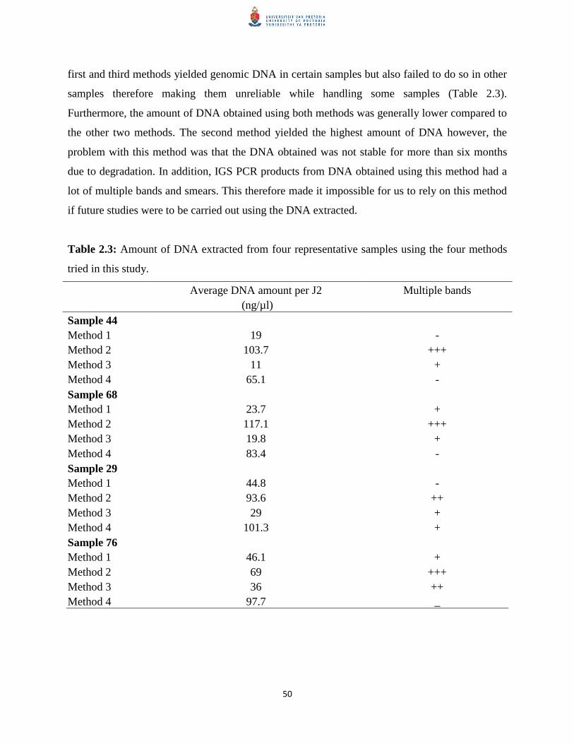
50
first and third methods yielded genomic DNA in certain samples but also failed to do so in other
samples therefore making them unreliable while handling some samples (Table 2.3).
Furthermore, the amount of DNA obtained using both methods was generally lower compared to
the other two methods. The second method yielded the highest amount of DNA however, the
problem with this method was that the DNA obtained was not stable for more than six months
due to degradation. In addition, IGS PCR products from DNA obtained using this method had a
lot of multiple bands and smears. This therefore made it impossible for us to rely on this method
if future studies were to be carried out using the DNA extracted.
Table 2.3: Amount of DNA extracted from four representative samples using the four methods
tried in this study.
Average DNA amount per J2 Multiple bands
(ng/µl)
Sample 44
Method 1 19 -
Method 2 103.7 +++
Method 3 11 +
Method 4 65.1 -
Sample 68
Method 1 23.7 +
Method 2 117.1 +++
Method 3 19.8 +
Method 4 83.4 -
Sample 29
Method 1 44.8 -
Method 2 93.6 ++
Method 3 29 +
Method 4 101.3 +
Sample 76
Method 1 46.1 +
Method 2 69 +++
Method 3 36 ++
Method 4 97.7 _

51
Method four, which is the modified WLB method, was much more reliable since using this
method it was possible to extract sufficient DNA yield from a number of samples used in this
study within a short period of time. There were also very few cases of multiple bands with IGS
PCR while using DNA from this method. Thus method four was selected as the best method and
subsequently used for all DNA extractions in the study.
2.8.3 Identification of Meloidogyne samples based on IGS region of the rDNA
Root-knot nematodes exhibit sequence divergence within the IGS region of their rDNA which
can be utilized for species discrimination (Blok, 2005). On the other hand, other regions of the
rDNA such as the ITS1 have little or no sequence divergence thus making it difficult to
differentiate various Meloidogyne species based on them (Blok, 2005). Therefore, Meloidogyne
species sampled during this study were identified using the IGS region of the rDNA among other
gene targets.
Genomic DNA obtained from at least five individual J2s from each of the 65 samples of the 78
collected was successfully amplified using primers (194/195) specific to the IGS region of the
rDNA. Different band sizes depending on the Meloidogyne species present in a sample were
obtained and compared to reference samples (L15, L16 and L32) from TJHI (Figure 2.5, Table
2.4). These band sizes ranged from 650bp as the smallest while 2000bp was the largest. In their
study Adam et al. (2007), IGS PCR for individual J2s gave different band sizes. They indicated
that tropical species yielded a PCR amplicon of 720bp for M. arenaria, 780bp for M. enterolobii,
700bp for M. hapla while M. fallax and M. chitwoodi gave 1600-1700bp. The size ranges
obtained in this study differed significantly to the band sizes obtained by those authors thus
sequencing and sequence analysis was undertaken. Thus the band size and corresponding
identity of Meloidogyne populations are indicated in Table 2.4. The identity was based on IGS-
rDNA after consensus sequences obtained were compared to those deposited in GenBank, NCBI
database for homology using BLAST search engine.

52
After IGS-PCR amplification all Meloidogyne species isolated during the study were identified
according to their amplicon sizes (Table 2.4). Cloning was carried out for all samples and in each
case, three representative clones per samples of different band sizes were selected, plasmid DNA
isolated using GeneJET™
plasmid Miniprep kit (Fermentas, Life Sciences) and sequenced in
both directions using the same amplification primers (for each of the three genes). In addition to
the amplicon sizes, results from all IGS-rDNA sequences obtained after being analyzed and
compared with sequences in GenBank, NCBI database using BLAST engine search were
recorded as shown below (Table 2.5). Therefore based on IGS-rDNA, we were able to identify
different Meloidogyne species as shown in Figure 2.6 below. The unidentified samples are those
which did not amplify on IGS PCR.

53
Table 2.4: Identity of isolated Meloidogyne species based on IGS-rDNA
Region
Approximate
Amplicon size
(bp)
Meloidogyne
spp
Number
identified
Mpumalanga, 650 M. hapla 1
N. West, Gauteng, Mpumalanga, 750-800 M. incognita 17
EFS,WFS, Limpopo, N. Cape1
Mpumalanga, N. West, WFS,EFS, 900-1000 M. javanica 18
Gauteng, N. Cape, Limpopo1
Mpumalanga, N. West, WFS, Gauteng 1000 M. arenaria 13
EFS, N. Cape1,
N. Cape, Mpumalanga, WFS1, 1100 Unidentified 13
WFS1 1200 M. artiellia 'like' 1
KZN1 780 M. enterolobii 10
EFS1 2000 M. chitwoodi 2
TJHI, UK 1000 M. arenaria L322
TJHI, UK 900-1000 M. javanica L162
TJHI, UK 750-800 M. incognita L152
1WFS; Western Free State, EFS; Eastern Free State, KZN; KwaZulu-Natal, N. Cape; Northern
Cape, N. West; North West
2Code of the reference samples

54
Table 2.5: BLAST results for isolated Meloidogyne species based on IGS-rDNA sequences
Meloidogyne species Max score e-value Max identity (%)
Meloidogyne arenaria 1103 0.00E+00 100
Meloidogyne hapla 511 3.00E-146 100
Meloidogyne incognita 1059 0.00E+00 100
Meloidogyne artiellia 26.5 7.00E+00 100
Meloidogyne javanica 1088 0.00E+00 99
Meloidogyne chitwoodi 883 0.00E+00 92
Meloidogyne enterolobii 699 0.00E+00 94

55
2.8.3.1 Phylogenetic analysis of Meloidogyne samples based on IGS sequences
To further confirm the identity of Meloidogyne populations preliminarily identified by BLAST
algorithm, phylogenetic analysis was done whereby consensus sequences obtained for IGS-
rDNA in this study and those retrieved from GenBank, NCBI database (Table 2.6) were aligned
over same length in ClustalW (with gap opening penalty for multiple alignments of 10 and
extension of 0.2) and MUSCLE (with gap opening penalty for multiple alignments of -12 and
extension of -1) using MEGA v 5.0 (Tamura et al., 2011). This was done to reveal regions of
similarity and dissimilarity between the sequences. Highly similar sequences for IGS-rDNA
were then aligned over the same lengths using MAFFT 5.3 (Katoh et al., 2005) fitted into the
jModel test for a suitable model (Posada and Crandall, 1998) before generating phylograms
(Figure 2.7) using Maximum likelihood (ML) and the Phylip 4.0 software. The best topology of
the ML tree was obtained by the TIM3 model as provided by the jModel test. During this
analysis, all phylograms were constructed using 1000 bootstrap replicates to assess their support
for each clade or phylogenetic branching (Landa et al., 2008).
Table 2.6: GenBank, NCBI accession numbers for Meloidogyne IGS reference sequences used
for the construction of phylogenetic relationships.
GenBank accession number Meloidogyne species
FJ555690.1 M. incognita
FJ555691.1 M. javanica
FJ555695.1 M. enterolobii
FJ555692.1 M. chitwoodi
FJ555693.1 M. fallax
FJ555694.1 M. hapla
FJ555689.1 M. arenaria

56
Figure 2.7: Maximum likelihood tree after an alignment of consensus sequences based on the
IGS rDNA region of various Meloidogyne species identified in this study. Newly obtained
sequences in this study are in bold. Analysis was done using 1000 bootstrap replicates. The
bootstrap support value for each clade is indicated on the nodes. An out-group was not used in
this phylograms since the primers (194/195) used are Meloidogyne species specific.

57
In Figure 2.7, putatively identified populations from our study grouped correctly with those
identified from other parts of the world. Meloidogyne arenaria sample (shown as Melo L32)
from TJHI UK was included in this study too. Meloidogyne species identified putatively as M.
artiellia based on BLAST was excluded from this phylogenetic analysis since it gave a very low
maximum score. Using the IGS-rDNA, 16 new sequences were generated from different
Meloidogyne species isolated in this study. These sequences were aligned together with seven
IGS sequences for various Meloidogyne species retrieved from GenBank resulting in a total of 23
sequences. Alignment and phylogenetic analysis of the 23 sequences resulted in several clades
and sub-clades which were separated with varying bootstrap support (BS) values in the
maximum likelihood analysis as follows: (i) M. javanica, M. incognita and M. arenaria
populations (BS = 100%), (ii) all M. enterolobii populations (BS = 90%), (iii) M. hapla
populations (BS = 100%) and (iv) M. chitwoodi populations (BS = 100%). In this analysis
(Figure 2.7), the tropical Meloidogyne species were distinguished by a bootstrap support of 83%
from the automictic species.
2.8.4 Identification of Meloidogyne samples based on D2-D3 (28S) region of the rDNA
The D2-D3 segments are positioned on the 26S (28S) region of the rDNA. The 28S region is a
common molecular signature that is used to carry out molecular identification related to most
Meloidogyne species and other members within the phylum Nematoda (Schmitz et al., 1998).
During this study, genomic DNA obtained from majority of the collected Meloidogyne species
was also successfully amplified with primers specific for the D2-D3 expansion segments within
the 26S (28S) region of the rDNA. All samples which amplified on D2-D3 gave a 950bp
amplification fragment during D2-D3 region amplifications (Figure 2.8). Amplified PCR
products obtained from D2-D3 PCR were excised from the gel, purified and cloned using
CloneJET™
kit (Fermentas, Life Sciences) according to the manufacturer‟s instructions.
Representative clones were sequenced in both directions using D2A and D3B primers.
Sequences obtained were manually checked and edited using BioEdit v 7.0.9 (Hall, 1999),
aligned using ClustalW and consensus sequences compared to those deposited in GenBank,
NCBI database using BLAST engine search in order to establish the identity of our Meloidogyne
population (Table 2.7).

58
Table 2.7: BLAST results for isolated Meloidogyne species based on D2-D3 28S sequences
Meloidogyne species Max score e-value Max identity (%)
Meloidogyne arenaria 1153 0.00E+00 99
Meloidogyne hapla 785 0.00E+00 100
Meloidogyne incognita 1337 0.00E+00 100
Meloidogyne javanica 684 0.00E+00 100
Meloidogyne chitwoodi 1284 0.00E+00 98
Meloidogyne enterolobii 1049 0.00E+00 96

59
2.8.4.1 Phylogenetic analysis of Meloidogyne samples based on 28S D2-D3 sequences
Phylogenetic analyses of Meloidogyne populations initially identified by BLAST algorithm
based on D2-D3 consensus sequences was done whereby consensus sequences obtained for D2-
D3 (28S) region of the rDNA in this study and those retrieved from GenBank, NCBI database
(Table 2.8) were aligned over the same lengths using MAFFT 5.3 (Katoh et al., 2005) and
analyzed using maximum parsimony (MP) (Tigano et al., 2005; Landa et al., 2008). For each
data set, both the un-weighted and weighted MP analyses were performed using PAUP* 4.0b 10
software (Swofford, 2003). Heuristic searches were performed (Subbotin et al., 2008) and
support for each clade (Figure 2.9) finally assessed by using MP analysis with 1000 replicates
(Landa et al., 2008)
Table 2.8: GenBank, NCBI accession numbers for Meloidogyne D2-D3 (28S) reference
sequences used for the construction of phylogenetic relationships.
GenBank accession number Nematode species
AY942624.1 M. incognita
JN005852.1 M. javanica
AY942629.1 M. enterolobii
AY593886.1 M. chitwoodi
AY593895.1 M. fallax
AY757849.1 M. hapla
AY942621.1 M. floridensis
EU443609.1 M. hispanica strain Servile
EU443610.1 M. hispanica strain Brazil
AY942632.1 M. morocciensis
EU364889.1 M. arenaria
JF826219.1 B. xylophilus

60
Figure 2.9: Maximum parsimony tree that has been rooted after an alignment of consensus
sequences based on the 28S D2-D3 rDNA region of various Meloidogyne species identified in
this study. Newly obtained sequences in this study are in bold. Bootstrap support for each clade
is indicated at the nodes.

61
In this phylogenetic analysis (Figure 2.9), Meloidogyne species identified from our study
grouped correctly with those identified from other parts of the world. The 28S D2-D3 sequences
for M. floridensis and M. incognita deposited in GenBank, NCBI database are highly similar and
this is why the two Meloidogyne species clustered together with the South African Meloidogyne
population previously identified as M. incognita based on IGS- PCR sequence analysis. Previous
studies have shown that the D2-D3 region is not useful for M. incognita but it does separate M.
javanica and M. arenaria with much greater confidence than the IGS region (Vivian Blok,
pers.comm). The 28S D2-D3 alignment was made up of 28 sequences, 16 of which were new
sequences from the study populations. Maximum parsimony analysis of the 28S D2-D3
sequences demonstrated varying bootstrap support values for various clades and sub-clades: (i)
M. chitwoodi populations (BS = 76%), (ii) M. chitwoodi and M. fallax populations (BS = 91%),
(iii) M. enterolobii populations (99%), (iv) M. incognita populations (BS = 98%), (v) M. hapla
populations (BS = 94%), M. javanica populations (BS = 100%) and M. arenaria populations (BS
= 100%). Therefore, the 28S D2-D3 region was able to consistently identify/confirm various
Meloidogyne species as identified using IGS-rDNA.
2.8.5 Identification of Meloidogyne samples based on COII region within the 16S rRNA.
Within the mitochondrial DNA of Meloidogyne species, the mitochondrial gene that codes for
the cytochrome oxidase subunit II (COII) is associated with a high rate of sequence evolution,
divergence and rearrangements (Blok, 2005). Based on this high rate of evolution, high copy
number in each cell and the assumption that the mtDNA is associated with maternal inheritance,
the mtDNA has been utilized as a diagnostic target and for studying lineages (Blouin, 2002). In
this study, COII-PCR amplifications gave a wide range of amplicon sizes (520bp-1600bp) using
the genomic DNA obtained from various Meloidogyne species sampled in this study (Figure
2.10). Amplification products obtained from PCR of the region between the COII and the 16S
rRNA (lRNA) gene of the mtDNA were excised from the gel, purified and cloned using
CloneJET™
kit (Fermentas, Life Sciences) according to the manufacturer‟s instructions.
Sequencing from representative clones was done in both directions using C2F3/1108 pair of
primers. Sequences obtained were manually checked and edited using BioEdit v 7.0.9 (Hall,
1999), aligned using ClustalW and consensus sequences obtained compared to those deposited in

62
GenBank, NCBI database using BLAST search algorithm in order to establish the identity of our
populations (Table 2.9).
The results obtained with COII were in agreement with those obtained in other studies where M.
mayaguensis (currently M. enterolobii) gave a PCR product of 705bp (shown in lane 6 in Figure
2.10) using primers C2F3 and 1108 (Powers and Harris, 1993). The different Meloidogyne
species in this study were demonstrated by various amplicon sizes within IGS and COII gene
amplifications. This was later confirmed through sequence and phylogenetic analyses. The
amplicon sizes for other Meloidogyne species not represented in the gel have been indicated in
Table 2.9.

63
Table 2.9: BLAST results for isolated Meloidogyne species based on COII 16S rRNA sequences
Meloidogyne species Amplicon
Size (bp) Max score e-value Max identity (%)
Meloidogyne arenaria 1100 1398 0.00E+00 100
Meloidogyne hapla 520 855 0.00E+00 89
Meloidogyne incognita 1500 1209 0.00E+00 100
Meloidogyne javanica 1600 1472 0.00E+00 90
Meloidogyne chitwoodi 520 747 0.00E+00 99
Meloidogyne enterolobii 705 1090 0.00E+00 100
2.8.5.1 Phylogenetic analysis of Meloidogyne samples based on COII sequences
To support the results obtained from BLAST search algorithm, phylogenetic relationships
between Meloidogyne populations in this study and reference sequences in GenBank, NCBI
database based on COII sequences (Table 2.10) were aligned over the same lengths using
MAFFT 5.3 (Katoh et al., 2005) and sequence data sets analyzed using maximum parsimony
(MP) (Tigano et al., 2005; Landa et al., 2008). For each data set, both the un-weighted and
weighted MP analyses were performed using PAUP* 4.0b 10 software (Swofford, 2003).
Heuristic searches were performed (Subbotin et al., 2008) and support for each clade (Figure
2.11) was finally assessed by using MP analysis with 1000 replicates (Landa et al., 2008).

64
Table 2.10: GenBank, NCBI accession numbers for Meloidogyne COII reference sequences
used for the construction of phylogenetic relationships.
GenBank accession number Nematode species
AY635611.1 M. incognita
AY635612.1 M. javanica
AY635613.1 M. enterolobii
JN241902.1 M. chitwoodi
JN241954.1 M. fallax
AY942850.1 M. hapla
AY635609.1 M. floridensis
JN241899.1 M. naasi
JN241955.1 M. marylandi
AY942849.1 M. morocciensis
AY942848.1 M. ethiopica
AY942851.1 M. paranaensis
JN241933.1 M. minor
AY942852.1 M. arabicida
AY757908.1 M. partityla
JN241929.1 M. graminicola
AY635610.1 M. arenaria
JQ514068.1 B. xylophilus

65
Figure 2.11: Maximum parsimony tree that has been rooted after an alignment of consensus
sequences based on the region between the COII and the 16S rRNA (lRNA) gene of various
Meloidogyne species identified in this study. Newly obtained sequences in this study are in bold.
Bootstrap support for each clade is indicated at the nodes.

66
In Figure 2.11, 33 sequences were aligned, 16 of which were from populations in this study. All
the study samples and those deposited in GenBank database formed clades and sub-clades of
varying bootstrap support values during maximum parsimony analysis as follows: (i) M.
graminicola, M. minor and M. naasi (BS = 77%), (ii) M. chitwoodi populations and M. fallax
(BS = 98%), (iii) M. hapla populations and M. partityla (BS = 88%), (iv) automictic
Meloidogyne species (M. chitwoodi, M. fallax, M. minor, M. naasi, M. marylandi, M. partityla
and M. hapla) formed a major clade with 83% bootstrap support, (v) M. enterolobii populations
(BS = 92%), (vi) M. javanica populations (BS = 92%), (vii) M. arenaria populations and M.
morocciensis (BS = 94%), (viii) M. incognita populations (BS = 100%) and (ix) All tropical
Meloidogyne species (including M. enterolobii) formed a major clade with 86% bootstrap
support.
All Meloidogyne species identified in this study were able to group closely with respective
Meloidogyne species identified from other parts of the world. Two unique clades; one consisting
of mainly the temperate species and another the tropical species were clearly identified in this
COII based phylograms. Like in IGS-rDNA phylograms, M. enterolobii formed its own clade but
closely related to tropical species.
2.8.6 Distribution of Meloidogyne species in South Africa
Based on the information obtained from sequence analyses of IGS, D2D3 and COII sequences,
the distribution of various Meloidogyne species in South Africa can be represented as shown in
Figure 2.12. Study findings indicated that most farms are dominated by the three common
tropical species; M. incognita, M. javanica and M. arenaria. Meloidogyne javanica was reported
as the most dominant species with a 24% occurrence in all the potato tubers collected.
Meloidogyne incognita and M. arenaria occurred in 23% and 17% of the potato tubers sampled
respectively. The highly damaging and resistance breaking Meloidogyne enterolobii was
reported in 14% of potato tubers sampled. Most of the affected samples by this nematode were
obtained from the KwaZulu-Natal potato growing region. It is not clear if this nematode has
spread to other potato growing areas or it is only restricted to this growing region.

67
The automictic Meloidogyne species were identified from a few areas where potatoes are grown
in South Africa. The Free State was the only region from where 3% of M. chitwoodi reported in
this study was isolated. Meloidogyne hapla had a 1% occurrence from all the samples surveyed.
This nematode was identified in samples from the Mpumalanga potato growing area. We were
not successful in getting samples from the Western Cape and the Eastern Cape and therefore we
have not been able to map the prevalence and distribution of Meloidogyne species in these areas.

68
2.9 Discussion
Accurate identification and in-depth understanding of the genetic diversity of Meloidogyne
species present in a given potato field is the first step in designing proper pest management
programmes (Powers et al., 2005). This can only be achieved through a regular, comprehensive
and accurate survey across all the 16 main potato growing regions in South Africa.
The first objective of this study was to detect and discriminate various Meloidogyne species
collected from various potato growing zones across the country based on IGS and D2-D3 regions
of the rDNA and the portion between the COII and the 16S rRNA gene of the mtDNA. The
rDNA was chosen in this study as the most appropriate target site because it has relatively
conserved and highly variable regions which have been used in identification and in construction
of phylogenetic relationships for root-knot nematodes and other species (Landa et al., 2008). The
mtDNA is also a well conserved target too with a relatively fast rate of sequence polymorphism
and rearrangements compared to the nuclear genome (Blouin, 2002).
Various stages of root-knot nematodes can be extracted from infected samples by using either the
centrifugal floatation technique or the Baerman method. The Baerman method is hinged on the
principle of motility of the second stage juveniles (J2s) and therefore it is the best method for
recovering J2s within a period of time (24-48hr) after preparing the samples (Bezooijen, 2006;
Coyne et al., 2009). On the other hand, the centrifugal floatation method is based on the principle
of the specific gravity of nematodes (Bezooijen, 2006). It separates nematodes from plant debris
through elimination which is achieved by use of kaolin (Fourie, unpublished). When nematodes
are centrifuged in a sucrose solution, they get suspended thus making it easy to recover them as
the supernatant. This method is suitable for extracting various developmental stages of
nematodes from infected samples within the shortest time. Therefore in this study, the centrifugal
floatation technique was chosen as the best method and used throughout this study to extract
various stages of root-knot nematodes from potato tubers which were collected (Coyne et al.,
2009).
Four methods of DNA extractions were compared. Of the four, the WLB modified DNA
extraction method adopted in this study was able to provide sufficient DNA amount from

69
individual J2s which was used for IGS, COII and D2-D3 amplifications. The procedure was also
quick since it took less than ninety minutes after nematode isolation to complete. Individual J2s
together with young developing females gave a high DNA yield compared to other
developmental stages using this method. This was in agreement with results obtained by Berry et
al. (2007) who indicated that the stage of nematode development used for DNA extraction,
among other factors has an influence in the amount of DNA yield. The simplicity and efficiency
of the modified method adopted in this study for DNA extraction is an aspect that can be
explored in routine laboratory diagnostic procedures to rapidly and accurately identify various
Meloidogyne species and other PPNs.
As demonstrated in other previous studies (Holterman et al., 2012) the IGS region of the rDNA
alone (at sequence level) was capable of preliminarily detecting and discriminating between
individual species collected in this study. This was in support of other studies that have been
carried out previously using IGS region as the basis to differentiate various Meloidogyne species
(Blok et al., 1997; Wishart et al., 2002). However, amplicon sizes based on IGS-rDNA alone are
not able to distinguish the closely related tropical Meloidogyne species (M. arenaria, M.
incognita and M. javanica) (Blok et al., 1997). Meloidogyne arenaria and M. javanica have
similar band sizes based on IGS region. Therefore it is important that other gene regions are used
in order to accurately distinguish between sibling Meloidogyne species.
The D2-D3 sequences were also able to give similar identities for Meloidogyne species as those
obtained using IGS region of the rDNA. This is besides the fact that D2-D3 primers used in this
study can amplify the 28S region of rDNA associated with Meloidogyne species and in other
genera for example Xiphinema (Gutierrez-Gutierrez et al., 2010). Amplifications of the 28S D2-
D3 segments were therefore used to indicate the presence of members from the phylum
Nematoda and at the same time support the results obtained by IGS-rDNA and COII sequences.
In this study, IGS sequences from most of the species identified were highly similar to the
reference populations deposited in GenBank, NCBI database. Moreover, IGS sequences obtained
for the Meloidogyne populations from South Africa were highly similar intraspecifically.
Furthermore, the use of COII in this study confirmed that our study samples were infected from a
diversity of Meloidogyne species which had been identified based on IGS and D2-D3 sequences

70
in this study. The cytochrome oxidase subunit II is a highly conserved region and therefore it
was easy to discriminate different Meloidogyne species in this study as it has also been
demonstrated in other studies (Blouin, 2002; Tigano et al., 2005). From the results obtained
using IGS, D2-D3 and COII sequences, various Meloidogyne species are distributed across the
16 major potato growing zones in South Africa. The three common tropical species; M.
incognita, M. javanica and M. arenaria dominate in this distribution while the temperate species
such as M. hapla and M. chitwoodi occur in Mpumalanga and the Free State respectively.
Meloidogyne enterolobii was isolated from KwaZulu-Natal but we were not able to establish if it
has spread to other potato growing zones in South Africa (Onkendi and Moleleki, 2012; in
press).
The second objective of this study was to construct phylogenetic relationships based on
consensus sequences derived from IGS-rDNA, D2-D3 region and the COII of 16S rRNA.
Phylogenetic relationships between various Meloidogyne species based on IGS, COII and D2-D3
demonstrated that the IGS and the D2-D3 regions of the rDNA can accurately resolve various
Meloidogyne species when used together (Blok, 2005). For the first time, we attempted to
construct phylogenetic relationships based on consensus sequences derived from IGS-rDNA of
various Meloidogyne species. Phylogenetic analysis of consensus sequences derived from IGS-
rDNA using ML analysis was able to group the tropical adapted species into one clade which is
completely distinct from the clade formed by the temperate species. Closely related apomictic
species (M. javanica, M. arenaria and M. incognita) grouped together into one clade with 100%
bootstrap support while automictic species (M. chitwoodi and M. fallax) grouped also together
into another clade with 100% bootstrap support. Populations of M. enterolobii (which is a
tropical species) formed an independent grouping which is slightly closer to the tropical species
than the temperate one. This was supported by a 94% bootstrap support using ML analysis.
Meloidogyne hapla which is a facultative parthenogenetic species was also clearly separated
during phylogenetic analysis to form an independent grouping in between the apomictic and
automictic species but closer to the automictic species. Other studies carried out previously also
suggest that M. hapla is more closely related to the automictic species than to apomictic species
based on percentage nucleotide base substitution using total genomic DNA (Castagnone-Sereno

71
et al., 1993). The phylograms for IGS sequences were constructed without an out-group since the
primer pair (194/195) used in this study is only specific to Meloidogyne genus.
Various Meloidogyne species identified in this study grouped correctly with those already
identified in various parts of the world based on phylogenetic analysis of the D2-D3 sequences.
The alignment based on 28S D2-D3 sequences from this study was 720bp long since all
Meloidogyne species in this study gave PCR products of the same size; 950bp. An alignment of
721bp was recorded in studies carried out by McClure et al. (2012). The analysis did not resolve
automictic and apomictic Meloidogyne species into separate and distinct clades despite the fact
that individual sequences were able to identify the species. This has also been witnessed in other
studies which have used this gene target for phylogenetic analysis of various Meloidogyne
species (Vovlas et al., 2007).
Further phylogenetic analyses based on COII showed that all tropical Meloidogyne species
grouped together to form a single clade with a bootstrap support of 86% while the temperate
apomictic species also grouped together to form a single clade with 93% bootstrap support.
Meloidogyne enterolobii is closely related to the tropical species than it is to M. hapla, M. fallax
and M. chitwoodi. This is why the species grouped with other tropical Meloidogyne species and
this was supported by 86% bootstrap support for the clade containing M. enterolobii and the
tropical species. This grouping was consistent with other studies that have been carried
elsewhere (McClure et al., 2012). The close relationship of M. enterolobii can be attributed to
the mode of reproduction since both M. enterolobii and most of the tropical Meloidogyne species
are mitotically parthenogenetic (Tigano et al., 2005). The unique PCR products (705bp for COII
and 780bp for IGS) easily differentiated M. enterolobii from other Meloidogyne species. Our
sequence analyses based on COII sequences in this study together with references isolates in
GenBank, NCBI showed that they were highly rich in AT nucleotides. McClure et al. (2012)
observed the same while analyzing COII sequences of various Meloidogyne from 238 golf
courses in Western United States.
Detection and discrimination of various closely related Meloidogyne species such as M. javanica,
M. incognita and M. arenaria in this study supports the importance of using multiple gene

72
targets such as IGS and D2-D3 regions of the rDNA together with the portion between the COII
and the 16S rRNA (lRNA) gene of the mtDNA to accurately resolve the identities of sibling
Meloidogyne species. Previous studies that attempted to differentiate M. javanica, M. arenaria
and M. incognita based only on band sizes from IGS PCR products were inconclusive since the
three apomictic species are closely related therefore producing band sizes that are almost similar
in size (Wishart et al., 2002; Holterman et al., 2012). Therefore, sequence data obtained for these
gene targets can be used to accurately identify, discriminate and establish genetic relationships
between various Meloidogyne species through the construction of phylogenetic relationships (De
Ley et al., 2002). The IGS primers used here are specific only to Meloidogyne species while the
D2-D3 primers are universal and therefore they can be used to identify other nematode species
besides various Meloidogyne species (Landa et al., 2008; Gutierrez-Gutierrez et al., 2010).

73
2.10 Conclusion
The current method of identifying different Meloidogyne species in South Africa based on
morphological and morphometrical characteristics can be challenging in resolving some of the
economically important and closely related species such as M. javanica, M. incognita and M.
arenaria. This method is not only tedious but also unable to accurately differentiate various
populations of certain Meloidogyne species which exhibit molecular heterogeneity (Randig et al.,
2002). Therefore there is need to integrate data from classical methods and that obtained from
molecular methods in root-knot nematode diagnostics. The use of molecular signatures such as
IGS, COII and 28S D2-D3 segments provides an accurate, reliable and rapid method of
identifying these phytoparasites as demonstrated in this study and others that have been carried
elsewhere (Blok et al., 1997; Tigano et al., 2005; Landa et al., 2008). In addition, these
molecular-based identification approaches can be employed in large scale surveillance of root-
knot nematodes in potato fields and other crops (Powers et al., 2005).
Identification of resistance breaking Meloidogyne populations of M. enterolobii and automictic
M. chitwoodi in this study re-emphasizes the need to put more efforts in carrying out more
surveillance of these parasites to contain them from spreading hence curb more losses in the
potato industry. Meloidogyne enterolobii is said to have a higher rate of reproduction compared
to other Meloidogyne species and it can also displace the three common tropical Meloidogyne
species in a case of co-infection therefore paving way for more losses (Blok et al., 2002).
Meloidogyne chitwoodi has also been reported to have resistance to systemic nematicides and
also to cause symptomless infections (OEPP/EPPO bulletin, 2009). This can potentially lead to
dissemination of M. chitwoodi to new areas through seed or symptomless plant materials. The
samples that were not identified in this study consistently failed to amplify with the Meloidogyne
species specific primers (194/195). We concluded that they might have been other plant-parasitic
nematodes from other nematode genera.
Phylogenetic analyses in this study revealed that apomictic species present in South African
potato farms are highly similar to those identified in other parts of the world. This was
demonstrated through the grouping of all apomictic species into one cluster based on the IGS,
D2-D3 and COII phylograms. The same trend was also noted with the automictic species (M.

74
chitwoodi) that was isolated from two samples. Therefore phylogenetic studies can also be used
in future to monitor the diversity of various Meloidogyne species in South Africa to see if they
are undergoing some evolutionary changes to cope with environmental pressures occasioned by
IPM programmes. Sequences data in GenBank, NCBI for various Meloidogyne species identified
in this study will serve as references for future studies. To our knowledge, this will be the first
time Meloidogyne sequences from a study of this nature in South Africa are being deposited in
GenBank, NCBI database.

75
2.11 References
Abebe, E., Mekete, T., & Thomas, W. K. (2011). A critique of the current methods in nematode
taxonomy. African Journal of Biotechnology, 10, 312-323.
Adam, M. A. M., Phillips, M. S., & Blok, V. C. (2007). Molecular diagnostic key for
identification of single juveniles of seven common economically important species of root-
knot nematode (Meloidogyne spp.). Plant Pathology, 56, 190-197.
Berry, S. D., Fargette, M., Morand, S., & Cadet, P. (2007). Reliability of PCR-based techniques
for detection and discrimination of plant-parasitic nematodes of sugarcane. Nematology, 9,
499-514.
Bezooijen, J. V. (2006). Methods and techniques for nematology (pp 25-26). Wageningen.
Blok, V. C., Phillips, M. S., & Fargette, M. (1997). Comparison of sequences from the ribosomal
DNA intergenic region of Meloidogyne mayaguensis and other major tropical root-knot
nematodes. Journal of Nematology, 29, 16-22.
Blok, V. C. (2005). Achievements in and future prospects for molecular diagnostics of plant
parasitic nematodes. Canadian Journal of Plant Pathology, 27, 176-185.
Blok, V. C., & Powers, Thomas O. (2009). Biochemical and molecular identification. In R. N
Perry, M. Moens, & J. L. Starr (eds.), Root-knot nematodes (pp. 98-118). CAB International.
Nosworthy Way. Wallingford, Oxfordshire OX10 8DE, UK.
Blok, V. C., Wishart, J. W., Fargette, M. F., Berthier, K. B., & Phillips, M. S. P. (2002).
Mitochondrial DNA differences distinguishing Meloidogyne mayaguensis from the major
species of tropical root-knot nematodes. Nematology, 4, 773-781.
Blouin, M. S. (2002). Molecular prospecting for cryptic species of nematodes: mitochondrial
DNA versus internal transcribed spacer. International Journal for Parasitology, 32, 527-
531.

76
Brown, W. M., George, M. J., & Wilson, A. C. (1979). Rapid evolution of animal mitochondrial
DNA. National Academy of Science, USA, 76, 1967-1971.
Carpenter, A. S., Hiatt, E. E., Lewis, S. A., & Abbott, A. G. (1992). Genomic RFLP analysis of
Meloidogyne arenaria race 2 populations. Journal of Nematology, 24, 23-28.
Castagnone-Sereno, P., Leroy, F., Bongiovanni, M., Zijlstra, C., & Abad, P. (1999). Specific
diagnosis of two root-knot nematodes, Meloidogyne chitwoodi and M. fallax, with satellite
DNA probes. Phytopathology, 89, 380-384.
Castagnone-Sereno, P., Esparrago, G., Abad, P., Leroy, F., & Bongiovanni, M. (1995). Satellite
DNA as a target for PCR-specific detection of the plant parasitic nematode Meloidogyne
hapla. Current Genetics, 28, 566-570.
Castagnone-Sereno, P., Piotte, C., Uijthof, J., Abad, P., Wajnberg, E., Vanlerberghe-Masutti, F.,
et al. (1993). Phylogenetic relationships between amphimictic and parthenogenetic
nematodes of the genus Meloidogyne as inferred from repetitive DNA analysis. Heredity, 70,
195-204.
Castillo, P., Vovlas, N., Troccoli, A,. Liebanas, G., Palomares-Rius, J. E., Landa, B. B. (2009). A
new root-knot nematode, Meloidogyne silvesteris n. sp (Nematoda: Meloidogynidae),
parasitizing European holly in northern Spain. Plant Pathology, 58, 601-619.
Coyne, D. L., Nicol, J. M., & Claudius-Cole, B. (2009). Practical plant nematology: a field and
laboratory guide (pp 40-42). International Institute of Tropical Agriculture.
De Ley, I. T., De Ley, P., Vierstraete, A., Karssen, G., Moens, M., & Vanfleteren, J. (2002).
Phylogenetic analyses of Meloidogyne small subunit rDNA. Journal of Nematology, 34, 319-
327.
De Ley, P., De Ley, I. T., Morris, K., Abebe, E., Mundo-Ocampo, M., Yoder, M., et al. (2005).
An integrated approach to fast and informative morphological vouchering of nematodes for
applications in molecular barcoding. Philosophical Transactions of the Royal Society of
Biological Sciences, 360, 1945-1958.

77
De Luca, F., Fanelli, E., Vito, M. D., Reyes, A., & Giorgi, C. D. (2004). Comparison of the
sequences of the D3 expansion of the 26S ribosomal genes reveals different degrees of
heterogeneity in different populations and species of Pratylenchus from the Mediterranean
region. European Journal of Plant Pathology, 110, 949-957.
De Luca, F., Reyes, A., Troccoli, A., & Castillo, P. (2011). Molecular variability and
phylogenetic relationships among different species and populations of Pratylenchus
(Nematoda: Pratylenchidae) as inferred from the analysis of the ITS rDNA. European
Journal of Plant Pathology, 130, 415-426.
De Luca, F., Reyes, A., Veronico, P., Di Vito M., Lamberti, F., & De Giorgi, C. (2002).
Characterization of the (GAAA) microsatellite region in the plant parasitic nematode
Meloidogyne artiellia. Gene, 293, 191-198.
Floyd, R., Abebe, E., Papert, A., & Blaxter, M. (2002). Molecular barcodes for soil nematode
identification. Molecular Ecology, 11, 839-50.
Fullando, A., Salazar, A., Barrena, E., & Ritter, E. (1997). Comparison of molecular patterns and
virulence behaviour of potato cyst nematodes. Fundam. Appl. Nematol, 20, 425-433.
Garate, T. (1991). Characterization of species and races of the genus Meloidogyne by DNA
restriction enzyme analysis. Journal of Nematology, 23, 414-420.
Gutierrez-Gutierrez, C., Palomares-Rius, J. E., Cantalapiedra-Navarrete, C., Landa, B. B.,
Esmenjaud, D., & Castillo, P. (2010). Molecular analysis and comparative morphology to
resolve a complex of cryptic Xiphinema species. Zoologica Scripta, 39, 483-498.
Hall, T. A. (1999). BioEdit: a user-friendly biological sequence alignment editor and analysis
program for windows 95/98/NT. Nucleic Acids Symposium Series, 41, 95-98.
Handoo, Z. A., Skantar, A. M., Carta, L. K., & Erbe, E. F. (2005). Morphological and molecular
characterization of a new root-knot nematode, Meloidogyne thailandica n. sp. (Nematoda:
Meloidogynidae), parasitizing ginger (Zingiber sp.). Journal of Nematology, 37, 343-353.

78
Holterman, M. M., Oggenfuss, M., Frey, J. E., & Kiewnick, S. (2012). Evaluation of high-
resolution melting curve analysis as a new tool for root-knot nematode diagnostics.
Phytopathology, 160, 59-66.
Hu, M. X., Zhuo, K., & Liao, J. L. (2011). Multiplex PCR for the simultaneous identification and
detection of Meloidogyne incognita, M. enterolobii, and M. javanica using DNA extracted
directly from individual galls. Phytopathology, 101, 1270-1277.
Hugall, A., Moritz, C., Stanton, J., & Wolstenholme, D. R. (1994). Low, but strongly structured
mitochondrial DNA diversity in root knot nematodes (Meloidogyne). Genetics, 136, 903-
912.
Hyman, B. C. (1990). Molecular diagnosis of Meloidogyne species. Journal of Nematology, 22,
24-30.
Katoh, K., Kuma, Ke-ichi., Toh, H., & Miyata, T. (2005). MAFFT version 5: improvement in
accuracy of multiple sequence alignment. Nucleic Acids Research, 33, 511-518.
Landa, B. B., Palomares-Rius, J. E., Vovlas, N., Carneiro, R. M. D. G., Maleita, C. M. N.,
Abrantes, I. M. D. O., et al. (2008). Molecular characterization of Meloidogyne hispanica
(Nematoda, Meloidogynidae) by phylogenetic analysis of genes within the rDNA in
Meloidogyne spp. Plant Disease, 92, 1104-1110.
Mahran, A., Tenuta, M., Shinners-Carenelly, T., Mundo-Ocampo, M., & Daayf, F. (2010).
Prevalence and species identification of Pratylenchus spp. in Manitoba potato fields and host
suitability of “Russet Burbank.‟‟ Canadian Journal of Plant Pathology, 32, 272-282.
McClure, M. A., Nischwitz, C., Skantar, A. M., Schmitt, M. E., & Subbotin, S. A. (2012). Root-
knot nematodes in golf course greens of the Western United States. Plant Disease, 96, 635-
647.
Niu, Jun-hai., Guo, Quan-xin., Jian, H.,Chen, Chang-long, Yang, D., Liu, Q., et al. (2011). Rapid
detection of Meloidogyne spp. by LAMP assay in soil and roots. Crop Protection, 30, 1063-
1069.

79
OEPP/EPPO bulletin. (2009). Meloidogyne chitwoodi and Meloidogyne fallax. OEPP/EPPO
Bulletin, 39, 5-17.
Onkendi E. M., & Moleleki, L. N. (2012). Detection of Meloidogyne enterolobii in potatoes in
South Africa and phylogenetic analysis based on intergenic region and the mitochondrial
DNA sequences. European Journal of Plant Pathology, DOI: 10.1007/s10658-012-0142-y.
Palomares-Rius, J. E., Vovlas, N., Troccoli, G., Landa, B. B., & Castillo, P. (2007). A new root-
knot nematode parasitizing sea rocket from Spanish Mediterranean coastal dunes:
Meloidogyne dunensis n. sp. (Nematoda: Meloidogynidae). Journal of Nematology, 39, 190-
202.
Petersen, D. J., & Vrain, T. C. (1996). Rapid identification of Meloidogyne chitwoodi, M. hapla,
and M. fallax using PCR primers to amplify their ribosomal intergenic spacer. Fundam. App.
Nematol, 19, 601-605.
Posada, D., & Crandall, K. A. (1998). MODELTEST: testing the model of DNA substitution.
Bioinformatics Application Note, 14, 817-818.
Powers, T. O., & Harris, T. S. (1993). A Polymerase chain reaction method for identification of
five major Meloidogyne species. Journal of Nematology, 25, 1-6.
Powers, T. O., Mullin, P. G., Harris, T. S., Sutton, L. A., & Higgins, R. S. (2005). Incorporating
molecular identification of Meloidogyne spp. into a large-scale regional nematode survey.
Journal of Nematology, 37, 226-235.
Powers, T. (2004). Nematode molecular diagnostics: from bands to barcodes. Annual Review of
Phytopathology, 42, 367-383.
Randig, O., Bongiovanni, M., Carneiro, R. M. D. G., & Castagnone-Sereno, P. (2002). Genetic
diversity of root-knot nematodes from Brazil and development of SCAR markers specific for
the coffee-damaging species. Genome, 45, 862-870.

80
Schimtz, B., Burgermeister, W., & Braasch, H. (1998). Molecular genetic classification of
central European Meloidogyne chitwoodi and M. fallax population. Nachrichtechnbl
Deutschen Pflanzenschutzd, 50, 310-317.
Semblat, J. P., Wajnberg, E., Dalmasso, A., Abad, P., & Castagnone-Sereno, P. (1998). High-
resolution DNA fingerprinting of parthenogenetic root-knot nematodes using AFLP
analysis. Molecular Ecology, 7, 119-125.
Skantar, A. M., Carta, L. K., & Handoo, Z. A., (2008). Molecular and morphological
characteristics of an unusual Meloidogyne arenaria population from traveller‟s tree,
Ravenala madagascariensis. Journal of Nematology, 40, 179-189.
Subbotin, S. A., Ragsdale, E. J., Mullens, T., Roberts, P. A., Mundo-Ocampo, M., & Baldwin, J.
G. (2008). A phylogenetic framework for root lesion nematodes of the genus Pratylenchus
(Nematoda): evidence from 18S and D2–D3 expansion segments of 28S ribosomal RNA
genes and morphological characters. Molecular Phylogenetics and Evolution, 48, 491-505.
Swofford, D. (2003). PAUP*: Phylogenetic analysis using parsimony (*and other methods),
version 4.0b 10. Sinauer Associates, Sunderland, Massachusetts.
Tamura, K., Peterson, D., Peterson, N., Stecher, G., Nei, M., & Kumar, S. (2011). MEGA5 :
Molecular evolutionary genetics analysis using maximum likelihood, evolutionary distance,
and maximum parsimony methods. Molecular Biology, 28, 2731-2739.
Tigano, M., Siqueira, K. D., Castagnone-Sereno, P., Mulet, K., & Queiroz, P. (2010). Genetic
diversity of the root-knot nematode Meloidogyne enterolobii and development of a SCAR
marker for this guava-damaging species. Plant Pathology, 59, 1054-1061.
Tigano, M. S., Carneiro, R. M. D. G., Jeyaprakash, A., Dickson, D. W. D., & Adams, B. J. A.
(2005). Phylogeny of Meloidogyne spp. based on 18S rDNA and the intergenic region of
mitochondrial DNA sequences. Nematology, 7, 851-862.

81
Wishart, J., Phillips, M. S., & Blok, V. C. (2002). Ribosomal intergenic spacer: a polymerase
chain reaction diagnostic for Meloidogyne chitwoodi, M. fallax and M. hapla.
Phytopathology, 92, 884-892.
Xu, J., Liu, P., Meng, Q., & Long, H. (2004). Characterization of Meloidogyne species from
China using isozyme phenotypes and amplified mitochondrial DNA restriction fragment
length polymorphisms. European Journal of Plant Pathology, 110, 309-315.
Zijlstra, C. (2000). Identification of Meloidogyne chitwoodi, M. fallax and M. hapla based on
SCAR-PCR: a powerful way of enabling reliable identification of populations or individuals
that share common traits. European Journal of Plant Pathology, 106, 283-290.
Zijlstra, C. (1997). A fast PCR assay to identify Meloidogyne hapla, M. chitwoodi, and M. fallax
and to sensitively differentiate them from each other and from M. incognita in mixtures.
Fundam. Appl. Nematol, 20, 505-511.
Zijlstra, C., Donkers-Venne, D. T. H. M., & Fargette, M. (2000). Identification of Meloidogyne
incognita, M. javanica and M. arenaria using sequences characterised amplified region
(SCAR) based PCR assay. Nematology, 2, 847-853.
Zijlstra, C., Van Hoof, R., & Donkers-Venne, D. (2004). A PCR test to detect the cereal root-
knot nematode Meloidogyne naasi. European Journal of Plant Pathology, 110, 855-860.
Zijlstra, C., Lever, A. E. M., Uenk, B. J., & Van Silfhout, C. H. (1995). Differences between ITS
regions of isolates of root-knot nematodes M. hapla and M. chitwoodi. Phytopathology, 85,
1231-1237.

82
CHAPTER THREE
Development of a high resolution melting curve (HRMC) analysis for tropical
Meloidogyne species in potatoes

83
3.0 Introduction
Mitotically parthenogenetic Meloidogyne species are usually the most common species present
in the tropics and the subtropics (IITA, 1981). They are a serious nematode problem to most
crops, both cultivated and non-cultivated (Lamberti, 1997). These species include M. javanica,
M. incognita and M. arenaria (Eisenback et al., 1981). Among the major six Meloidogyne
species (M. chitwoodi, M. hapla, M. arenaria, M. incognita, M. javanica and M. fallax) that
parasitize crops worldwide, the tropical Meloidogyne species are associated with substantial
vegetable losses in the tropics (Lamberti, 1997; Koenning et al., 1999; Trudgill and Blok, 2001).
Meloidogyne arenaria, M. javanica and M. incognita were first reported in South Africa in 1951
and have since spread to all the sixteen major potato growing areas of South Africa (Kleyhans et
al., 1996; Fourie et al., 2001). This distribution was also observed in the recent survey conducted
on root-knot nematodes in potatoes (Chapter two).
The tropical Meloidogyne species are closely related to each other making most of the
morphological and biochemical based diagnostic methods challenging in accurately
distinguishing them from one another (Landa et al., 2008). Furthermore, in the past, based on
morphological and morphometric characters, some studies failed to distinguish M. arenaria and
M. hispanica (Kleynhans, 1993).
In the recent past, accurate detection and discrimination of tropical Meloidogyne species has
been achieved through the development of DNA-based diagnostic assays (Zijlstra et al., 2000;
Tigano et al., 2005). Among these methods is the real-time PCR assay which has greatly
revolutionized molecular assays due to its accuracy, flexibility, reproducibility and robust nature
(Gachon et al., 2004). The use of conventional PCR assays improved accuracy and sensitivity of
diagnostics relative to morphological and biochemical approaches (Blok, 2005). However,
laborious post PCR procedures such as electrophoresis, use of insensitive and carcinogenic
intercalating dyes (such as ethidium bromide), end point determination of PCR product and
irreproducibility of results demerits wide adoption of conventional PCR in nematode
quantification and characterization compared to qPCR (Gachon et al., 2004; Cao et al., 2005).

84
Real-time PCR involves the use of highly sensitive and nucleotide sequence specific fluorescent
dyes which hybridize to the target DNA sequence segment as dictated by the primers being used.
Usually the amount of fluorescence from these fluorescent dyes is directly related to the
amplification product being produced (Bohm et al., 1999). Unlike in conventional PCR where
the final signal does not always reflect the initial template concentration, qPCR allows template
quantification at the exponential phase, where the amplification efficiency is at the peak and the
reaction is more reproducible since the template is the only limiting factor. The fluorescence
emitted is directly related to a threshold number of cycles (Ct) and is usually determined from
the linear part of the area within which amplification is taking place with the best efficiency
(Gachon et al., 2004). Therefore the total number of individual nematodes present in a given
sample can be estimated through direct correlation with the number of target DNA copies in the
sample (Madani et al., 2005). This is another specific benefit of using real-time PCR over
conventional PCR.
Real-time PCR chemistry can be either intercalating dyes or fluorescently labelled probes
(Gachon et al., 2004). Intercalating dyes are designed to fluoresce when they are bound to the
DNA template. The fluorescence of intercalating dyes is usually observed during the extension
period of PCR. The common examples of intercalating dyes include SYBR® Green I dye,
SYTO9 and LC Green (Hawkins, 2010). On the other hand, fluorescently labelled probes are
specific to the target DNA and they bind to the PCR products after amplification has taken place.
They are divided into two broad groups; hydrolysis (quenching) and hybridization (Gachon et
al., 2004). They include, among others, TaqMan®, molecular beacons and scorpions™
(Hawkins, 2010).
The use of SYBR green I dye is often associated with the challenge of non-specificity since this
dye binds to any double stranded DNA, including primer dimers (Madani et al., 2005). Therefore
in the case of foreign DNA, the amplification signal generated will be equated to both the
specific sequence and the unspecific sequence in the sample (Gachon et al., 2004). In contrast,
the use of sequence specific hydrolysis probes such as TaqMan® or quenching probes such as
scorpion™ or molecular beacons with specific primers results in very specific real-time PCR
assays (Zijlstra and Van Hoof, 2006).

85
Real-time PCR assays have the capacity to detect low concentrations of nucleic acids as opposed
to other molecular methods. In addition they can determine the absolute amount of the target
DNA using a calibration curve which has significantly made qPCR assays a better option for
quantification of unknown samples (Gachon et al., 2004). Several quantitative real-time PCR
assays have been developed for detection of specific nematode species from a mixture (Berry et
al., 2008). Toyota et al. (2008) designed real-time PCR assays which could be used successfully
to detect and quantify the cyst nematode Globodera rostochiensis and M. incognita from the soil.
To accurately detect low levels of M. minor in the soil and plant materials De Weerdt et al.
(2011) developed hydrolysis TaqMan® probes together with specific ITS-rDNA based primers
that could detect a single second stage juvenile of M. minor. All these studies have greatly
improved the accuracy of detecting and identifying these phytoparasites from different crops and
this is important in formulating informed nematode control management practices (Madani et al.,
2005).
In real-time PCR, SYBR green I dye remains the most routinely used chemistry for detection and
quantification assays due to its ability to bind to any double stranded DNA, lack of sequence
specificity and for being cost effective (Madani et al., 2005). Although inherent with non-
specificity, SYBR green I dye can be useful in performing melt curve analysis on the PCR
products to give reliable results (Madani et al., 2005). The possibility to distinguish PCR
products from different Meloidogyne species based on their unique melting temperature has been
adopted in genotyping through a method known as high-resolution melting curve (HRMC)
analysis (Holterman et al., 2012).
High-resolution melting curve (HRMC) analysis involves PCR amplification followed by slow
melting of PCR products while monitoring fluorescence decline (Reed et al., 2007). The
temperature at which DNA melts and separates into single strands is dependent on its length and
nucleotide sequence, each sequence producing a unique thermal transition (Bates et al., 2002).
Due to sequence polymorphisms of different Meloidogyne species, it is expected that DNA from
these phytoparasites amplifies to give PCR products which melt at different temperatures, each
species giving a distinct dissociation curve (Holterman et al., 2012).

86
Real-time PCR assays involving SYBR green I dye have been used in accurately identifying
Meloidogyne species from a mixed population of nematodes (Berry et al., 2008). Holterman et
al. (2012) successfully employed HRMC using SYBR green I dye to distinguish the cold
adopted Meloidogyne species such as M. chitwoodi, M. fallax and M. hapla from tropical
species. However, according to our knowledge a comprehensive study focusing on quantification
and the use of HRMC to specifically detect tropical Meloidogyne species has not been done to
date. Tropical Meloidogyne species are equally important phytoparasites due to the damage that
they cause to host crops (Whitehead, 1969; IITA, 1981). Tropical Meloidogyne species are
regarded as one of the highly damaging root-knot nematodes worldwide and therefore an
accurate method of quantification and characterization is a pre-requisite to better disease
management. Currently characterization and determination of individual nematode count of
Meloidogyne species is often based on morphological features and biochemical methods which
are often challenging in distinguishing the closely related tropical Meloidogyne species (Landa et
al., 2008).
The aim of this study was therefore to develop a real-time polymerase chain reaction (qPCR)
assay based on SYBR green I dye and high-resolution melting curve (HRMC) analysis to
quantify and characterize the tropical Meloidogyne species isolated from various potato growing
regions in South Africa. The study objectives were to use Meloidogyne arenaria as the test
species and also to compare the melting temperature (Tm) values of the tropical Meloidogyne
species.
3.1 Materials and methods
3.1.1 Biological material
Root-knot nematode infected potato tubers and vegetable samples submitted to The Agricultural
Research Corporation (ARC) from various parts of the country were used in this study (Table
3.1). Samples were processed to isolate second stage juveniles (J2s) and other developmental

87
stages of Meloidogyne species using the centrifugal floatation method according to Bezooijen
(2006) with modifications as outline (Chapter two, section 2.3).
Table 3.1: Meloidogyne species used in this study
Meloidogyne species Code Origin Host
M. arenaria A7 Mpumalanga1 Potato
M. arenaria 54 Limpopo1 Potato
M. arenaria C Tarlton, Gauteng1 Carrot
M. arenaria 68 Free State1 Potato
M. chitwoodi 51 Free State1 Potato
Unknown 30 Free State1 Potato
M. arenaria L32 French West Indies2 Unknown
M. incognita L15 Thailand (Race 1)2 Unknown
M. javanica L16 Crete2 Unknown
1 Region in South Africa
2 Dr. Vivian C Blok, Scottish Crop Research Institute, Invergowrie, Dundee DD2 5DA, Scotland,
UK
3.1.2 DNA extraction
Using Peter‟s 1ml slide, different J2 numbers (1, 2, 4, 8, 16, 32 and 64) from isolated M.
arenaria were counted ten times under a magnification of ×10 using the Olympus SZX2-ILLK,
Japan dissecting microscope, aspirated from the blended solution of the sample together with
plant fragments into 0.5ml eppendorf tubes and the mixture crushed using a micro-pestle/blunt
end of a pipette tip. The resulting solution was then incubated at -80°C for 10min, heated at 95°C
for 10min, spun at 13500rpm for 2min and finally the supernatant collected as genomic DNA.
Genomic DNA obtained from different groups of J2s was quantified using the Nanodrop
spectrophotometer (Nanodrop Technologies, Inqaba Biotechnologies, South Africa).

88
3.1.3 Quantification of target DNA from second stage juveniles using qPCR
To determine if real-time PCR assay is capable of quantifying target DNA from individual J2s of
M. arenaria in a sample with plant debris, genomic DNA extracted from different J2 numbers (1,
2, 4, 8, 16, 32 and 64) was serially diluted to obtain six groups of DNA dilutions. Starting with
98.10ng/µl of DNA from a single juvenile nematode, the DNA concentration was subjected to
serial dilutions to reduce it to 25ng/µl. Further dilutions were made to obtain 13ng/µl, 6.0ng/µl,
3ng/µl, 1.5ng/µl and 0.75ng/µl for each group of J2 numbers. Six independent real-time PCR
assays were then performed in a total volume of 10µl using each of the DNA dilutions as a
template, 5µl of SYBR green I dye, 0.3µM each of the JMV primers; JMV1, JMV2, JMVhapla
and JMVtrp (Table 3.2) (Wishart et al., 2002). Amplification with the cocktail of JMV primers
using the LightCycler 480 (Roche, Basel, Switzerland) was carried out using the following
profiles; initial denaturation at 95°C for 3min, followed by 35 cycles of 95°C for 30s, 55°C for
30s and 72°C for 90s. After PCR phase, data for HRMC was measured during a temperature
ramp from 65°C to 95°C for 10min with an acquisition of five units of data per °C. High-
resolution melting curve (HRMC) analysis was incorporated to confirm the identity of M.
arenaria populations through the characteristic Tm values.
3.1.4 IGS PCR amplification
To initiate HRMC analysis for characterizing M. arenaria juveniles and other tropical species,
IGS PCR was essential. The IGS-rDNA region was amplified using primers 194 and 195 (Table
3.2) as adopted by Blok et al. (1997). All PCR amplifications were performed in a final volume
of 25µl mixture containing 30ng crude gDNA, 200µM dNTPs (Fermentas), 0.4µM primers, 0.5U
Taq DNA polymerase (Fermentas) and 10× Taq DNA polymerase reaction buffer with 20mM
MgCl2 supplement (Fermentas). Thermal amplification profile included initial denaturation at;
94°C for 2min, followed by 45 cycles of denaturation at 94°C for 30s, annealing at 50°C for 30s
and extension at 72°C for 90s with a final extension at 72°C for 10min in a Biometra Analytica
Jena thermocycler (Göttingen, Germany).

89
3.1.5 Nucleotide sequencing and species identification
From each sample that was amplified, five microlitres of the PCR product were size separated in
2% (w/v) agarose gel, electrophoresed in 1× TAE (2M Tris-acetate base, 5.1% v/v glacial acid
and 0.5M EDTA pH 8.0) buffer. For visualization, all PCR products were mixed with GelRed®
as the staining dye. The gel was run at 100V for 35min before being observed under UV-
illumination, UVP Model M-15 UV transilluminator (Vilber Lourmat, Paris, France). The
remaining 20µl of the PCR products were cleaned using Wizard® SV Cleanup System (Promega)
according to the manufacturer‟s instructions. For populations which showed multiple bands, each
band was excised from the gel and purified using the same protocol adopted for PCR products
(Handoo et al., 2005).
To standardize the amount of DNA in subsequent reactions, DNA concentration from purified
PCR products was measured using a Nanodrop spectrophotometer (Nanodrop Technologies,
Inqaba Biotechnologies, South Africa). Purified PCR products were sequenced directly in both
directions using the same primers (194/195) with a terminator ready reaction kit (BigDye®;
Perkin-Elmer Applied Biosystems, UK) according to the manufacturer‟s instructions. After
sequencing PCR, sodium acetate precipitation was done to remove the free dNTPs and ddNTPs
from the sequencing reaction and also to precipitate the DNA. In this procedure, 16µl of 100%
ethanol was used to precipitate the DNA at a high concentration (3M) of sodium acetate (salt).
Finally, 150µl of 70% ethanol was used to remove excess sodium acetate from the precipitated
DNA. The resulting samples were sequenced using the multicapillary sequencer (model
ABI3500xl genetic analyzer; Applied Biosystems) at the University of Pretoria, South Africa.
Species identification was done by manually editing raw sequences using BioEdit 7.0.9 software
(Hall, 1999) before alignments were done with ClustalW and MUSCLE using MEGA v 5.0
(Tamura et al., 2011) and consensus sequences obtained compared directly to reference
sequences deposited in GenBank, NCBI database for homology using BLAST search engine
(Castillo et al., 2009).

90
3.1.6 SCAR PCR amplification
To confirm the results obtained from sequencing, all samples identified as M. arenaria were
subjected to sequence characterized amplified region (SCAR) PCR (Zijlstra et al., 2000).
Amplification was done using primers Far and Rar as used by Zijlstra et al. (2000). Furthermore,
study samples were subjected to SCAR PCR using primers Far/Rar, Fjav/Rjav and MI-F/MI-R
for the three common tropical Meloidogyne species (Zijlstra et al., 2000; Meng et al., 2004). All
the PCR conditions were as previously described with IGS PCR only that the annealing
temperature was changed to 61°C for M. arenaria, 64°C for M. javanica and 62°C for M.
incognita. Annealing was done for 30s.
3.1.7 Characterization of Meloidogyne species using HRMC analysis
To carry out HRMC studies, 0.5ng of each purified IGS PCR product was used together with the
cocktail of JMV primers (JMV1, JMV2, JMVhapla and JMVtrp) on a LightCycler 480 (Roche,
Basel, Switzerland). All populations identified as M. javanica, M. arenaria, M. incognita and M.
chitwoodi were used in 10µl final reactions consisting of 0.5ng of DNA template, 5µl of SYBR
green I dye, 0.3µM each of the JMV primers; JMV1, JMV2, JMVhapla and JMVtrp (Table 3.2)
(Wishart et al., 2002). Amplification with the mixture of JMV primers using the LightCycler 480
(Roche, Basel, Switzerland) was carried out using the following profiles; initial denaturation at
95°C for 3min, followed by 35 cycles of 95°C for 30s, 55°C for 30s and 72°C for 90s. After PCR
phase, data for HRMC was measured during a temperature ramp from 65°C to 95°C for 10min
with an acquisition of five units of data per °C.

91
Table 3.2: List of oligonucleotide primers used for real-time PCR studies
Primer code 5'-3' sequence Reference
JMV1 GGATGGCGTGCTTTCAAC Wishart et al., 2002
JMV2 TTTCCCCTTATGATGTTTACCC Wishart et al., 2002
JMVhapla AAAAATCCCCTCGAAAAATCCACC Wishart et al., 2002
JMVtrp GCKGGTAATTAAGCTGTCA Wishart et al., 2002
194 TTAACTTGCCAGATCGG ACG Blok et al., 1997
195 TCTAATGAGCCGTACGC Blok et al., 1997
Far TCGGCGATAGAGGTAAATGAC Zijlstra et al., 2000
Rar TCGGCGATAGACACTACAACT Zijlstra et al., 2000
Fjav GGTGCGCGATTGAACTGAGC Zijlstra et al., 2000
Rjav CAGGCCCTTCAGTGGAACTATAC Zijlstra et al., 2000
MI-F GTGAGGATTCAGCTCCCCAG Meng et al., 2004
MI-R ACGAGGAACATACTTCTCCGTCC Meng et al., 2004
3.1.8 Analysis of unknown test sample using HRMC analysis
To evaluate the use of HRMC analysis in characterizing other Meloidogyne species, an unknown
sample (sample 30) was used in this assay and compared to known samples (samples 51 and
L32). Genomic DNA from the three samples was subjected to IGS PCR using primers 195/ 194
with the thermal profile outlined in section 3.1.4 and the obtained PCR products purified using
Wizard® SV Cleanup System (Promega) according to the manufacturer‟s instructions.
Standardization of DNA amount was done by measuring the DNA concentration using a
Nanodrop spectrophotometer (Nanodrop Technologies, Inqaba Biotechnologies, South Africa).
Of the obtained DNA amount, 0.5ng/µl was used in each of the six independent real-time PCR
assays as outlined in section 3.1.7 to generate characteristic melting profiles. To confirm the
species identity of the unknown sample based on HRMC, direct sequencing of the purified PCR
products was done as outlined in section 3.1.5. Obtained raw sequences were manually edited
using BioEdit 7.0.9 software (Hall, 1999) before alignments were done with ClustalW and
MUSCLE using MEGA v 5.0 (Tamura et al., 2011) and consensus sequences obtained compared

92
directly to reference sequences deposited in GenBank, NCBI database for homology using
BLAST search engine (Castillo et al., 2009).
3.1.9 Statistical analysis
Data obtained was statistically analyzed for variance (ANOVA) and mean comparison by student
paired t- test using JMP v.5 software (SA Institute Inc., Cary, NC, USA). In this analysis, all the
six independent experiments were analyzed together.

93
3.2 Results
3.2.1 Quantification of target DNA from second stage juveniles using real-time qPCR
Real-time qPCR can be used in a beneficial way to quantify various pathogens based on Ct
values and also identify them based on their unique Tm values that are recorded during HRMC
analysis. Quantification of target DNA from the infective stages (J2s) of root-knot nematodes in
infected potato tubers is very critical for risk assessment. Establishing the presence of low traces
of DNA from individual J2s in infected potato tubers which are asymptomatic can also play a
crucial role in the surveillance and containment of quarantined and all other Meloidogyne species
(Viaene et al., 2007). Therefore, there is a need to develop an accurate and reliable technique that
is capable of quantifying target DNA of individual J2 nematodes in potato tubers. This method
should be able to quantify target DNA from individual J2s in both tubers showing characteristic
symptoms and also in the asymptomatic ones. This is especially important for the seed industry
where controlling the dissemination of root-knot nematodes through seed tubers that are
symptomless is crucial.
In this study, for each number of J2s extracted from the potato tuber samples, sufficient DNA
was obtained which could be quantified using the Nanodrop spectrophotometer. Starting with
25ng/µl of DNA as the standard DNA amount from each group of juvenile numbers, the serially
diluted DNA from all samples provided enough templates to run at least six independent real-
time PCR assays (Table 3.3) using the JMV primers and the LightCycler 480 (Roche, Basel,
Switzerland).

94
Table 3.3: Table showing means for One-way ANOVA for logarithm of DNA amount and the
Ct values obtained from juveniles using the JMV primers (P < 0.05).
DNA conc.
(ng/µl)
Log of
DNA
Assays Ct Mean Std Error Lower
95%
Upper
95%
0.75 -0.1249 6 32.26 0.37949 31.482 33.045
1.5 0.1761 6 32.33 0.26834 31.779 32.884
3.0 0.4771 6 31.63 0.32865 30.956 32.309
6.0 0.7782 6 31.18 0.26834 30.627 31.733
13.0 1.1139 6 30.82 0.26834 30.269 31.374
25.0 1.3979 6 29.69 0.26834 29.139 30.244
Real-time PCR was able to detect consistently 1.5ng/µl diluted from the average 25.0ng/µl of
DNA originating from a single juvenile translating to 1.53/100th
of the total amount of DNA
detectable from a single juvenile. From the results obtained in Figure 3.1, there was a significant
correlation in a positive way between different target DNA amounts from the juvenile nematode
and the Ct values (R2
= 0.953, P < 0.05, paired t-test).

95
With low concentrations (0.75ng/µl), the Ct means tended to be skewed and therefore they could
not be plotted on a graph. Therefore this informed the exclusion of the Ct mean for 0.75ng/µl
DNA from the individual juvenile nematode. Overall, the calibration curve indicated that this
quantification assay was able to detect as low as 1.53/100th
of an individual juvenile nematode.
The high (R2
> 0.953) and significant (P < 0.05) positive correlation between target DNA
concentration and Ct values using real-time PCR indicated that this assay can be used to detect
and quantify target DNA from individual juvenile nematodes in vegetable or potato tubers.
3.2.2 IGS PCR amplification and species identification
The intergenic spacer (IGS) region is one of the relatively well conserved regions within the
rDNA in most Meloidogyne species (Vahidi and Honda, 1991). However there are reports of
insufficient polymorphisms in closely related tropical Meloidogyne species (Blok et al., 1997;

96
Blok, 2005). Samples shown in Table 3.1 in this study were subjected to IGS PCR amplifications
before being sequenced to verify their identity. The PCR amplicon size for M. arenaria study
sample (A7) was compared to reference samples L15, L16 and L32 obtained from TJHI (Figure
3.2). Meloidogyne arenaria could be distinguished from M. incognita which typically had a
smaller fragment (800bp). However, we could not distinguish M. arenaria from M. javanica
based solely on fragment size. To distinguish the two, PCR fragments were sequenced and
sequences compared to those deposited in GenBank database using BLAST search engine. We
confirmed that the identity of the samples was closely related to M. arenaria species (Max score;
1103, e-value; 0.00E+00, identity; 100%). IGS PCR results for other samples not included.

97
3.2.3 SCAR PCR amplification
Sequence characterized amplified region (SCAR) markers involves the use of species specific
primers designed from random amplified polymorphism DNA (RAPD) (Zijlstra et al., 2000).
Using SCAR PCR markers which are species specific, one can distinguish the common tropical
Meloidogyne species; M. incognita, M. javanica and M. arenaria from one another (Zijlstra et
al., 2000). In this study SCAR markers for Meloidogyne javanica gave a 720bp amplicon size,
M. incognita 999bp amplicon while M. arenaria gave a 420bp band size as observed by Adam et
al. (2007). Only the SCAR PCR marker (Far/Rar), specific for M. arenaria was able to give a
PCR product (500bp) with the positively identified M. arenaria populations (Zijlstra et al.,
2000). Meloidogyne arenaria samples (A7 and 68 in lanes 2 and 3 respectively) in this study
together with SCRI L32 sample which was being used as a positive control, amplified with M.
arenaria specific SCAR primers to give an amplicon size of 500bp slightly bigger than expected
(Figure 3.3). As expected, no amplicon was present for the two negative controls (M. javanica
SCRI L16 and M. incognita SCRI L15 in lanes 4 and 5 respectively) using M. arenaria specific
primers. Therefore we concluded that indeed all samples identified as M. arenaria in this study
were correctly identified. The gel picture shows only selected representatives.

98
3.2.4 Characterization of Meloidogyne species using HRMC analysis
Having quantified target DNA and positively identified M. arenaria samples using real-time
PCR, IGS PCR, SCAR PCR, sequencing and BLAST analysis, we next developed HRMC
analysis to characterize and distinguish M. arenaria from the other closely related tropical
Meloidogyne species. The IGS PCR products for various Meloidogyne species in this study were
purified and quantified using the Nanodrop spectrophotometer. A standardized DNA amount of
0.5ng/µl from the IGS PCR products was then used consistently as the DNA template in real-
time PCR. Using the JMV primers, HRMC was applied to evaluate the Tm values of various
Meloidogyne species in each real-time PCR assay. Reproducibility of HRMC was also evaluated
during these assays. From the six independent assays, the average Tm values for M. arenaria
samples in this study (A7, C, 54, 68 and L32) ranged between 79.18 to 79.33 (Table 3.4).
Meloidogyne arenaria samples produced specific melting peaks (79.3183 ± 0.0295°C, P < 0.05)
clearly distinguishing themselves from other Meloidogyne species (M. incognita; 79.5025 ±

99
0.0224°C and M. javanica; 79.96 ± 0.0459°C). Meloidogyne samples (L15, L16 and L32)
obtained from TJHI, UK were included in the assay as positive controls with which we compared
our study samples. Sample L15 recorded a mean melting temperature (Tm) value of 79.5025 ±
0.0224°C, L16; 79.96 ± 0.0459°C and L32; 79.184 ± 0.0742°C (P < 0.05) (Figure 3.4). The
range of Tm values obtained in this study indicates that the assay has to be done at least twice for
one to confidently confirm the identity of any given Meloidogyne species.
Table 3.4: Real-time PCR results for the study samples. The average melting temperatures
generated from M. arenaria study samples and reference samples were obtained from six
independent real-time PCR assays.
Sample
Meloidogyne species Number of
independent assays Average Tm Values
A7a M. arenaria 6 79.33
C1 M. arenaria 6 79.33
54b M. arenaria 6 79.33
68c M. arenaria 6 79.27
L32d M. arenaria 6 79.18
L16d M. javanica 6 79.96
L15d M. incognita 6 79.50
51e M. chitwoodi 6 83.43
aMpumalanga,
bLimpopo,
cFree State,
dTJHI positive control samples,
eFree State,
1Tarlton,
Gauteng.

100
Despite the fact that all M. arenaria samples used in this study were highly similar, both the
SCRI M. arenaria sample (L32) and sample 68 gave slightly lower Tm values, 79.18 and 79.29
respectively compared to other M. arenaria study samples (79.33). Sample 68 was obtained from
the Free State which is in the South as opposed to the other three M. arenaria study samples
which were obtained from the North. It will be worthy to investigate what factors contributed to
this observed difference.
3.2.5 Analysis of unknown test sample using HRMC analysis
Verification of HRMC method to see if it can be applied in diagnostic laboratories for routine
identification of samples was of essence in this study. When the samples which amplified with

101
the JMV primers were separated on agarose gel (2%) to determine their band sizes, it was found
out that the unknown sample (30) had a band size (499bp in lane 1) similar to that of the M.
chitwoodi study sample (51), slightly different from M. arenaria sample (L32) with the band size
513bp (in lane 3) (Figure 3.5).
In addition, sequencing of the IGS PCR products of the unknown sample and sample 51 was
done based on the IGS region of the rDNA. Sequences obtained were compared for homology to
all Meloidogyne sequences deposited in GenBank, NCBI database using BLAST search engine.
Results obtained for samples 30 and 51 confirmed that they have a 99% sequence homology with
M. chitwoodi (max score; 883 and e-value; 0.00E +00).

102
3.3 Discussion
Accurate quantification and characterization of various root-knot nematodes in infected potato
tubers is increasingly becoming more important in deciding and implementing proper control
programmes. To carry out accurate quantification and characterization, it entails adopting
reliable and robust methods for root-knot nematode isolation before they are subjected to other
stages of the process. In this study, the centrifugal floatation method was chosen as the most
appropriate method to isolate root-knot nematodes from infected samples. This was due to the
fact that the method is quick and efficient in specifically isolating root-knot nematodes from a
variety of samples including potato tissues (Coyne et al., 2009). We have also been able to use
this method successfully to isolate root-knot nematodes (Chapter 2).
During quantification assays in this study, the high (R2
> 0.953) and significant (P < 0.05)
positive correlation between target DNA concentration and Ct values in this study using real-
time PCR indicated that this assay can be used to quantify as low as 1.53/100th
of DNA
associated with individual juvenile nematodes during quantifications. Subbotin et al. (2005) were
able to detect as low as 1/100th
of the fourth stage juvenile of the nematode Ditylenchus dipsaci.
In addition, studies done by Berry et al. (2008) on three different nematode species
(Meloidogyne javanica, P. zeae and X. elongatum) demonstrated a detection limit of 1/40th
of the
body of each nematode species. In this study, amplification assays of DNA extracted from
individual J2s demonstrated that quantification of target DNA from individual J2s from potato
tubers and vegetable samples can be done in a single step to yield reliable results. However, there
is a detection limit with this assay.
We were able to differentiate M. arenaria from other Meloidogyne species at sequence level
using IGS-rDNA based primers which are specific to root-knot nematodes (Blok et al., 1997).
This was the first step in laying a proper ground for characterizing M. arenaria using real-time
PCR assays. Confirmation of the identities of these Meloidogyne samples using SCRI-UK M.
arenaria positive control sample, species specific primers in SCAR PCR amplifications and
sequence results gave us more confidence to utilize the DNA purified from the IGS PCR
products in designing high resolution melting curve (HRMC) analysis for accurate
characterization of M. arenaria.

103
Real-time PCR assays with a cocktail of JMV primers was rapid and efficient in differentiating
various Meloidogyne species and therefore it was easy to carry out HRMC analysis during these
assays (Wishart et al., 2002; Holterman et al., 2012). The JMV primers are designed from IGS
sequences within the rDNA of Meloidogyne genus and they give different band sizes for each
Meloidogyne species (Wishart et al., 2002). The presence of M. arenaria results into an amplicon
about 513bp in size while that of M. chitwoodi gives an amplicon of about 499bp in size
(Holterman et al., 2012). The identity of Meloidogyne species was confirmed in our study by
subsequent sequencing of the resultant PCR products.
High resolution melting curve analysis is a novel method which can be used to characterize
various Meloidogyne species based on the melting temperatures (Tm) of their PCR products.
With a multiplex PCR approach, HRMC is capable of distinguishing different Meloidogyne
species within one assay (Bates et al., 2002). The melting temperature (Tm) is defined as the
temperature at which a half of the double stranded DNA melts while a half of it is still intact
(Bates et al., 2002). On a particular gene target, different Meloidogyne species have variable
nucleotide composition which results in the unique melting temperature of their PCR products.
In this study we were able to detect and differentiate the closely related tropical Meloidogyne
species which is challenging while using only the band sizes in IGS PCR. Furthermore, unlike in
SCAR PCR where the primers can only target one Meloidogyne species in one assay, the JMV
primers were useful (in a multiplex approach) in detecting and distinguishing various
Meloidogyne species within one real-time PCR assay. Moreover, incorporation of HRMC
technique in this study provided a reliable and quick method of distinguishing various
Meloidogyne species in the study samples.
Analysis of all the six independent real-time PCR assays with M. arenaria samples and other
Meloidogyne species allowed us to indicate with greater confidence that M. arenaria positive
samples have a melting peak of 79.3183 ± 0.0295°C (P < 0.05). Meloidogyne arenaria was
found in this study to have a lower (Tm) value compared to the other two mitotically
parthenogenetic Meloidogyne species (M. javanica and M. incognita). During this study all M.
arenaria samples gave an average melting peak which was different from peaks of other

104
Meloidogyne species used. On the other hand, data analysis of all the individual results of other
Meloidogyne species showed that they demonstrated a certain pattern in terms of their Tm values.
This re-affirmed the concept of reproducibility and reliability of HRMC in diagnostics (Krypuy
et al., 2006; Jas et al., 2012). The JMV primers were able to give us a single melting peak for
study samples as expected (Holterman et al., 2012).
Immense agreement between our sequence data, SCAR PCR results and HRMC analysis is a
good indication that HRMC technique can be used in routine laboratory characterization of M.
arenaria and other Meloidogyne species. High resolution melting curve analysis is a fast and
reliable method which can be multiplexed to detect several Meloidogyne species at the same
time. This process has a high potential for reproducibility and it can be done in one single step to
realize results rather than the two step approach adopted in this study.

105
3.4 Conclusion
Root-knot nematodes (Meloidogyne spp.) are highly damaging pathogens to a wide range of
crops (Trudgill and Blok, 2001). Different nematode species require unique approaches for their
control and management. It is therefore imperative to have simple, accurate and reliable
techniques for quantifying and characterizing these pathogens even in asymptomatic samples.
Usually, determining potato tuber infection by root-knot nematodes, typically involves making
visual inspection for presence of symptoms such as galls (Coyne et al., 2006). This seems to be
an easy and faster way of identifying infected tubers particularly when infestation is high. But for
surveillance of quarantined and all other Meloidogyne species, these methods rarely detect
latently infected potato seed tubers. Currently this is a big problem in seed production where
latently infected tubers often get distributed resulting in the distribution of root-knot nematodes
to new areas. Accurate quantification and characterization of various Meloidogyne species from
latently infected tubers is important to the seed industry in controlling the dissemination of root-
knot nematodes from one area to another.
The development of the high resolution melting curve (HRMC) analysis method for
quantification and characterization of M. arenaria in this study will greatly improve on accurate
quantification and characterization of this pathogen to curb potato losses incurred in South
Africa. Quantification and detection of target DNA as low as 1.53/100th
of a single juvenile
nematode in this study can be improved on to carry out future surveillance of root-knot
nematodes in potato tubers or vegetable samples. With this quantification technique, root-knot
nematodes such as M. chitwoodi, which do not cause visible symptoms in infected tubers can be
detected. High resolution melting curve (HRMC) analysis provides an alternative way of
characterizing Meloidogyne species compared to conventional PCR. It is capable of resolving the
identities of closely related Meloidogyne species where PCR alone is unable to do so.
Furthermore, HRMC analysis can be used to obtain results in a rapid way with ordinary primers
and fluorescence probes such as SYBR green I dye without using labeled probes which are
usually very expensive. This technique can also be used in combination with other approaches
such SCAR PCR to analyze bulky samples.

106
3.5 References
Adam, M. A. M., Phillips, M. S., & Blok, V. C. (2007). Molecular diagnostic key for
identification of single juveniles of seven common and economically important species of
root-knot nematode (Meloidogyne spp.). Plant Pathology, 56, 190 -197.
Bates, J. A., Taylor, E. J. A., Gans, P. T., & Thomas, J. E. (2002). Determination of relative
proportions of Globodera species in mixed populations of potato cyst nematodes using PCR
product melting peak analysis. Molecular Plant Pathology, 3, 153-161.
Berry, S. D., Fargette, M., Spaull, V. M., Morand, S., & Cadet, P. (2008). Detection and
quantification of root-knot nematode (Meloidogyne javanica), lesion nematode (Pratylenchus
zeae) and dagger nematode (Xiphinema elongatum) parasites of sugarcane using real-time
PCR. Molecular and Cellular Probes, 22, 168-76.
Bezooijen, J. V. (2006). Methods and techniques for nematology (pp 25-26). Wageningen.
Blok, V. C., Phillips, M. S., & Fargette, M. (1997). Comparison of sequences from the ribosomal
DNA intergenic region of Meloidogyne mayaguensis and other major tropical root-knot
nematodes. Journal of Nematology, 29, 16-22.
Blok, V. C. (2005). Achievements in and future prospects for molecular diagnostics of plant
parasitic nematodes. Canadian Journal of Plant Pathology, 27, 176-185.
Bohm, J., Hahn, A., Schubert, R., Bahnweg, G., Adler, N., Nechwatal, J., et al. (1999). Real-time
quantitative PCR: DNA determination in isolated spores of the mycorrhizal fungus Glomus
mosseae and monitoring of Phytophthora infestans and Phytophthora citricola in their
respective host plants. Journal of Phytopathology, 147, 409-416.
Cao, A. X., Liu, X. Z., Zhu, S. F., & Lu, B. S. (2005). Detection of the pinewood nematode,
Bursaphelenchus xylophilus using a real-time polymerase chain reaction assay.
Phytopathology, 95, 566-571.

107
Castillo, P., Vovlas, N., Troccoli, A., Liebanas, G., Palomares-Rius, J. E., & Landa, B. B.
(2009). A new root-knot nematode, Meloidogyne silvestris n. sp. (Nematoda:
Meloidogynidae), parasitizing European holly in northern Spain. Plant Pathology, 58, 606-
619.
Coyne, D. L., Nicol, J. M., & Claudius-Cole, B. (2009). Practical plant nematology: a field and
laboratory guide (pp 40-42). International Institute of Tropical Agriculture.
Coyne, D. L., Tchabi, A., Baimey, H., Labuschagne, N., & Rotifa, I. (2006). Distribution and
prevalence of nematodes (Scutellonema bradys and Meloidogyne spp.) on marketed yam
(Dioscorea spp.) in West Africa. Field Crops Research, 96, 142-150.
De Weerdt, M., Kox, L., Waeyenberge, L., Viaene, N., & Zijlstra, C. (2011). A real-time PCR
assay to identify Meloidogyne minor. Journal of Phytopathology, 159, 80-84.
Eisenback, J. D., Hirschmann, H., Sasser, J. N., & Triantaphyllou, A. C. (1981). A guide to the
four most common species of root-knot nematodes (Meloidogyne spp.), with a pictorial key.
Plant Pathology (pp. 1-51). Departments of Plant Pathology and Genetics North Carolina
State University and The United States Agency for International Development.
Fourie, H., McDonald, A. H., & Loots, G. C. (2001). Plant-parasitic nematodes in field crops in
South Africa. 6. Soybean. Nematology, 3, 447-454.
Gachon, C., Mingam, A., & Charrier, B. (2004). Real-time PCR: what relevance to plant studies?
Journal of experimental Botany, 55, 1445-1454.
Hall, T. A. (1999). BioEdit: a user- friendly biological sequence alignment editor and analysis
program for windows 95/98/NT. Nucleic Acids Symposium Series, 41, 95-98.
Handoo, Z. A., Skantar, A. M., Carta, L. K., & Erbe, E. F. (2005). Morphological and molecular
characterization of a new root-knot nematode, Meloidogyne thailandica n. sp. (Nematoda:
Meloidogynidae), parasitizing ginger (Zingiber sp.). Journal of Nematology, 37, 343-353.

108
Hawkins, S. (2010). Real-time PCR (qPCR). Agenda. https://www.bioline.com. pdf. Accessed
24 October 2011.
Holterman, M. M., Oggenfuss, M., Frey, J. E., & Kiewnick, S. (2012). Evaluation of high-
resolution melting curve analysis as a tool for root-knot nematode diagnostics.
Phytopathology, 160, 59-66.
IITA. (1981). Proceedings of the third research planning conference on root-knot nematodes,
Meloidogyne spp. International Meloidogyne Project (pp. 1-286).
Jas, R. M., Vasudevan, R., Ismail, P., Gafor, A. H. A., Moin, S., & Eshkoor, S. A. (2012).
Amplification of real-time high resolution melting analysis PCR method for polycystic
kidney disease (PKD) gene mutations in autosomal dominant polycystic kidney disease
patients. African Journal of Biotechnology, 11, 6750-6757.
Kleyhans, K. P. N., Van den Berg, E., Swart, A., Marais, M., & Buckley, N. H. (1996). Plant
nematodes in South Africa. (p. 165). Pretoria, South Africa: Business Print.
Kleynhans, K. P. N. (1993). Meloidogyne hispanica Hirschmann, 1986 and M. ethiopica
whitehead, 1968 in South Africa (Nemata: Heteroderidae). Phytophylactica, 25, 283-288.
Koenning, S. R., Overstreet, C., Noling, J. W., Donald, P. A., Becker, J.O., & Fortnum, B. A.
(1999). Survey of crop losses in response to phytoparasitic nematodes in the United States
for 1994. Journal of Nematology, 31, 587-618.
Krypuy, M., Newnham, G. M., Thomas, D. M., Conron, M., & Dobrovic, A. (2006). High
resolution melting analysis for the rapid and sensitive detection of mutations in clinical
samples: KRAS codon 12 and 13 mutations in non-small cell lung cancer. BMC Cancer, 6, 1-
12.
Lamberti, F. (1997). Plant nematology in developing countries: problems and progress. Plant
nematode problems and their control in the Near East region (pp. 1-2). Agriculture and
consumer protection, FAO. http://www.fao.org/docrep/V9978E/v9978e05.htm. Accessed 15
March 2011.

109
Landa, B. B., Palomares-Rius, J. E., Vovlas, N., Carneiro, R. M. D. G., Maleita, C. M. N.,
Abrantes, I. M. D. O., et al. (2008). Molecular characterization of Meloidogyne hispanica
(Nematoda, Meloidogynidae) by phylogenetic analysis of genes within the rDNA in
Meloidogyne spp. Plant Disease, 92, 1104-1110.
Madani, M., Subbotin, S. A., & Moens, M. (2005). Quantitative detection of the potato cyst
nematode, Globodera pallida, and the beet cyst nematode, Heterodera schachtii, using real-
time PCR with SYBR green I dye. Molecular and Cellular Probes, 19, 81-86.
Meng, Q., Long, H., & Xu, J. H. (2004). PCR assays for rapid and sensitive identification of
three major root-knot nematodes, Meloidogyne incognita, M. javanica and M. arenaria.
Acta Phytopathologica, 34, 204-10.
Reed, G. H., Kent, J. O., & Wittwer, C. T. (2007). High-resolution DNA melting analysis for
simple and efficient molecular diagnostics. Pharmacogenomics, 8, 597-608.
Subbotin, S. A., Madani, M., Krall, E., Sturhan, D., & Moens, M. (2005). Molecular diagnostics,
taxonomy, and phylogeny of the stem nematode Ditylenchus dipsaci species complex based
on the sequences of the internal transcribed spacer-rDNA. Phytopathology, 95, 1308-1315.
Tamura, K., Peterson, D., Peterson, N., Stecher, G., Nei, M., & Kumar, S. (2011). MEGA5:
Molecular evolutionary genetics analysis using maximum likelihood, evolutionary distance,
and maximum parsimony methods. Molecular Biology, 28, 2731-2739.
Tigano, M. S., Carneiro, R. M. D. G., Jeyaprakash, A., Dickson, D. W. D., & Adams, B. J. A.
(2005). Phylogeny of Meloidogyne spp. based on 18S rDNA and the intergenic region of
mitochondrial DNA sequences. Nematology, 7, 851-862.
Toyota, K., Shirakashi, T., Sato, E., Wada, S., & Min, Y. Y. (2008). Development of a real time
PCR method for the potato-cyst nematode Globodera rostochiensis and the root-knot
nematode Meloidogyne incognita. Soil Science and Plant Nutrition, 54, 72-76.

110
Trudgill, D. L., & Blok, V.C. (2001). Apomictic, polyphagous root-knot nematodes:
exceptionally successful and damaging biotrophic root pathogens. Annual Review of
Phytopathology, 39, 53-77.
Vahidi, H., & Honda, B. M. (1991). Repeats and subrepeats in the intergenic spacer of rDNA
from the nematode Meloidogyne arenaria. Molecular Gen Genet, 227, 334-336.
Viaene, N. V., Mahieu, T. M., & De La Pena, E. (2007). Distribution of Meloidogyne chitwoodi
in potato tubers and comparison of extraction methods. Nematology, 9, 143-150.
Whitehead, A. G. (1969). The distribution of root-knot nematodes (Meloidogyne spp.) In tropical
Africa. Nematologica, 15, 315-333.
Wishart, J., Phillips, M. S., & Blok, V. C. (2002). Ribosomal intergenic spacer: a polymerase
chain reaction diagnostic for Meloidogyne chitwoodi, M. fallax and M. hapla.
Phytopathology, 92, 884-892.
Zijlstra, C., & Van Hoof, R. A. (2006). A multiplex real-time polymerase chain reaction
(Taqman) assay for the simultaneous detection of Meloidogyne chitwoodi and M. fallax.
Phytopathology, 96, 1255-1262.
Zijlstra, C., Donkers-Venne, D. T. H. M., & Fargette, M. (2000). Identification of Meloidogyne
incognita, M. javanica and M. arenaria using sequences characterised amplified region
(SCAR) based PCR assay. Nematology, 2, 847-853.

111
CHAPTER FOUR
General conclusions and future prospects

112
The presence of Meloidogyne species across different potato growing regions poses a significant
economic threat to the potato industry. The annual economic loss experienced as a result of
rejecting infected potato tubers, high cost of inputs and decreased revenue can be averted
through proper understanding of the identity, genetic diversity, distribution and parasitism nature
of these Meloidogyne species (Powers et al., 2005). In addition, a broad understanding of how
these phytoparasites interact with other soil-borne pathogens will play a pivotal role in scaling
down incidences of complex diseases that are exacerbated as a result of synergism between
Meloidogyne species and opportunistic pathogens such as Fusarium solani after root-knot
nematode infection (Martins et al., 2011; Wesemael et al., 2011).
Several methods, among others, use of morphological traits, biochemical and molecular
techniques are being used in South Africa to routinely identify various Meloidogyne species
(Fourie et al., 2001). The accuracy, robustness and reliability of these methods vary from one
another and very often, it is important that the most appropriate method for resolving these
highly polyphagous species is chosen (Blok and Powers, 2009). Factors such as affordability,
availability of skilled personnel and research interest also significantly influence the method of
choice in root-knot nematode diagnostics. Use of morphological traits has been the most
preferred method for a number of years but due to the need to resolve accurately and rapidly
some of the sibling mitotically parthenogenetic species, molecular methods are increasingly
becoming the preferred choice (Blok and Powers, 2009). Lack of sufficient taxonomic data for
new/emerging Meloidogyne species is also becoming a challenge in employing classical methods
of identification to accurately resolve their identity (Conceição et al., 2012).
In this study, a comprehensive molecular approach was adopted to identify, unravel genetic
diversity and develop a real-time PCR technique for various Meloidogyne species isolated in
South Africa. This approach proved accurate in determining the identity of various Meloidogyne
species some of which were previously misidentified using morphological methods.
Meloidogyne species such as M. enterolobii, M. hispanica and M. ethiopica whose perineal
patterns are often similar to those of the common tropical species, can often be misidentified
(Brito et al., 2004; Landa et al., 2008; Conceição et al., 2012). This implies that there may be

113
more Meloidogyne species which have been misidentified in South Africa and elsewhere in the
continent due to over-reliance on classical methods for identification (Landa et al., 2008).
This study sought to utilize a PCR-based tool for the identification of Meloidogyne species
infesting potatoes in South Africa. The three key gene regions used were the IGS, D2-D3 of 28S
and the COII. Using these three genes, different Meloidogyne species present in South African
potatoes were identified and their distribution mapped according to the different growing
regions. As expected, the three tropical species were dominant and more widely distributed.
However, there were other interesting results that were highlighted by our mapping survey. For
example, detection of the temperate-adapted species such as M. chitwoodi in the Free State
region and M. hapla in Mpumalanga was noteworthy. These results compare well with other
studies which have been carried out in South Africa to identify various Meloidogyne species
(Kleyhans et al., 1996). Another significant finding was the identification of M. enterolobii in
potato seed growing regions of KwaZulu-Natal. A manuscript profiling the identification of this
important root-knot nematode in South African potato growing regions has already been
accepted for publication in the European Journal of Plant Pathology. Meloidogyne chitwoodi and
M. enterolobii are listed on the OEPP/EPPO alert list as quarantine organisms (OEPP/EPPO
bulletin, 2009; OEPP/EPPO bulletin, 2011). They highly damage most crops and they are
invasive due to their ability to persist in the event of conventional methods of control.
Meloidogyne chitwoodi has been shown to be resistant to most conventional nematicides and
also to cause symptomless infestations in the affected crops (OEPP/EPPO bulletin, 2009). This
therefore makes it easy for the seed industry to disseminate M. chitwoodi if proper mechanisms
of screening are not enforced. Meloidogyne enterolobii on the other hand has been shown to be
the most aggressive in comparison to other tropical root-knot nematode species (Brito et al.,
2004). This is primarily due to its ability to overcome resistance genes such as the Mi-1 gene in
tomato (Kiewnick et al., 2009). The resistance breaking ability of this species is an important
factor that gives the nematode the ability to reproduce well and cause more galling than any
other tropical species even in crops with resistance (Cetintas et al., 2007). This study therefore
affirmed that these species are distributed in some of the potato farms and they are likely to
spread to new areas and impact negatively on potato production if not contained.

114
In this study, genetic diversity of different Meloidogyne species in South Africa was
demonstrated through phylogenetic analyses based on three key genes; IGS, D2-D3 of 28S and
the COII. All these three genes unanimously supported the observation that Meloidogyne
populations in South African potatoes have a highly similar sequence homology with those
which have been isolated in other parts of the world (Blok et al., 1997; Schmitz et al., 1998;
Berry et al., 2008). A manuscript detailing the distribution and genetic diversity of root-knot
nematode species in South African potato growing regions and the wealth of information
generated by the study has been submitted to Plant Pathology and is currently in press.
Root-knot nematodes are microscopic organisms which are not easy to identify from newly
infected potato tubers. Infected seed tubers which are asymptomatic but harbouring Meloidogyne
species can inadvertently disseminate these phytoparasites to new areas thus perpetuating the
problem (Viaene et al., 2007). Furthermore, a wrong diagnosis can be costly as it may lead to
revenue loss due to rejection of table and processing potato tubers in international markets
(Holterman et al., 2012). In this study, it was found that the closely related tropical Meloidogyne
species are the dominant in South African potato fields. This created a necessity to develop a
diagnostic technique for quantifying and characterizing the closely related tropical Meloidogyne
species which are widely distributed in South Africa. In addition, the need to resolve wrongly
identified Meloidogyne species such as M. hispanica based on morphological traits motivated the
development of this technique (Landa et al., 2008). In the study the use of HRMC and real-time
PCR was developed for identification and quantification of tropical Meloidogyne species
infesting potato tubers. Using these two techniques, we were able to show that Meloidogyne
arenaria populations produced specific melting peaks (79.3183± 0.0295°C, P < 0.05) thus
distinguishing themselves from M. incognita (79.5025± 0.0224°C, P < 0.05) and M. javanica
(79.96 ± 0.0459°C, P < 0.05). Real-time PCR was also able to detect 1.53/100th
of a nematode
using second stage juveniles.
Therefore the development of an accurate detection and quantification technique using high
resolution melting curve (HRMC) analysis for tropical Meloidogyne species in real-time PCR
was a successful attempt that can be extended to other Meloidogyne species. High resolution
melting curve (HRMC) analysis is a highly sensitive, rapid and reliable method which can be

115
used routinely to analyze bulky samples in diagnostic laboratories and also screen for quarantine
species in potato seed tubers (Holterman et al., 2012). High resolution melting curve (HRMC)
analysis can also be used to study and monitor mutations and SNP differences in various
Meloidogyne species if applied the same way as it has been done with other microorganisms
(Krypuy et al., 2006).
Finally most of the Meloidogyne species in South Africa and many parts of Africa have not been
studied comprehensively (De Waele and Elsen, 2007). Therefore, there is need to accurately
identify various Meloidogyne species infesting potatoes (and other crops) in other parts of Africa
using this molecular approach. This will provide stakeholders with more information on the
identity, genetic diversity and the parasitism trends of these phytoparasites. Information obtained
will be vital for the seed industry to tame the spread of Meloidogyne species and also in
designing informed pest control management practices which will consequently curb revenue
losses attributed to these phytoparasites.
Future work will also entail carrying out more studies to evaluate resistance of various
commercially grown potato cultivars to root-knot nematodes. Development of commercially
resistant cultivars can be a great breakthrough in controlling various root-knot nematodes. There
is also need to carry out more molecular studies on different genes for the mitotically
parthenogenetic Meloidogyne species with a view of trying to improve on nematode diagnostics.
Meloidogyne species are closely related and they have little sequence divergence within the ITS
region of the rDNA (Blok, 2005). An understanding on this will greatly inform the idea of
designing various molecular techniques which can be used routinely in detecting root-knot
nematodes in any infected sample including the latently infected.

116
4.1 References
Berry, S. D., Fargette, M., Spaull, V. M., Morand, S., & Cadet, P. (2008). Detection and
quantification of root-knot nematode (Meloidogyne javanica), lesion nematode (Pratylenchus
zeae) and dagger nematode (Xiphinema elongatum) parasites of sugarcane using real-time
PCR. Molecular and Cellular Probes, 22, 168-76.
Blok, V. C., Phillips, M. S., & Fargette, M. (1997). Comparison of sequences from the ribosomal
DNA intergenic region of Meloidogyne mayaguensis and other major tropical root-knot
nematodes. Journal of Nematology, 29, 16-22.
Blok, V. C. (2005). Achievements in and future prospects for molecular diagnostics of plant
parasitic nematodes. Canadian Journal of Plant Pathology, 27, 176-185.
Blok, V. C., & Powers, Thomas O. (2009). Biochemical and molecular identification. In R. N
Perry, M. Moens, & J. L. Starr (eds.), Root-knot nematodes (pp. 98-118). CAB International.
Nosworthy Way. Wallingford, Oxfordshire OX10 8DE, UK.
Brito, J., Powers, T. O., Mullin, P. G., Inserra, R. N., & Dickson, D. W. (2004). Morphological
and molecular characterization of Meloidogyne mayaguensis isolates from Florida. Journal
of Nematology, 36, 232-240.
Cetintas, R., Kaur, R., Brito, J. A., Mendes, M. L., Nyczepir, A. P., & Dickson, D. W. (2007).
Pathogenicity and reproductive potential of Meloidogyne mayaguensis and M. floridensis
compared with three common Meloidogyne spp. Nematropica, 37, 21-32.
Conceição, I. L., Tzortzakakis, E. A., Gomes, P., Abrantes, I., & José, M. (2012). Detection of
the root-knot nematode Meloidogyne ethiopica in Greece. European Journal of Plant
Pathology, 134, 451-457.
De Waele, D., & Elsen, A. (2007). Challenges in tropical plant nematology. Annual Review of
Phytopathology, 45, 457-485.

117
Fourie, H., McDonald, A. H., & Loots, G. C. (2001). Plant-parasitic nematodes in field crops in
South Africa. 6. Soybean. Nematology, 3, 447-454.
Holterman, M. M., Oggenfuss, M., Frey, J. E., & Kiewnick, S. (2012). Evaluation of high-
resolution melting curve analysis as a tool for root-knot nematode diagnostics.
Phytopathology, 160, 59-66.
Kiewnick, S., Dessimoz, M., & Franck, L. (2009). Effects of the Mi-1 and the N root-knot
nematode-resistance gene on infection and reproduction of Meloidogyne enterolobii on
tomato and pepper cultivars. Journal of Nematology, 41, 134-139.
Kleyhans, K. P. N., Van den Berg, E., Swart, A., Marais, M., & Buckley, N. H. (1996). Plant
nematodes in South Africa. (p. 165). Pretoria, South Africa: Business Print.
Krypuy, M., Newnham, G. M., Thomas, D. M., Conron, M., & Dobrovic, A. (2006). High
resolution melting analysis for the rapid and sensitive detection of mutations in clinical
samples: KRAS codon 12 and 13 mutations in non-small cell lung cancer. BMC Cancer, 6, 1-
12.
Landa, B. B., Palomares-Rius, J. E., Vovlas, N., Carneiro, R. M. D. G., Maleita, C. M. N.,
Abrantes, I. M. D. O., et al. (2008). Molecular characterization of Meloidogyne hispanica
(Nematoda, Meloidogynidae) by phylogenetic analysis of genes within the rDNA in
Meloidogyne spp. Plant Disease, 92, 1104-1110.
Martins, G. V., Moreira, S. R., Mussi-Dias, V., Felipe Da Silveira, S., & Dolinski, C. (2011).
Guava decline: a complex disease involving Meloidogyne mayaguensis and Fusarium
solani. Phytopathology, 159, 45-50.
OEPP/EPPO bulletin. (2009). Meloidogyne chitwoodi and Meloidogyne fallax. OEPP/EPPO
Bulletin, 39, 5-17.
OEPP/EPPO bulletin. (2011). Meloidogyne enterolobii. OEPP/EPPO Bulletin, 41, 329-339.

118
Powers, T. O., Mullin, P. G., Harris, T. S., Sutton, L. A., & Higgins, R. S. (2005). Incorporating
molecular identification of Meloidogyne spp. into a large-scale regional nematode survey.
Journal of Nematology, 37, 226-235.
Schimtz, B., Burgermeister, W., & Braasch, H. (1998). Molecular genetic classification of
central European Meloidogyne chitwoodi and M. fallax population. Nachrichtechnbl
Deutschen Pflanzenschutzd, 50, 310-317.
Viaene, N. V., Mahieu, T. M., & De La Pena, E. (2007). Distribution of Meloidogyne chitwoodi
in potato tubers and comparison of extraction methods. Nematology, 9, 143-150.
Wesemael, W. M. L., Viaene, N., & Moens, M. (2011). Root-knot nematodes (Meloidogyne
spp.) in Europe. Nematology, 13, 3-16.

119
Appendices
Appendix I
Centrifugal floatation method for root-knot nematode extraction
Procedure
1. Peel potato tubers that are suspected to be infected. The depth of peeling should be
approximately 1cm deep.
2. Chop the peeled sample into 1cm pieces and then weigh it to obtain a 100g subsample.
3. Add 1% Sodium hypochlorite to cover the subsample and then add distilled water to reach
the 250ml mark before macerating in a domestic blender for 35s. This is aimed at
releasing the nematodes from the sample
4. Decant the suspension of root knot nematodes and potato fragments on a set of nested
sieves (710µm mesh sieve through 38µm mesh sieve)
5. The potato pieces on the 710µm mesh sieve are thoroughly washed with running and tap
water before being discarded
6. Wash thoroughly the suspension on other mesh sieves before collecting the residue on the
38µm mesh-sieve in a beaker
7. Divide the sample into two equal parts and add 1 teaspoonful of kaolin into each, stir well
and centrifuge at 3500rpm for 7min
8. Discard the supernatant gently and add sucrose solution (450g/l) to the centrifuge tubes
and then spin at 3500rpm for 3min
9. Decant the supernatant into a 38µm mesh-sieve, rinse well with tap water to remove the
sucrose solution. Collect 50ml of the residue in a sample bottle for examination and
counting of the nematodes under a microscope.

120
Appendix II
Research outputs arising from this work
a)
Detection of Meloidogyne enterolobii in potatoes in South Africa and phylogenetic analysis
based on intergenic region and the mitochondrial DNA sequences
Edward M. Onkendi1 and Lucy N. Moleleki
1*
Abstract
Root-knot nematodes (Meloidogyne spp.) are a major problem facing crop production globally
including potatoes. During the 2011/2012 potato growing season, root-knot nematode infested
potato tubers were obtained from different potato growing regions in South Africa for
identification of Meloidogyne spp. Using the intergenic region of the ribosomal DNA (IGS-
rDNA) together with the region between the cytochrome oxidase small subunit II (COII) and the
16S rRNA gene in the mitochondrial DNA (mtDNA), five of the 78 composite samples received
produced amplicon sizes of 705bp for COII and 780bp for IGS typical of M. enterolobii. These
five samples were from the Kwa-Zulu Natal potato producing region. Nucleotide sequencing and
phylogenetic analysis of the COII and IGS fragment showed that the five Meloidogyne
populations were 100% similar and they clustered closely with those of M. enterolobii in the
GenBank database. The high damage potential of resistance-breaking populations of
Meloidogyne species is a threat to profitable potato production and will require effective pest
management programmes to be put in place.
Key words: Meloidogyne spp., rDNA region, mtDNA, emerging species

121
b)
Distribution and genetic diversity of root-knot nematodes (Meloidogyne spp.) in potatoes
from South Africa
E. M. Onkendi1 and L. N. Moleleki
1*
Abstract
A molecular based assay was employed to rapidly analyse and accurately identify various root-
knot nematodes (Meloidogyne spp.) parasitizing potatoes (Solanum tuberosum) in South Africa.
Using the intergenic region (IGS) and the 28S D2-D3 expansion segments within the ribosomal
DNA (rDNA) together with the region between the cytochrome oxidase subunit II (COII) and the
16S rRNA gene of the mtDNA, 78 composite potato tubers collected from seven major potato
growing provinces were analysed and all Meloidogyne species present identified. During this
study M. incognita, M. arenaria, M. javanica, M. hapla, M. chitwoodi and M. enterolobii were
identified. The three tropical species; M. javanica, M. incognita and M. arenaria were identified
as the dominant species, occurring almost in every region sampled. Meloidogyne hapla and M.
enterolobii occurred in Mpumalanga and KwaZulu–Natal respectively while M. chitwoodi was
isolated from two growers located within the Free State. Results presented here form part of the
first comprehensive surveillance study of root-knot nematodes to be carried out on potatoes in
South Africa using a molecular based approach. The three genes were able to distinguish various
Meloidogyne populations from one another, providing a reliable and robust method for future use
in diagnostics within the potato industry for these phytoparasites.
Key words: Molecular identification, IGS, rDNA region, mtDNA

122
c)
Threat of invasive root-knot nematodes (Meloidogyne spp.) in Africa
Edward M. Onkendi1, George M. Kariuki
2 and Lucy N. Moleleki
1*
Abstract
Meloidogyne species are increasingly becoming a serious nematode problem in Africa due to the
significant losses they cause on various crops. The direct and indirect damage caused by various
Meloidogyne species results in delayed maturity, toppling, reduced yields and quality of crop
produce, high costs of production and loss of income. In addition, the entry of invasive and
resistance-breaking Meloidogyne species has thrown into disarray various effective pest
management programmes therefore putting food security and forest cover of the continent at risk.
It is likely that more losses may be experienced in the future due to the withdrawal of effective
nematicides such as methyl bromide. To mitigate this, there are currently intensive global efforts
to search for alternative methods to control these pathogens. Out of more than 92 Meloidogyne
species which have been identified and described in various parts of the world, most of the
tropical species have been identified in various parts of Africa with 14 different species being
recorded in South Africa alone. Therefore for Africa to adequately address the threat of, invasive
and resistance-breaking Meloidogyne species, an accurate assessment and understanding of the
species present, genetic diversity, population structure, parasitism mechanisms and the overall
threat posed by these parasites is important. This will inform regular surveillance and pest
management programmes for these Meloidogyne species.
Key words: Meloidogyne, identification, invasive, resistance-breaking, pest management.

123
d) Other outputs
Characterisation of Pectobacterium wasabiae causing blackleg and soft rot diseases in South
Africa
Lucy N. Moleleki* . Edward M. Onkendi . Aobakwe Mongae . Gugulethu C. Kubheka
Abstract
Pectolytic bacteria were isolated from potato tubers and stems showing tuber soft rot and
blackleg symptoms. Approximately half (52 %) of the isolates could grow at both 27 and 37 °C
while another half (48 %) failed to grow at 37 °C. All isolates could be amplified with primers
specific to the pectate lyase (pel) gene. Carbon utilisation profiles could not conclusively identify
these isolates. PCR amplification using primers specific for Pectobacterium carotovorum subsp.
brasiliensis was positive for all isolates that grew at 37 °C. However, the group that did not grow
at 37 °C failed to amplify with P. atrosepticum specific primers. To characterise this group of
isolates, the intergenic transcribed spacer region (ITS) was amplified and PCR products digested
with two restriction enzymes (RsaI and CfoI) to generate ITS-PCRRFLP profiles. The profiles of
these new isolates were compared to those of the type strains of other pectolytic bacteria. Profiles
of five of the selected atypical strains generated with the enzyme CfoI appeared to be most
similar to those of P. wasabiae type strain. Phylogenetic analysis using concatenated partial gene
sequences of housekeeping genes mdh and gapA clustered these isolates together with those of
P. wasabiae reference strains thus confirming their identity. These strains were virulent on
potato tubers and stems but did not elicit hypersensitive response on tobacco plants. This is the
first report of P. wasabiae causing soft rot and blackleg of potatoes in South Africa.
Keywords: T3SS . Soft rot Erwinia . Emerging soft rot Enterobacteriaceae