Embed Size (px)
Citation preview
® The Biotechnology Education Company ®
EDVO-Kit
DNA Fingerprinting: 109 Identification of DNA Restriction Fragmentation Patterns
See Page 3 for storage instructions.
EXPERIMENT OBJECTIVE:
The objective of this experiment is to develop a basic understanding of DNA fingerprinting. Variations in restriction enzyme cleavage patterns
obtained from different DNA molecules will be analyzed and the possible perpetrator of a crime will be identified using
the logic of DNA fingerprinting.
EDVOTEK, Inc. • 1-800-EDVOTEK • www.edvotek.com
EVT I 00202AM

2
DNA Fingerprinting - ID of DNA Restriction Fragmentation Patterns
Table of Contents
Experiment Components
Experiment Requirements
Background Information
Experiment Procedures
Experiment Overview and General Instructions
Agarose Gel Electrophoresis
Study Questions
Instructor's Guidelines
Notes to the Instructor and Pre-Lab Preparations
Experiment Results and Analysis
Study Questions and Answers
Appendices
Material Safety Data Sheets
All components are intended for educational research only. They are not to be used for diagnostic or drug purposes, nor administered to or consumed by humans or animals.
THIS EXPERIMENT DOES NOT CONTAIN HUMAN DNA None of the experiment components are derived from human sources.
Page
3
3
4
10
12
13
15
21
22
23
34
EDVOTEK,The Biotechnology Education Company, and lnstaStain are registered trademarks of EDVOTEK, Inc .. Ready-to-Load, UltraSpec-Agarose and FlashBlue are trademarks of EDVOTEK, Inc.
The Biotechnology Education Company® • 1-800-EDVOTEK • www.edvotek.com EVT I 00202AM

DNA Fingerprinting - ID of DNA Restriction Fragmentation Patterns
Experiment Components
DNA samples are stable at room temperature. However, if the experiment will not be conducted within one month of receipt, it is recommended that the DNA samples be stored in the refrigerator.
DNA samples do not require heating prior to gel loading.
READY-TO-LOAD™ DNA SAMPLES FOR ELECTROPHORESIS
A DNA from crime scene cut with Enzyme 1 B DNA from crime scene cut with Enzyme 2 C DNA from Suspect 1 cut with Enzyme 1 D DNA from Suspect 1 cut with Enzyme 2 E DNA from Suspect 2 cut with Enzyme 1 F DNA from Suspect 2 cut with Enzyme 2
REAGENTS & SUPPLIES
• UltraSpec-Agarose™ powder • Concentrated electrophoresis buffer • FlashBlue™ DNA Stain • lnstaStain® Blue cards • Practice Gel Loading Solution • 1 ml pipet • Microtipped Transfer Pi pets
iJ@® Experiment
Note: If you ordered Experiment# I 09-Q, the experiment components include lnstaStain® Ethidium bromide instead of Flash Blue™ and lnstaStain® Blue DNA stains.
Requirements
• Horizontal gel electrophoresis apparatus • D.C. power supply • Automatic micropipets with tips • Balance • Microwave, hot plate or burner • Pipet pump • 250 ml flasks or beakers • Hot gloves • Safety goggles and disposable laboratory gloves • Small plastic trays or large weigh boats (for gel destaining) • DNA visualization system (white light) • Distilled or deionized water
EDVOTEK - The Biotechnology Education Company® 1-800-EDVOTEK • www.edvotek.com
FAX: (301) 340-0582 •email: [email protected] EVT I 00202AM 3

4
i] ®® Experiment
DNA Fingerprinting - ID of DNA Restriction Fragmentation Patterns
Background Information
DNA typing (also called DNA profile analysis or DNA fingerprinting) is the process whereby the genomic DNA of an organism is analyzed by examining several specific, variable DNA sequences located throughout the genome. In humans, DNA fingerprinting is now used routinely for identification purposes.
Human DNA fingerprinting was pioneered by Dr. Alex Jeffreys at the University of Leicester in 1984 which led to the apprehension of a murderer in the first DNA fingerprinting conviction in September 1987 in the UK. Two months later, the first U.S. conviction based on DNA fingerprinting occurred in Orlando, Florida. Since then, the use of DNA fingerprinting has led to thousands of criminal convictions, as well as dozens of exonerations.
In contrast to earlier methodologies, such as blood typing which can only exclude a suspect, DNA fingerprinting can provide positive identification with great accuracy. In addition to criminal identification cases, DNA fingerprinting is now used routinely in paternity determinations and for the identification of genetic disease "markers". It is also used for the identification of human remains, such as in war casualties, and was used extensively to identify victims of the September 11, 2001 terrorist attacks on the World Trade Center, the Pentagon, and passengers in the plane which crashed in a field near Shanksville, Pennsylvania.
Human cells contain two types of DNA. The first type is cellular chromosomal DNA, which is packaged in 23 sets of chromosomes in the nucleus of the cell. This DNA, obtained from both parents, reflects the combined parental genetic inheritance of an individual. DNA fingerprinting utilizing cellular DNA involves analysis of the sequence of two alleles for a particular gene.
The second type of DNA is different from cellular DNA and is present only in the mitochondria, which are the energy-producing organelles of the cell. Mitochondrial DNA is inherited maternally by both males and females and is extremely useful in the analysis of specific cases where fraternal linkages are important to determine. For example, a brother, sister, half brother or half sister who share the same mother would inherit the same mitochondrial DNA. Identification is determined by sequencing certain regions within mitochondrial DNA, which is a single circular chromosome composed of 16,569 base pairs.
DNA fingerprinting developed by Dr. Jeffreys utilizes cellular chromosomal DNA submitted to restriction enzyme digestion and Southern blot analysis. When human DNA is digested by a restriction enzyme, a very large number of DNA fragments are generated. When separated by agarose gel electrophoresis, the numerous DNA fragments appear as a "smear" on the gel. Labeled probes are used to detect Restriction Fragment Length Polymorphic (RFLP) regions within DNA, which will be described in greater detail. DNA RFLP analysis is statistically very accurate but requires relatively large amounts of DNA and takes several days to perform.
In recent years, the use of the RFLP method has been overtaken by the Polymerase Chain Reaction (PCR) method because of two important advantages. The first is the sensitivity of PCR, which allows for DNA fingerprinting identification using much smaller amounts of DNA. This is because PCR is able to amplify DNA to facilitate analysis. The second advantage is the speed of PCR analysis, which allows critical questions to be answered more quickly compared to Southern Blot analysis. One PCR cycle has three steps, resulting in a doubling of the amount of DNA (see Figure 1).
Duplication of this document, in conjunction with use of accompanying reagents, is permitted for classroom/laboratory use only. This document, or any part, may not be reproduced or distributed for any other purpose without the written consent of EDVOTEK, Inc.
Copyright © 1989, 1992, 1994, 1997, 1998, 2000, 2004, 2007, 2009, EDVOTEK, Inc., all rights reserved. EVT I 00202AM
The Biotechnology Education Company® • 1-800-EDVOTEK • www.edvotek.com

DNA Fingerprinting - ID of DNA Restriction Fragmentation Patterns
Background Information
QJ
u a
{
~ ~~
Target Sequence
l'-1
t
.w;1 3'
.. ,..;15' - = Separation of 2 DNA strands Primer 1
c:::J = Primer 2
SV.w
i 3wN
..... ~ 3 : J Denature 94°C
°'l'M!llllllll:==::::::l--_,.HW.M 5
Extension 720(
Figure I: The Polymerase Chain Reaction
The Polymerase Chain Reaction (PCR) method amplifies target sequences of DNA, which are referred to as AMRFLPs. PCR made it possible for very small amounts of DNA found at crime scenes to be amplified for DNA fingerprinting analysis. A specific set of two primers is used to prime DNA polymerase to synthesize many copies of the targeted areas of DNA.
Many important concepts of molecular biology can be conveyed in the context of DNA Fingerprinting methods. In this experiment, emphasis is placed on concepts related to RFLP analysis. The experiment activities will focus on the identification of DNA by analyzing restriction fragmentation patterns separated by agarose gel electrophoresis.
Duplication of this document, in conjunction with use of accompanying reagents, is permitted for classroom/laboratory use only.
Experiment i]@@
This document, or any part, may not be reproduced or distributed for any other purpose without the written consent of EDVOTEK, Inc. Copyright © 1989, 1992, 1994, 1997, 1998, 2000, 2004, 2007, 2009, EDVOTEK, Inc., all rights reserved. EVT I 00202AM
The Biotechnology Education Company® • 1-800-EDVOTEK • www.edvotek.com 5

6
i] ®@ Experiment
DNA Fingerprinting - ID of DNA Restriction Fragmentation Patterns
Background Information
USE OF RESTRICTION ENZYMES IN DNA FINGERPRINTING
DNA fingerprinting involves the electrophoretic analysis of DNA fragment sizes generated by restriction enzymes. Restriction enzymes are endonucleases which catalyze the cleavage of phosphodiester bonds within both DNA strands. The sites of cleavage occur in or near very specific palindromic sequences of bases called recognition sites, which are generally 4 to 8 base pairs in length.
The two most commonly used restriction enzymes for DNA profile analysis are Hae Ill and Hinf I, which are 4-base and 5-base cutting enzymes. The examples in the figure 2 show recognition sites for various restriction enzymes.
The size of the DNA fragments generated depends on the distance between the recognition sites. In general, the longer the DNA molecule, the greater the probability that a given recognition site will occur. Human DNA is very large and contains approximately three billion base pairs. A restriction enzyme having a 6-base pair recognition site, such as Eco RI, would be expected to cut human DNA into approximately 750,000 different fragments.
DNA is highly polymorphic - that is, no two individuals have exactly the same pattern of restriction enzyme recognition sites in their DNAs. A large number of alleles exist in the population. Alleles, which are alternate forms of a gene, result in alternative expressions of genetic traits which can be dominant or recessive.
Chromosomes occur in matching pairs, one of maternal and the other of paternal origin. The two copies of a gene (alleles) at a given chromosomal locus represent a composite of the parental genes constituting an individual's unique genotype. It follows that alleles have differences in their base sequences which consequently creates differences in the distribution and frequencies of restriction enzyme recognition sites. Other differences in base sequences between individuals can occur because of mutations and deletions. Such changes can also create or eliminate a recognition site.
Polymorphic DNA refers to chromosomal regions that vary widely from individual to individual. By examining several of these regions within the genomic DNA obtained from an individual, one may obtain a "DNA fingerprint" for that individual. The most commonly used polymorphisms are those that vary in length; these are known as Fragment Length Polymorphisms (FLPs). The main reason for the occurrence of RFLPs is because of variations in length of a given segment of genomic DNA between two restriction enzyme recognition sites among individuals of the same species.
Duplication of this document, in conjunction with use of accompanying reagents, is permitted for classroom/laboratory use only. This document, or any part, may not be reproduced or distributed for any other purpose without the written consent of EDVOTEK, Inc.
Copyright © 1989, I 992, I 994, I 997, 1998, 2000, 2004, 2007, 2009, EDVOTEK, Inc., all rights reserved. EVT I 00202AM
The Biotechnology Education Company® • 1-800-EDVOTEK • www.edvotek.com

DNA Fingerprinting - ID of DNA Restriction Fragmentation Patterns
Background Information
Likewise, RFLP can occur in "intergenic" or noncoding regions of DNA and is known as Variable Number of Tandem Repeats (VNTRs). In this case, segments of DNA that contain sequences from 2 to 40 bases in length repeat in tandem manner many times. The number of segments or "core unit" repeats varies among individuals of the same species while the restriction enzyme cut sites are not altered. VNTR loci are very polymorphic. There are potentially hundreds of alleles at a single locus and therefore they are very useful in DNA fingerprinting. Ten to fifteen percent of mammalian DNA consists of sets of repeated, short sequences of bases that are tandemly arranged in arrays. The length of these arrays (the amount of repeated sets) varies between individuals at different chromosomal loci.
TGTTTAITGTTTAITGTTTA. ...... .. variable number
When these sequences in DNA are flanked by recognition sites, the length of the repeat will determine the size of the restriction enzyme fragment generated. There are several types of these short, repetitive sequences and they have been characterized.
DNA FINGERPRINTING USING SOUTHERN BLOTS
Agarose gel electrophoresis is a procedure used to analyze DNA fragments generated by restriction enzymes. The gel consists of microscopic pores that act as a molecular sieve. Samples of DNA are loaded into wells made in the gel during casting. Since DNA has a negative charge at neutral pH, it migrates through the gel towards the positive electrode during electrophoresis. DNA fragments are separated by the gel according to their size. The smaller the fragment the faster it migrates. After electrophoresis, the DNA can be visualized by staining the gel with dyes. Restriction enzyme cleavage of relatively small DNA molecules, such as plasmids and viral DNAs, usually results in discrete banding patterns of the DNA fragments after electrophoresis. However, cleavage of large and complex DNA, such as human chromosomal DNA, generates so many differently sized fragments that the resolving capacity of the gel is exceeded. Consequently, the cleaved DNA is visualized as a smear after staining and has no obvious banding patterns.
RFLP analysis of genomic DNA is facilitated by Southern Blot analysis. After electrophoresis, the DNA fragments in the gel are denatured by soaking in an alkali solution. This causes double-stranded DNA fragments to be converted into single-stranded form (no longer base-paired in a double helix). A replica of the electrophoretic pattern of DNA fragments in the gel is made by transferring (blotting) them to a sheet of nylon membrane. This is done by placing the membrane on the gel after electrophoresis and transferring the fragments to the membrane by capillary action or suction by vacuum. The DNA, which is not visible, becomes permanently adsorbed to the membrane, and can be manipulated easier than gels.
Duplication of this document, in conjunction with use of accompanying reagents, is permitted for classroom/laboratory use only.
Experiment iJ@®
This document, or any part, may not be reproduced or distributed for any other purpose without the written consent of EDVOTEK, Inc. Copyright© 1989, I 992, I 994, 1997, 1998, 2000, 2004, 2007, 2009, EDVOTEK, Inc., all rights reserved. EVT 100202AM
The Biotechnology Education Company® • 1-800-EDVOTEK • www.edvotek.com 7

8
i]@@ Experiment
DNA Fingerprinting - ID of DNA Restriction Fragmentation Patterns
Background Information
r
A
B
c
f 1 1
.... ~f-~------ Probe
~ 30 Kb
-~------ Probe f ~ f --==v==----
40 Kb
f --....-1-------f- Probe
--==v==----50 Kb
Genotypes 234567
======= ---=---
50Kb C
40 Kb B 30Kb A
Probe overlaps both the variable region, as well as adjacent part of the genome. Arrows show restriction enzyme sites with probe for Southern Blot analysis. PCR can also be used to detect variable nucleotide regions.
Lane I Lane 2 Lane 3 Lane 4 Lane 5 Lane 6 Lane 7
DNA Marker Homozygous Copies Heterozygous VNTR Heterozygous VNTR Homozygous Copies Heterozygous VNTR Homozygous Copies
(Lanes 3, 4, and 6 represent different combinations of the three VNTRs.)
Figure 3: RFLP analysis demonstrating Variable Numbers of Nucleotide Tandem Repeats (VNTR).
Analysis of the blotted DNA is done by hybridization with a labeled DNA probe. In forensic RFLP analysis, the probe is a DNA fragment that contains base sequences which are complementary to the variable arrays of tandemly repeated sequences found in the human chromosomes. Probes can be labeled with isotopic or non-isotopic reporter molecules, such as fluorescent dyes used for detection. A solution containing the single-stranded probe is incubated with the membrane containing the blotted, singlestranded (denatured) DNA fragments. Under the proper conditions, the probe will only base pair (hybridize) to those fragments containing the complementary repeated sequences. The membrane is then washed to remove excess probe. If the probe is isotopically labeled to the membrane, it is then placed on an x-ray film for several hours. This process is known as autoradiography. Only DNA fragments that have hybridized to the probe will reveal their positions on the film because the localized areas of radioactivity cause exposure. The hybridized fragments appear as discrete bands (fingerprint) on the film and are in the same relative positions as they were in the agarose gel after electrophoresis. Only specific DNA fragments, of the hundreds of thousands of fragments present, will hybridize with the probe because of the selective nature of the hybridization (base pairing) process.
In forensic cases, DNA samples can be extracted and purified from small specimens of skin, blood, semen, or hair roots collected at the crime scene. DNA that is suitable for analysis can also be obtained from dried stains of semen and blood. The RFLP analyses performed on these samples is then compared to samples obtained from the suspect. If the RFLP patterns match, it is then beyond reasonable doubt that the suspect was at the crime scene. In practice, several different probes containing different types of repetitious sequences are used in the hybridizations in order to satisfy certain statistical criteria for absolute, positive identification. To assure positive identification in criminal cases, 13 different loci are compared between a suspect and evidence DNA obtained from the crime scene.
Duplication of this document, in conjunction with use of accompanying reagents, is permitted for classroom/laboratory use only. This document, or any part, may not be reproduced or distributed for any other purpose without the written consent of EDVOTEK, Inc.
Copyright© 1989, 1992, 1994, 1997, 1998, 2000, 2004, 2007, 2009, EDVOTEK, Inc., all rights reserved. EVT I 00202AM
The Biotechnology Education Company® • 1-800-EDVOTEK • www.edvotek.com

DNA Fingerprinting - ID of DNA Restriction Fragmentation Patterns
Background Information
In this experiment, DNAs are pre-digested by restriction enzymes and the fragmentation patterns serve as the individual fingerprint. The DNA fragmentation patterns can be analyzed directly in the stained agarose gel, which eliminates the need for a Southern blot. In this hypothetical case, DNA obtained from two suspects are cleaved with two restriction enzymes in separate reactions. The objective is to analyze and match the DNA fragmentation patterns after agarose gel electrophoresis and determine if Suspect 1 or Suspect 2 was at the crime scene.
THIS EXPERIMENT DOES NOT CONTAIN HUMAN DNA.
Collection of DNA Extraction of DNA
Figure 4 DNA Fingerprinting by RFLP Analysis
DNA cut into fragments by restriction enzymes
4. DNA fragments separated by agarose gel electrophoresis
5. DNA denatured into single strands 6. Blot DNA onto a nylon membrane
(Southern Blot) ...,,____........._ __
7. Nylon membrane soaked with probes that bind to target DNA fragments and detected.
8. Computer analysis
Duplication of this document, in conjunction with use of accompanying reagents, is permitted for classroom/laboratory use only.
Experiment i]@@
This document, or any part, may not be reproduced or distributed for any other purpose without the written consent of EDVOTEK, Inc. Copyright© 1989, 1992, 1994, I 997, 1998, 2000, 2004, 2007, 2009, EDVOTEK, Inc., all rights reserved. EVT I 00202AM
The Biotechnology Education Company® • 1-800-EDVOTEK • www.edvotek.com 9

~ ::l
""C cu u 0 :i... Q. ..... c cu E "i:: cu c. >< w
10
i]@@ Experiment
DNA Fingerprinting - ID of DNA Restriction Fragmentation Patterns
Experiment Overview and General Instructions
EXPERIMENT OBJECTIVE:
The objective of this experiment is to develop a basic understanding of DNA fingerprinting. Variations in restriction enzyme cleavage patterns obtained from different DNA molecules will be analyzed and the possible perpetrator of a crime will be identified using the logic of DNA fingerprinting.
LABORATORY SAFETY
1. Gloves and goggles should be worn routinely as good laboratory practice.
2. Exercise extreme caution when working with equipment that is used in conjunction with the heating and/or melting of reagents.
3. DO NOT MOUTH PIPET REAGENTS - USE PIPET PUMPS .
4. Exercise caution when using any electrical equipment in the laboratory.
5. Always wash hands thoroughly with soap and water after handling reagents or biological materials in the laboratory.
LABORATORY NOTEBOOK RECORDINGS:
Address and record the following in your laboratory notebook or on a separate worksheet.
Before starting the Experiment:
• Write a hypothesis that reflects the experiment. • Predict experimental outcomes.
During the Experiment:
• Record (draw) your observations, or photograph the results.
Following the Experiment:
• Formulate an explanation from the results. • Determine what could be changed in the experiment if the experiment
were repeated. • Write a hypothesis that would reflect this change.
Duplication of this document, in conjunction with use of accompanying reagents, is permitted for classroom/laboratory use only. This document, or any part, may not be reproduced or distributed for any other purpose without the written consent of EDVOTEK, Inc.
Copyright © 1989, I 992, I 994, I 997, I 998, 2000, 2004, 2007, 2009, EDVOTEK, Inc., all rights reserved. EVT I 00202AM
The Biotechnology Education Company® • 1-800-EDVOTEK • www.edvotek.com

DNA Fingerprinting - ID of DNA Restriction Fragmentation Patterns
Experiment Overview: Flow Chart
@ Removeend blocks & comb, then submerge
gel under buffer in electrophoresis
chamber ( -)
@ Attach safety cover,connect leads to power
source and conduct electrophoresis
@ Analysis on white
light source
CJ) Prepare LO agarose gel in
casting tray
•
After electrophoresis, l ••"""' gel lo• '"''"'"" +
@ FlashBlu~~M §' DNAsta~
( -)
123456
(+)
Gel pattern will vary depending upon experiment.
Duplication of this document, in conjunction with use of accompanying reagents, is permitted for classroom/laboratory use only.
Experiment i]@@
This document, or any part, may not be reproduced or distributed for any other purpose without the written consent of EDVOTEK, Inc. Copyright© 1989, 1992, 1994, 1997, 1998, 2000, 2004, 2007, 2009, EDVOTEK, Inc., all rights reserved. EVT I 00202AM
The Biotechnology Education Company® • 1-800-EDVOTEK • www.edvotek.com
m ><
"C tD ~-3 tD :l r+ .,, a @ Q. c nJ
11
~ :::J
"C cu v 0 :r...
Q..
+" c: cu E
·;:: cu c. >< w
12
iJ©® DNA Fingerprinting - ID of DNA Restriction Fragmentation Patterns Experiment
Agarose Gel Electrophoresis
For gels to be stained with FlashBlue TM or lnstaStain® Blue, prepare gels according to Appendix A
For gels to be stained with lnstaStain® Ethidium bromide, prepare gels according to Appendix B.
Step-by-step guidelines for agarose gel preparation are summarized in Appendix D.
Reminders:
During electrophoresis, the DNA samples migrate through the agarose gel towards the positive electrode. Before loading the samples, make sure the gel is properly oriented in the apparatus chamber.
Black +
Red
Prepare the Gel
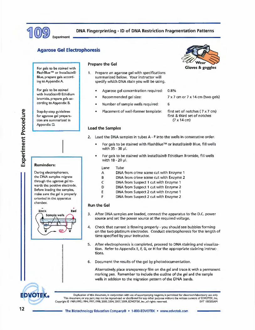
1. Prepare an agarose gel with specifications summarized below. Your instructor will specify which DNA stain you will be using.
• Agarose gel concentration required: 0.8%
• Recommended gel size: 7 x 7 cm or 7 x 14 cm (two gels)
• Number of sample wells required: 6
• Placement of well-former template: first set of notches ( 7 x 7 cm) first & third set of notches
(7 x 14 cm)
Load the Samples
2. Load the DNA samples in tubes A - F into the wells in consecutive order.
• For gels to be stained with FlashBlue™ or JnstaStain® Blue, fill wells with 35 - 38 µI.
• For gels to be stained with lnstaStain® Ethidium Bromide, fill wells with 18 - 20 µI.
Lane Tube A DNA from crime scene cut with Enzyme 1 B DNA from crime scene cut with Enzyme 2 C DNA from Suspect 1 cut with Enzyme 1 D DNA from Suspect 1 cut with Enzyme 2 E DNA from Suspect 2 cut with Enzyme 1 F DNA from Suspect 2 cut with Enzyme 2
Run the Gel
3. After DNA samples are loaded, connect the apparatus to the D.C. power source and set the power source at the required voltage.
4. Check that current is flowing properly- you should see bubbles forming on the two platinum electrodes. Conduct electrophoresis for the length of time specified by your instructor.
5. After electrophoresis is completed, proceed to DNA staining and visualization. Refer to Appendix E, F, G, or H for the appropriate staining instructions.
6. Document the results of the gel by photodocumentation.
Alternatively, place transparency film on the gel and trace it with a permanent marking pen. Remember to include the outline of the gel and the sample wells in addition to the migration pattern of the DNA bands.
Duplication of this document, in conjunction with use of accompanying reagents, is permitted for classroom/laboratory use only. This document, or any part, may not be reproduced or distributed for any other purpose without the written consent of EDVOTEK, Inc.
Copyright © 1989, 1992, 1994, 1997, 1998, 2000, 2004, 2007, 2009, EDVOTEK, Inc., all rights reserved. EVT I 00202AM
The Biotechnology Education Company® • 1-800-EDVOTEK • www.edvotek.com

DNA Fingerprinting - ID of DNA Restriction Fragmentation Patterns
Study Questions
1. Define FLP's and give their significance.
2. What is the most likely cause of Restriction Fragment Length Polymorphisms?
3. What are Variable Number of Tandem Repeats (VNTRs)?
4. Who are the only individuals possessing the same DNA fingerprints?
5. List the steps involved in DNA fingerprinting from extraction of DNA through the matching of a suspect to a crime scene sample.
6. What type of human cells can be utilized for this technique?
Duplication of this document, in conjunction with use of accompanying reagents, is permitted for classroom/laboratory use only.
Experiment i]@@
This document, or any part, may not be reproduced or distributed for any other purpose without the written consent of EDVOTEK, Inc. Copyright© 1989, 1992, 1994, 1997, 1998, 2000, 2004, 2007, 2009, EDVOTEK, Inc., all rights reserved. EVT I 00202AM
The Biotechnology Education Company® • 1-800-EDVOTEK • www.edvotek.com 13

DNA Fingerprinting - ID of DNA Restriction Fragmentation Patterns
®
The Biotechnology Education Company® • 1-800-EDVOTEK • www.edvotek.com
14 EVT I 00202AM