Embed Size (px)
Citation preview
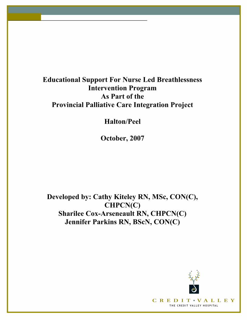
Educational Support For Nurse Led Breathlessness Intervention Program
As Part of the Provincial Palliative Care Integration Project
Halton/Peel
October, 2007
Developed by: Cathy Kiteley RN, MSc, CON(C), CHPCN(C)
Sharilee Cox-Arseneault RN, CHPCN(C) Jennifer Parkins RN, BScN, CON(C)

2
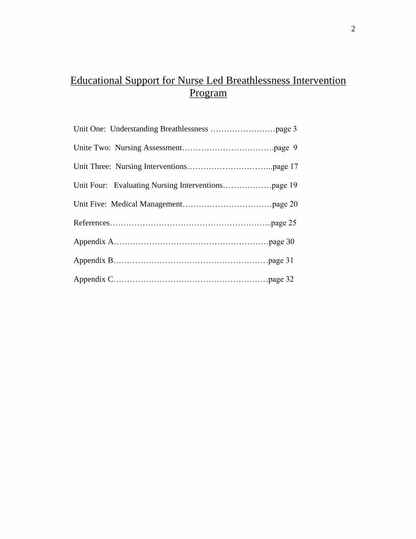
Educational Support for Nurse Led Breathlessness Intervention
Program
Unit One: Understanding Breathlessness ……………………page 3
Unite Two: Nursing Assessment…………………………….page 9
Unit Three: Nursing Interventions…………………………..page 17
Unit Four: Evaluating Nursing Interventions………………page 19
Unit Five: Medical Management……………………………page 20
References…………………………………………………...page 25
Appendix A…………………………………………………page 30
Appendix B…………………………………………………page 31
Appendix C…………………………………………………page 32

3
Unit One
Understanding Breathlessness
Traditionally, dyspnea has been approached using a biomedical framework, such
as looking at cause and effect. An alternative approach for assessing and managing
dyspnea exists which considers involving the mind, body experience and meaning of the
symptom for the individual experiencing the symptom. The community nurse is in an
opportune position to apply this as she/he engages caring relationships with individuals
and their families. The therapeutic relationship that the nurse engages in with these
clients provides the basis of continuity of care, partnership, reciprocity and mutual
inquiry which are the foundation of this intervention. Improved patient outcomes can be
realized when the meaning of breathlessness can be understood from the perspective of
the individual experiencing it (Krishnisamy, Corner, Bredin, Plant and Bailey, 2001).
Definitions
Breathlessness is best understood as symptom which has complex physical psychological
emotional and functional influences
(O’Driscoll, Corner, Bailey, 1999).
Breathlessness is a subjective experience described as an unpleasant or uncomfortable
awareness of the need to breathe (CCO, 2005).
Dyspnea us a subjective experience of breathing discomfort that consist of qualitatively
distinct sensations that vary in intensity…dyspnea derives from interactions among
physiological, psychological, social, and environmental factors (American Thoracic
Society, 1999).
Dyspnea is a subjective experience of difficult labored and uncomfortable breathing that
occurs when the demand for ventilation exceeds the individual’s ventilation capacity
(Brown, Carrueri, Janson-Bjerklie, Dodd, 1986).
Background and Statistics
Overall incidence of breathlessness reveals 21-90% (Brurea et al., 2000)
Breathlessness can occur with any cancer but is know to be prevalent in individuals with
lung cancer. It is a symptom that can increase as the disease progresses.
Bredin and colleagues (1999) state that between 10% to 15% of patients with lung cancer
have breathlessness at diagnosis and 65% will have the symptom at some point during
their illness.
4
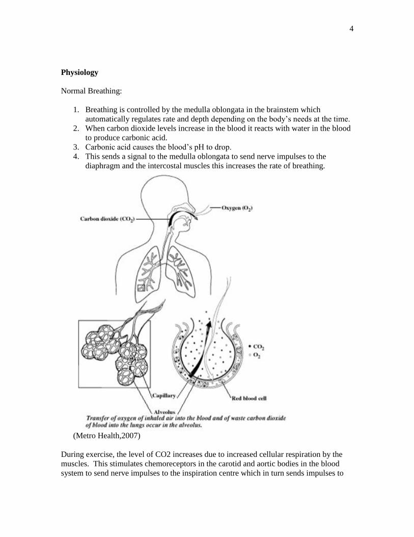
Physiology
Normal Breathing:
1. Breathing is controlled by the medulla oblongata in the brainstem which
automatically regulates rate and depth depending on the body’s needs at the time.
2. When carbon dioxide levels increase in the blood it reacts with water in the blood
to produce carbonic acid.
3. Carbonic acid causes the blood’s pH to drop.
4. This sends a signal to the medulla oblongata to send nerve impulses to the
diaphragm and the intercostal muscles this increases the rate of breathing.
(Metro Health,2007)
During exercise, the level of CO2 increases due to increased cellular respiration by the
muscles. This stimulates chemoreceptors in the carotid and aortic bodies in the blood
system to send nerve impulses to the inspiration centre which in turn sends impulses to

5
the diaphragm and intercostal muscles through the phrenic and thoracic nerves. The
diaphragm and intercostal muscles contract at a higher rate.
During rest, the level of CO2 is lower, so the breathing rate is lower. It is the build up of
CO2 making the blood acidic that causes a need for increased respiratory rate, rather than
lack of oxygen (Wikipedia, 2007).
The respiratory center in the medulla oblongata responds to stimuli from four sources:
Chemoreceptors: in the aorta, carotid arteries and medulla sense changes in PO2, PCO2,
and pH and transmit signals back to the respiratory centre to adjust breathing. The
peripheral receptors (those in the carotid and aorta) are most sensitive to changes in PO2.
When PO2 decreases respirations increase. However, please note hypoxia must be fairly
profound before this change in respiratory pattern is seen.
Mechanoreceptors: are located in the diaphragm and chest wall and they sense changes
in the work of breathing. When an increased work load is sensed, the respiratory centre
stimulates the diaphragm and respiratory muscles and attempts to expand the lungs.
Vagal receptors: in the airways and lungs also influence breathing. Afferent impulses
are generated when 1) stretch receptors in the lungs are stimulated as the lungs expand. 2)
irritant receptors in the bronchial wall are stimulated or 3) C fibres in the interstitium of
the lungs respond to an increase in pulmonary interstitial or capillary pressure.
Cortical areas within the brain: affect breathing by allowing individuals to consciously
increase or decrease their respiratory rate. It also appears the chemoreceptors,
mechanoreceptors, and respiratory centre, send messages to the higher brain centres
leading to a cognitive awareness of the ventilatory demand.
Physiology of DyspneaPhysiology of Dyspnea
6
Pathophysiology of Dyspnea
Precise origin of the sensation remains unknown.
1) Increased effort to overcome a certain load (For example, obstructive or
restrictive lung disease pleural effusion).
2) Increase in respiratory muscle required to maintain a normal workload (Muscle
wasting, cachetic).
3) Increase in ventilatory requirements (Hypoxemia, hypercapnia, metabolic
acidosis, and anemia).
In many cancer patients, different proportions of the three abnormalities may coexist,
thereby making the pathophysiologic interpretation of the intensity of dyspnea more
complex.
The etiology of breathlessness is very complex.
Breathlessness can be classified by the time course: it is acute, subacute, and
progressive.
Or by etiology, direct effects of the tumour, effects of the treatment or, indirect
effects.
Direct effect of the tumor
Primary or metastatic tumor
Pleural effusion
SVC
Carcinamatous lymphangitis
Atelectasis
Phrenic nerve palsy
Tracheal obstruction
Carcinomatous infiltration of the chest wall
Effects of therapy
Radiation fibrosis
Bleomycin, methotrexate, cyclophosphamide induced fibrosis
Adriamycin induced cardiomyopathy
Post pneumectomy
Indirect cancer related
Anemia
Cachexia
Rib fracture
Fever
Asthma
Pulmonary emboli
7
Heart failure
Obesity
Psychosocial distress
Cancer related dyspnea usually is multifactorial resulting from cancer and perhaps
exacerbated by cancer therapy or other factors.
Impacts of Breathlessness
The literature reveals that there are many unmet needs in relation to symptom
management for individuals with cancer and their families. Breathlessness is
frequently seen as a presenting symptom in lung cancer and a sign of advancing
tumour.
The experience of dyspnea is debilitating because it interferes with an individual’s
activities of daily living, such as their physical and psychological functioning.
Difficulty breathing causes people to stop the activity in which they were engaged in.
Dyspnea results in increased tension and apprehension or anxiety. Chronic dyspnea
causes fatigue.
Dyspnea leads to multiple threats and barriers to normal life and can have a
devastating effect on physical functioning, personal and sexual behaviours, self care,
household and work activities and social roles.
In addition, dyspnea negatively affects emotions and decreases comfort and peace of
mind.
8
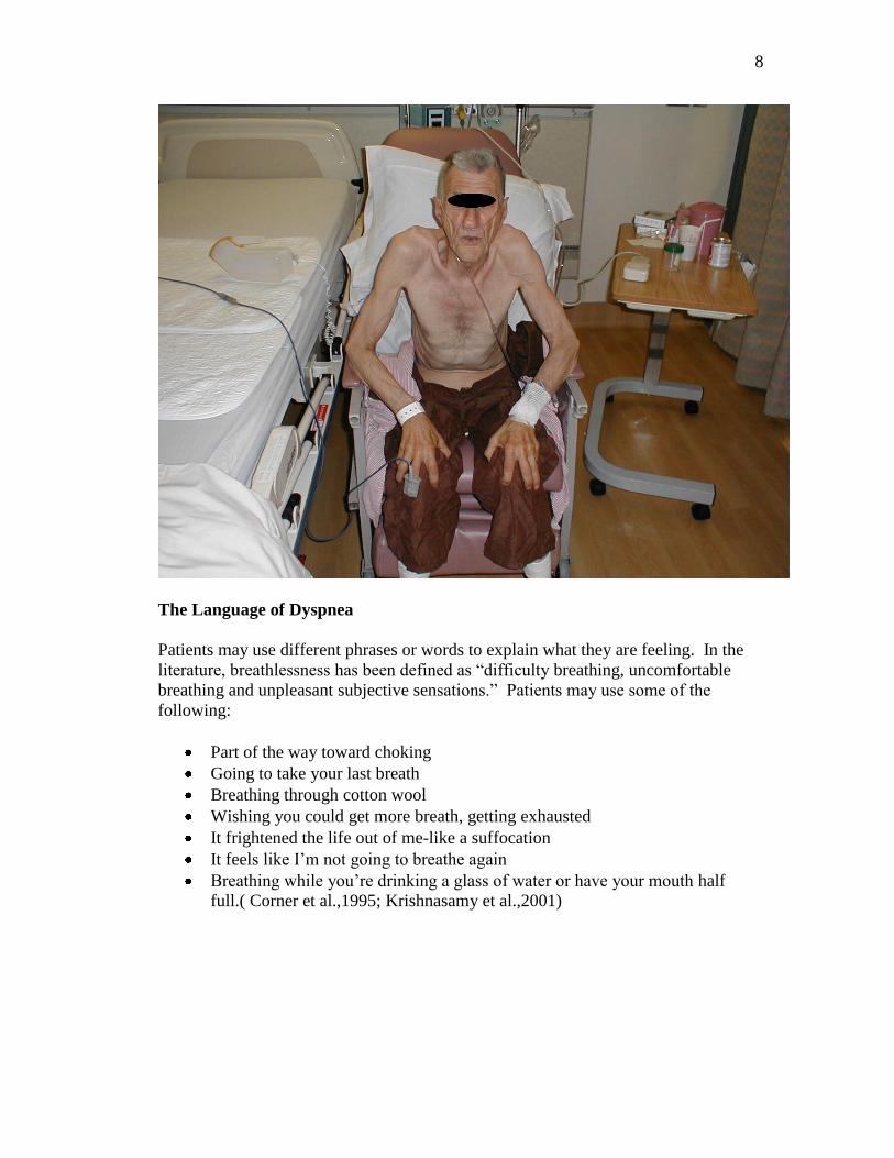
The Language of Dyspnea
Patients may use different phrases or words to explain what they are feeling. In the
literature, breathlessness has been defined as “difficulty breathing, uncomfortable
breathing and unpleasant subjective sensations.” Patients may use some of the
following:
Part of the way toward choking
Going to take your last breath
Breathing through cotton wool
Wishing you could get more breath, getting exhausted
It frightened the life out of me-like a suffocation
It feels like I’m not going to breathe again
Breathing while you’re drinking a glass of water or have your mouth half
full.( Corner et al.,1995; Krishnasamy et al.,2001)
9
Unit Two
Assessment
It is important to determine the etiology of dyspnea as dyspnea( or worsening
breathlessness) is frequently multifactoral and a result of the occurrence of several
factors. Assessment for this symptom tends to be generally poorly conducted and is
therefore frequently inadequately treated because the symptom is so subjective and
under-reported.
The assessment of breathlessness must be holistic and include the physical, social and
emotional components. Breathlessness can be a very frightening experience and it
can cause profound anxiety and stress.
Dyspnea assessment
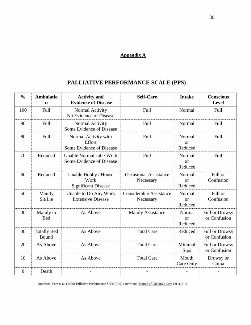
PPS:
This is a valid and reliable tool to guide the assessment of the palliative patient’s
functional performance.
The PPS provides a framework for measuring progressive decline over the course of
illness. It serves as a communication tool for the team. It can also be used to reassess
care and care requirements. For example patients who score between 0-40% usually
require increased hands-on nursing and their family members often need more
support compared to those patients with higher PPS scores (See Appendix A).
Level of distress:
Individuals with lung cancer have been reported to have heightened levels of
psychological distress (Sarna et al., 2005), however, it is important to determine the
level of distress a patient is experiencing. For example, a patient with longstanding
dyspnea may rate severity as 7/10 but may have little distress in relation to how long
they have had the symptom. In contrast a patient, who has not previously
experienced dyspnea, may rate as a 3/10, but may be very distressed.
Objective observation may alert the nurse that the client is having difficulty
breathing; however the degree of distress is subjective. The most simplistic and
concise method of assessing this is to simply ask the patient:
“Are you distressed?”
“How distressed are you?” and ask them to identify: -not at all
-somewhat
-very much
Onset, Duration and Severity:
Is onset new? Or a change to the usual pattern? A patient with chronic COPD
may have lived with dyspnea for sometime and developed compensation
mechanisms, whereas new onset may indicate the need for emergency medical
intervention.
10
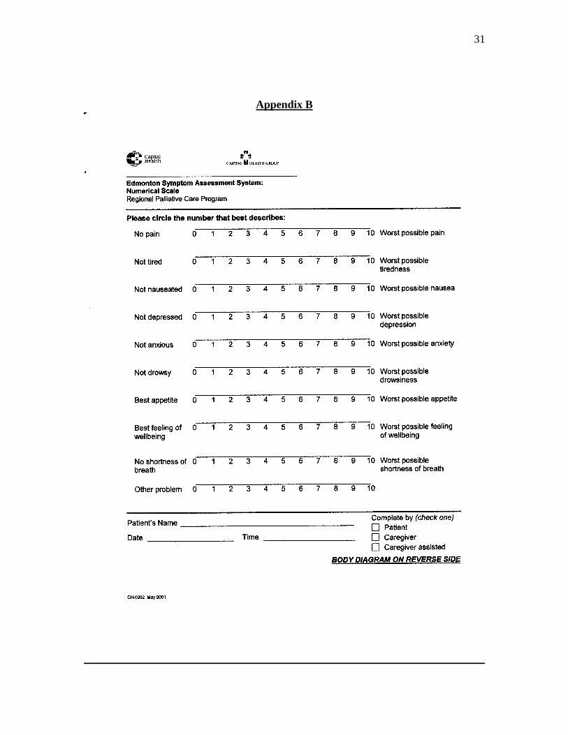
ESAS
The Edmonton Symptom Assessment System (ESAS) is a valid and reliable self-
assessment tool that monitors nine common symptoms experienced by cancer
patients; pain, tiredness, nausea, depression, anxiety, drowsiness, appetite, well
being and breathlessness (See Appendix B)
On a scale of 0-10 with 0 being no shortness of breath and 10 being the worst
possible shortness of breath, how do you rate your shortness of breath? Is the
onset sudden or progressive?
Is the pattern constant or intermittent?
According to the collaborative care plan, the severity of dyspnea can be categorized
according to ESAS scores as follows:
Mild Dyspnea
ESAS (0-3)
-Usually can sit and lie quietly
-May be intermittent or persistent
-Worsens with exertion
-No anxiety or mild anxiety during shortness of breath
-Breathing not observed as laboured
-No cyanosis
Moderate Dyspnea
ESAS (4-6)
-Usually persistent
-May be new or chronic
-Shortness of breath worsens if walking or with exertion; settles partially with rest
-Pauses while talking every 30 sec
-Breathing mildly laboured
Progressive Severe Dyspnea
ESAS (7-10)
-Often acute or chronic
-Worsening over days/weeks
-Anxiety present
-Often; awakes suddenly with shortness of breath
-May have cyanosis
-May have onset of confusion
-Laboured breathing awake and asleep
-Pauses while talking q 5-15 sec
11
Acute Exacerbation or Very Severe Dyspnea (Treat as a medical emergency)
-Sudden onset (minutes to hours)
-High anxiety and fear
-Agitation with very laboured respirations
-Air hunger-pauses while talking or unable to speak
-Exhausted
-Total concentration on breathing
-Cyanosis usually
-May be cold /clammy
-May or may not have respiratory congestion
-May or may not have acute chest pain
-May or may not be diaphoretic
-May or may not be confused
Precipitating and relieving factors:
What things make you breathless?
The oxygen cost diagram is a visual analogue scale consisting of a 100mm vertical line.
Everyday activities such as walking, shopping, and bed making are listed at various
points along the line. The activities are listed proportionately to the oxygen cost e.g. the
amount of oxygen needed to perform the activity. The oxygen cost diagram quantifies
breathlessness severity. It puts a number to the breathlessness symptom and it can be
used to evaluate response to treatment or to assess changes in breathlessness. (McGavin,
Artvinli, Naoe, & McHardy, 1978)
What do you do to improve your breathlessness?
-modify activity
-positioning
-breathing exercises
-medications (including use of O2)
Associated symptoms:
Do you experience any other symptoms or feelings when the shortness of breath occurs?
Chest pain:
Respiratory pain is usually sharp in nature and is aggravated by deep breathing or
coughing and occurs in 30-50% of patients with lung cancer diagnosis (Tyson, 2005).
Chest pain is often seen in patients with peripheral tumors although the etiology of chest
pain is not always clear.
In some patients chest pain may be directly related to chest wall or pleural invasion
12
Established methods of pain control should be used in these patients irregardless of the
etiology of pain.
Cough:
Cough is the most common presenting symptom and occurs in 50-75% of patients with
lung cancer (Tyson, 2005).
Assess cough in respect to:
Recent onset
Chronic cough
Irritating cough
Chronic cough plus production of large volumes of purulent sputum
Sputum
White mucoid sputum (Asthma and bronchitis)
Purulent green or yellow, foul smelling (Respiratory infection)
Blood (Carcinoma of lung, pulmonary embolism) hemoptysis is defined as blood
that is coughed up, it may be mixed with sputum. In 25-35% of lung patients,
hemoptysis is present at diagnosis. It usually occurs in patients who have
centrally located tumors as a result of tumor invading blood vessels or tumor
necrosis (Tyson, 2005)
Frothy white or pink (Pulmonary edema)
Thick Viscid (Life-threatening asthma)
Most important pearl about the assessment of dyspnea is that objective signs often do not
match the patient’s perception. Thus, the subjective report as in pain is the gold standard
for assessing this symptom.
Fever
Many patients with lung cancer display signs or symptoms of pneumonia, chest pain,
cough and fever (Tyson 2005).
Is there a presence of fever greater than 38.0 C?
Is the patient currently on chemo therapy for lung cancer?
If the client is currently on chemotherapy, consultation with oncology team is
necessary to rule out febrile neutropenia and/or related sepsis.
Physical assessment
General appearance
How does client appear on first visual?
Note: posture, body language, facial expression
Respiratory rate and vital signs
Monitor for RR above 24 rpm
Elevated heart or tachycardia

13
Color
Note whether ashen, pale, cyanotic, flushed
Use of accessory muscles
Note: Indrawing, substernal/suprasternal indrawing, diaphragmatic breathing
(belly breathing)
Stridor
A type of wheeze that is caused by the partial obstruction of the larynx or trachea, it
primarily occurs on inspiration and tends to high pitched.
Lung Auscultation
Auscultation of the lungs is the most important examining technique for assessing air
flow through the tracheobronchial tree. Auscultation involves:
1) Listening to the sounds generated by breathing.
2) Listening for any added or adventitious sounds.
3) If abnormalities are suspected, listening to sound of the patient’s spoken or whispered
voice as they are transmitted through the chest wall.
Breath Sounds
Patterns of breath sounds can be identified by their intensity, pitch and relative duration
of their inspiratory and expiratory phases.
Normal breath sounds are:
Vesicular, or soft and low pitched. They are heard through inspiration and continue
without pause through expiration and then fade away about one third of the way through
expiration.
Bronchovesicular: with and expiratory and inspiratory sounds about equal in length, at
times are separated by a silent interval, differences in pitch and intensity are often more
easily detected during expiration.
Bronchial: or louder and higher in pitch with short silence between inspiratory and
expiratory sounds. Expiratory sounds last longer than inspiratory sounds.
Tracheal breath sounds, very loud harsh sounds that are heard by listening over the
trachea and the neck.
Listen to the breath sounds with the diaphragm of the stethoscope after instructing the
patient to breathe deeply through an open mouth.

14
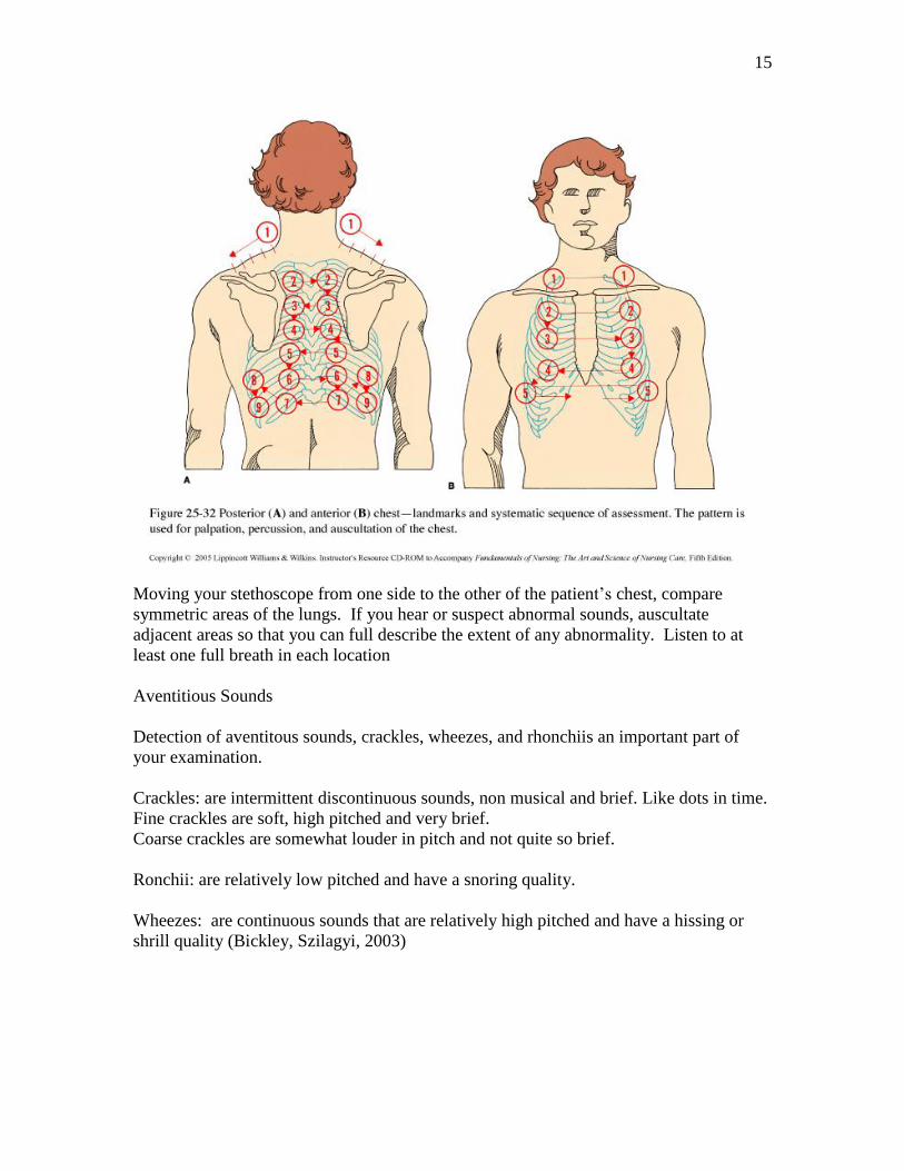
15
Moving your stethoscope from one side to the other of the patient’s chest, compare
symmetric areas of the lungs. If you hear or suspect abnormal sounds, auscultate
adjacent areas so that you can full describe the extent of any abnormality. Listen to at
least one full breath in each location
Aventitious Sounds
Detection of aventitous sounds, crackles, wheezes, and rhonchiis an important part of
your examination.
Crackles: are intermittent discontinuous sounds, non musical and brief. Like dots in time.
Fine crackles are soft, high pitched and very brief.
Coarse crackles are somewhat louder in pitch and not quite so brief.
Ronchii: are relatively low pitched and have a snoring quality.
Wheezes: are continuous sounds that are relatively high pitched and have a hissing or
shrill quality (Bickley, Szilagyi, 2003)

16
Wheezing is the result of the vibration of a narrowed airway as air passes through it. In
patients with lung cancer, wheezing often is caused by a lesion in the main stem bronchii.
The wheezing is localized and may be associated with a cough. This should be
differentiated from generalized wheezing, which usually is caused by bronchospasm
(Tyson, 2005).
Mental status
Degree of alertness
Alert
Confusion
Drowsy/stuporous
Psychological
Ask about the presence of anxiety or fear.
How does the breathlessness make the client feel?
The nurse may refer to anxiety scores and sense of well being on the ESAS as an
approach to addressing this, or opening the discussion.
Krishnasamy and colleagues (2001) present breathlessness as a symptom with physical
and emotional influences; therefore, it would be important to consider the presence of
anxiety, fear and uncertainty that the client may be experiencing along with the symptom
of breathlessness. The parallel model of care works in collaboration with the traditional
biomedical model of care. This approach then helps to identify the meaning of the
symptom by examining the psychological impact.
Anxiety does not cause breathlessness, but breathlessness left untreated will always result
in anxiety.
Diagnostic tests
O2 sat if able. Objective measurement rarely matches the subjective experience.
Therefore, this is helpful for obtaining data but not for expressing the significance of the
symptom for patient.
O2 saturation <90% indicates a need for immediate nursing intervention and consultation
with the medical team.
17
Unit Three
Nursing Interventions
Refer to “Toolkit for Breathlessness Management” for detailed interventions.
This toolkit elaborates on the Provincial Palliative Care Integration Project’s
Collaborative Care Plans and numerous best evidence random controlled trial studies
as well as the MacMillan UK based breathlessness education program. This Toolkit is
both for the management of patients experiencing breathlessness and also for those
who are at risk but not yet experiencing the symptom.
1. Assessment
2. Training in Breathing Control Techniques
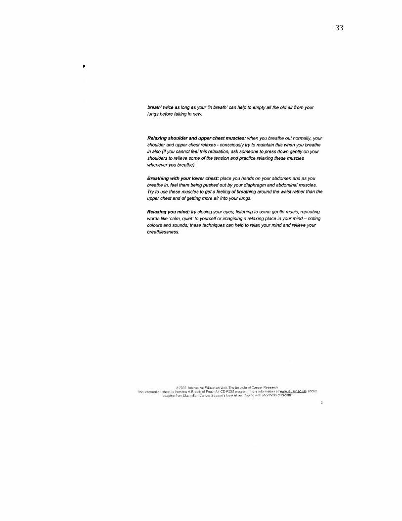
(See Attached Patient Fact Sheet: Coping with Shortness of Breath)
Bredin et al. (1999) conducted a randomized controlled study with
interventions carried out by specialist nurses including training in
breathing control techniques, progressive muscle relaxation and distraction
exercises. The findings showed that patients attending the clinics for
breathlessness experienced improvements in breathlessness, performance
status and physical and emotional states.
Gallo-Silver and Pollack (2000) wrote an article that reviewed breathing
techniques and provided detailed information about them. This suggested
that the breathing retraining has been proven to be beneficial, especially
when initiated at the earliest possible stage.
3. Goal Setting and Developing a Therapeutic Nurse Client Relationship
(See Attached Patient Fact Sheet: Managing Breathlessness)
Hately and colleagues (2003) conducted a study in which individuals with
lung cancer engaged in an intervention program that consisted of a range
of strategies including breathing retraining, simple relaxation techniques,
activity pacing and psychosocial support. The study outcomes revealed at
study entry 97% of patients were experiencing breathlessness at least once
or twice per day, 73% several times daily, and 27% most or all of the time.
At final visit, 27% were experiencing dyspnea several times daily and only
3% experiencing it most or all of the time.
4. Support and Advice on Managing Dyspnea
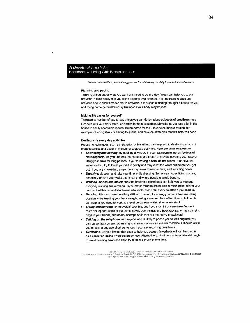
(See Attached Patient Fact Sheet: Living with Breathlessness)
Krishnasamy et al, (2001) considered the methodological and
philosophical issues that arose during a multicentre, randomized
controlled trial of a new nursing intervention to manage breathlessness
with patients with primary lung cancer and concluded that the intervention

18
is not about following a series of steps. The physical and emotional
components of the intervention are interdependent, as they are for the
individuals experiencing them. The method involved in working with
each patient involved the focus on breathing retraining and strategies for
minimizing functional limitations as well as engaging in the transpersonal
caring relationship to understand the meaning of the symptom for the
patient.
19
Unit Four
Evaluating Nursing Interventions
The literature supports that individuals with lung cancer who have received nursing
interventions for breathlessness management, focused within the areas of symptom
meaning, advice on ways to manage, breathing retraining and supportive counselling
experienced improvements in breathlessness ratings, ability to manage activities of daily
living, performance status and psychological well being. (Corner et al., 1996; Bredin et
al., 1996; Hately et al., 2003).
Initial dyspnea assessment should be recorded and patient stated goals should
be recorded
Nursing interventions should be recorded
Subsequent breathlessness scores should be recorded using the dyspnea
assessment to indicate the effectiveness of the intervention
Feedback from patients and family members should also be included
The patient goals should included their perception of decreased breathlessness
The goal should also include decreased perception of distress related to
breathlessness

20
Unit Five
Medical Management
After dyspnea assessment is complete, collaboration with the medical team may be
indicated for advice on further diagnostic testing and/or procedures, oxygen therapy and
pharmacology. In situations when there is a new onset or change to the patient’s regular
breathing pattern, an acute episode or exacerbation of breathlessness, new onset chest
pain, and/or fever greater than 38 C, consultation with the medical team will be indicated
(CCO, 2005).
Treatment of the Underlying Cause
Pleural Effusion
Thoracentesis, tenchkoff catheters, chest tubes and/or pleurodesis may be
indicated.
A chest x-ray will show a pleural effusion, but it can be difficult to differentiate
an effusion from collapse/consolidation or a mixture of the two. An ultrasound
scan will confirm the presence of a pleural effusion, and many radiologists will
mark a site for aspiration if requested. Chest x-ray or ultrasound should be
performed if a clinically-diagnosed pleural effusion has not been confirmed
radiologically and the clinical signs are not straightforward.
The aspiration of a pleural effusion can give symptomatic relief from dyspnea. A
pleural effusion large enough to cause dyspnea will be detectable clinically.
Aspiration of 300-500 ml fluid will usually give some symptomatic improvement
but up to 1.5 litres may be aspirated in some cases (Watson, Lucas, Hoy, & Back,
2005)
Anemia
Transfusion of packed cells if it is felt that this could improve dyspnea.
Sometimes a therapeutic trial is required to determine this
Cardiac
Diuretics, ace inhibitors, and digoxin should be considered
Pulmonary Emboli
Anticoagulation therapy should be considered
Infection
Treat with antibotics, expectorant, physio
Airway Obstruction
Radiotherapy is an option. Corticosteriods may be useful. Also, stenting, laser
treatment, and brachytherapy
21
Lymphangitis Carcinomatosis
Can only be diagnosed on x-ray and even this may not be diagnostic; suspect
when consistent severe breathlessness at rest or on exertion and widespread fine
crepitations in the lungs.
Consider corticosteroids, diuretics and/or bronchodilators
Radiation Fibrosis
Radiotherapy of tumors located within or around the thoracic cavity usually
results in partial irradiation of the surrounding normal lung tissue. Fibrosis
develops together with loss of capillaries, increases in the thickness of the
alveolar septa, and obliteration of the alveolar space (Bruner, Haas, Gosselin-
Acomb, 2005). Corticosteriods can be tried
(Alberta Palliative Care Resource, 2001).
Pharmacology
Medications have been explored as a method of relieving dyspnea by altering the
perceptual sensitivity and/or by exerting respiratory depressive effects (American
Thoracic Society, 1999).
Oxygen Therapy
Patients who are hypoxemic on room air are quite likely to benefit from oxygen therapy.
Most medical professionals recommend oxygen for patients with hypoxic dyspnea to
achieve and maintain an oxygen saturation greater than 88% (Dudgeon, 2002; Houlihan,
Inzeo, Joyce, & Tyson, 2005). However, the usefulness of oxygen for management of
patients with cancer who have nonhypoxic dyspnea is questioned in the literature (Bruera
& Ripamonti, 1998; Houlihan et al., 2005). Oxygen may acutely reduce exertional
dyspnea; however, an individual response to oxygen cannot be predicted with precision.
Thoughtful clinicians must always consider that oxygen can tie the patient down, it can
be claustrophobic, it may cause difficulty talking, and it may be associated with dry
mouth and increased risk of mouth sores.
As per the Dyspnea Management Guideline (2003), “Start humidified oxygen prn if the
patient is hypoxic (SaO2<92%) or if deemed helpful by the patient (Up to 6 L/min by
nasal prongs)” (KFL&A, p. 1).
In the community setting it is important to consider how patients can qualify for home
oxygen therapy when consulting with the medical team:
1. Individuals must have chronic hypoxemia on room air at rest (Pa02 of 55 mmHg
Or less, or SaO2 of 88 per cent or less).
Individuals with persistent PaO2 in the range of 56-60 mmHg may be considered
22
Candidates for long-term oxygen therapy if any of the following medical
conditions are present; cor pulmonale, pulmonary hypertension, persistent
erythrocytosis.
Individuals with PaO2 consistently in the range of 56 to 60 mmHg (SaO2 of
89 to 90 %) on room air may be considered candidates for funding if exercise
limited by hypoxemia and documented to improve with supplemental oxygen,
nocturnal hypoxemia.
2. Special consideration is given to Ontario residents at the end stage of a terminal
disease (i.e. life expectancy<3 months) who are receiving end of life care and
require home oxygen therapy. Individuals who are identified as receiving end of
life care will receive funding assistance for a maximum period of 90 days. The
start date of the coverage for palliative funding will be based on the physician’s
prescription date. There are no extensions for palliative care funding beyond the
90-day funding period. If home oxygen therapy is required after the 90-day
funding period, the client must apply to the regular funding program and the
eligibility criteria must be met (Ministry of Health and Long Term Care, 2005).
Pa02 levels are measured by arterial blood gases and can be ordered by the physician and
obtained by a Respiratory Therapist when the patient is in to the hospital for
appointments.
Opioid Therapy
Opioids are known respiratory depressants that reduce the central processing of neural
signals within the central nervous system. Opioids have been shown to modulate dyspnea
in acute bronchoconstriction. Opiates may alleviate dyspnea by blunting perceptual
responses so that for a given stimulus, the intensity of respiratory sensation is less
(American Thoracic Society, 1999). Cancer Care Ontario’s clinical practice guideline for
the management of dyspnea in cancer patients recommends, “systemic opioids, by the
oral or parenteral routes, can be used to manage dyspnea in advanced cancer patients and
nebulized morphine should not be used to treat dyspnea” (2006, p. 1).
It is important to distinguish between patients who have not been taking opioids regularly
for at least several days and patients who are already taking opioids regularly for pain and
dyspnea (Gallagher, 2003). The KFL&A (2003) dyspnea management guideline for
palliative care patients states that:
If the patient is not taking an opioid, initiate short-acting morphine 2.5-5.0 mg po
q4h and 2.5 mg po q2h prn for breakthrough (if the SC route is needed, divide the
PO dose by half)
Titrate up by 25% every 3 to 5 doses until dyspnea is relieved
If the patient is taking an opioid with q 4h dosing, increase this dose by 25%
If the patient is taking a long acting opioid, change back to q4h dosing and
increase this dose by 25%
Titrate short-acting opioid by 25% every 3 to 5 doses until dyspnea is relieved
23
If significant opioid side effects are present (e.g. nausea, drowsiness, myoclonus)
consider switching to another opioid (p. 1).
An increased HS dose may be helpful as dyspnea often is perceived as worse at
nightime.
Administration of liquid forms of morphine or dilaudid may allow for titration in
smaller increments.
There is no evidence that nebulized opioids are of any benefit (CCO,2006).
Anxiolytics
Anxiolytics have the potential to relieve dyspnea by depressing hypoxic or hypercapnic
ventilatory responses as well as by altering the emotional response to dyspnea. However,
several controlled studies with various benzodiazepines have failed to demonstrate
consistent improvement in dyspnea over placebo and the active drug tended to be poorly
tolerated (American Thoracic Society, 1999).
Nozinan has been shown to have some dyspnea management properties.
The KFL&A (2003) dyspnea management guideline for palliative care patients states
that:
To treat agitation with severe dyspnea consider Nozinan (Methotrimeprazine) 5
mg po/sc q4-6h prn and titrate to a maximum of 25 mg q4-6h prn
For all patients, if significant anxiety is present consider lorazepam 0.5-1.0 mg
po/iv/sc/sl q 30min prn for anxiety. (Carefully!) If the patient is already taking a
higher dose of lorazepam or another benzodiazepine, then dose appropriately.
Monitor for paradoxical agitation or excessive somnolence.
For all patients with very congested breathing consider glycopryrrolate 0.1-0.2 mg
sc q4h prn or scopolamine 0.3-0.6 mg sc q2-3h prn (p. 2).
Bronchodilators
Bronchodilators are useful therapeutic tools when dyspnea is exacerbated by reversible
airway obstruction. Their use should be considered in patients who are smokers or ex-
smokers and those with a history of bronchitis. Other obstructive causes could be a cold,
bronchial tumor, superior vena cava obstruction, radiation pneumonitis and pulmonary
fibrosis. They can be very effective in the patient with lymphangitic carcinomatosis
(Watson, Lucas, Hoy, & Back, 2005).
Consider:
B Adrenergic stimulants (e.g. salbutamol) (of note, excessive use can cause
cardiac stimulation)
Anticholinergics (e.g. atrovent)
Methylxanthines (e.g. theophylline) (of note, recommended with COPD, CHF,
can cause cardiac arrhythmias)

24
Corticiosteroids
Steroids are thought to reduce the edema associated with tumor. Breathlessness may
improve if due to multiple lung metastases, stridor due to airway obstruction, superior
vena cava obstruction and lymphangitic carcinomatosis (Watson, Lucas, Hoy & Back,
2005)
Consider:
Dexamethasone 4-8 mg po od for a one week trial if there is no improvement stop
Intractable Dyspnea
Dyspnea in the terminal phase can be quite severe. The management in this phase follows
standard symptom management principles. In addition, in the final hours of expected
life, appropriate sedation and comfort measures to manage the symptoms is warranted
(Yarbro, Frogge, & Goodman, 2004)
Reduce excessive secretions with scopolamine, hyoscyamine or atropine
Implement oxygen therapy if required
Institute sedation as needed (ONS,2007)
Midazolam (Versed)
In a study by Navigante and colleagues (2006), there was some evidence to support the
use of benzodiazepines for intractable dyspnea, such as midazolam. Midazolam is used
mainly for its properties of sedation.
Always remember, a holistic supportive approach can greatly improve breathlessness
management in patients. An extensive dyspnea assessment is the cornerstone of
determining the underlying cause of the symptom. Based on this assessment and the
essence of the therapeutic relationship, effective interventions such as teaching,
counselling in collaboration with the medical team can be successfully implemented to
improve symptom management.

25
References
Alberta Cancer Board. (2001). Alberta palliative care resource (2nd
ed.). Retrieved
October, 2007 from the Alberta Cancer Board Web Site:
www.albertapalliative.net
American Thoracic Society. (1999). Dyspnea, Mechanisms, assessment, and
management: A consensus statement. American Journal of Respirology Critical
Care Medicine, 159, 321-340.
Bickley, L. S., & Szilagyi, P. G. Bates' guide to physical examination and history taking.
Lippincott Williams & Wilkins. (Philadelphia, Pennsylvania) ** 2003; 8 Ed 862 p,
Bredin, M., Corner, J., Krishnasamy, M., Plant, H., Bailey, C., & A’Hern, R. (1999).
Multicentre randomized controlled trial of nursing intervention for breathlessness in
patients with lung cancer. BMJ, 318, 901-903.
Brown, M. L., Carrieri, V., JansonBjerklie, S., & Dodd, M. J. (1986). Lung cancer and
dyspnea: The patient's perception. Oncology nursing forum, 13(5), 19-24.
Bruera, E., Schmitz, B., Pither, J., Neumann, C. M., & Hanson, J. (2000). The frequency
and correlates of dyspnea in patients with advanced cancer. Journal of pain and
symptom management, 19(5), 357-362.
Bruera, E., & Ripamonti, C. (1998). Dyspnea in patients with advanced cancer. In A.
26
Berger, R.K., Portenoy, & D.E. Weissman (Eds.), Principles and practice of
supportive oncology (pp. 295-308). Philadelphia: Lippincott Williams &
Wilkins.
Bruner, D., Haas, M., & Gosselin-Acomb, T. (2005). Manual for radiation oncology
nursing practice and education (3rd
ed.). Pittsburgh, PA: ONS Publishing
Division.
Canadian Cancer Society. (2007). Canadian cancer statistics 2007. Retrieved Sept. 11,
2007 from the Canadian Cancer Society Web Site:
www.cancer.ca/ccs/internet/frontdoor/0,,3543_longID-en,00.html
Cancer Care Ontario. (2005). Nursing telephone practice and symptom management
guidelines: Breathlessness guideline. Retrieved September 5, 2007 from the
Cancer Care Ontario Web Site:
www.cancercareontario.on.ca/index_OtherPracticeGuidelines.htm
Cancer Care Ontario. (2006). The management of dyspnea in cancer patients: A clinical
practice guideline. Retrieved September 5, 2007 from the Cancer Care Ontario
Web Site: www.cancercareontario.on.ca/index_OtherPracticeGuidelines.htm
Corner, J., Plant, H., A’Hern, R., & Bailey, C. (1996). Non-pharmacological intervention
for breathlessness in patients with lung cancer. Palliative Medicine, 10, 299-305.
Dudgeon, D.J. (2002). Managing dyspnea and cough. Hematology/Oncology Clinics of
North America, 16, 557-577.
27
Gallagher, R. (2003). An approach to dyspnea in advanced disease. Canadian Family
Physician, 49, 1611-1616.
Gallo-Silver, L, Pollack, B. (2000). Behavioural interventions for lung cancer-related
breathlessness. Cancer Practice, 8(6), 268-273.
Hately, J., Laurence, V., Scott, A., Baker, R., & Thomas, P. (2003). Breathlessness
clinics within specialist palliative care settings can improve the quality of life
and functional capacity of patients with lung cancer. Palliative Medicine, 17, 410-
417.
Hood, L.E. & Harwood, K. (2004). Dyspnea. In C. Henke Yarbro, M. Hansen Frogge, &
M. Goodman, Cancer symptom management (3rd
Ed.). Sudbury, Massachusetts:
Jones and Barlett Publishers.
Houlihan, N., Inzeo, D., Joyce, M., & Tyson, L. (2005). In N. Houlihan, Site specific
cancer series (pp. 103-124). Pittsburgh, Pennsylvania: ONS Publishing Division.
Institute of Cancer Research. (2007). A breath of fresh air: An interactive guide to
Managing breathlessness in patients with lung cancer. Sutton, UK: Author.
KFL&A Palliative Care Integration Project. (2005). Palliative care integration project:
Resource manual. Kingston, Ontario: Author.
Krishnasamy, M., Corner, J., Bredin, M., Plant, H., & Bailey, C. (2001). Cancer nursing
practice development: understanding breathlessness. Journal of Clinical Nursing,
10, 103-108.
Martelli-Reid, L. (2006). Nursing management of breathlessness; oral presentation.
28
McGavin, C.R., Artvinli, M., Naoe, H., & McHardy, G. (1978). Dyspnoea, disability, and
distance walked:Comparison of estimates of exercise performance in respiratory
disease. BMJ, 2, 241-243.
Metro Health. Normal Breathing Diagram. Retrieved October 30, 2007 from Metro
Health Web Site: www.metrohealh.org/body.cfm?id=1553
Ministry of Health and Long Term Care. (2005). Assistive devices program: Ministry of
health and long term care.. Queen’s Printer for Ontario: Author.
Navigante, A.H., Cerchietti, L.C., Castro, M. A., Lutteral, M.A., & Cabalar, M.E. (2006).
Midazolam as adjuncant therapy to morphine in the alleviation of severe dyspnea
perception in patients with advanced cancer. Journal of Pain and Symptom
Management, 31, 38-47.
O’Driscoll, M., Corner, J., & Bailey, C. (1999). The experience of breathlessness in lung
cancer. European Journal of Cancer Care, 8, 37-43.
Oncology Nursing Society. (2007). Dyspnea: What can nurses do to assist people with
cancer-related dyspnea? Putting evidence into practice series. Pittsburgh: PA:
ONS Publishing Division.
Sarna, L., Brown, J., Cooley, M., Williams, R., Chernecky, C., Padilla, G., et al. (2005).
Quality of life and meaning of illness of women with lung cancer. Oncology
Nursing Forum, 32(1), 9-19.
Tyson. (2005). Patient Assessment. In N. Houlihan, Site-specific cancer series: Lung
cancer (pp. 35-44). Pittsburgh, Pennsylvania: ONS Publishing Division.

29
University of California San Diego. (2005). Breathless patient picture insert. Retreived
October 30, 2007 from the University of California San Diego Web Site:
http://medicine.ucsd.edu/clinicalmed/lung.htm
Watson, J. (2005). Caring science as sacred science. Philadelphia, PA: Davis.
Watson, M., Lucas, C., Hoy, A., Back, I. (2005). Oxford handbook of palliative care.
Oxford, NY: Oxford University Press.
Wikipedia. (2007). Breath: Definition. Retrieved Sept. 15, 2007 from the Wikipedia Web
Site: www.wikipedia.ca

30
Appendix A
PALLIATIVE PERFORMANCE SCALE (PPS)
Anderson, Fern et al. (1996) Palliative Performance Scale (PPS) a new tool. Journal of Palliative Care 12(1), 5-11
% Ambulatio
n
Activity and
Evidence of Disease
Self-Care Intake Conscious
Level
100 Full Normal Activity
No Evidence of Disease
Full Normal Full
90 Full Normal Activity
Some Evidence of Disease
Full Normal Full
80 Full Normal Activity with
Effort
Some Evidence of Disease
Full Normal
or
Reduced
Full
70 Reduced Unable Normal Job / Work
Some Evidence of Disease
Full Normal
or
Reduced
Full
60 Reduced Unable Hobby / House
Work
Significant Disease
Occasional Assistance
Necessary
Normal
or
Reduced
Full or
Confusion
50 Mainly
Sit/Lie
Unable to Do Any Work
Extensive Disease
Considerable Assistance
Necessary
Normal
or
Reduced
Full or
Confusion
40 Mainly in
Bed
As Above Mainly Assistance Norma
or
Reduced
Full or Drowsy
or Confusion
30 Totally Bed
Bound
As Above Total Care Reduced Full or Drowsy
or Confusion
20 As Above As Above Total Care Minimal
Sips
Full or Drowsy
or Confusion
10 As Above As Above Total Care Mouth
Care Only
Drowsy or
Coma
0 Death - - - -

31
Appendix B

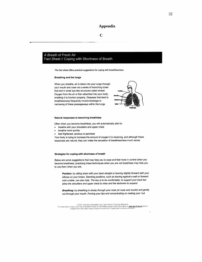
32
Appendix
C
33
34

35
36
37