Embed Size (px)
Citation preview

join us...JUNE 7–11, 2015
LYON, FRANCE
isAkoscongress
10TH Biennial
2015
ISA
KO
S N
ew
Sl
et
te
r 2
01
3 •
VO
lu
me
II
INSIDE 6 ISAKOS & FIFA – A GOld TeAm
8 ISAKOS COnGrATulATeS AwArdeeS
18 ISAKOS HOnOrAry memberS
Femoral head as seen during hip Arthroscopy in a 15 year old boy with Perthes. Impingement mechanism was addressed prior to subsequent PAO.
Cur
rent
Con
cept
s on
Art
hros
copy
, Kne
e Su
rger
y &
Ort
hopa
edic
Spo
rts
med
icin
e

2
EDITOROmer Mei-Dan, MD, USA
EDITORIAL BOARDNadim Aslam, FRCS Orth, UNITED KINGDOMMandeep S. Dhillon, Prof., MS, FAMS, INDIALucio S. R. Ernlund, MD, MSc, BRAZILNorimasa Nakamura, MD, PhD, JAPANDavid Sadigursky, MD, BRAZILGonzalo Samitier, MD, PhD, USADaniel A. Slullitel, MD, Prof., ARGENTINALuis Alberto Vargas, MD, USAJames H. Lubowitz, MD, USA, Past Editor
inthis issueEditor’s Message ..........................................1Presidential Message ....................................2ISAKOS Office Update ...................................3ISAKOS & OREF Grant Winner Announcement .............................4ISAKOS and FIFA ..........................................62013 ISAKOS Congress Wrap Up ..................7Case Corner ................................................24Current Concept Article ...............................26Worst Case Scenario ..................................34Pearls & Pitfalls – Surgical Technique ........37Lifestyle ......................................................42Tip of the “Day” .........................................44In A Nutshell ...............................................45Approved ISAKOS Courses ..........................46ISAKOS Asks ...............................................47Upcoming ISAKOS Collaborative Courses ....47Upcoming Approved Courses ......................48
ISAKOS would like to thankall who attended the 9th Biennial ISAKOS Congress in Toronto, Canada! The city of Toronto played host to more than 3,300 ISAKOS Congress participants, exhibitors and their guests during the week of May 12 – 16, 2013.
ISAKOS Congress Program Chairman, Marc Safran, MD, USA and the ISAKOS Program Committee developed a diverse and exceptional educational program including more than 280 international experts on a variety of topics ranging from general arthroscopy, to the use of biologics, to the pearls and pitfalls of surgical techniques. All major specialty areas were covered from concussion to the foot and ankle!
The 9th Biennial ISAKOS Congress offered course participants a packed schedule of Instructional Course Lectures, symposia, debates, lectures, surgical demonstrations and scientific paper presentations. More than 250 scientific papers were presented at the podium, in addition to more than 500 electronic posters. Presentations on topics such as hip arthroscopy, ACL reconstruction and rotator cuff repair were especially well attended. Special thanks are due to all ISAKOS Congress presenters – your vitality and variety help to make the ISAKOS Congress the outstanding educational event that it has become!
ISAKOS participants also had the option to expand their educational opportunities with the ISAKOS Congress Pre-Courses and Sports Rehabilitation Concurrent Course. Special thanks to the chairs of these courses! More information on these events can be found on page 16 of this Newsletter.
The ISAKOS Executive Committee and Board of Directors wish to thank all who attended the 2013 ISAKOS Congress. It is your participation and diverse perspectives that make the ISAKOS Congress the extraordinary educational event that it is!
International Society of Arthroscopy, Knee Surgery and Orthopaedic Sports Medicine
2678 Bishop Drive, Suite 250San Ramon, CA 94583 USA
Telephone: (925) 807 – 1197Fax: (925) 807 – 1199Email: [email protected]
01 Marc Safran, ISAKOS Program Chair 2013 and Moises Cohen, ISAKOS President 2011 – 2013
01

ISAKOS NEWSLETTER 2013: Volume II 1
As summer continues in the northern hemisphere, and winter cools the south, we are still digesting our recent memories from the long-awaited ISAKOS Congress in Toronto.
As with previous Congresses, the meeting in Toronto was a huge success, bringing together surgeons from all over the globe to interact scientifically and socially, to share experiences and thoughts, differences and similarities.
In my opinion, this is what ISAKOS is all about…
Big world, small (but rapidly growing!) community. Interaction without boundaries, resulting in a fruitful experience for us all. New friends, new places to visit, new techniques to adopt, and constant evolution.
With our eyes on the future ISAKOS Congress in Lyon in 2015, we have decided to broaden the ISAKOS voice to many more aspects of life that are important and relevant to us. This is in keeping with our society’s spirit and our ever expanding interests far beyond simply orthopaedics.
ISAKOS members are well read and up to date with the scientific literature, and as your global voice we are aware that the ISAKOS Newsletter is not meant to be a substitute for high impact orthopaedic journals. However, we would like to generate a global discussion and common language, to provide the community with cutting edge information that is not usually available in the purely scientific forums. We aspire to share life experiences and to let our role models pass on their wisdom, in order to eliminate the need to reinvent the wheel all over again, or fight battles which have already been won.
In the era of Facebook, Twitter and streamlined communication, where information is available in small digestible bites, the ISAKOS Newsletter is moving to quick, take-away messages, so you can consume it, sip by sip, with your morning espresso or in between cases.
As the new Editor of the ISAKOS Newsletter, my vision is for the ISAKOS Newsletter to be something you can pick up and read for a few minutes at a time, and still be provided with interesting, relevant information. We hope it will provoke thought perhaps on topics slightly broader than typical journals.
There are the things that surround us or we use everyday, but we do not always stop to think what are they made of or how they work (see ‘In a Nutshell’, Page 45).
• As we progress and develop in our careers, we are all curious to know how our mentors deal with both family and professional life decision making (jump to ‘Life Style’, Page 42).
• We have also included advice from the more organized or experienced physicians among us, that we hope you find helpful to learn how they keep five balls in the air, stay productive, on schedule, safe and happy! (see Robert Marx’s OR checklist on Page 39).
• Everyone could use tips on how to get through a busy professional day without stumbling or fading, an issue which many of us have been dealing with (without too much success) for years (Visit our Nutritionist on Page 44).
I’d like to take this opportunity to thank James Lubowitz, USA, the legendary Editor in Chief of this Newsletter, for six outstanding years of excellence. Jim has turned this Newsletter into an established and respected global forum, making it hard for anyone to fill these large shoes.
Finally, we would like congratulate our incoming new President, Masahiro Kurosaka, and send a round of applause to ISAKOS’ Executive Director, Michele Johnson, and her amazing team, Donna Festo, Katie Anderson, Hilary Matthews, Kathleen Reyes, April Warden and Beverlee Galstan, who will take us with great vision and expertise into this new and exciting 2013 – 2015 term.
Enjoy!
Omer Mei-Dan, MD, USA
EDITOR’S MESSAGE
01 Marc Safran, ISAKOS Program Chair 2013 and Moises Cohen, ISAKOS President 2011 – 2013

ISAKOS NEWSLETTER 2013: Volume II 2
I would like to begin my first ISAKOS Presidential Message, with a congratulations to the entire membership of ISAKOS on the wonderful success of the recent 9th Biennial ISKAOS Congress, held on May 12 – 17, 2013 in Toronto, Canada.
Special thanks are owed to Marc Safran, MD, USA and his extraordinary Program Committee, for creating a diverse and energetic scientific program for participants of all specialties. I would like to offer additional thanks to all paper presenters, and faculty members for making the Congress a diverse educational event, and creating a collaborative and interactive atmosphere.
As the President of ISAKOS, I am grateful for the dedication and hard work of my predecessors Moises Cohen (Brazil), and Freddie Fu (USA). I look forward to continuing their great work with the collaboration of the entire ISAKOS Membership for my 2013 – 2015 presidential term.
When I was named as part of the ISAKOS presidential line in 2009, I began to reflect on the future of ISAKOS. What will ISAKOS be? Who can shape the future or ISAKOS? How can we fulfill our commitments to our members?
With an eighteen-year history, I always compare ISAKOS to a teenager – young, talented, and energetic, with with an ambition to change the world! ISAKOS is an international society embracing 4,032 members, which makes it a unique, living, motivated society with huge potential and tremendous fortune. However, potential and fortune will not develop on their own – we need wisdom and a deep sense of history to forsee the potential and grow! I was very happy when I found the future is in the younger generation’s hand…. they are the fortune of ISAKOS.
The ISAKOS traveling fellowship program has witnessed fruitful achievements in the winning physicians. I was inspired by the creativity and passion from those young people when they were traveling around the world and learning from others. Without any doubt, they will be the leaders in their countries in the coming future, and we feel the responsibility to establish a wider platform for more and more young people to show their talent and aspiration. The ISAKOS Upper Extremity and Patellofemoral Traveling Fellowships are fantastic opportunities, and ISAKOS is thrilled to announce the opening of the Masaki Watanabe Arthroscopy Traveling Fellowship! For more information on these fellowships, please visit the ISAKOS Congress Awards website - http://isakos.com/meetings/2015congress/Awards.aspx.
Education is at the core of ISAKOS’ mission to “advance the worldwide exchange and dissemination of education, research and patient care in arthroscopy, knee surgery and orthopaedic sports medicine.” This is why the Education Committee and Program Committee are essential when we discuss ISAKOS’ future. I am humbled to see the continuing hard work and tireless contributions from my predecessors and colleagues in the purpose of making the ISAKOS an international, diverse, and committed organization. One such example of the ISAKOS Education Committee’s efforts are the four booklets published and distributed at the 2013 ISAKOS Congress. These booklets are on a variety of topics ranging from Rotator Cuff to the Meniscus, and I encourage
you to log in to ISAKOS Global Link to read them if you were unable to attend the ISAKOS Congress.
In the next two years, we still need the vivid participation and volunteerism from all of our members, by unifying the strength, sharing the values, and keeping the forward-looking mindset in heart, to do the right things, to shape a brighter future for ISAKOS.
Masahiro Kurosaka01 From left: Freddie Fu, ISAKOS President 2009 – 2011
Marc Safran, ISAKOS President 2017 – 2019 Masahiro Kurosaka, ISAKOS President 2013 – 2015 Moises Cohen, ISAKOS President 2011 – 2013 Philippe Neyret, ISAKOS President 2015 – 2017
01
PRESIDENT’S MESSAGE

ISAKOS NEWSLETTER 2013: Volume II 3
ISAKOS Global Linkwww.isakos.com/globallinkGlobal Link is ISAKOS’ online education resource. Navigate by anatomic location, sports medicine, or search by keyword to access content from archived ISAKOS Congress presentations, surgical demonstrations, committee projects and current concept articles. The ISAKOS Global Link currently includes the latest research presented at the ISAKOS Congress, as well as resources for Standard Terminology, Definitions, Classifications and Scoring Systems for Arthroscopy, and a video series for Normal Arthroscopic Anatomy.
New Content Now Available: 2012 Upper Extremity Consensus on Rotator Cuff Disease: Known Facts and Unresolved Issues
Global Connection Campaignwww.isakos.com/campaignISAKOS is focused on expanding international education in arthroscopy, knee surgery and orthopaedic sports medicine in an organized and collaborative effort with national societies and industry partners. To do so, ISAKOS launched the ISAKOS Global Connection: A Campaign for Education, Research and Collaboration, a fundraising campaign that will help shape the future of our Society mission. At the core of this endeavor is a determination to provide high quality and accessible educational programs to a global audience. Through partnerships with ISAKOS corporate supporters, we are given the opportunity to reach a variety of audiences through education and training, to raise funds for research and patient education, and to promote access to high-quality care. The Godfather Initiative was also created in order to provide meaningful benefits to individuals who donate to the Global Connection Campaign, as well as to individuals interested in ISAKOS membership, but who may not currently have the resources to become members. To learn more about the Godfather Initiative, visit isakos.com/campaign/godfathers.
ISAKOS
ISAKOS Thrives Online to Better Serve Its Members
ISAKOS has numerous online outlets for members and users to learn and gain from all the Society has to offer. The Society constantly strives to develop programs to better serve our members, as well as to reinforce involvement and partnership.To maximize your ISAKOS Membership you are invited to access the following ISAKOS online portals and programs:
myISAKOSwww.isakos.com/myISAKOSAs a replacement to the “Members Only” section of the ISAKOS website, the myISAKOS Dashboard reshapes the way ISAKOS
members and non-members access their ISAKOS accounts. ISAKOS Members can now conveniently manage their profile information, subscriptions and access their member benefits. Non-members can use myISAKOS to browse public ISAKOS resources and benefits, manage their account information as well as apply for membership.
myCongresswww.isakos.com/2015congress/myCongressmyCongress was developed as a gateway to the ISAKOS Congress for its attendees and participants. With myCongress, users are
able to register, manage program participation, review or edit submitted abstracts and more. Through this dashboard, one can also request a Visa letter or CME certificate, download important Congress documents as well as find links to book accommodations and tours. With this single portal, meeting attendees and participants can easily access everything needed to participate in the ISAKOS Biennial Congress.
ISAKOS Welcomes Your Suggestions! We’d love to hear your thoughts and ideas. Please feel free to send a message to [email protected]
These wonderful opportunities are created exclusively with the ISAKOS Member in mind. ISAKOS encourages you to explore these online tools and hopes that they make your involvement with ISAKOS much more engaging and convenient.
myCongress
ISAKOS OFFICE UPDATE
myISAKOSwww.isakos.com/myISAKOSAs a replacement to the “Members Only” section of the ISAKOS website, the myISAKOS Dashboard reshapes the way ISAKOS
members and non-members access their ISAKOS accounts. ISAKOS Members can now conveniently manage their profile
myISAKOS

ISAKOS NEWSLETTER 2013: Volume II 4
Clinical Application of Quantitative Assessment of the Pivot Shift – A Multicenter Study November 1, 2012 – June 1, 2013
Principle Investigator:Volker Musahl, MDPittsburgh, PA, USA
A. Specific AimsThe Specific Aims for the study remain unchanged. They are:
Specific Aim 1: Determine the relationship in ACL-deficient knees between quantitative measurements during the pivot shift test and clinical grading of ACL injury (partial vs. isolated vs. combined injury)
Specific Aim 2: Determine the relationship in ACL-reconstructed knees between quantitative measurements during the pivot shift test at time-zero following reconstruction and post-operative patient-reported and clinician-measured outcomes at 2 years
Specific Aim 3: Determine the relationship in ACL deficient knees between quantitative measures of joint kinematics as evaluated with inertial sensors compared to a new video-based iPad application.
B. Investigator meetings1. (Bologna, Italy) – In October 2012 we had a two-day investigator meeting at the Rizzoli Institute in Bologna, Italy. Investigators from all Institutions were present (Gulio Marcheggiani Muccioli, Cecilia Signorelli, Nicola Lopomo, all Bologna; Yuichi Hoshino, Ryosuke Kuroda, all Kobe; Kristian Samuelsson, Gothenburg; James Irrgang, Volker Musahl, all University of Pittsburgh). During this meeting we introduced the two devices (accelerometer and iPad-image analysis) to the investigators. We examined two patients pre-operatively while awake in the office using both devices. The next day we had the opportunity to test both subjects under anesthesia and observe ACL reconstruction. Time lines were set for obtaining IRB approval and tentative start date for enrolment. We discussed translation of our outcome tools and IRB forms in each institution’s respective language. Finally, we reviewed and finalized a manual of operations and procedures (MOOPs).
2. (Toronto, Canada) – In May 2013, the investigators met during the ISAKOS Congress 2013 in Toronto, Canada. Investigators from all Institutions were present (Stefano Zaffagnini, Jon Karlsson, Hakur Björnsson, Yuichi Hoshino, Ryosuke Kuroda, Daisuke Araki, James Irrgang, and Volker Musahl). During this meeting the investigators discussed progress of the study including issues regarding recruitment and retention, data collection and implementation of the central data collection system. Additionally, the investigators discussed the administrative issues surrounding the distribution of funds to the sites. Finally, the investigators discussed preliminary study data and preparation of abstracts for upcoming professional conferences.
The next investigator meeting is being planned for October 2013 in Gothenburg, Sweden.
C. Manual of Operations and ProceduresDuring the investigator meeting in Bologna, the investigators agreed on the manual of operations and procedures (MOOPS). The MOOPs includes the study protocol, study flow chart, study sites, training plan, communications plan, recruitment, screening and eligibility criteria, informed consent and patient confidentiality, description of study intervention, participant evaluations and follow-up, participant retention, concomitant procedures, data and safety monitoring activities, study compliance, data collection and study forms, data flow, regulatory documents, retention of study documentation, administrative forms, data management, quality control procedures, study completion and closeout procedures, policies, maintenance of the MOOP, and references.
During the investigator meeting in Toronto, the investigators agreed to change the one of the inclusion criteria. Patients will be eligible to participate in the study if they undergo surgery within 1 year of injury.
ISAKOS & OREF Grant Winner Announcement

ISAKOS NEWSLETTER 2013: Volume II 5
D. Studies and ResultsIRB approval has been obtained for Rizzoli Institute (December 6, 2012), University of Pittsburgh (December 15, 2012), Kobe University (January 7, 2013) and Sahlgrenska University (February 26, 2013). Additionally IRB approval for the Coordinating Center at the University of Pittsburgh was obtained on November 30, 2013. Enrolment began at the Rizzoli Institute December 17, 2012, University of Pittsburgh January 8, 2013, and Kobe University February 1, 2013. As of June 1, 2013, 22 subjects have been scheduled by the investigators to undergo ACL reconstruction. All 22 subjects were tested pre-operatively and post-operatively under EUA. Outcome scores were obtained pre-operatively.
E. Adverse EventsThere are no adverse events to report.
F. SignificanceSuccessful completion of this study will provide quantification of the pivot shift test. The data from this study will be useful for diagnosis, surgical planning and assessing outcome of treatments for ACL injuries. The measurement of tibial translation and acceleration during the pivot shift test coupled with the standardized pivot shift test maneuver could provide consistent evaluation of rotatory knee laxity in ACL deficient and reconstructed knees. These new non-invasive technologies could be used for patient-individualized and functional treatment of ACL injury.
G. PlansWe plan to continue with subject recruitment and follow-up as proposed in the original application. An electronic data capture system has been developed for this project for all data forms.
H. Related Publications & PresentationsB. Muller, M. Hofbauer, Y. Hoshino, P. Araujo, M. Ahlden, J.J. Irrgang, F.H. Fu, V. Musahl Image Analysis for Quantification of the Pivot Shift Test and Development of an iPad Application (ISAKOS E-poster 9063)
Y. Hoshino, N. Lopomo, M. Ahlden, M. Wolf, S. Zaffagnini, F.H. Fu, V. Musahl Quantitative Measurement of the Pivot Shift is Improved using a Standardized Technique and Accelerometer (ISAKOS E-poster 8098)
P. Araujo, Y. Hoshino, M. Ahlden, B. Muller, J.J. Irrgang, F.H. Fu, V. Musahl Quantitative Parameters of the Pivot Shift – Correlation and Clinical Use (ISAKOS E-poster 7826)
N. Lopomo, C. Signorelli, Y. Hoshino, M. Ahlden, P. Araujo, F.H. Fu, V. Musahl, M. Marcacci, S. ZaffagniniA standardized Technique in Performing Pivot Shift Test on the Knee Joint Provided More Consistent Acceleration Cure Shape, Allowing to Highlight Side-to-side Differences (ISAKOS Paper 8818)
V. Musahl, M. Hofbauer, B. Muller, M. Wolf, D. Araki, Y. Hoshino, J.J. Irrgang, F.H. Fu A Standardized Pivot Shift Test and Newly Developed iPad Application can Quantify ACL Injury in Patients (ISAKOS Paper 9079)
01 Fig 1 Recruitment Summary as of June 1 201302 Fig 2 Actual vs goal recruitment as of June 1 2013
ISAKOS & OREF Grant Winner Announcement
01
02

ISAKOS NEWSLETTER 2013: Volume II 6
ISAKOS and FIFA – A Gold TeamLuis A. Vargas, MD, PhD, USA Moises Cohen, MD, PhD, BRAZIL
In May 2013, the first ever meeting between FIFA’s Chief Medical Officer, Professor Jiri Dvorak, and the Board of Directors of ISAKOS was held in Toronto just hours before the kick off of the Pre-Course ISAKOS & FIFA: Key Issues and Challenges in Safety and Health in Soccer.
The meeting enabled both sides to exchange ideas for mutual cooperation in the near future and the signing of an agreement to produce two important events. The first one to be held in Sao Paulo, Brazil February 2014, and the second one in 2016 in the city of Lyon, during the 10th Biennial ISAKOS Congress.
Another major project to be undertaken is a traveling fellowship involving physicians from regions indicated by FIFA Board and the FIFA centers of excellence, with the aim of disseminating and maintaining the FIFA / ISAKOS standard of excellence in areas which have not yet been created. The possibility of using a single universal database in medicine and soccer was also discussed.
FIFA has a design of medical care and surgeries for athletes in underserved areas, an important social and humanitarian gesture.
The two entities have in common among themselves the great global penetration, one spreading the science of football and the other, the science of medicine.
We believe it is a perfect partnership and certainly will score many goals.
Just over a year ago, in conversation with Bert Mandelbaun MD, DHL (Physician US. Soccer Federation National Teams), we had the idea to create something together involving ISAKOS and FIFA, and the Congress in Toronto could be the first step to start a very promising relationship.
We realized that both have the same goals, focus on injury prevention and improving standards of care for athletes worldwide with the aim of protecting players’ health, working on globalization to bring knowledge to communities, not yet well supported and preserve the level of excellence in the places where they were already present.
The idea of holding a course in partnership was submitted to FIFA Medical Board and the Program Committee of ISAKOS and was immediately accepted and supported by all.
At the 2013 ISAKOS Congress in Toronto, a football specific pre-course was successful, thanks to the participation of several important people very well known in the football world, including some members of the Medical Board of FIFA, such as Dr. Jiri Dvorak, FIFA Chief Medical Officer, Dr. Bert Mandelbaum and physiotherapist Mario Bizzini, who conducted the program FIFA 11+.
01 Standing: Benno Ejnimann, Jon Karlsson, João Espregueira Mendes, Luis Vargas, Paulo Lobo, Marc Safran, and Willem Van Der Merwe
Sitting: Philippe Neyrert, Jiri Dworak, Moisés Cohen, Mario Bizzini, and Masahiro Kurosaka
ISAKOS and FIFA
01

ISAKOS NEWSLETTER 2013: Volume II 7Volume II
On behalf of the ISAKOS Executive Committee and Board of Directors, I would like to personally thank all who attended the 9th Biennial ISAKOS Congress, held in Toronto, Canada! Toronto was a fantastic host city, and we hope you enjoyed the variety of activities the city had to offer.
The 2013 ISAKOS Congress had more than 3,000 attendees. While not an ISAKOS record, attendance by participants from 83 different countries proves the value physicians see in attending an ISAKOS Congress. More than 1800 abstracts were submitted for the 2013 ISAKOS Congress, and only 800 were accepted for presentation as a scientific paper or electronic poster. This represents a 44% acceptance rate, and is demonstrative of the competition to participate in the ISAKOS Congress program. We are thrilled that the ISAKOS Congress is a highly respected and sought after meeting to attend and present at.
Special thanks are due to the more than 750 presenters who contributed electronic posters, scientific paper presentations and lectures to the ISAKOS Congress – we could not have accomplished our ambitious scientific program without your efforts!
On a personal note, I would like to thank the Pre-Course chairs for their hard work in developing their individual programs. The Pre-Courses were attended by nearly 600 people, and represented a wide variety of topics, including Advances in Knee: Patellofemoral Instability, ACL Reconstruction and Meniscal Repair, chaired by Allen Anderson, David Parker, and Willem van der Merwe; Clinical Research Methods: From Idea to Publication, chaired by Robert Marx and Stephen Lyman; International Update on Surgical Controversies of the Shoulder and Elbow, chaired by Guillermo Arce and Felix Savoie, III; and ISAKOS & FIFA – Key Issues and Challenges in Safety and Health in Soccer, chaired by Bert Mandelbaum and Moises Cohen. Online courses based on the content provided by these courses are currently under development! For more information on each of these courses, please see page 16.
ISAKOS continued the presentation of a Sports Rehabilitation Concurrent Course during the 2013 ISAKOS Congress. This course, entitled Global Perspectives for the Physical Therapist and Athletic Trainer, was geared towards physicians, athletic trainers, physiotherapists, and coaches concerned with the management or prevention of injuries for the athlete. This course was very successful with more than 130 participants! Special thanks to chairs, Trevor Birmingham, James Irrgang and Lynn Snyder-Mackler for their assistance in planning this very informational course. For more information on the Sports Rehabilitation Concurrent Course, please see page 17.
The ISAKOS Program Committee worked diligently to create a program with a variety of preventative, innovative and exciting topics, we are pleased to say we achieved our goal! We hope you enjoyed the 2013 ISAKOS Congress, and took new information home to use in your practice.
I would also like to thank and recognize the ISAKOS Office Team for their outstanding organization, professionalism and support.
We look forward to the next ISAKOS Congress and the next opportunity to engage in the worldwide exchange and dissemination of knowledge that ISAKOS is famous for.
M AY 1 2 – 1 6 , 2 0 1 3 • T O R O N T O , C A N A D A
ISAKOS CONGRESS
9TH BIENNIAL
2013 ISAKOS Congress Wrap Up

ISAKOS NEWSLETTER 2013: Volume II 8
2013 ISAKOS Congress Wrap Up
ISAKOS Congratulates Award & Fellowship WinnersThe International Society of Arthroscopy, Knee Surgery and Orthopaedic Sports Medicine proudly presented the following awards at the 9th Biennial ISAKOS Congress held May 12 – 16, 2013 in Toronto, Canada.
John J. Joyce AwardSPONSORED BY SMITH & NEPHEW
In 1981, Dr. John J. Joyce III offered a monetary prize for the best arthroscopy paper read by an orthopaedic surgery resident or fellow during the Scientific Program of the 4th Congress of the International Arthroscopy Association in Rio de Janeiro. With characteristic generosity, he endowed a prize to be awarded at every IAA Congress thereafter. John Joyce created the award with the intention to stimulate and reward younger members who contribute high-quality data and presentations. A committee comprised of members of the ISAKOS Arthroscopy Committee selects first and second place prize-winning papers from manuscripts presented at the ISAKOS Biennial Congress.
1st Place“The Second Fracture: Just an X-Ray Clue for a Ruptured Anterior Cruciate Ligament?”
Steven Claes, MD, BELGIUM
2nd Place “Pain-Related Cytokines in Osteochondral Lesions of the Talus: A Role for Prognosis and Risk Factors for Arthritis?”
Ariel Palanca, BS, MD, USA
Richard B. Caspari AwardSPONSORED BY DEPUY SYNTHES MITEK SPORTS MEDICINE
Beginning at the 2003 ISAKOS Congress in Auckland, New Zealand, a monetary prize in honor of Richard B. Caspari was awarded to the best upper extremity paper read at the scientific program of the Congress. A panel composed of members of the ISAKOS Upper Extremity Committee selects the prize-winning paper read at the ISAKOS Biennial Congress.
1st Place – TIE“Expression Profile of Collagen Genes in Shoulder Instability”
Paulo Santoro Belangero, MD, BRAZIL
“ Biomechanical, Cell Biological and Magnetic Resonance Imaging (MRI) – Morphological Results Comparing Single- and Double-Row Repair”
Mike H. Baums, MD, PhD, GERMANY

ISAKOS NEWSLETTER 2013: Volume II 9Volume II
2013 ISAKOS Congress Wrap Up
Scientific Research Award SPONSORED BY ÖSSUR
Beginning at the 2007 ISAKOS Congress in Florence, Italy, a monetary prize was awarded to the best scientific paper presented during the scientific program of the Congress. A panel composed of members of the ISAKOS Scientific Committee selects the prize-winning paper from manuscripts presented at the ISAKOS Biennial Congress.
1st Place“Endoscopic Transphyseal Anterior Cruciate Ligament Reconstruction in Children Using Live Donor Hamstring Tendon Allograft”
Justin P. Roe, FRACS, AUSTRALIA
2nd Place“A Standardized Pivot Shift Test and Newly Developed iPad Application Can Quantify ACL Injury in Patients”
Volker Musahl, MD, USA
Albert Trillat Young Investigator’s Award SPONSORED BY STRYKER
Established in memory of Professor Albert Trillat, past President and founder of the International Society of the Knee, this award provides recognition for a young researcher who has done outstanding clinical laboratory research contributing to the understanding, care or prevention of injuries to the knee. A panel composed of members of the ISAKOS Knee Committee reviews the award applications and the winning manuscript is presented at the ISAKOS Biennial Congress.
2013 Winner“Biomechanical Consequences of a Complete Radial Tear Near the Medial Meniscus Posterior Root Attachment Site: In-Situ Pullout Repair Restores Derangement of Joint Mechanics”
Jeffrey R. Padalecki, MD, USA
Achilles Orthopaedic Sports Medicine Research Award SPONSORED BY DJO GLOBAL
This ISAKOS award recognizes researchers who have done outstanding clinical or laboratory research in the field of sports medicine, such as the care and prevention of injuries. A panel composed of members of the ISAKOS Orthopaedic Sports Medicine Committee reviews the award applications and the winning manuscript is presented at the ISAKOS Biennial Congress.
2013 Winner “The FIFA 11+ Programme is Effective in Preventing Injuries in Elite Male Basketballers: A Cluster Randomised Controlled Trial”
Umile Giuseppe Longo, MD, MSc, PhD, ITALY
Patellofemoral Research Excellence Award THE PATELLOFEMORAL FOUNDATION & ISAKOS
The Patellofemoral Research Excellence Award was established in 2003 to encourage outstanding research leading to improved understanding, prevention and treatment of patellofemoral pain or instability. A panel composed of representatives from the ISAKOS Knee and Scientific Committees, The International PF Study Group and The Patellofemoral Foundation review the award applications and the winning manuscript is presented at the ISAKOS Biennial Congress.
2013 Winner“A Simulation of the Optimal Femoral Insertion Site in Medial Patellofemoral Ligament Reconstruction”
Shinya Oka, MD, JAPAN

ISAKOS NEWSLETTER 2013: Volume II 10
Patellofemoral Traveling Fellowships THE PATELLOFEMORAL FOUNDATION & ISAKOS
This travel award was developed to promote better understanding and communication regarding patellofemoral pain. This opportunity is awarded on a competitive basis to an orthopaedic surgeon interested in the study and advancement of understanding of the patellofemoral joint. Preference is given to those who have established an academic record of accomplishment. The Patellofemoral Foundation and ISAKOS will provide a stipend to permit visits to several centers, worldwide, that offer opportunities to learn about the complexities of patellofemoral pain. The fellows will write a report of the experience, which will be considered for publication in Arthroscopy: The Journal of Arthroscopic and Related Surgery.
2013 – 2014 WinnersLaurie Hiemstra, MD, PhD, FRCSC, CANADA
Shital Parikh, MD, USA
Upper Extremity Traveling Fellowships
This fellowship was developed to promote better understanding and communication regarding injuries or conditions involving the structures of the Upper Extremity. This opportunity is available on a competitive basis to an orthopaedic surgeon between the ages of 35 and 45 years, interested in the study and advancement of understanding of injuries to the Upper Extremity. Preference is given to those who have established an academic record of accomplishment. A stipend will be provide to permit visits to several centers, worldwide, that can match their facilities with the applicant’s interest. The fellow will write a report of the experience which will be considered for publication in Arthroscopy: The Journal of Arthroscopic and Related Surgery.
2013 – 2014 WinnersBernard Kuo Hung Lin, MBBS, MRCS(Edin), MMED(Ortho), FRCS (Edin) (Ortho), SINGAPORE
Vasyl Makarov, MD, PhD, UKRAINE
Welcome ReceptionISAKOS thanks all who participated in the 2013 ISAKOS Congress Welcome Reception. Congress participants and their guests enjoyed global food stations representing Italy, China, Greece, India and of course, Toronto, Canada. Attendees also delighted in a Chinese dragon dance parade and photo opportunities with Canadian Mounties.
2013 ISAKOS Congress Wrap Up

ISAKOS NEWSLETTER 2013: Volume II 11Volume II
RegistrationMore than 3,300 people attended the 2013 ISAKOS Congress.
Surgical DemonstrationsNumerous surgical demonstrations were sponsored by various companies, and provided an up-close look at specific techniques and procedures.
2013 ISAKOS Congress Wrap Up
ISAKOS NEWSLETTER 2013: Volume II 12
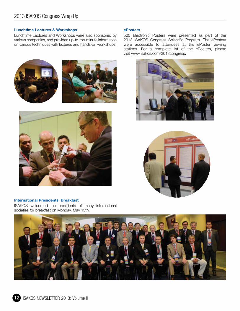
Lunchtime Lectures & WorkshopsLunchtime Lectures and Workshops were also sponsored by various companies, and provided up-to-the-minute information on various techniques with lectures and hands-on workshops.
International Presidents’ BreakfastISAKOS welcomed the presidents of many international societies for breakfast on Monday, May 13th.
ePosters500 Electronic Posters were presented as part of the 2013 ISAKOS Congress Scientific Program. The ePosters were accessible to attendees at the ePoster viewing stations. For a complete list of the ePosters, please visit www.isakos.com/2013congress.
2013 ISAKOS Congress Wrap Up

ISAKOS NEWSLETTER 2013: Volume II 13
Donors ReceptionISAKOS hosted a reception for those who have graciously donated to the ISAKOS Global Connection Campaign.
ISAKOS Booth and Resource Area
President’s Dinner2011 – 2013 ISAKOS President, Moises Cohen, hosted an elegant dinner at the Fairmont Royal York Hotel for his closest friends and industry colleagues.
2013 ISAKOS Congress Wrap Up

ISAKOS NEWSLETTER 2013: Volume II 14
Controversies in PRP Use: The Instructional Course at ISAKOS 2013
Prof. Mandeep S. DhillonPresident Indian Arthroplasty AssociationPresident Indian Foot & Ankle SocietyVice President Indian Association of Sports MedicineChandigarh, INDIA
Platelet Rich Plasma seems to have taken the Orthopaedic, Sports Medicine, and Plastic and Reconstructive surgery communities by storm. The enthusiasm and interest generated in recent years seems to far outstrip the medical research, which is still an ongoing process, and many things are yet not adequately defined in relation to efficacy, quantification, usage and regulatory aspects.
I googled the words platelet rich plasma one day prior to the ICL at the ISAKOS meeting, and was amazed at the number of hits; I got 5.5 million hits on this subject alone! The interest in this is definitely immense, but in the words of Allan Mishra, “the most read article is probably the one in the New York Times” Famous sportsmen using this treatment for early return to sports have ranged from top Golfers (Tiger Woods) to famous American footballers like Hines Ward and Troy Polamalu of the Pittsburg Steelers, and the pressure on doctors to use this methodology has been immense.
To put this subject in a scientific perspective and look at the most recent research through the opinions of world leaders, Dr. Rogerio Teixeira da Silva from Sao Paulo Brazil, coordinated this ICL at ISAKOS 2013 Toronto, and invited the top minds in PRP focused research, namely Steven P. Arnoczky from Michigan USA, (who has been at the forefront of numerous path-breaking research projects), Allan Mishra from Stanford, (who initiated the interest in this topic by his pioneering work on Tennis Elbow many years ago), and Nicola Mafulli from London (who is a World leader in tendon related research).
In the words of Rogerio Teixeira da Silva, Past President – Brazilian Orthopedic Sports Medicine Society….
“The main objective of the PRP ICL at ISAKOS, in Toronto, was to perform a broad discussion in this very controversial topic. We had a good audience for an ICL early in the morning, showing that the topic continues to have the attention of the orthopedic surgeons, mainly the ones who deal with sports injuries. From the basics (with Steve Arnoczky’s lecture) to the always welcome counterpoint view showed by Nicola Maffulli, we had the opportunity to see high level scientific presentations. Also we had an outstanding contribution from Alan Mishra, showing his 8 year experience with the study of PRP treatment in tendinopathy. I focused my presentation on the more recently published papers where PRP was employed in different clinical situations, and opened the opportunity for the audience to think about new treatment strategies and research projects on this topic. It was a great pleasure to chair this very interesting ICL in ISAKOS Toronto 2013.”
Steven P. Arnoczky, gave a comprehensive talk on the basic aspects of PRP, the gist of which is paraphrased below
Platelet Rich Plasma (PRP) is defined as an increase in the concentration of platelets above that found in the same volume of whole blood. Platelets (and the liquid plasma component of blood they are suspended in) contain over 1100 bioactive factors involved in the healing and regeneration of connective tissues. PRP has a long history of safety and efficacy in dental and maxillofacial surgery as well as cosmetic surgery. In considering the basic science of PRP, it is important to recognize that all PRP products are not the same and they can vary in 1) the concentration of platelets (as a function of whole blood drawn), 2) the inclusion / exclusion of white blood cells, 3) the use of an exogenous platelet activation agent, and 4) the level of fibrin production. These variables may have an effect on the indications and outcomes of PRP use in sports related injuries and may explain the variable results presented in the literature regarding the efficacy of PRP. Therefore, the classification system proposed by Dr. Allan Mishra (which categorizes the 4 variables noted above) should be employed when publishing outcome studies using PRP so as to more clearly define the efficacy of specific PRP product in a specific clinical application. It is also important to remember that most growth factors do not have a linear dose-response curve so having a higher concentration of platelets does not always guarantee a more robust biologic response. The precise concentration of platelets in PRP (as well as the frequency and mode of application) that can effectively (and consistently) augment healing in the many applications that have been advocated for PRP have yet to be determine. Therefore, more well-designed and appropriately analyzed level 1 studies are still needed to identify the role of PRP in the treatment of sports injuries.
2013 ISAKOS Congress Wrap Up

ISAKOS NEWSLETTER 2013: Volume II 15
Dr. Allan Mishra then spoke in his usual understated manner, and presented data from a prospective, randomized trial of 230 patients evaluating the efficacy of PRP for chronic tennis elbow. That study revealed a significant improvement in patients treated with needling and PRP compared to needling alone in terms of reported pain and elbow tenderness. The overall success rate of needling with PRP compared to PRP alone was 84% vs 68% at six months. (p = 0.012). This study used a formulation of PRP that contained increased concentrations of platelets and increased white blood cells compared to baseline. This formulations was injected in an unactivated fashion. (Type 1A PRP prepared via the Biomet GPS PRP System, Warsaw, IN USA; see classification system below) This paper has been accepted for publication to TheAmerican Journal of Sports Medicine.
Dr. Mishra stated “The data will hopefully help patients and clinicians as they navigate the decisions they need to make with regard to treating chronic tennis elbow. Finally, it was a pleasure to reconnect with friends from India including Dr. Mandeep Dhillion, one of the authors of the recent excellent investigation using PRP for knee osteoarthritis. I look forward to future meetings and continuing collaborations. Please consider joining with me on the Biologic Orthopedic Society on LinkedIn (BiologicOrtho.com) as we work toward better solutions for challenging problems”.
Mishra PRP Classification System (Mishra et al 2012)
PRP Classification
White Blood Cells Activated?
Type 1 Increased over Baseline No
Type 2 Increased over Baseline Yes
Type 3 Minimal or No WBCs No
Type 4 Minimal or No WBCs Yes
A: > 5x PlateletsB: < 5x Platelets
Dr. Mishra’s classification is presented above
Then came the turn of Nicola Maffulli, Professor of Sports and Exercise Medicine at London, and a highly published author; he played the Devil’s advocate and brought into focus the lack of significant evidence based studies to validate many points. I asked Nicola to give the gist of his beautifully presented talk, and his words are paraphrased below.
“Platelet rich plasma (PRP) therapies aim to improve the process of tissue repair through local delivery of autologous bioactive agents to influence critical mechanisms such as inflammation, angiogenesis or extracellular matrix synthesis. PRP has been used for just about any and all conditions in orthopaedic sports medicine, and some practitioners swear by it. I must confess that I am still cautious: I am fascinated by this new technology, and by the opportunity that PRP may afford to get my athletes back to health and fit to fight faster. However, I am aware of the fact that there is still relatively little level I evidence in favour of PRP. Indeed, the well-performed level-1 studies seem to paint a different view from what has been outlined in the press. The early studies were impressive, and PRP seems to be effective in the management of tennis elbow. However, subsequent well performed randomized controlled trials in Achilles tendinopathy form Holland and our own in rotator cuff tears do not show any advantages. Another randomized controlled trial using PRP in open repair of Achilles tendon ruptures from Sweden shows that it is at best of no use, and possibly harmful. I am aware of another trial in rotator cuff repair, which shows early advantages, and of another two that show no advantages. We performed systematic reviews with the Dutch group and with the group who introduced the concept of PRP in the management of musculoskeletal injuries, led by Drs Anitua, Sanchez and Andia, and found that the scientific evidence is just not there. The same applies to muscle injuries. Therefore, at present, I am happy to perform studies on PRP (and we are doing so), but I do not use it in clinical practice, at least not yet!”
These three talks were followed by presentation of clinical results from various published studies by the ICL Chairman, Rogerio da Silva; he looked at the pros and cons, and subsequently initiated an excellent discussion.
As a member of the audience, I was impressed by the number of people who attended this ICL as well as the quality of the presentations and the inquisitive questions that were subsequently asked.
I congratulate ISAKOS for picking this topic for the ICL and Dr. da Silva for getting the best minds in this field on to the podium together, which in itself is a monumental task
2013 ISAKOS Congress Wrap Up

ISAKOS NEWSLETTER 2013: Volume II 16
ISAKOS Congress Pre-CoursesClinical Research Methods Chairs: Robert Marx and Stephen Lyman,
ISAKOS Scientific Committee
For the 9th Biennial ISAKOS Congress, the ISAKOS Scientific Committee organized the first ISAKOS Clinical Research Methods Pre-Course. Attendees were given a copy of the Journal of Arthroscopy Clinical Research Methods Supplement and treated to a series of lectures by experienced orthopedic clinical investigators from around the globe. This short-course was co-chaired by Stephen Lyman, PhD, and Robert Marx, MD, MSc, FRCSC both from the Hospital for Special Surgery (USA). The course covered the life of a clinical research project from conceiving an answerable research question (Lyman) to writing a publishable paper (Jon Karlsson MD PhD, Sweden). Bias is a constant threat to the accuracy of clinical research findings, explained very well by Dr. Daniel Wheelan (Canada). The advantages of disadvantages of observational studies designs including case control studies (Jacques Menetrey MD, Switzerland) and large prospective cohort studies (Kurt Spindler MD, USA) were explored. Data capture options were explored by Dr. Lyman while Dr. Norimasa Nakamura (Japan) addressed objective measurement of patient outcomes in a clinical setting. A talk by Dr. Marx on surgeon equipoise in surgical trials was enlightening to all attendees and illicted a robust discussion of the potential pitfalls of randomized study designs in surgical research. Drs. Lyman and Karlsson wrapped up the session with an insightful lesson into visualizing data in scientific manuscripts and an editor’s view of manuscript preparation (Dr. Karlsson is the editor-in-chief of KSSTA).
Advances in Knee: Patellofemoral Instability, ACL Reconstruction and Meniscal RepairChairs: Allen Anderson, David Parker, and
Willem van der Merwe, ISAKOS Knee Committee
As a prelude to the ISAKOS 2013 congress in Toronto, the ISAKOS knee committee organized a pre-course in Advances in Sports Medicine Knee Surgery. The course was divided into sessions dedicated to Anterior Cruciate Ligament, Patellofemoral surgery, and Meniscal surgery, with the morning sessions involving talks and discussion, and the afternoon involving live surgery demonstrations. In the morning the ACL session included talks on primary and revision surgery and pediatric reconstruction, the Patellofemoral session included talks on management of acute and recurrent injuries, and the Meniscal session included talks on repair techniques and meniscal transplant. In the afternoon, the audience was able to observe and ask questions about live surgical demonstrations of MPFL reconstruction, Tibial tubercle osteotomy and Trochleoplasty, Double Bundle ACL reconstruction and Extraarticular Reconstruction, as well as Meniscal Root Repair.
The course was certainly an incredibly comprehensive program that was expertly covered over the course of a single day by the collection of world renowned speakers. It was wonderful to see representation from America, Europe, Asia, Africa, Australia and New Zealand amongst the faculty, and the program was well organized and chaired by Allen Anderson, Willem van der Merwe, and David Parker. The course was sold out, and the feedback from the audience has been overwhelmingly positive. The Knee Committee would like to thank all of those who contributed, and is now working to put together a Knee Surgery Pre-course for the congress in Lyon in 2015, which we hope will be similarly successful!
2013 ISAKOS Congress Wrap Up
Apply for an ISAKOS Arthroscopy Traveling Fellowship! Arthroscopy Traveling Fellowship! Arthroscopy Traveling Fellowship! Arthroscopy Traveling Fellowship! Arthroscopy Traveling Fellowship! Arthroscopy Traveling Fellowship! Arthroscopy Traveling Fellowship! Arthroscopy Traveling Fellowship! Arthroscopy Traveling Fellowship! Arthroscopy Traveling Fellowship! Arthroscopy Traveling Fellowship! Arthroscopy Traveling Fellowship! Arthroscopy Traveling Fellowship! Arthroscopy Traveling Fellowship! Arthroscopy Traveling Fellowship! Arthroscopy Traveling Fellowship!
Dr. Masaki Watanabe developed the first device for minimally invasive surgery. Created in honor of Dr. Watanabe’s accomplishments, the Masaki Watanabe Arthroscopy Traveling Fellowship Award is a new traveling fellowship sponsored by the ISAKOS Arthroscopy Committee. The fellowship will provide funding for two young arthroscopic surgeons to learn more about the current practice of arthroscopic surgery from well-respected experts in the field.
Application Deadline: January 31, 2014
Apply Today at www.isakos.com/awards

ISAKOS NEWSLETTER 2013: Volume II 17Volume II
International Update on Surgical Controversies of the Shoulder and ElbowChairs: Guillermo Arce and Felix Savoie, III,
ISAKOS Upper Extremity Committee
The ISAKOS Upper Extremity Committee was pleased to offer a half day surgical skills based pre-course on Surgical Controversies of the Shoulder and Elbow. This pre-course was designed to allow participants to evaluate the optimal use of diverse techniques for a variety of upper extremity surgical procedures, and formulate surgical protocols for upper extremity procedures that integrate strategies to avoid potential complications. The chairs would like to thank our Surgical Demonstration faculty – Guillermo Arce, MD ARGENTINA, Gregory Bain, MB BS, FRACS, PhD AUSTRALIA, Klaus Bak, MD DENMARK, Luigi Pederzini, MD ITALY, Felix Savoie, III, MD USA, Hiroyuki Sugaya, MD JAPAN, and W. Jaap Willems, MD, PhD NETHERLANDS. This diverse and international faculty presented their unique perspectives on surgical techniques, as well as pearls and pitfalls. Featured procedures included Instability with Arthroscopic Reconstruction, SLAP Repair, Biceps Tenodesis, Open Distal Biceps Repair, Arthroscopic Radial Head Excision, Arthroscopic Subscapularis Repair, Double Row Rotator Cuff Repair, and Arthroscopic AC Reconstruction.
These surgical demonstrations are currently under development as ISAKOS Global Link online courses – we encourage you to log into ISAKOS Global Link in the coming months to take these courses if you were unable to participate in the Upper Extremity pre-course.
ISAKOS & FIFA – Key Issues and Challenges in Safety and Health in SoccerChairs: Bert Mandelbaum and Moises Cohen
ISAKOS was pleased to partner with the Fédération Internationale de Football Association (FIFA) to present a course specifically dedicated the treatment and management of soccer players. As the most popular sport in the world, there is a unique need for specific information on the treatment of soccer athletes. This course provided a great opportunity for athletes, team physicians, athletic trainers and coaches to learn more about the treatment of soccer players from a diverse faculty. ISAKOS thanks FIFA Medical Director Jiri Dvorak for his participation in the course – Dr. Dvorak’s lecture on “Football for Health: Exercise is Medicine, Prevention of Injuries and Disease” was a highlight of the course. Our diverse faculty allowed this course to focus on the entire player, including issues related to concussion, patient population specific injuries (women and children), and shoulder lesions; as well as more common soccer related injuries related to the knee and ankle. Panel based case discussions were also a highlight of the pre-course, as physicians presented cases from their own practices. Thank you to all who participated!
This course was sponsored by an educational grant from
ISAKOS Sports Rehabilitation Concurrent Course: Global Perspectives for the Physical Therapist and Athletic TrainerChairs: Trevor Birmingham, BSc PT, PhD, CANADA;
Moises Cohen, MD, PhD BRAZIL; James Irrgang, PT, PhD, ATC, FAPTA USA; Lynn Snyder-Mackler, PT, ScD, FAPTA USA
On behalf of the course chairs, ISAKOS would like to thank all who participated in the ISAKOS Sports Rehabilitation Concurrent Course: Global Perspectives for the Physical Therapist and Athletic Trainer. This unique course brought together orthopaedic surgeons, physiotherapists and athletic trainers to discuss international concerns related to sports rehabilitation.
The international faculty focused on issues related to current developments in the management of knee, shoulder and elbow, hip, foot and ankle and muscle injuries in athletes. Course participants felt that following the course they are better able to evaluate and manage sideline or onsite issues in sports medicine, and address controversial issues concerning return to play in athletic events. The diverse faculty also presented different modalities and treatment strategies utilized around the world when dealing with similar injuries.
This course was sponsored by an educational grant from
2013 ISAKOS Congress Wrap Up

ISAKOS NEWSLETTER 2013: Volume II 18
ISAKOS Honorary Members
2013 ISAKOS Congress Wrap Up
Werner Müeller, MD
Written by: Roland Jakob, ISAKOS Past President (2001 – 2002)
Werner Müeller studied Medicine in Basel, Paris and Vienna obtaining his degree in 1959. He acquired his Orthopaedic training in Basel with Nissen and Chapchal, the former being a disciple of former Prof. Sauerbruch, known for his strength and severity – whose assistants were not allowed to get married while working with him. We feel happy with Werner that, times had changed by the time he came in the department because he already was married to Ursula and later had four sons with her. Starting in 1970, Prof. Morscher became his Orthopaedic Chief with Prof. Martin Allgöwer, known for his leading function in the AO, being Chief of the Surgery Department. Werner was named Head of Orthopaedic Traumatology, although his main interest was less the broken bones; it was the torn ligaments and restoring stability with full function of the joints that attracted him! In 1978 he left University to become Chief of his own unit of Orthopaedics in Bruderholzspital – Basel which later became the mecca for ligament pathology of the knee in Switzerland. 1982 he became Privatdocent and 1990 Associate Professor of the University of Basel.
Werner is famous for his book The Knee. Form, Function and Ligament Reconstruction, published in 1982 / 83, containing the anatomic relations with the primary and secondary stabilizing functions of the knee ligaments. It appeared in six languages. It was the foundation and basis of the courses on applied knee anatomy he was giving in Basel and at many other sites in which he has remained unbeaten.
Dr. Müeller wrote over 150 articles and was Co-Editor of the ESSKA Journal. He was member of fifteen Medical Societies, and has been honored with eleven Honorary Memberships with various medical societies.
Werner Müller became Founding President of ESSKA in 1982 and headed the second ESSKA Congress in Basel in 1986. Until today he has held many Guest Lectures of which numerous Honorary Lectures over the world.
Together with his intimate friend John Feagin, he started the AOSSM-ESSKA Traveling Fellowship in 1985 and on his initiative they decided to found a group to devote energy and time to the development of a common system of classification – the IKDC, a system still valid today and being constantly adapted.
He has eight grandchildren and remains happily married to Ursula.
01
02

ISAKOS NEWSLETTER 2013: Volume II 19Volume II
2013 ISAKOS Congress Wrap Up
Paolo Aglietti, MD1942 – 2013
Written By: Matteo Denti, ITALY and Francesco Giron, ITALY
Paolo Aglietti was born in a small village, Fucecchio, not far from the Tuscan coast, in 1942. In 1970 he completed his formal medical and general orthopaedic training and thereafter followed a highly distinguished career. In 1971 and 1973 he was awarded fellowships in adult hip reconstruction and adult knee reconstruction respectively at the Hospital for Special Surgery in New York. It was here that his strong and enduring friendships with Dr. Edoardo Salvati and Dr. John N. Insall, both pioneers in their respective fields of modern hip and knee replacements, began.
He returned to Italy, where in 1975 he undertook the role of Assistant Professor of Orthopaedics and Traumatology at the University of Perugia. In 1979 he was appointed Associate Professor at the University of Florence. He was then appointed Professor of Orthopaedics and Traumatology in 1990 and Chief of the Orthopaedic Clinic, including the residency program in 1996. He served as Director until his retirement in 2009.
Paolo was a founding member of both the Italian Arthroscopy Society (SIC) and the Italian Society of Knee Surgery (SICG). It was he who promoted the integration of the Italian knee, cartilage and sports trauma societies under the newly founded Italian Society of Knee Surgery, Arthroscopy, Sports Traumatology, Cartilage and Orthopaedic Technologies (SIGASCOT).
He was a member of SIOT, ISAKOS, AAOS, AOA, ESSKA, ICRS, Herodicus and the ACL study group. He was also an honorary member of the Knee Society. 2007 saw his election as President of the International Society of Arthroscopy, Knee Surgery and Orthopaedic Sports Medicine (ISAKOS) and in 2008 he was inducted into the Hall of Fame of the American Orthopaedic Society for Sports Medicine.
His contributions to orthopaedic surgery are impressive. He has published in the most influential orthopaedic journals commencing with his studies on total knee replacement with Edoardo Salvati, and total hip replacement with John N. Insall. These studies took place during his time at the Hospital for Special Surgery in New York. His study on “New Patellar Prosthesis” is still considered a keystone paper. His subsequent articles dealt with techniques for treatment of patellar chondromalacia and malalignment, the varus osteoarthritic knee and osteonecrosis of the knee. He was also a pioneer of knee arthroscopy and published several articles on arthroscopic meniscectomies.
Paolo will be remembered for his numerous contributions to knee arthroplasty and ACL reconstruction surgery. He co-operated with Dr. Insall to develop and evaluate the outcome results of the Insall-Burstein knee replacement and subsequently the MBK knee prosthesis. Also he will be remembered for his studies on ACL surgery comparing the results of patellar and hamstring tendon grafts and his pioneer study on the new double bundle reconstruction technique.
Paolo was acknowledged as both a master surgeon and rigorous researcher, and numbered amongst his special talents the ability to choose appropriately the correct indication for each individual patient who consulted with him. His investigations and studies were astute and brilliant. His wealth of knowledge was evident by the clear and concise presentations and lectures he gave.
In both public and private, Paolo was a shy man and not one given to easy friendships; however he was always frank in his opinions and suggestions in spite of being sometimes considered rough. Notwithstanding this he was considered a role model by his colleagues and co-workers alike.
In the summer season, during his precious free time away from the rigors of his academic commitments he enjoyed sailing in the Mediterranean with his wife Chiara on board their boat and would enjoy recounting these memories with us.
01 Roland Jakob; Werner Müeller; Moises Cohen02 Roland Jakob; Werner Müeller; Moises Cohen, John Feagin; John Bergfeld03 Paolo Aglietti, MD
03

ISAKOS NEWSLETTER 2013: Volume II 20
John Feagin, MD
Written by: John A. Bergfeld, ISAKOS Past President (2005 – 2007)
It is an honor to welcome Dr. John Feagin, USA into the fellowship of Honorary Member of International Society of Arthroscopy, Knee Surgery and Orthopaedic Sports Medicine. “He who desires to practice surgery must first go to war”. That is a Hippocrates quote and certainly applies to John Feagin.
John was born in San Antonio, Texas. Following an outstanding high school student career at Texas Military Academy, he went on to West Point where he graduated in 1955. He then served active duty for 2 years before attending Duke University Medical School, graduating in 1961 with honors. John was the first active duty Army officer to attend medical school. To get the Army to recognize that a West Point graduate would be a gung ho medical Army officer was no small effort.
Upon completion of medical school, John served an orthopaedic residency at Walter Reed Army Hospital and then volunteered for a tour of duty in Vietnam. He returned the United States Military Academy where he served as a staff orthopaedic surgeon and team physician to the West Point Cadets.
Following his tour of duty at the Military Academy, John did a fellowship in joint replacement with Sir John Charnley and returned to the United States to serve as the Chief of the US Army’s Joint Replacement program at Lederman Hospital. John then returned to West Point as the Commanding officer at the Kellerman Army Hospital at West Point. During this tenure, he oversaw the introduction of women to the military academy and set the standards for the women’s participation in athletics and the physical conditioning demanded of all West Point Cadets.
During John’s tenure as team physician at West Point, he recognized the importance of the anterior cruciate ligament in the athlete’s knee in spite of the Godfathers of orthopaedics telling us that it wasn’t important. John presented the results of his anterior cruciate ligament primary repairs in 1972 at the American Academy of Orthopaedic Surgeons, a follow up which proved that the primary repair of the ligament was only temporarily successful. This work of John’s set the stage for the substitution of the injured anterior cruciate ligament which is the standard of care today.
In 1979 John was faced with the decision as to whether to move up to the rank of Army General and become an administrator or retire from active duty and continue to do orthopaedic surgery. John chose to retire from the Army and went to Jackson Hole, Wyoming to follow his passion of skiing and practice sports orthopaedic surgery.
John continued his academic association with Duke University where he eventually moved and has been appointed Clinical Professor of Orthopaedic Surgery. Currently he serves as emeritus professor of Orthopaedic Surgery at Duke University and Director of the Duke University Feagin Leadership Program.
In addition to John’s academic career and contributions to surgery of the knee, John was a founding member and eventually President of the American Orthopaedic Society for Sports Medicine. He has served on the Board of Trustees of the United States Military Academy. He has delivered 17 endowed named lectureships around the world. He was awarded the Distinguished Graduate of the United States Military Academy. John has also served as team physician for the US Ski Team.
In addition to all of this work, John, along with Werner Müeller from Basel, Switzerland, fathered the exchange of traveling fellows, initially between ESSKA and AOSSM and now the exchange between AOSSM, ESSKA, APKASS and SLARD.
His innovativeness also is responsible for starting the Anterior Cruciate Ligament Study Group. He chaired the first meeting held in 1975 while John was still at West Point.
John now divides his time between Duke University in North Carolina and Vail, Colorado where he lives with his wife Marty and their families.
Truly John is an icon of orthopaedic Sports Medicine, an innovative surgeon, team physician, mentor to many of us. ISAKOS is honored to welcome John to a well-deserved Honorary membership in International Society of Arthroscopy, Knee Surgery and Orthopaedic Sports Medicine.
ISAKOS Honorary Members
2013 ISAKOS Congress Wrap Up
01 John Feagin & John Bergfeld
01

COMPLETE THE ONLINE CONGRESS EVALUATION TO DOWNLOAD YOUR CME CERTIFICATE
Log in to myCongress to get started today!
WE LOOK FORWARD TO SEEING YOU IN LYON IN 2015!
Abstract Submission & Award Applications for the 2015 Congress are now open!
myCongress
Thank you for your participation in the 2013 ISAKOS Congress.the 2013 ISAKOS Congress.
JUNE 7–11, 2015LYON, FRANCE
isAkoscongress
10TH Biennial
2015
www.isakos.com / 2013congress / myCongress

ISAKOS NEWSLETTER 2013: Volume II 22
2013 ISAKOS Congress Wrap Up
Prevention of Sports Injuries
Lucio Ernlund, MD, MScISAKOS Newsletter Editorial BoardMedical Director Coritiba FCInstituto de Joelho e OmbroCuritiba, Parana, BRAZIL
Injury prevention is the number one role of the Sports Medicine Doctor. Prevention has low cost as compared to treatment. Prevention starts with a systematic screening protocol, PPPE (Pre participation Physical Examination), when doctors get to know the athlete and his / her predisposing factors.
Accordingly to van Mechelen, 1992, prevention should follow this sequence:
1 Establish the extent of the injury: incidence and severity;2 Establish the etilogy and mechanisms of the injury;3 Introduce a prevention measure;4 Assess its effectiveness by repeating step 1
The multifactorial nature of sports injuries makes it difficult to prevent with 100% success. Meeuwisse, Bahr, Holme and Krosshaug (on different publications) showed an interesting model of potential causative factors for injuries.
The overall level of injury to professional footballers has been showed to be around 1000 times higher than for industrial occupations generally regarded as high risk. (Hawkins and Fuller Br J Sports Med 1999.)
A variety of injury specific and sport specific preventive exercise programs exist. Various authors have established a strong relationship between the frequent use of standardized warm-up programs and reduction of the risk of injuries to up to 50%. (Myklebust, 2003; Mandelbaum,2005; Olsen, 2005; Soligard, 2008; Kiani, 2010; LaBella, 2011; Walden, 2012)
Although all published data on the efficacy of prevention protocols, Doctors must struggle to convince team coach and trainers that these exercises are as important as the technical training itself.
It is important to show not only the medical point of view but also the economical point of view. The real costs involved with an athlete on the Medical Department (salaries, consequencial damage, etc) are extremely high.
Effective Prevention Program1 Sport specific exercises2 Variation and progression of exercises3 Warm-up program4 Strength 5 Balance6 Plyometrics
Results Coritiba FC1 Decrease 21% of overall injury rate2 Decrease 60% overall injury rate Coritiba FC 2006 – 20103 Increase injury rate after program was left aside, due to
increase on games calendar (76 last season)
Conclusion1 Injury rate in football (soccer) is too high2 Muscle injuries and ankle sprains are most
frequent injuries3 Mid season most dangerous moment4 Low cost / high efficacy5 Program should involve all team from athletes to coach6 On the practical point of view, not so ease to perform on
a professional Brazilian Team. Too many championships, long season (State tournament, Brazilian Cup, Brazilian Championship, Libertadores da America), 76 games last season (10 months)
7 Long trips (Brazil huge country), more than 60,000 Km driving / flying every season. Last year athletes slept over 150 days away from home.
8 Most of the teams change at least 30% of their players from one season to the other (new comers may not have been involved on prevention programs before, expect delays on the progression)
9 Although all the difficulties team doctors may face, he / she should proceed emphasizing the importance of the injury prevention programs
ABOVE ALL: IT IS GREAT TO BE A WINNER

ISAKOS NEWSLETTER 2013: Volume II 23
Header?
congress content FIVE MEETING DAYS WITH:
250 Scientific Papers and 500 Electronic Posters
Sports Rehabilitation Concurrent Course
Instructional Course Lectures
Lunchtime Lectures and Workshops
Surgical Demonstrations
Hands-On Workshops
Congress Awards
Technical Exhibits
CME Certification
JUNE 7–11, 2015LYON, FRANCE
isAkoscongress
10TH Biennial
2015
awardsawardsawardsAWARDS AND FELLOWSHIPS
John Joyce Award Richard B. Caspari Award Scientific Research Award Albert Trillat Young Investigator’s Award
Achilles Orthopaedic Sports Medicine Research Award
Patellofemoral Research Excellence Award
The Upper Extremity Traveling Fellowship
The Patellofemoral Traveling Fellowship
The Arthroscopy Traveling Fellowship
cme hoursThe 10th Biennial ISAKOS Congress will be planned and implemented in accordance with the essential areas and policies of the Accreditation Council for Continuing Medical Education (ACCME) through joint sponsorship.
ONLINE ABSTRACT SUBMISSIONISAKOS is pleased to announce the Call for Abstracts for the 2015 Congress.
To submit an abstract, visit www.isakos.com/2015congress
Abstract Submission Opens September 1, 2013
Apply Online for Congress Awards
join us...join us...
International Society of Arthroscopy, Knee Surgery and Orthopaedic Sports Medicine2678 Bishop Drive, Suite 250San Ramon, CA 94583 USA
www.isakos.com

ISAKOS NEWSLETTER 2013: Volume II 24
Case Presenter: Omer Mei-Dan, MD USA
20 y.o. active male with no previous complaints was snowboard jumping and went out of control. Upon landing on his knees he remembers feeling his right hip dislocating with, what seemed to be, immediate spontaneous reduction. He states that he rested for 30 minutes before resuming snowboarding for one more run. Pain limited him from continuing and he stopped activity. He has been limping since injury, two weeks ago, and though has crutches, has not been using them and has been weight-bearing. The hip would wake him at night due to discomfort more than pain and would click and catch on him. Sitting has not been especially difficult for him and he has continued to drive since the accident. Physical exam: 5 feet 11 inches tall, 165 lbs male. Currently, he walks with a guarded, limping gait. He has no leg length discrepancy and present with no signs of joint laxity. He is fit looking. NV exam is normal. ROM at 90 degrees of hip flexion presents with 45 degrees ER, 15 degrees IR. Abduction is 40 degrees. All position generate joint discomfort, even without provocative maneuvers.
What would you do?Scope now? Scope later? At all? Open procedure?
Recommended weight bearing status?
Other recommendations?
Case Response: Rodrigo Mardones Petermann Santiago, CHILE
I will offer this patient an immediate surgical treatment.However I recommend to somebody that is not an expert on Hip scope to wait until capsule has healing so avoid extravasation (4 – 6 weeks). It is important to perform the full procedure in less then 2 hours and be very careful with the water flow. Main complication to avoid is peritoneal fluid extravasation.I will perform all at the same time:Treatment of his minimal FAI (may have small retroversion since the AP pelvis looks little bit hipo lordotic / outlet plus had a plane anterior offset mainly reactive typically seen in a pincer type plus reactive cam).Labral repair if there is some anterior damage. For sure posterior bone labral repair for the posterior wall. If it is too small to repair I will debride it and repair the labrum over the new bone edge. For small fragment like this I will pass throughout the fragment with my anchors (drill trough) and will suture the fragment with the labrum against the defect (some support by the anchor and some by the suture). Our group actually has perform this twice with very good results. If the fragment is bigger (not in this case) would use small cannulated screws, arthroscopicaly. More than 1/3 of the wall I will probably proceed with an open procedure.Will not suture the capsule.I will protect with partial weight bearing for 4 – 6 weeks after surgeryWill use CPM for the hospital stay but if the patient can rent, it will prescribe for 4 weeks. Otherwise will use stationery bike twice a day for 6 weeks (15 min a session) plus PT stage one (isometric and passive motion) plus an abductor pillow for 4 weeks at night.
After 6 weeks will put him in our regular post FAI program.
Case Response: Marc R. Safran, MD Stanford University, CA, USA
I had a very similar case in an NFL running back – treated non-op on crutches…and the following season had one of his best years productivity wise, and no hip symptoms.
Sunday, June 9, 13
CASE CORNER

ISAKOS NEWSLETTER 2013: Volume II 25
Case Response: Michael Dienst Munich, GERMANY
Imaging indicates a posterior rim fracture with minimal displacement likely after posterior subluxation of the right hip joint. The rim fragment is continuous with the superior rim but shows an increasing gapping of the fracture further posterior and medial. Integrity of the Teres Lig., the acetabular labum and the articular cartilage cannot be sufficiently assessed on the presented MR imaging. The head and neck areas show mild bone edema without touching the weightbearing surface of the femoral head, thus a significant direct bruise of the cartilage areas are less likely. On the lateral radiograph, there is mild loss of offset more at the neck than at the head. The alpha angle is around 45° thus in the normal range. The distal loss of offset is usual an indicator of Pincer-FAI and retroversion, which however is not present on the pelvis ap radiograph and CT scans.With respect to the minimal displacement and posteromedial location of the facture and missing previous complaints, conservative treatment would be an option. If that was preferred by the patient, weight bearing should be limited to about 20kg for 6 weeks and subsequently increased by 10 kg weeks coming back to full weight bearing at 10 – 12 weeks. In ordert to prevent stretching of the posterior capsule and further displacement of the posterior fragment, flexion should be limited to 90° for the first 6 weeks. Continuous passive motion therapy should be recommended to at least 6 weeks to prevent adhesions.Because of the young age of the patient, some fracture displacement, the loss of neck offset with the risk of further damage by impingement sports and the high risk of non imaged further intraarticular damage, I would recommend immediate arthroscopy. The risk of fluid extravasation at 2 – 3 weeks would be small, and connections to the retroperitoneum unlikely because of the fracture location. Further waiting would reduce the option to arthroscopically reduce the fracture. During arthroscopy, further potential damage to the Teres Lig., articular cartilage and particularly the anterior labrum (traction injury by capsule or direct impinging injury during subluxation) need to be evaluated and treated. If the fracture gaps proves relevant, the perilabral bony surface of the posterior rim needs to be exposed, the fracture reduced and compressed with one to two screws. At the end of the procedure, the offset of the femoral neck should be increased. Postoperative treatment would be similar to that of conservative treatment.
Case Response: Victor Ilizaliturri Mexico City, MEXICO
The diagnosis for this case is very evident. The patient presented a traumatic posterior subluxation of the right hip that resulted in a posterior wall fracture.As much as the diagnosis is clear, the treatment strategy is controversial.In a more conservative treatment strategy, the patient can be advised to use crutches for protected weight bearing and a brace to allow limited range of motion, a CT-Scan can then be repeated at 3 to 4 weeks and the situation re-assessed then.One must take into consideration that the patient is very symptomatic with pain in every range of motion. The imaging of the hip shows a posterior wall fracture with minimal displacement.Because the patient is young and active in sports, and has pain and minimal displacement, I believe that the fracture should be fixed immediately with arthroscopic assistance using cannulated screws. Loose bodies should be removed arthroscopically from the fracture line and labral repair performed (most of these lesions will be associated with some type of labra injury). Most of these cases are also associated with chondral damage and this is usually treated by removal of unstable cartilage and microfracture if needed.Fluid extravasation may be a concern to some surgeons because of the acetabular fracture. In this particular case, the fracture is of the posterior wall and there is no fracture that communicates the joint with the intra-pelvic space, therefore I believe the procedure can be performed with the standard monitoring of the abdomen throughout the operation.With early intervention the patient will benefit because he will return to normal and sport activity sooner, the possibility of third body wear produced by fragments coming from the fracture line is reduced which may result in better prognosis for the joint. Also early arthroscopic assessment of the joint will be important to understand the amount of intraarticular damage.In the other hand a more conservative strategy like mentioned earlier may also be a valid choice, however it would not be my course of action.
CASE CORNER

ISAKOS NEWSLETTER 2013: Volume II 26
Prevention of Injuries in Brazilian Soccer
David Sadigursky, MDMember of the Department of Knee Surgery at Orthopedic and Traumatological Clinic (COT), Salvador - Bahia - BrazilMaster in Orthopedics from the University of São Paulo (IOT HC FMUSP)
The FIFA World Cup 2014 will be hosted by Brazil. As it approaches the preparations are intensifying in the country, where soccer is the most popular sport. Soccer is a sport that involves great physical contact and also short, quick and continuous movements such as acceleration, deceleration, change of direction, jumps and pivots. With the increasing number of games and tournaments held in Brazil, the consequence is that a significant incidence of injuries occurs as well. Their rate has been estimated at 70.7 per 1,000 game hours, as demonstrated in a study during the 2011 Copa América (America Cup) – The Latin America national teams championship.
Due to the World Cup, the training routine of the soccer players significantly increases. This fact, along with the number of local competitions, has caused equally serious concern with the amount of injuries those athletes end up suffering, which may keep them away from work.
Cohen et al while studying eight Brazilian professional soccer teams in a period of two years showed that 72% of the lesions occur in the lower limb, 16% to the head and trunk and 6% in the upper limbs, with a predominance of thigh injuries with 34.5%, ankle joints by 17% and knee joints at 11.8%. Among the injuries that require temporary removal, the muscle lesions are the most frequent, with 39.2% of cases, while fractures and swellings represent only 5.4%. The highest incidence of injuries happen to players who perform in midfield and forward positions, and the injuries accounted for 39% of all injuries, being followed by contractures, strains and sprains.
As commented Dr. Andre Pedrinelli, from the First FIFA Excellence Medical Center, Sports Medicine group at the University of São Paulo’s Institute of Orthopedics and Traumatology: “Logically, the concept of prevention should not be restricted to strength and balance exercising only, but also to soccer players’ regular clinical exams. This becomes an active search for possible risk factors.
To make it possible, it is necessary to implement an epidemiological tracking system of those lesions, because only then will we know where to focus our efforts. The Brazilian Football Confederation (BFC) already uses an on-line system where injuries that occur in soccer matches can be documented for both the Premier and Secondary leagues of Brazilian Soccer. The São Paulo State Soccer Federation is implementing a similar system in the state of São Paulo”.
Despite the fact that the thigh is usually the spot of highest incidence of lesions, knee injuries are the most common among severe ones, leading to a longer period of absence, highlighting the ACL (anterior cruciate ligament) as the most frequently injured ligament. It usually occurs due to non-contact mechanisms, which are perceived as incapacitating. This leads to surgical treatment, keeping the athlete away from competitions for at least 4 months. During a professional soccer league season in Brazil, more than 5/6 of the athletes have some sort of injury and the average number of injuries per athlete is almost two in a season. Thus, as demonstrated by Raymundo JLP et al, efforts should be made to promote the implementation of mechanisms which can prevent these injuries. Promoting pre-season sessions is crucial to the preparation of the athletes in regard to strength gain and optimal physical performance. This measure significantly contributes to the prevention of injuries, as stated by Heidt et al. In the literature, several risk factors and prevention measures are found, and previous injuries and inadequate rehabilitation are identified as risk factors for the occurrence of future injury. Recurrent lesions to athletes make up to 25% of cases. Medical teams, concerned with the distancing of professional athletes, have intensified their participation to prevent it from occurring. Technological advances have occurred in the last decade and have been relevant when it comes to the physical preparation of these athletes, and have resulted in greater demand at their peak performance.
01
CURRENT CONCEPTS

ISAKOS NEWSLETTER 2013: Volume II 27
As for the prevention of ACL injuries in soccer players, there is evidence that neuromuscular and proprioceptive training prevent the onset of these lesions. Brito J (2009) proposes a specific program of conditioning, education and training dedicated to the prevention of ACL injuries in soccer players, involving neuromuscular and proprioceptive training. The adoption of techniques adjusted to perform the skills of each modality is highly recommended. Before the matches, a dynamic warm-up is proposed, set to prepare the athlete for the intensity of the training. It is important to point out that, the submitted program can be implemented in resource-poor countries, such as Brazil, for not requiring sophisticated equipment and can thus be applied in any soccer club. The recent FIFA Medical Assessment and Research Centre – F Marc-developed prevention protocols, as protocol named 11+, for instance. This program was developed to assist health teams, in achieving a standard training, prior to soccer matches. To implement the program, no more than 15 minutes are necessary.
The F-MARC 11 + program, involves 10 exercises to strengthen the muscles CORE, development of the thigh muscles, preventing incorrect posture and gaining stability. The element 11 was intended for a “fair play” culture, since many injuries are caused by fouls. Junge et al conducted a study in Switzerland, where 60% of the coaches applied FIFA’s warm-up program, showing that the players suffered 12% fewer injuries in league competitions and 25% fewer injuries during training when compared with other athletes.
The program starts with running activities followed by 6 strength, speed, balance and coordination exercises. In total, there are 15 specific exercises distributed in 3 parts, ranging in difficulty. The third part involves another running session when the warm-up is finished. The effectiveness of this program was also demonstrated by Soligard et al (1718) who, by applying the program, was able to observe a decrease of 1/3 of injuries in general and a half in severe injury. Thus, the F-MARC recognizes the strength of CORE as a key element of effective programs for injury prevention in soccer, the balance and neuromuscular control, the eccentric hamstring training, the pilometria and the agility.However, injuries in soccer go beyond individual skills. The opposing player plays an important role in the incidence of these lesions. Thus, the F-MARC has created harsher punitive measures for players who commit serious fouls, such as the elbow to the head strike, recognized as the main cause of concussion. Consequently, this foul has to be penalized with a red card after the board’s decision in the 2006 FIFA World Cup. The effect of this measure can be noted in the decrease of contact injuries. The material with all the necessary tools to train all those involved in professional soccer has become available on the website of FIFA (www.FIFA.com/medical).
There is strong scientific evidence that neuromuscular training programs specific to each sport should be able to prevent knee and ankle injuries. Thus, further studies should be conducted worldwide, in order to evaluate the implementation of uniformed prevention programs involving the variables of each region and the standardization of the protocols used as the basis for the evaluation of athletes, such as the protocol developed by F-MARC, during competition seasons and practice phases, seeking the most evidence, especially when it comes to both professional teams and under-18 teams, with the possibility of extending the program to amateur teams, with the creation of a specific format for this type of athlete.
Um programa de aquecimento completo para prevenir lesões no futebol
MANUAL
FIFA 11+
01 Fig 1 FIFA 11+ prevention program – F-MARC (18)
CURRENT CONCEPTS

ISAKOS NEWSLETTER 2013: Volume II 28
CURRENT CONCEPTS
Patellofemoral Disorders: Are They a Female Issue?
Elizabeth A. Arendt, MD Minneapolis, MN, USA
Patellofemoral (PF) injury and disease are commonly thought to be more prevalent in females. Literature to support this conclusion is sparse. This article investigates possible sex differences in three categories of PF disorders: PF pain, patella dislocations, and (isolated) PF arthritis.
Patellofemoral Pain / Anterior Knee PainIs anterior knee pain (AKP) more common in females? One study investigated constitutional features such as sex, age, body composition, athletic activity and duration of symptoms as possible predictive factors in the outcome of non-operative management of AKP. Results did not show sex as a determinant for AKP. Younger age was the only predictive factor for favorable outcomes.A two-year prospective study evaluated motor performance, joint laxity, limb alignment and muscle flexibility of physical education students with no prior history of knee problems. Of the 282 students assessed (151 M/131F; average age, 18.6 years), AKP developed in 7% of the males and 10% of the females. DeHaven and Litner reported on college-age athletes presenting to the training room over a 7-year period; PF pain was reported in 18.1% of males and 33.2% of females.
An established link exists between PF pain and clinical overload. Dye and associates4 have advanced a pain theory centering on biological load acceptance, i.e. PF pain reflects the loss of tissue homeostasis, i.e., biological tissue exceeding load acceptance capacity. With this model, the role of motor control and strengthening in reducing pain is better understood.
Differences in muscle development between males and females are well known, with females having less muscle mass, different muscle fiber composition and differences in muscle stiffness about the knee. Testosterone increases muscle fiber mass, fiber recruitment and type 2 fiber development. Muscle stability also shows variances between the sexes; joint load is influenced by muscle strength and fatigue. These neuromuscular differences between males and females, particularly manifest after puberty, may explain discrepancies in the rates of PF pain and injury. Males may have an advantage in regard to knee stability, strengthening of knee musculature, and by association, may be less prone to AKP.
Another consideration is pain perception; females are more sensitive to pain and have different physical responses to pain than males. In experimental settings, females are more sensitive to pain stimuli, less tolerant of pain, and more able to discriminate between different pain pathways. Several studies suggest a sex difference in pain perception. Males tolerate more pain than females in experimental settings. High ‘male-identifying’ men tolerate more pain than high and low ‘female-identifying’ women. However, low male identifying men are equal to women. This highlights the roles of sex and gender norms in pain-reporting behaviors.
02
01

ISAKOS NEWSLETTER 2013: Volume II 29
Patella Dislocations Historically, PF instability was felt to be primarily a disorder of females. (Figure 1) However, a systematic review of primary traumatic patella dislocations reveals a nearly equal female to male distribution. Of 22 articles reviewed, the total number of first-time PF dislocators was 1765, with a male / female ratio of 46% / 54%, average age 21.5 years. A second study looking at the epidemiology and natural history of acute patella dislocations suggests a similar incidence of males and females in primary patella dislocations, with a preponderance of recurrent dislocations occurring in females. In this study, risk was highest among females age 10 – 17 years.
A report of 127 patients with primary dislocations at two Finnish trauma centers followed for 7 years revealed a slight female preponderance (64%). In analyzing risk factors, the authors found two risk factors for recurrent instability were initial contralateral instability and young age. Females with open tibial epiphysis at the time of the initial dislocation had the worst prognosis for recurrent instability.
A seminal study from Lyon, France analyzed factors of patella instability using standardized imaging to identify factors related to patella instability. In their review of 110 patients, 83 were female (75%). In reviewing risk factors for patella instability, the authors found that trochlear dysplasia, as defined by the crossing sign, was present in 96% of patients with objective patella instability. (Figure 2)
A recent study analyzed imaging of patella instability patients compared to a control group. The goal was to identify sex-related differences in the anatomy of lateral patellar dislocations. The authors found that trochlear dysplasia and the TT-TG distance is more pronounced in women who experience patellar dislocation.
A cautious assumption of the current literature suggests that females are more likely to suffer recurrent patella dislocations than their male counterparts. Reasons for this are likely multi-factorial; including anatomic and neuromuscular factors. We know that compared with males, females display lower knee flexion angles with activities, with greater knee valgus angles and quadriceps activation. There is a higher prevalence of dysplastic distal femora among females.
Understanding sexual dimorphism in neuromuscular and anatomic risk factors is key. The clinician should be especially vigilant with their female patients when discussing re-injury risk, inclusive of known anatomic factors of instability, and be aware of potential neuromuscular factors during rehabilitation back to sporting activities after a patella dislocation.
(Isolated) Patellofemoral ArthritisPF arthritis has been a relatively undiagnosed as a cause of knee pain. (Figure 3) Though studies looking at sex as a risk for isolate PF arthritis are sparse, the available clinical studies show an overwhelmingly female preponderance. McAlindon et al. reviewed radiographs of 273 subjects with complaints of knee pain; isolated PF arthritis was present more than twice as often in females (24%) vs. males (11%). The incidence of combined medial and PF compartment arthritis showed equal incidence between males (7%) and females (6%). Iwano et al. and colleagues reviewed a series of 108 knees in 69 patients with PF arthritis; 93% were female. In a large French multi-center review of 578 patients with isolated PF arthritis, 72% of the patients were female.
ConclusionReview of the clinical literature to date suggests sexual dimorphism in the presentation of PF disorders, with an overrepresentation of these disorders in females. However, to date there has not been shown a reason to alter our treatments of these disorders based on the patient’s sex. Continued study of PF disorders and their treatments should include sex as a variable, in hope of providing better prevention, treatment and care of these disorders.
01 Fig 1 Axial MR image of an acute patella dislocation depicting classic bone bruising, torn MPFL, and large effusion
02 Fig 2 True lateral Radiograph depicting Type B Trochlea Dysplasia with Crossing sign and supratrochlear bump
03 Fig 3 An axial radiograph showing loss of lateral patallofemoral joint space indicative of patellofemoral arthritis
03
CURRENT CONCEPTS

ISAKOS NEWSLETTER 2013: Volume II 30
CURRENT CONCEPTS
Recurrent Anterior Shoulder Instability with Glenoid Bone Loss and Subscapularis Insufficiency: Successful Surgical Treatment with Distal Tibia Allograft and Split Pectoralis Major Tendon Transfer
Jonathan T. Bravman, MDAssistant ProfessorCU Sports MedicineDivision of Sports Medicine and Shoulder SurgeryDepartment of OrthopaedicsUniversity of Colorado
Charles T. Crellin BS, Armando F. Vidal MD
Introduction:Anterior shoulder instability is a common problem, particularly amongst the young, active sporting population. The resultant capsulolabral injury, coined a Bankart tear, can often be accompanied by bone loss on either the humeral side (Hill-Sachs lesion) or on the glenoid side, such as in the case acutely with a bony Bankart or chronically with attritional wear of the anterior glenoid due to repetitive subluxation.
The role of glenoid bone loss in relation to the success or failure of anterior stabilization procedures was initially noted in 1961 when Rowe identified increased failures in his patients with greater than 30% glenoid bone loss. This concept was revisited in the setting of arthroscopic Bankart repair through the work of Burkhart and DeBeer who coined the term “inverted Pear” glenoid to describe the appearance of critical glenoid bone loss, which has been further lowered in biomechanical study to the “critical” threshold of 19 – 20%.
In addition to bony deficiency and failure of the repaired capsulolabral complex, subscapularis deficiency has been identified as a complicating issue in the management of failed open stabilization procedures. This is specifically dependent upon the manner in which access to the anterior shoulder is obtained (ie: subscapularis split vs. partial takedown vs. tenotomy). Though there have been several reports of subscapularis rupture in association with acute instability, it was Nevaiser that pointed out that subscapularis rupture should be considered in all cases of recurrent instability as well as in older patients following traumatic anterior dislocation.
This report demonstrates a case of complex, recurrent anterior shoulder instability following two failed attempts at open capsullorhaphy with resultant critical glenoid bone loss and subscapularis insufficiency. A novel surgical approach utilizing a fresh osteochondral distal tibia allograft for glenoid reconstruction and a split pectoralis major tendon transfer to address subscapularis insufficiency is presented.
Case Report:A 38 year old healthy female presented for evaluation of recurrent left anterior shoulder instability and pain. She was an active-duty soldier and initially sustained a traumatic anterior shoulder dislocation 18 years prior to presentation in a fall down several stairs. This was treated at the time with an open Bankart repair and capsullorhaphy. She reports that the shoulder became recurrently unstable within a year, prompting a return to the OR for a revision open anterior shoulder stabilization, 20 months following the first procedure.
She states the shoulder “never felt stable” and she had progressively increasing instability with multiple subluxation and frank dislocation events requiring reduction. She presented to our Shoulder Service with complaints of constant 7/10 pain as well as 3 – 4 instability episodes per week with a subjective shoulder value of 30%.
Exam demonstrated a healed deltopectoral incision without atrophy with forward flexion to 160°abduction to 100°, external rotation in adduction to 95° (vs 45° on the right), and internal rotation to L2. She had 5/5 strength of the supraspinatus and infraspinatus with an asymmetric, weak belly press and lift-off with lag. She had positive anterior apprehension with relocation as well as a 3+ anterior load shift.
01a 01b
01c

ISAKOS NEWSLETTER 2013: Volume II 31
CURRENT CONCEPTS
Plain film imaging was obtained (Figure 1). MRI demonstrated a recurrent Bankart lesion with further capsulolabral injury consistent with an anterior labral periosteal sleeve avulsion (ALPSA) as well as insufficiency and near complete fatty degeneration of the subscapularis.(Figure 2,3) By both CT and MRI anterior glenoid bone loss was calculated to be 22% using the circle method.(Figure 4)
A decision was made to proceed with revision stabilization utilizing a fresh oseochondral distal tibia allograft for bony glenoid reconstruction which was fixated utilizing 2 parallel 3.5mm fully threaded cortical screws with a washer (Synthes Inc, West Chester, PA, USA).(Figure 5) as described by Provencher, et al.
As the subscapularis was not able to be mobilized or repaired, a pectoralis transfer was performed. A modification of the technique described by Resch, et al was performed based on the anatomic study of Fung, et al where the anterior and posterior leaflets of the pectoralis major attachment to the humerus were separated and the anterior leaflet (including the clavicular head and upper 3 to 5 sternal head attachments) was mobilized and transferred.(Figure 6) The split pectoralis was then passed subcoracoid, anterior to the musculocutaneous nerve and posterior to the conjoint tendon and was secured to the lesser tuberosity with two 4.5mm double-loaded suture anchors (Arthrex Inc, Naples, FL, USA).
Formal postoperative rehabilitation commenced at 2 weeks with institution of gentle passive range of motion (ROM) limited to 90 degrees of flexion and abduction with “subscapularis precautions” (no active internal rotation or passive external rotation beyond neutral) x 6 weeks. Sling immobilization was discontinued at 6 weeks with commencement of progressive active ROM and strengthening thereafter.
05a
05b
05c
02 03
04

ISAKOS NEWSLETTER 2013: Volume II 32
CURRENT CONCEPTS
Recurrent Anterior Shoulder Instability with Glenoid Bone Loss and Subscapularis Insufficiency: Successful Surgical Treatment With Distal Tibia Allograft and Split Pectoralis Major Tendon Transfer
CT arthrogram performed 7months post operatively demonstrated anatomic restoration of the glenoid contour with healing of the distal tibia allograft and excellent integrity of her pectoralis transfer.(Figure 7) At most recent follow up, 1 year from surgery, the patient demonstrated return of full ROM and full strength with no recurrence of instability. Pain was rated at 1-2/10 with a subjective shoulder value of 90%, stating she was “very happy she had surgery”. She demonstrated no cosmetic deformity of her chest in the region of pectoralis harvest and had normalization of her previous hyper-external rotation and now a symmetric belly press and absence of apprehension / relocation.(Figure 8)
Decision Making and Discussion: This case illustrates a successful, novel treatment of complex recurrent anterior shoulder instability with critical glenoid bone loss and subscapularis insufficiency, a particular clinical situation that to our knowledge has yet to be reported in the currently available literature. Though limited to a single case in this setting, we anticipate that with heightened awareness, this may be more frequently encountered in the future.
In the current case, it was felt that reconstruction of the glenoid bone loss was of paramount importance and thus consideration was given first to bony glenoid reconstruction. Our procedure of choice in this setting is typically an autologous transfer of the coracoid to the anterior glenoid (Latarjet procedure). However, part of the success of the Laterjet procedure hinges on the “sling effect” of the conjoined tendon and the inferior half of the subscapularis. In the absence of a functional subscapularis, the advantage of the “sling effect” was lost. Additionally, in a young patient with several failed surgical procedures, there was concern that she may require additional interventions, possibly for arthroplasty, at some point in the future and this would be complicated by the distortion in the native anatomy created by the Laterjet procedure. Thus, free iliac crest autograft and allograft options were presented to the patient and she wished to proceed with allograft reconstruction. Distal tibia allograft was chosen due to the recent reports of its excellent articular congruency and graft availability.
06a
06b
07a
07b

ISAKOS NEWSLETTER 2013: Volume II 33
01 Fig 1 AP (a), Scapular Y (b) and Axillary views of the left shoulder were obtained which demonstrated blurring of the anterior glenoid margin (arrow)
02 Fig 2 Axial MRI demonstrating capsulolabral tear and irreparable subscapularis insufficiency
03 Fig 3 Sagital MRI demonstrating near complete fatty degeneration of the subscapularis – indicating an irreparable rotator cuff tear.
04 Fig 4 Sagital Image demonstrating 22% glenoid bone loss as calculated by the “circle method” – beyond what is felt to be a “critical “ sized glenoid defect.
05 Fig 5 Postoperative AP, Scapular Y and Axillary views with distal tibia allograft in place.
06 Fig 6 Intraoperative image demonstrating separation of the anterior and posterior leaflets of pectoralis major tendon (a) and demonstration of pectoralis transfer passed subcoracoid prior to fixation to the lesser tuberosity (b)
07 Fig 7 Axial CT arthrogram demonstrating anatomic glenoid reconstruction with healed allograft and excellent integrity of the pectoralis transfer (a) and Sagital CT arthrogram demonstrating glenoid reconstruction
08 Fig 8 Clinial post-operative photographs demonstrating no cosmetic deformity of the chest following pectoralis transfer, full motion with resolution of her prior recognized hyper-external rotation and a now symmetric belly-press.
Additionally, the decision to utilize a free graft for glenoid reconstruction was complimented by the decision that a pectoralis transfer would likely be required for treatment of this irreparable subscapularis tear. It has been well demonstrated that pectoralis major transfers perform superiorly when transferred in a subcoracoid fashion, more closely mirroring the vector of the native pull of the subscapularis it is being utilized to compensate for. Thus, it was felt to be preferable to not perform Latarjet reconstruction, in order to preserve the conjoint tendon and coracoid to take advantage of this biomechanical principle. Furthermore, based on the anatomic study performed by Fung, et al demonstrating that the pectoralis major is indeed made up of anterior and posterior leaflets without a rotational component to the terminal tendon insertion, the techniques described by both Resch and Gerber was modified. This modification allows transfer of the anterior leaflet of the pectoralis major tendon, encompassing the clavicular and upper 3 to 5 sternal attachments, preserving the remaining posterior leaflet, which in this thin female was felt to be beneficial for primarily cosmetic purposes of her upper chest.
Though failure of open treatment for shoulder instability with glenoid bone loss or subscapularis insufficiency has been well reported, to our knowledge, this particular constellation of findings has not been reported together previously. What makes this patient situation unique is the specific set of conditions that led to ultimate decision making regarding the use of free graft for glenoid reconstruction and pectoralis major tendon transfer with the modification of previously described techniques as stated above. This case demonstrates a successful and novel treatment strategy in this complicated recurrent instability patient that may be useful to surgeons treating this particular set of reasons for failure of open repair in recurrent anterior shoulder instability.
08a 08b
08c
CURRENT CONCEPTS

ISAKOS NEWSLETTER 2013: Volume II 34
Air Embolus During Clavicle Internal Fixation with a Plate
Gregory Bain, MBBS, FRACS, FA(Ortho)A, PhD1,2
Kevin Eng (FRACS)1,2
Matthias A Zumstein, MD3
1 Department of Orthopedics and Traumatology, University of Adelaide, South Australia, AUSTRALIA
2 Department of Orthopaedics and Trauma, Modbury Public Hospital and Royal Adelaide Hospital, South Australia, AUSTRALIA
3 Shoulder and Elbow Unit, Dept. Orthopaedic Surgery Traumatology, University of Bern, Inselspital, Bern, SWITZERLAND
There was a fatality in Brisbane, Queensland during internal fixation of a clavicle fracture, which was reviewed in the Queensland Coroner’s Court. At the request of Dr. Phil Duke, then President of the Shoulder and Elbow Society of Australia, we reviewed the coroner’s records, reviewed the literature, and published an article in JBJS in 2013.
This article provides a brief discussion of this devastating case.
We provide this important information in the hope that it may help prevent a recurrence of this unfortunate event.
IntroductionClavicle fractures are common, and there is a trend towards internal fixation, especially if there is shortening of > 2cm. Reported major complications include, subclavian vessel thrombosis, arterial injury, pseudoaneurysm and neurological injury.
Case ReportA 34yo man sustained an isolated midshaft clavicle fracture, which was managed with ORIF one month following the fracture. The patient was positioned supine with a sandbag under the affected shoulder. A laryngeal mask was used. The clavicle was fixed with a 6 hole locking distal lateral clavicle plate positioned superiorly. A Bristow elevator was placed on the inferior surface of the clavicle whilst drilling. Locking screws with a locking guide were utilised. The final screw was the most medial. On withdrawal of the drill profuse low pressure bleeding was noted.
The plate was removed and the bleeding subclavian vein controlled. However, the patient went into shock despite hemorrhage control and fluid resuscitation.
The patient was intubated. A chest tube was inserted but no pneumothorax was identified. A vascular surgeon aspirated air from the right atrium via a central line. The patient deteriorated and was declared deceased.
The coroners report identified;– Air in the right atrium – A 25mm perforation of the subclavian vein directly below
the most medial screw hole.
Reported cause of death: “air embolism and severe haemorrhage”.
DiscussionThe risk of penetration and subsequent air emboli depends on the:1 Anatomy of the subclavian vein and artery2 Clavicle dimensions3 Surgical instruments and technique
Anatomy Of Subclavian Vein And ArteryThe subclavian vessels begin posterosuperiorly and pass inferior to the clavicle at the lateral end. (Figure 1 – 3) The vein lies anterior to the artery, closer to the posterior border of the clavicle. The subclavian vein is only 5 mm behind the clavicle in its medial third, and may even be adherent to the clavicle, particularly if the anatomy is distorted such as in cases of revision surgery, infection or non union.
01
02
03
WORST CASE SCENARIO

ISAKOS NEWSLETTER 2013: Volume II 35
WORST CASE SCENARIO
The characteristics of venous and arterial bleeding are different. Venous bleeding is continuous, dark and lower pressure. It may be difficult to control, and instrumentation of the vein will tend to tear the wall. Arterial bleeding is high pressure and pulsatile. It can be clamped and repaired. Venous bleeding is a more life threatening and more difficult to manage.
Clavicle DimensionsThe clavicle dimensions are highly variable. The smallest diameter may be as little as 6.7mm in the mid diaphysis. If the subclavian vein is adherent to the clavicle, a drill penetration of just over 7 mm may damage the vein.
Pre-Operative AssessmentPre-operative assessment is important in all surgery. Complex cases, such as those with extensive comminution, previous surgery, tumors or extensive osteoporosis may be at higher risk of vascular injury. Assessment of the plain radiographs should be performed in all cases. CT angiography in complex cases will assist in understanding the details of the fracture and vascular structures.
Surgical Instruments And TechniqueDrill bits and screws may damage the vessels either acutely or by chronic irritation. The risk of drill penetration depends on depth and trajectory.
Depth control:Drill stops are becoming more common, however, the authors strongly caution on the use of drill stops as this can lead to an assumption of 100% safety. The depth of the clavicle is highly variable, the surgeon may drill obliquely across the bone, and not be at its widest position. If the vein is adherent to the clavicle, then it could still be injured. The surgeon must not under any circumstance assume that the stop will ensure safety!
Blunt retractors are used but require more dissection, and in this case were not effective. Unicortical screw fixation would virtually eliminate risk of vessel penetration but may lead to construct failure from screw “pull-out”.
Trajectory: The drill and screws should be aimed away from the subclavian vessels where practical. A superior plate (screw trajectory superior to inferior) is safest medially. An anterior plate may be best for the middle and lateral thirds where the vein is inferior. New generation plates are likely to be designed with a “twist, ” which would allow screws to be correctly directed to minimise risk of vascular injury.. Moulded pelvic reconstruction plates have previously been used in this fashion.
Alternative devices:No vascular injuries have been reported with intramedullary nailing. A non union would alter the canal and make passage of a nail difficult.
04b
04a05
01 Fig 1 CT Angiogram: The right SCV (white vessel) is seen passing directly below the second Quarter of the clavicle. The left subclavian artery is also marked.
02 Fig 2 Superior view of left clavicle, dimensions and vessels: Median width and length of the dry bone clavicles are shown (mm). The position of the axillary vein (V) and artery (A) are represented as a ratio of clavicle length. 0.33 is 1/3 of length measured from sternum. S, Sternal end; AC, acromial end. At risk area is the 2nd quarter. Modified from Galley, Watt, Bain JSES 2009.
03 Fig 3 The dangerous trajectory in the middle of the clavicle. Note the vessels lie posterior to the clavicle medially and inferior to the clavicle laterally [8]. Both lateral and medial the vein is closer to the clavicle than the artery. (From Sinha JBJS Am 2011)
04a Fig 4a Anterior view of left clavicle and vessels: 4a Height of clavicle (mm) at various positions. Note considerable variability. Narrowest clavicle was 7mm.
04b Fig 4b Anterior view of left clavicle and vessels: 4b The minimum distance from the superior clavicle to the superior aspect of the axillary artery (A) was 22 mm. However, the vein lies closer. NOTE: the vein can lie on the posterior periostium of the intact clavicle. A, axillary artery; S, Sternal end; CC, costocoracoid membrane; SM, subclavius muscle; V, axillary vein; AC, acromial end. Modified from Galley, Watt, Bain JSES 2009.
05 Fig 5 Concept of the curved wave plate that directs the screws away from the vessels. Medial screws from superior to inferior, Middle screws are unicortical, and lateral screws from anterior to posterior. (Concept has been presented in open meeting, therefore not patent protected)

ISAKOS NEWSLETTER 2013: Volume II 36
Air Embolus During Clavicle Internal Fixation with a Plate Air Emboli Risk factors for air emboli involve any event that increases the pressure gradient between the surgical site (atmospheric pressure) and venous system (right atrial pressure). This includes negative intrathoracic pressure, hypovolemia, and positioning where the operative site is higher than the right atrium. The subclavian vein retains its patency even in hypovolemia due to its soft tissue attachments, allowing large volumes of air to enter if injured.
Measures to prevent air emboli include, maintaining normovolemia, avoiding venous injury, strict haemostasis and positive pressure ventilation. Conversely, positive pressure ventilation may cause venous engorgement resulting in an enlarged subclavian vein, increasing the risk of iatrogenic injury.
If venous injury is suspected the wound should be immersed in saline to prevent air entry. The patient should be immediately placed into a trendelenberg position with the left side down, and the vessel controlled. A right atrial catheter can aspirate air and reduce bubble size. The author’s current approach to minimise risk is outlined in table 1 and 2.
Table 1. Recommendations to prevent vascular injury with fixation of clavicle fractures
Pre-operative assessment
Assess risk factors such as a small clavicle, medial fracture, osteoporosis, comminution, non-union, take down of malunion, previous surgery or infection.
Perform a 3D CT angiogram in high risk patients.
Advise the anaesthetist of the risk of vessel penetration and air embolus prior to the case and just prior to drilling the clavicle.
Measure clavicle dimensions in complex cases.
Anaesthetic Advise the anaesthetist of risk pre-operatively and just prior to drilling.
Positive pressure ventilation
Surgical exposure
Subperiosteal dissection of the clavicle, so that the position of the drill and screws at time of perforation of the second cortex can be visualised.
Do not breach the posterior periostium, (as the vessels may be adherent) as it acts as a layer between the instruments and vessels.
Fixation Medial clavicle – Superior plateMid clavicle – Unicortical screwsLateral clavicle – Anterior plate
Drilling of clavicle
Superior to inferior on medial clavicleAnterior to posterior on lateral clavicleUse a new, or sharp drill bit for each case.
The drill is advanced slowly, with attention being paid to the change in pitch as the drill is almost through the second cortex.
Table 2. Management of suspected vascular injury
Anaesthetist Aggressive fluid resuscitation Position patient trendelenburg and left side down
Orthopaedic Surgeon
High index of suspicionEarly referral to a vascular surgeon if injury is suspectedFlood wound with salineControl haemorrhage
AcknowledgementsWe extend our condolences to the family. Acknowledgement to the Queensland Coroner’s Office for providing information and to the operating surgeon who supporting this review and for this information being made available. The authors have no conflict of interest in this paper.
WORST CASE SCENARIO
Apply for an ISAKOS Teaching Center Scholarship!ISAKOS is pleased to offer a Teaching Center Scholarship as part of the ISAKOS Global Connection Campaign. The scholarship is designed to give ISAKOS Members an opportunity to visit an ISAKOS Teaching Center to expand their education in the specialties of Arthroscopy, Knee Surgery and Orthopaedic Sports Medicine. Please visit the ISAKOS website to view eligibility requirements.
Application Deadline: October 1, 2013
www.isakos.com/teachingwww.isakos.com/teaching
Apply Today

ISAKOS NEWSLETTER 2013: Volume II 37
Prophylactic Antibiotics for Arthroscopic Surgery – To Give or Not To Give?
Noam Reshef, MD, Orthopedic Sports Surgeon, Orthopedic Sports Service, Department of Orthopedic Surgery, Ziv Medical Center, Tzfat, ISRAEL
Arthroscopic procedures are extremely common in the world of orthopedic surgery while the actual rate of associated infections is considered fairly low. Studies show that infection rates for ambulatory arthroscopic procedures range from 0.13% to 5.7%, depending on which is the operated joint. However, no consensus currently exists on whether the use of prophylactic antibiotic in arthroscopic procedures is efficacious.
The AAOS considers infection prevention a major point of interest, and published guidelines for prophylactic antibiotics in total joint replacement exist. This is currently not the case for arthroscopic surgeries. Recent studies showed substantial bacterial adherence to different types of synthetic sutures commonly used for rotator cuff repair. Despite the lack of evidence-based literature or society guidelines, and considering the fairly small rates of infection reported with arthroscopic procedures, the question regarding whether to use prophylactic antibiotics prior to arthroscopic cases seems more relevant than ever.
As aforementioned, the body of literature discussing this topic is currently not substantial. Mehta et al found an infection rate of 3.4% in arthroscopic shoulder procedures. Ferkel et al reported 8 superficial infections and 2 deep infections in their first 612 ankle arthroscopy cases. According to these authors, both deep infections appeared to correlate with the lack of prophylactic antibiotics. Oak et al reported rates of 0.01% – 0.2% infection in hip arthroscopy, attributing this low rate to meticulous preparation and draping and the use of preoperative antibiotics. At the same time, Bert et al, in a retrospective comparative study showed that the infection rate for simple knee arthroscopy ranged from 0.15% with prophylactic antibiotic treatment compared to 0.16% without the use of antibiotics.
Another factor to consider is the “envelope of sterility”, defined as… The “envelope of sterility” in the operating theater should remain unbroken, especially if no antibiotic prophylaxis is administered. The surgeon can control several variables in the OR. These include proper scrubbing technique, personal sterile technique and case length. Unfortunately, the surgeon cannot control poor surgical instrument sterility or infected ventilator or ventilation tubes. Therefore, the surgeon should always consider an accidental breakage in the envelope of sterility.
In the evaluation of surgical site infection outbreak, Babcock et al found the main causes of infection to be intra articular steroid injection and preoperative skin shaving with a razor. They also found that a single scrub nurse was associated with 75% of the “coagulase negative” positive cultures cases.
In addition, patient-related variables cannot be fully controlled by the surgeon, even though these must be properly addressed. Kirchhof et al recommended a list of risk factors that increases a patient’s risk for infection. Underlying malignancy, rheumatoid arthritis, nicotine abuse, immunodeficiency, renal dialysis, liver cirrhosis, obesity, increased age, uncontrolled diabetes and immunosuppression related to medication are all considered risk factors. The type of arthroscopy is also considered an important variable. Although not evidence-based, arthroscopy for cruciate ligament reconstruction or rotator cuff repair are considered longer cases and are known to have an increased risk of infection due to implant usage.
The issue of the increased costs due to the usage of prophylaxis is also a point to consider. However, one must bear in mind that the costs of prolonged hospitalization, expensive antibiotics and recurrent surgery due to septic arthritis are much higher than the cost of a single dose of simple antibiotics. The most common cause of joint infection is Staphylococcus species, both gram positive and gram negative. As with total joint replacement, a single dose of Cefazolin or Clindamycin (if cephalosporin allergy is known) is considered sufficient, while another dose should be given if surgery time extends beyond 3 – 4 hours.
The use of antibiotics does not come without possible complications and raises the risks of allergic reaction and diarrhea, with or without the relationship to Clostridium Difficile. Though infection rates currently appear to be low, a joint infection following arthroscopic surgery is a devastating outcome, both for the patient and the surgeon. After dealing with the infection, the subjective outcome scores tend to be lower than for patients with an uneventful post op course.
In summary, whether to give prophylactic antibiotics is not yet an evidence-based decision. The surgeon should consider his patient’s risk factors, his familiarity with the surgical facility he is operating in and its ability to maintain the envelope of sterility, and to always evaluate his own personal sterile technique. Prolonged and complicated cases, cases with implant usage and patients with co morbidities are all good reasons for preoperative antibiotic prophylaxis. For the remainder of patients, a single dose of Cefazolin or Clindamycin will not substantially increase the overall expenses. Additionally, it will give the surgeon an extra precautionary measure to avoid accidental sterility problems and thus decrease the risk of infection, as well as peace of mind.
PEARLS & PITFALLS – SURGICAL TECHNIQUE

ISAKOS NEWSLETTER 2013: Volume II 38
Why I Use a Checklist in the Operating Room
Robert Marx, MDHospital for Special Surgery,
New York, NY, USA
After I read Atul Gawande’s book, The Checklist Manifesto, a few years ago, I was convinced of the utility of a checklist in the operating room to improve safety. Gawande’s data from his multi-center, multi-national prospective comparative study published in the New England Journal of Medicine is persuasive, but the concept of a checklist is also intuitively appealing. I have used checklists for many things in my personal life outside the operating room, so it was logical that a checklist would help me get things right where it matters the most.
There are many advantages to using a checklist, including ensuring that we are operating on the correct patient and the correct limb. However, one of my favorite parts of the checklist is the introduction of each individual by name and role at the beginning of each operation. The introductions are something that put all members of the operating room team on equal footing as members of the team taking care of the patient. It is critical that everyone introduce themselves before every case by name and role. If this is abandoned due to familiarity, it can be awkward and intimidating for those who are not familiar to the team to speak up and introduce themselves. By making the introductions a routine, those who are new or unfamiliar to the team are not singled out. It also allows me to learn the first and last names of all the people I work with. Since the implementation of the checklist, I have learned the names of everyone in the operating room, and I now greet them by name every day. I believe this makes them feel more valued in their job and more eager to facilitate the surgery and take better care of the patients. They also feel more comfortable to speak up if they see something in the room that isn’t right. This improved communication in the operating room has been shown to result in better outcomes for patients.
Interestingly, the most stubborn opponent to the use of a checklist in the operating room is often the surgeon. The anesthesiologist, nursing staff, and surgical technicians generally have no problem complying with the implementation of a checklist. I believe surgeons have several motivations for resisting the checklist, none of which are reasonable. By implementing a checklist and involving multiple individuals, the operating room staff is intentionally converted from a “surgeon and his helpers” into a “team”. This teamwork concept has been demonstrated to improve patient safety and decrease the risk of surgical error. Engaging everyone in the operating room and making them part of the team caring for the patient means team members are more likely to report potential problems. For example, a unit assistant who cleans the operating room and delivers the equipment will feel a greater sense of responsibility and have a greater sense of pride in their work, making them more likely to have the correct equipment in the room. This prevents delays during the case and a more prolonged operating time. Also, the unit assistant may identify something as minor as having the arm board on the wrong side of the operating room table for an upper extremity case. It is often small mistakes such as this that lead to a cascade of errors that can culminate in a disaster, such as wrong side or wrong procedure surgery.
Some surgeons, however, may feel that they are now merely a member of the team and not “in charge.” In my experience, nothing could be further from the truth. Engaging and empowering all team members improves both the atmosphere of the operating room and patient care. I have also overheard surgeons saying that a checklist wastes time. In my experience, the checklist has actually saved time. For example, during the preoperative checklist, we routinely discuss what instruments and implants will be needed for the operation. Often, I will mention fixation devices or surgical instruments that will be required but were not in the room and not readily available. This discussion allows the operating room staff the opportunity to get the equipment before I need it. Even at our hospital where we do only orthopedic surgery, we may have to wait five or ten minutes to get a piece of equipment when it was not anticipated. The checklist takes less than one minute to complete prior to surgery.
Best of all, unlike many surgical innovations, there is no cost to the surgical checklist. All it requires is commitment and leadership from the surgeon.
PEARLS & PITFALLS – SURGICAL TECHNIQUE

ISAKOS NEWSLETTER 2013: Volume II 39
PEARLS & PITFALLS – SURGICAL TECHNIQUE
SURGICAL SAFETY CHECKLIST
ROOM PREPARATION CIRCULATOR AND SCRUB CONFIRMLatex allergy / precautions? Yes NoBetadine allergy? Yes NoEquipment Yes Supplies Yes N / AInstrumentation Yes N / AImplants Yes N / AAllograft Yes N / A
patient’s ID? Yes N / A
Blood, Type and Screen or Type and Cross Yes N / AAnti-venous thromboembolism compression available? Yes N / ASpinal Cord Monitoring Yes N / A
ANESTHESIA TIME OUT NURSE AND ATTENDING ANESTHESIOLOGISTPlease stop everything for the ANESTHESIA TIMEOUT!The patient’s name is and the procedure is /site= .Do you anticipate any special anesthetic needs? (yes / no)The patient is allergic to Is this the appropriate time to administer antibiotics?The surgical site has been marked. (yes / no)What is the anesthetic plan and site of block (if applicable) .The blood products available are (or state not applicable).Final step: dispense block needle (if applicable) and stay with patient until block is initiated.
SURGICAL TIME OUT NURSE INITIATES WITH ATTENDING SURGEON PRESENT BEFORE SKIN INCISION
Please stop everything for the TIMEOUT!Team members introduce themselves by name and roleThe patient’s name is and the procedure is /site= .Has the site been marked? (yes / no)The radiological studies open are for ?The implant system or special equipment for this case is .Is the tourniquet (if applicable) functioning and connected? (yes / no)Has the initial count been done? (yes / no)Have antibiotics been given within the last 60 minutes? (yes / no)The blood products available are (or state not applicable).
SIGN OUT NURSE INITIATES BEFORE SURGEON LEAVES OPERATING ROOM
Time for sign out / debriefing!The procedure was .The specimens are ?The first closure count was correct? (yes / no)Was there any equipment or instrument problems?Are there any post op concerns including DVT prophylaxis?
Color Code: CIRCULATOR AND SCRUB CONFIRM NURSE AND ATTENDING ANESTHESIOLOGIST ATTENDING SURGEON MUST BE PRESENT03/21/2011

ISAKOS NEWSLETTER 2013: Volume II 40
Pectoralis Major Repair Technical Note Expanding our therapeutic armoury
Gonzalo Samitier, MD, PhDSports Medicine Fellow at Henry Ford Health System CLAWSON, MI USA
Surgical Technique and Senior Author: Kevin W Farmer, MDAssistant Professor Sports Medicine Division at University of Florida, GAINESVILLE, FL, USA
IntroductionPectoralis major rupture is an injury related to certain sports or activities as weight lifting (especially during bench pressing), American football, wrestling and rugby. As former Shoulder & Elbow fellow at University of Florida I was exposed to a wide variety of shoulder, elbow and sports procedures, some of them, interesting and novel techniques as the one described below and developed by the senior author KF.Pectoralis major function consist in adduction, internal rotation, and flexion of the humerus. Origin comes from the sternum, medial clavicle, aponeurosis of the external oblique muscle and the cartilaginous area of the first six ribs; all them form a complex composed of multilaminar segments that merge into a bilaminar tendon. Classical mechanism of injury is in full extension and external rotation of the shoulder while trying to resist a sudden forceful load directed in an anterior to posterior direction. This overloads the maximally contracted muscle during eccentric movement. Classical clinical presentation for this injury is sudden severe pain in the arm and shoulder, with or without an audible “snap”, motion accompanied with pain, ecchymosis, swelling, and weakness; Physical examination shows asymmetric anatomy on the axillary fold presenting a thinning on the affected area or even an area of depression at the deltopectoral groove and bulging at pectoralis origins when tensioning is created on the muscle. Muscle strenght shows weakness in adduction and internal rotation of the arm. For diagnosis, radiographs usually do not show abnormalities except in bony avulsion cases; MRI is considered the gold standard imaging modality.
Surgical TechniqueA modified and inferiorly positioned deltopectoral incision about 5 cm in length is utilized. The cephalic vein is displaced laterally and dissection is carried until the conjoined tendon is identified. With blunt dissection medially, the clavicular and sternal head are identified, the type of rupture is analyzed and a decision about how to proceed is made. Often a large seroma is encountered around the tendon. The tendon and / or muscle belly are usually retracted medially approximately 2.5 cm from its insertion point along with, occasionally, a thin veil of tissue / fascia still attached to the humeral insertion; but the majority of the muscle tendon unit is normally torn and retracted. We first bluntly dissect superficially as well as posteriorly to the ruptured heads until a 360-degree release is obtained, then two number 2 ultra-resistant Fiberwire © (Arthrex, Inc. Naples, FL) locked sutures are placed within the bulk of the muscle tendon unit in a Krackow fashion to be able to bring this back to the insertion point without undue tension. After creating a trough in the humerus at the anatomic insertion site lateral to the biceps tendon, holes are drilled for two Peek Swivelock© 5.5 anchors loaded with #2 fiberwire© (Arthrex, Inc. Naples, FL), appropriately drilled and tapped into place.
Then the two prefixed sutures in the tendon can be tied with each other and the preloaded sutures of anchors are also tied in the same manner over the pectoralis insertion tendon. At this point it is advisable to test the external rotation and stability of the construct. Finally, the deltopectoral interval is closed with absorvable sutures and non-absorvable intracutaneous sutures are used for the skin.
01
PEARLS & PITFALLS – SURGICAL TECHNIQUE

ISAKOS NEWSLETTER 2013: Volume II 41
DiscussionMost authors in order to restore function and full strength now recommend acute surgery when complete tears occurs, especially in individuals who want to resume their athletic upper extremity activities. Conservative treatment, however, might be satisfactory in elderly and inactive persons or in intramuscular ruptures injury in the typical cases of direct trauma.
Options for repair have varied widely. The way of preparing the tendon usually makes use of Krakow or Kessler sutures and the most common options for fixation used are transosseous sutures and / or suture anchors. However, others have been described in the past, such as suturing the tendon to the periosteum, the remaining tendon or the clavipectoral fascia, screws with spiked plastic washers and even barbed staples. Musculo-tendinous junction ruptures are better repaired using mattress sutures.
The transosseous technique has demostrated good long-term clinical outcomes and is considered the “Gold Standard”, it allows for increased tendon to bone surface healing. Suture anchors is becoming a popular technique as biomechanical and clinical studies, reported no significant differences comparing the transosseous option; however pros and cons has been described with both repair constructs. The main disadvantage described with transosseous technique is that sutures are tied over two bone bridges, 1 cm lateral to the pectoralis insertion area in a thinner bone with increased risk of local fracture and fixation failure as well as greater dissection and soft tissue stripping is required compared to the suture anchor technique. Retracted and / or chronic ruptures can be problematic as well with transosseous technique as tendon length is important to pass through the humerus.
Disadvantages of the suture anchor repair include cost, local host reaction to metal or biodegradable anchors, and decreased suture area contact of tendon to bone. Also, metal anchors, if used, could interfere with imaging.
ConclusionThe previously described surgical technique attempts to combine the best of both options; knotless suture anchors allows the tendon end to be brought into the trough similar to the transosseous technique, yet has the strength inherent to the suture anchor technique. By tying the pre-loaded sutures to themselves as well as the sutures in the tendon, we enhance the strength of the construct. Surgeons may want to add this technique to their repertoire as we believe this could lead to improved long-term outcomes.
01 Fig 1 Pectoralis major tendon hold with two number 2 ultra-resistant Fiberwire © (Arthrex, Inc. Naples, FL) locked sutures in a Krackow fashion. The humeral insertion site was properly prepared, creating a trough with a rounded burr at the anatomic insertion site, lateral to the biceps tendon.
02 Fig 2 Insertion of one of the knotless Peek Swivelock© 5.5 anchor previously loaded with one of the #2 fiberwire© (Arthrex, Inc. Naples, FL) sutures from the pectoralis major tendon.
03 Fig 3 Final result of the pectoralis major repair. The anatomic insertion site was respected, lateral to the bicipital groove which is shown in the picture
02
03
PEARLS & PITFALLS – SURGICAL TECHNIQUE

ISAKOS NEWSLETTER 2013: Volume II 42
Marc J. Philippon, MD Vail, CO, USA
A1 I believe in a balanced life and for me my family is very important. We also have an ethical obligation to help our patients. There are a few sports-related hip injuries that require emergency surgeries (dislocations, open fractures, infections etc.). If the athlete does not fit these criteria, I would make my recommendations after reviewing the studies and a good dialogue with the patient and his agent. I would also tell them that I need to perform a full physical exam to complete my recommendations. If he needs an elective surgery and they still want to have surgery the next day, I would refer them to a surgeon that I trust and decide to stay with my family. I do not think that a few days make a difference in the life of a professional athlete and to defer the surgery for a few days, can actually be of great benefit. It allows more time for a better decision-making process.
A2 Balance and Spirituality are essential to me. I like to run every morning. In addition 4 – 5x / week, I find time to swim / ski / bike and I am about to re-start playing ice hockey. I spend as much time with my family as I can. I like to travel, play tennis, ski and do some Aikido with them. I enjoy watching them compete in tennis and performing Ballet. Most importantly, enjoying your work and having a great team with you, help tremendously avoiding unnecessary stress.
Here are their answers to the below two questions.
Q1 You are on a long planned family vacation for a week, celebrating one of your kids birthday, relaxing and having fun. As you sit for breakfast to drink coffee with your lovely wife, LeBron James’ agent calls, reports on an injury he had just suffered, and ask you to operate on him...Tomorrow. What would you do....?
Q2 How do you relive the stress involved with daily practice so you don’t wear off?
We have talked to few of the busiest and most renowned surgeons in the world, trying to get their take on some of the most important aspects of our career: decision making
LIFESTYLE

ISAKOS NEWSLETTER 2013: Volume II 43
LIFESTYLE
C. Niek Van Dijk, MD Amsterdam, NETHERLANDS
A1 One way or the other I will make it work.
A2 These are the type of “problems” that give me energy.
Annunziato Amendola, MD Iowa City, IA, USA
A1 It depends, if you have an agreement with the team or organization, I think you have to honor your obligations. If not, then would have to weigh the pros and cons, why the surgery could not wait a few days. It would be difficult for me to say yes without knowing all the circumstances. As physicians we all like to help people, but on the other hand many people can perform the same surgery as good or better than I can.
A2 I really like what I do. Daily practice is not stressful if you enjoy what you do every day, if you don’t, it is stressful. Should do what you do well, do the right thing, and ask for help when you don’t really feel you can do the right thing. As a result my conscience is free of guilt and pressure, therefore it is not stressful. Keep in perspective what is important and what makes one happy, if you do that then you don’t deviate from what is important.
João Espregueira-Mendes, MD, PhD Porto, PORTUGAL
A1 An orthopaedic surgeon related with the treatment of athletes must be prepared to deal with codependent domains of his life. It takes obviously a long, often hard, and committed way. We have been doing it in a team’s strategy between our group and my family. When a change of plans comes up, understanding normally overcomes their disappointment. For years this scenario is well worked in my mind while receiving the solicitation to perform surgery, with short notice, in an inconvenient moment. I will be immediately dealing with logistics in order to analyze the best options to take me there. I also would be in touch with my team to manage all the arrangements needed. Medical care of a high level football player entails different concerns and communication (player, family, manager, directors, coach and so on). In my experience with a high level athlete, rush is always involved, expected, even needed when someone calls to operate a soccer player, yesterday! Nonetheless, there are strong reasons to cope with associated urgency and demand for excellence of care. This rush must not make us forget the human being with a quiet and smooth preparation dealing carefully with the player expectations. If through an arthroscope’s light we can bring hope again to an athlete, I think it is worth returning from vacation a few days earlier than expected…
A2 My physic or emotional balance is difficult to conciliate with professional activities. I believe that there is a drive force with origin in a strong intrinsic motivation, familiar support, social responsibility and mission spirit. I rescue strengths and fight stress with tennis, submarine diving, Indo-Portuguese art and the Portuguese discovery time history. Nevertheless, my body and stress resilience keep remembering me that time goes by…

ISAKOS NEWSLETTER 2013: Volume II 44
LIFESTYLE
Tip of the “Day”
Jacqueline R. Berning, PhD, RD, CSSD (CSSD= Board Certified Specialist in Sports Dietetics)Professor, Department of BiologyUniversity of Colorado-Colorado Springs
Member of the Sports Science and Safety Committee US Lacrosse25 years as the Sport Dietitian for the Denver Broncos18 years as the Sport Dietitian for the Cleveland Indians8 years as the Sport Dietitian for the Colorado Rockies17 years as the Sport Dietitian for the University of Colorado-Boulder Athletics Department17 years as the Sport Dietitian for USA Swimming*CSSD=Board Certified Specialist in Sports Dietetics
How do I maintain high energy level and not loose weight during a full surgery day without having time to eat? Nutritional AdviceWhether you are an orthopedic surgeon or an elite athlete, working or training for long periods of time without consuming food affects energy metabolism and results in a lack of fuel for performance and changes in body composition.
Energy MetabolismThe preferred fuel source for the body is carbohydrates in the form of glucose. Carbohydrates are stored in the liver and muscles. Muscle glycogen is only used for muscular work while liver glycogen is released into the circulatory system and used systemically throughout the body. When the liver is fully loaded with carbohydrates, it can store about 16 – 18 hours worth of glucose.
If a surgeon is scheduled for 12 hours of surgery starting at 7am after an overnight fast and without eating breakfast, surgery starts with decreasing blood glucose. If the fast continues throughout the morning, liver glycogen becomes exhausted and ketone levels start to rise. Although most cells can use fatty acids for energy, the central nervous system and red blood cells only use glucose for energy. As blood glucose decreases, there is a corresponding decrease in the insulin to glucagon ratio which signals the release of Amino Acids (AA), particularly from muscles, to be oxidized and converted into glucose (gluconeogenesis) to maintain vital bodily functions. The use of AA to maintain energy results in rapid weight loss impacting body mass and body composition.
Changes in Body CompositionIn general, individuals who have restricted eating patterns during the day are more likely to binge eat compared to those who eat moderately throughout the day. This type of low calorie eating during the day leaves a surgeon feeling so hungry; they over consume and eat more than they ordinarily would at the end of a long surgical day. Additionally, the weight loss and long periods of fasting reduce the rate of metabolism. This Yo-Yo type of metabolism, using protein to fuel the body during the day and overloading on calories at the end of the day along with a slowing metabolic rate, will, over-time, change body composition by increasing body fat and decreasing lean muscle mass.
Food Plan to Maintain Lean Body Mass and Energy Realizing that orthopedic surgeons have long days in surgery and not much time in between cases stress the importance to eat throughout the day. The nutrient composition of these meals and snacks should focus on whole-grain carbohydrate along with lean protein and healthy fats.
Try these suggestions to maintain energy and lean body mass during long surgical days:
TIME EXAMPLE OF FOODS
6am – 7am Oatmeal with almonds Or 2 hard-boiled eggs
10am Greek Yogurt with fruit Or P-nut butter or Almond butter sandwich on whole grain bread
1pm Recovery shake such as Gatorade Protein shake or recovery shake
4pm Cottage cheese with vegetables like celery, carrots sticks, cut up peppers
Evening meal Grilled Salmon with wild rice, asparagus, and salad with low fat dressing Or Lean Roast beef with medium baked potato, spinach salad with low fat dressing
Of course the success of any diet plan is to PLAN and PACK before you need the snack or meal. Try packing foods the night before having foods close by that you can grab and go.

ISAKOS NEWSLETTER 2013: Volume II 45
IN A NUTSHELL
Cecilia Pascual-Garrido, MDUniversity of Colorado, Denver, CO, USA
What is “PEEK”?PEEK (Polyetheretherketone) is a strong thermoplastic polymer (plastic). The properties of PEEK make it an ideal material for use in orthopedic implants. It closely matches the mechanical properties of the bone (tensile yield strength, shear strength and modulus). Clinically, is highly biocompatible (no cytotoxicity has been observed when implanted) and stays as a permanent implant with ability to be revised easily. PEEK material was initially used in Orthopedics for Spine implants. Lately, its application has widely spread in the field of Sports Medicine in the form of Suture anchors and screws. Compared to bioabsorbable suture anchors such as: PLAA and PGA, PEEK is non-absorbable. It offers the advantage of excellent postoperative imaging (similar to bioabsorbable anchors) with stable fixation without the complication associated with the degradation of the polymer. No published data to date have been reported on complications or failures with implants manufactured with PEEK
What is “sensitivity to latex”?Latex allergy is defined as a previous reaction to latex. If rash or other symptoms after contact with latex were developed, that will be considered as a latex sensitivity. Latex allergies or sensitivity may cause reactions ranging in severity from skin redness or a rash to sneezing or even anaphylaxis, a potentially life-threatening condition. It is estimated that 6% of the general population suffers from latex allergy and 15% of health-care workers. Patients with spina bifida (myelomeningocele) are at the highest risk of latex allergy because of repeated exposure of mucous membranes to latex during surgeries and procedures. The prevalence of latex allergy in these patients range from 20 to 67% and their risk of anaphylaxis in the operating room is 500 times higher than rest of the population. The prevalence of anaphylactic reactions during the perioperative period and during medical procedures overall has been defined poorly. Different reports have suggested an incidence between 1 in 10.000 to 1 in 20.000. Between 1988 and 1992, the FDA received more than 1000 reports of latex allergic reactions. Most of the cases were associated with use of gloves or latex balloon-tipped barium enema catheters. Fifteen deaths occurred, all of which were related to the use of the latex balloon-tipped catheter. The deaths prompted the CDC and the FDA to issue a medical alert and recall these products.
The hospital, especially the OR staff, should be prepared to deal with a patient who suffers from latex sensitivity. Latex-sensitive patients undergoing surgery should be scheduled as the first case of the day, when aerosolized latex particles are at a low. If unmanaged, latex allergies can have a profound and unnecessary impact on hospital resources due to post-operative complications or operating room teardown costs. If latex gloves are worn even during the set-up of the O.R., a last minute discovery of a patient’s latex allergy will require a teardown of the O.R. set up which would result in significant cost implications. Many hospitals are now implementing latex-free environment in order to avoid these unpredictable issues.
What is “the process of allograft irradiation”?Allograft irradiation is part of the secondary sterilization normally used during allograft tissue processing.
Sterilization has been defined as the process or act of inactivating or killing all forms of life, especially microorganisms. Sterilization by gamma irradiation is normally used in order to minimize the risk of blood-borne diseases transmission such as hepatitis, bacterial or fungal infection, and HIV. The virucidal and bactericidal effects of gamma irradiation are created by two mechanisms. The primary mechanism is direct alteration of nucleic acids leading to genome dysfunction and destruction. A secondary mechanism is the generation of free radicals, primarily from liquid water.Initially, human tissue allografts were irradiated with 2.5 to 5 Mrad (25-50kGy), considered as “high dose radiation”, which compromised graft structural integrity and resulted in high failures rate. More recent secondary sterilization protocols have employed lower irradiation doses, typically from 1 to 1.8 Mrad (10-18kGy), and are frequently termed “low dose radiation”.. Studies conducted on low-dose irradiated allograft suggest that the pre-implantation biomechanical properties are not altered when allografts are irradiated at these lower levels. Gamma irradiation is very effective against bacteria at doses of 1.5 to 2.5 Mrad.However, gamma irradiation is much less effective against viruses. Some studies, have estimated that more than 3.6 Mrad may be needed to inactivate all but 1 in 1,000,000 HIV-infected cells. Gamma radiation, together with aseptic harvesting, antibiotic soaks, multiple cultures, and low-dose gamma irradiation (<3.0 Mrad), has become the most commonly used process for producing a sterile graft. Despite the risk of HIV infection from allogeneic grafts, there has been only one reported case in which HIV was proved to be transferred from an infected donor.Any surgeon using allografts should make a point of being familiar with the exact techniques and standards used by the bank supplying the grafts. Surgeons should feel comfortable that everything reasonable has been done to ensure that the grafts they use are of the highest quality available.

ISAKOS NEWSLETTER 2013: Volume II 46
San Diego Shoulder 30th Annual CourseSan Diego Shoulder Institute held its 30th Annual Course: Arthroscopy, Arthroplasty, and Fractures on June 12 – 15, 2013 at the Hilton San Diego Bayfront. Approximately 650 attendees representing 26 countries attended the historical event. The San Diego Shoulder Course is recognized as being the best shoulder course in the world, and this year was no exception. Moving to a new location in the heart of San Diego, California, USA, the venue provided additional features for expanded learning, networking, and social activities. The educational symposium included the top quality features of what the San Diego Shoulder Course is known for: comprehensive lectures by world-recognized experts in the Orthopaedic field, break-out skills workshops and cadaveric laboratory sessions hosted by highly skilled surgeons, a robust exhibit area, and case study sessions. New features this year included small group round table discussions focusing on Total Shoulder Arthroplasty and Instability, a latarjet demonstration, and a break-out session at PETCO Baseball field hosted by Padre Baseball team physicians, the team manager, and staff. This unique session provided customized and innovative insight in the prevention and treatment of shoulder injuries. Working together with major providers of orthopaedic equipment and instrumentation, San Diego Shoulder Institute was able to provide the hands-on cadaveric experience and all other labs at the central location of the hotel.
Of special and memorable note: San Diego Shoulder Institute’s Board of Directors hosted a special tribute to James C. Esch, MD, President and Course Chair. The tribute honored Dr. Esch and his accomplishments for 30 years of educational excellence. The session encompassed the history of San Diego Shoulder Institute. Perhaps many don’t realize that the course began as an informal group gathering sponsored by Dr. Esch due to his personal drive to teach and improve the quality of shoulder care. The small gathering, originally held in 1983, grew over the years to its now eminent stature. Dr. Lanny Johnson (a leading pioneer of shoulder arthroscopy), and Dr. Howard Sweeney (Dr. Esch’s mentor) participated in the tribute.
Many course attendees expressed that although they had attended the course for repetitive years, this years’ offering and locale was the best course in history. The Hilton San Diego Bayfront is within walking distance to key features of the city of San Diego, and nestled on the San Diego harbor.
You won’t want to miss next year’s course: June 18 – 21, 2014 at the Hilton San Diego Bayfront. Registration will open in early December. For additional information regarding the course, or to view the tribute to Dr. Esch, visit San Diego Shoulder Institute’s web site at www.shoulder.com
APPROVED ISAKOS COURSES

ISAKOS NEWSLETTER 2013: Volume II 47
Upcoming ISAKOS Collaborative Courses3rd IHKS (Indonesian Hip & Knee Society) Scientific MeetingAugust 22 – 25, 2013 | Jakarta, INDONESIA
International Congress of Indian Arthroscopy Society (IIAS)September 20 – 22, 2013 | Mumbai, INDIA
ISAKOS & FIFA Collaborative Course on Football MedicineFebruary 13 – 14, 2014 | Sao Paulo, BrazilThis one and a half day course will be chaired by Dr. Moises Cohen, Brazil, Dr. Philippe Neyret, France, and Dr. Joao Espregueira-Mendes, Portugal. These chairs are currently collaborating with the FIFA Medical Officers to determine the course content and agenda. This course will be open to registration by physicians, and will immediately precede the FIFA Medical Meeting.
ISAKOS & ISKSAA and IAAMarch 1 – 2, 2014 | Chennai / Bangalore, IndiaThis course is being developed in collaboration with the Indian Arthroscopy Association and the International Society for Knowledge for Surgeons on Arthroscopy and Arthroplasty, under the guidance of local hosts Parag Sancheti and Dinshaw Pardiwala.
International Forum on Orthopaedic Sports Medicine & Arthroscopic Surgery – an ISAKOS Collaborative Course with CMA, COA and CSSMMay 8 – 10, 2014 | Shanghai, ChinaISAKOS looks forward to partnering again with the Chinese Medical Association, Chinese Orthopaedic Association and Chinese Society for Sports Medicine for the International Forum on Orthopaedic Sports Medicine and Arthroscopic Surgery. The Local Organizer of this course will be Dr. Shiyi Chen. This course will include didactic sessions, as well as hands on surgical skills labs.
ISAKOS, ESSKA & ASTAOR Collaborative CourseSeptember 9 – 12, 2014 | Moscow, RussiaISAKOS will partner with ASATOR and ESSKA, under the direction of local host Professor Andrey Korolev, for a collaborative course in Moscow. This course program is still under development, but will likely include didactic sessions, as well as a hands-on component (model or cadaver workshop).
Visit www.isakos.com for more information on ISAKOS Collaborative Courses
UPCOMING ISAKOS COLLABORATIVE COURSES
ISAKOS ASKSTell us about yourself, and we will tell you where you sit in the crowd.
The ISAKOS Newsletter Poll questions are available on the ISAKOS homepage – www.isakos.com. Results and additional comments will be published in the next ISAKOS Newsletter.
1 What is your primary ACL graft? 1. Auto BTB 2. Auto Hamstrings 3. 50 / 50 4. Allograft
2 How long does your average Cuff repair take?
1. < 45m 2. 60m 3. 90m 4. > 2h
3 How many weeks off do you take a year? 1. 2 2. 3 3. 5 4. 7 5. > 8
4 Do you work in a private or academic institution?
1. Academic only 2. Private only 3. 50 / 50
5 How involved are you in research? 1. Not interested, don’t publish at all 2. I publish 1 – 2 research works a year 3. I Conduct several studies a year and
publish 4 – 5 papers 4. I spend much time on research
work and publish more than 7 papers a year
I spend much time on research work and publish more than

ISAKOS NEWSLETTER 2013: Volume II 48
UPCOMING ISAKOS APPROVED COURSES
IHKS 3rd Scientific MeetingGran Melia HotelJakarta, INDONESIA August 22 – 25, 2013Chair(s): Edi Mustamsir, MD, PhDFor further information, please contact:
Nicolaas C. [email protected]: +62 – 855 – 8851111Fax: +62 – 21 – 52920303www.ihksmeeting.com
ICRS 2013 – 11th World Congress of the International Cartilage SocietySwissotel Grand EfesIzmir, TURKEY September 15 – 18, 2013Chair(s): Wiltrud Richter (DE),
Kai Mithoefer (USA)For further information, please contact:
Stephan [email protected]: +41 – 44 – 5037370www.cartilage.org
The Napa Course 2013Silverado ResortNapa, USA September 18 – 20, 2013Chair(s): Mark Getelman, MD;
Carlos Guanche, MD; Wes Nottage, MD
For further information, please contact:Paige [email protected]: +01 – 336 – 287 – 9895Fax: +01 – 336 – 766 – 0318www.thenapacourse.com
International Congress of Indian Arthroscopy Society (IIAS) 2013Grant Hyatt Hotel, and K E M HospitalMumbai, INDIA September 19 – 22, 2013Chair(s): Dr. Roshan Wade,
Organising Secretary GeneralFor further information, please contact:
Dr. Roshan [email protected]: +91 – 022 – +919820050701Fax: +91 – 022 – 24136051
International Course for Shoulder Arthroscopy and SurgeryUniversity Hospital Rechts Der Isar MunichMunich, GERMANY September 23 – 25, 2013Chair(s): Professor Andreas B. Imhoff MDFor further information, please contact:
Frank Martetschläger MD / Claudia [email protected]: +49 – 761 – 69699 – 15Fax: +49 – 761 – 69699 – 11www.sportortho.de
1st Sub-Saharan African Combined Knee and Shoulder Congress 2013Elephant Hills ResortVictoria Falls, ZIMBABWE October 5 – 6, 2013Chair(s): Dr. Richard von Bormann
Dr. Dinos KastanosFor further information, please contact:
Dr. Richard von [email protected]: +27 – (0)21 – 5541598Fax: +27 – 21 – 5542974www.saknee.co.za
2013 Korea Arthroscopy Society Annual Meeting With Arthroscopy Master of ASIA Konkuk University Medical Center of KoreaSeoul, KOREA October 5, 2013Chair(s): Beom Koo LeeFor further information, please contact:
Jin Goo [email protected]: +82 – 2 – 2270 – 0140Fax: +82 – 2 – 2270 – 0083www.korarthro.com
5th International Hip Arthroscopy MeetingAlte KongresshalleMunich, GERMANY October 10 – 12, 2013Chair(s): Michael Dienst MD,
Richard Villar FRCSFor further information, please contact:
Michael [email protected]: +49 – 89 – 206082 – 0Fax: +49 – 89 – 206082 – 333www.ishacongress.com/
Artromost 2013Hotel Baltschug KempinkiMoscow, RUSSIA November 30 – December 1, 2013Chair(s): Milenin OlegFor further information, please contact:
Oleg [email protected]: +7 – 926 – 2232835Fax: +7 – 495 – 9170989www.artromost.ru
ICRS Focus Meeting 2013 – Stem Cells and ScaffoldsCongress Centre of the Rizzoli Orthopaedic InstituteBologna, ITALY December 5, 2013Chair(s): Christoph Erggelet (CH)
Maurilio Marcacci (IT)For further information, please contact:
[email protected]: +41 – 44 – 5037373Fax: +41 – 44 – 5037372www.cartilage.org/index.php?pid=249
SFA Annual CongressPalais des CongrèsBordeaux, FRANCE December 5 – 7, 2013Chair(s): H. ThomazeauFor further information, please contact:
Corine [email protected]: +33 – 1 – 40257401Fax: +33 – 1 – 42290688www.sofarthro.com
Robert W. Metcalf, MD and Arthroscopy Association of North America (AANA)Snowbird Ski and Summer ResortSnowbird, USA January 22 – 25, 2014Chair(s): Robert T. Burks, MD For further information, please contact:
Sue [email protected]: +1 – 801 – 587 – 5457Fax: +1 – 801 – 587 – 7149www.metcalfmeeting.org
3rd Biennial International Congress of the Iranian Society of Arthroscopy, Knee and Sports Traumatology (ISKAST)Kish Island International Convention CenterKish Island, IRAN February 4 – 7, 2014Chair(s): Dr Fariborz Azizi,
Dr. Mohammad Naghi Tahmasebi
For further information, please contact:Fariborz AziziTel: +98 – 912 – 3028773Fax: +98 – 21 – 44484299www.iskast2014.com
XIII International Arthroscopy Meeting – Argentina 2014Hilton Buenos Aires HotelCaba, ARGENTINA June 4 – 7, 2014Chair(s): Dr. Daniel SlullitelFor further information, please contact:
Laura Espó[email protected]: +54 – 11 – 4811 – 2089Fax: +54 – 11 – 4811 – 2389www.artroscopia.com.ar
Football Medicine Strategies for Joint and Ligament InjuriesMiCo - Milano CongressiMilan, ITALY July 22 – 23, 2014Chair(s): Peter Brukner
Stefano Della Villa For further information, please contact:
Giulia IndelicatoTel: +39 – 051 – 2986878Fax: +39 – 051 – 6133197www.FootballMedicineStrategies.com

DYONICS™ PLATINUM Blades
LEGION™ Primary Knee with VERILAST™ Technology
BIORAPTOR™ CURVED OSTEORAPTOR™ CURVEDSuture Anchors and Guide System
FAST-FIX™ 360Meniscal Repair System
VISIONAIRE™ Patient Matched Instrumentation
Committed to being the world’s premier provider of advanced surgical devices.
Smith & Nephew, Inc.Andover, MA 01810 USA
T +1 978 749 1000www.smith-nephew.com
™ Trademark of Smith & Nephew. Certain marks Reg. US Patent and Trademark Office. ©2012 Smith & Nephew. All rights reserved. Printed in USA. 06/12 2960 Rev. A
HEALICOIL™ PKSuture Anchor
Advanced Surgical Devices
When you demand performance from start to finish…We’ve got you covered. Our wide array of knee, shoulder and hip repair solutions gives surgeons a comprehensive product offering for a wide range of techniques. From patient positioning and innovative fixation to joint replacement solutions, we have what it takes to overcome your specific challenges.
To learn more about our comprehensive product offerings and solutions, go to www.smith-nephew.com.
COR12_2960A_Solutions_ASD_ISAKOS_Summer_8.5x11.indd 1 6/27/12 1:31 PM

ISAKOS
JUNE 7–11, 2015LYON, FRANCE
isAkoscongress
10TH Biennial
2015
International Society of Arthroscopy, Knee Surgery and Orthopaedic Sports Medicine2678 Bishop Drive, Suite 250San Ramon, CA 94583 USA
Telephone: (925) 807–1197Fax: (925) 807–[email protected]
JoinISAKOSJoin
ISAKOSJoin
ISAKOSJoin
ISAKOS
myISAKOSYour Personal Portal to ISAKOS
• ISAKOS Members can access Members Only benefits, manage their account information, subscriptions and more.
• Non-Members can edit their information profile and browse ISAKOS resources and benefits.
www.isakos.com/myISAKOS
myISAKOS