Embed Size (px)
Citation preview
Edaravone alleviates Alzheimer’s disease-typepathologies and cognitive deficitsShu-Sheng Jiaoa,1, Xiu-Qing Yaoa,1, Yu-Hui Liua,1, Qing-Hua Wanga, Fan Zenga, Jian-Jun Lub, Jia Liub, Chi Zhua,Lin-Lin Shena, Cheng-Hui Liua, Ye-Ran Wanga, Gui-Hua Zenga, Ankit Parikhb, Jia Chena, Chun-Rong Lianga, Yang Xianga,Xian-Le Bua, Juan Denga, Jing Lia, Juan Xua, Yue-Qin Zengc, Xiang Xud, Hai-Wei Xue, Jin-Hua Zhongb, Hua-Dong Zhoua,Xin-Fu Zhoub,c,2, and Yan-Jiang Wanga,2
aDepartment of Neurology, Daping Hospital, Third Military Medical University, Chongqing 400042, China; bSchool of Pharmacy and Medical Sciences andSansom Institute, Division of Health Sciences, University of South Australia, Adelaide, SA 5000, Australia; cInstitute of Molecular and Clinical Medicine,Kunming Medical University, Kunming 650031, China; dResearch Institute of Surgery, Daping Hospital, Third Military Medical University, Chongqing 400042,China; and eSouthwest Eye Hospital, Southwest Hospital, Third Military Medical University, Chongqing 400038, China
Edited by Thomas C. Südhof, Stanford University School of Medicine, Stanford, CA, and approved March 10, 2015 (received for review December 4, 2014)
Alzheimer’s disease (AD) is one of most devastating diseasesaffecting elderly people. Amyloid-β (Aβ) accumulation and thedownstream pathological events such as oxidative stress play crit-ical roles in pathogenesis of AD. Lessons from failures of currentclinical trials suggest that targeting multiple key pathways of theAD pathogenesis is necessary to halt the disease progression. Herewe show that Edaravone, a free radical scavenger that is marketedfor acute ischemic stroke, has a potent capacity of inhibiting Aβaggregation and attenuating Aβ-induced oxidation in vitro. Whengiven before or after the onset of Aβ deposition via i.p. injection,Edaravone substantially reduces Aβ deposition, alleviates oxida-tive stress, attenuates the downstream pathologies includingTau hyperphosphorylation, glial activation, neuroinflammation,neuronal loss, synaptic dysfunction, and rescues the behavioraldeficits of APPswe/PS1 mice. Oral administration of Edaravonealso ameliorates the AD-like pathologies and memory deficits ofthe mice. These findings suggest that Edaravone holds a promiseas a therapeutic agent for AD by targeting multiple key pathwaysof the disease pathogenesis.
Alzheimer’s disease | Edaravone | amyloid-β | BACE1 | oxidative stress
Alzheimer’s disease (AD) is the most common form of de-mentia among the elderly, and the incidence increases with
the aging population worldwide, causing a huge social and eco-nomic burden for families and societies (1, 2). Accumulatingevidence indicates that amyloid-β (Aβ) and its oligomers playcentral roles in the pathogenesis of AD (3). Despite significantprogress that has been made toward understanding the pathogen-esis of AD in recent years, no efficient disease-modifying thera-peutics are available for the management of AD (4). In recent years,a number of drug candidates targeting Aβ through immunotherapyor using secretase inhibitors have proceeded to clinical trials but allfailed to improve cognitive functions in patients (5). Clearly, lessonshave been learned through failed clinical trials, indicating that adrug targeting a single target or pathway does not work on thiscomplex disease (6). Aβ, overproduced and accumulated in ADbrains, triggers subsequent pathological events such as synapticdegeneration, Tau-hyperphosphorylation, oxidative stress, neuro-inflammation, neurite degeneration, and neuronal loss (7, 8). Thesesecondary pathological events can form vicious cycles themselvesand accelerate the disease progression (9–11). Therefore, we pro-posed that it is critical to discover novel drugs, which target multiplekey pathways in the pathogenesis of AD, to improve or halt theprogression of the disease (6).As a series of new drugs for AD failed in clinical trials, it is
necessary to choose drugs with both an established safety profileand a mechanism-based rationale for future clinical trials. Oneapproach is to screen current drugs approved by regulatorybodies for other indications and reposition them for AD (12). Inthe present study, we took such an approach and investigated the
potential therapeutic effect of Edaravone, an oxygen radicalscavenger that is currently used for the treatment of acute is-chemic stroke (13, 14). Oxidative imbalance is a manifestation ofAD even preceding Aβ deposition and neurofibrillary tangle(NFT) (15). Aβ is a highly redox active peptide that generatesreactive oxygen species (ROS) (16, 17). ROS is one of the keyfactors, which promote several Aβ-driven vicious cycles andpropagate the pathogenesis of AD (9–11). Previous study foundthat Edaravone was able to attenuate Aβ-induced oxidative stressand neurotoxicity (18, 19). Aβ accumulation and aggregation intoamyloid plaques in the brain are considered to trigger the ADpathogenesis. In the present study, we found that Edaravonecan interact with Aβ and is competent in inhibiting Aβ aggre-gation and disaggregating preformed Aβ fibrils, suggesting thatEdaravone is a scavenger for both ROS and Aβ. In animalmodels, we found that Edaravone, given before or after theonset of Aβ deposition, reduced Aβ burden in the brain andcerebral arterioles by inhibiting Aβ deposition and reducingBACE1 processing of the amyloid-β precursor protein (APP),attenuated oxidative stress and neuroinflammation, inhibitedTau hyperphosphorylation, protected brain neurons from loss
Significance
Alzheimer’s disease (AD) is a devastating disease that results inthe progressive cognitive deficits of elderly and has becomeone of major social and economic burdens worldwide. There isno effective drug or therapy to prevent or halt the progressivecognitive dysfunctions due to the complex mechanisms such asaccumulation of amyloid-β (Aβ), increase in oxidative stress,and formation of neurofibrillary tangle that drive the de-velopment of the disease. We found here that Edaravone, adrug that has been used for ischemic stroke, is able to preventand treat AD by targeting multiple pathways of AD patho-genesis and rescuing the cognitive deficits of a mouse model ofAD. Our study suggests Edaravone is a promising drug candi-date for AD.
Author contributions: X.-F.Z. and Y.-J.W. designed research; S.-S.J., X.-Q.Y., Y.-H.L., Q.-H.W.,F.Z., J.-J.L., J. Liu, C.Z., L.-L.S., C.-H.L., Y.-R.W., G.-H.Z., A.P., J.C., C.-R.L., Y.X., X.-L.B., J.D., J. Li,J.X., Y.-Q.Z., X.X., H.-W.X., J.-H.Z., H.-D.Z., X.-F.Z., and Y.-J.W. performed research; Y.-Q.Z.,X.X., and H.-W.X. contributed new reagents/analytic tools; S.-S.J., X.-Q.Y., Y.-H.L., Q.-H.W.,F.Z., Y.X., X.-L.B., Y.-Q.Z., H.-W.X., and H.-D.Z. analyzed data; and S.-S.J., Y.-H.L., X.-F.Z.,and Y.-J.W. wrote the paper.
The authors declare no conflict of interest.
This article is a PNAS Direct Submission.
Freely available online through the PNAS open access option.1S.-S.J., X.-Q.Y., and Y.-H.L. contributed equally to this work.2To whom correspondence may be addressed. Email: [email protected] [email protected].
This article contains supporting information online at www.pnas.org/lookup/suppl/doi:10.1073/pnas.1422998112/-/DCSupplemental.
www.pnas.org/cgi/doi/10.1073/pnas.1422998112 PNAS | April 21, 2015 | vol. 112 | no. 16 | 5225–5230
NEU
ROSC
IENCE
and synaptic degeneration, and finally rescued the cognitivedeficits of aged APPswe/PS1dE9 (APP/PS1) mice.
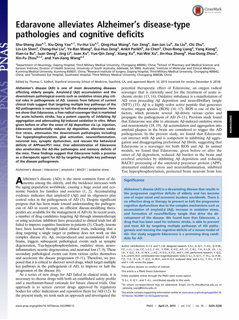
ResultsEdaravone Inhibits Aβ Aggregation and Antagonizes Aβ Neurotoxicityin Vitro. Previous studies suggested that several natural antioxi-dants such as curcumin and grape-derived polyphenolics caninhibit aggregation of Aβ (20, 21). Based on these findings, wespeculate that Edaravone (the structure shown in Fig. 1A), anoxygen radical scavenger, might also be able to interfere with Aβaggregation. Using Thioflavin T (ThT) fluorescence assay, wefound that Edaravone, when incubated with Aβ monomers orpreformed Aβ fibrils, dose-dependently reduced Aβ fibrillation-induced fluorescence intensity (Fig. 1 B and C). Western blotassays further showed that Edaravone inhibited the formation ofAβ fibrils during incubation (Fig. 1 D and E), which was revealedby the bands on the bottom of the loading wells without movingto the gel (arrow in Fig. 1D). Loaded with a fresh sample withoutprior incubation, soluble oligomers are the predominant Aβspecies, and only faint fibrils were seen in the loading well (Fig.1D). Edaravone preincubated with Aβ dose-dependently in-creased the soluble Aβ oligomer species (Fig. 1F). Moreover,transmission electron microscopy (TEM) assays visually con-firmed that Edaravone suppressed the fibrillation of Aβ anddisaggregated the preformed Aβ fibrils (Fig. 1G–J). To elucidatethe Edaravone binding epitope in Aβ42, we did Aβ42 fragment
competition assays using ThT fluorescence measurement. Wefound that only the peptide of the Aβ42 fragment amino acid(aa) 13–18, among seven peptides that cover the entire Aβ aasequence, had an ability to increase the Aβ fibril fluorescence,which was otherwise suppressed by Edaravone (Fig. S1A), in-dicating this peptide may compete for the binding site of Edaravonein Aβ42 and suggesting Edaravone may bind on the Aβ42 sequenceaa 13–18. No peptides interfered with the formation of Aβ42 fibrils(Fig. S1B) or formed any fibrils themselves (Fig. S1C). The putativeEdaravone binding epitope aa 13–18 is within the β strand regionof Aβ42 (Fig. S1D).Based on the above findings, we examined whether Edaravone
can protect neurons from Aβ-induced neurotoxicity. In humanneuroblastoma SH-SY5Y cells, Edaravone dose-dependentlyprotected neurons from cell death (Fig. S2 A and B) and neuritecollapse (Fig. S2C) triggered by Aβ. Furthermore, using neonatalprimary cortical neurons as another cell model, we also foundthe protective effects of Edaravone against neurite collapse (Fig.S2 D and E), cell death (Fig. S2 F and G), and ROS production(Fig. S2H) triggered by Aβ. These results suggest that Edaravonehas a potent capacity of inhibiting Aβ aggregation and neutral-izing the toxicity of Aβ in vitro.
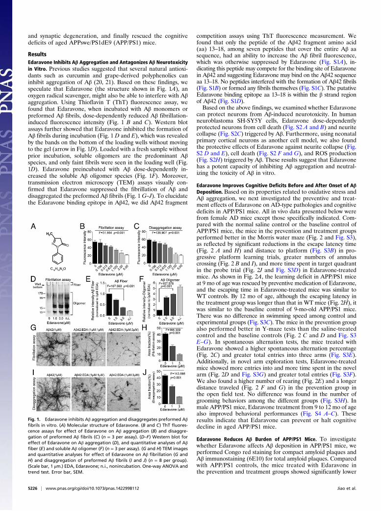
Edaravone Improves Cognitive Deficits Before and After Onset of AβDeposition. Based on its properties related to oxidative stress andAβ aggregation, we next investigated the preventive and treat-ment effects of Edaravone on AD-type pathologies and cognitivedeficits in APP/PS1 mice. All in vivo data presented below werefrom female AD mice except those specifically indicated. Com-pared with the normal saline control or the baseline control ofAPP/PS1 mice, the mice in the prevention and treatment groupsperformed better in the Morris water maze (Fig. 2 and Fig. S3),as reflected by significant reductions in the escape latency time(Fig. 2 A and H) and distance to platform (Fig. S3B) in pro-gressive platform learning trials, greater numbers of annuluscrossing (Fig. 2 B and I), and more time spent in target quadrantin the probe trial (Fig. 2J and Fig. S3D) in Edaravone-treatedmice. As shown in Fig. 2A, the learning deficit in APP/PS1 miceat 9 mo of age was rescued by preventive medication of Edaravone,and the escaping time in Edaravone-treated mice was similar toWT controls. By 12 mo of age, although the escaping latency inthe treatment group was longer than that in WT mice (Fig. 2H), itwas similar to the baseline control of 9-mo-old APP/PS1 mice.There was no difference in swimming speed among control andexperimental groups (Fig. S3C). The mice in the prevention groupalso performed better in Y-maze tests than the saline-treatedcontrol and the baseline controls (Fig. 2 C and D and Fig. S3E–G). In spontaneous alternation tests, the mice treated withEdaravone showed a higher spontaneous alternation percentage(Fig. 2C) and greater total entries into three arms (Fig. S3E).Additionally, in novel arm exploration tests, Edaravone-treatedmice showed more entries into and more time spent in the novelarm (Fig. 2D and Fig. S3G) and greater total entries (Fig. S3F).We also found a higher number of rearing (Fig. 2E) and a longerdistance traveled (Fig. 2 F and G) in the prevention group inthe open field test. No difference was found in the number ofgrooming behaviors among the different groups (Fig. S3H). Inmale APP/PS1 mice, Edaravone treatment from 9 to 12 mo of agealso improved behavioral performances (Fig. S4 A–C). Theseresults indicate that Edaravone can prevent or halt cognitivedecline in aged APP/PS1 mice.
Edaravone Reduces Aβ Burden of APP/PS1 Mice. To investigatewhether Edaravone affects Aβ deposition in APP/PS1 mice, weperformed Congo red staining for compact amyloid plaques andAβ immunostaining (6E10) for total amyloid plaques. Comparedwith APP/PS1 controls, the mice treated with Edaravone inthe prevention and treatment groups showed significantly lower
Fig. 1. Edaravone inhibits Aβ aggregation and disaggregates preformed Aβfibrils in vitro. (A) Molecular structure of Edaravone. (B and C) ThT fluores-cence assays for effect of Edaravone on Aβ aggregation (B) and disaggre-gation of preformed Aβ fibrils (C) (n = 3 per assay). (D–F) Western blot foreffect of Edaravone on Aβ aggregation (D), and quantitative analyses of Aβfiber (E) and soluble Aβ oligomer (F) (n = 3 per assay). (G and H) TEM imagesand quantitative analyses for effect of Edaravone on Aβ fibrillation (G andH) and disaggregation of preformed Aβ fibrils (I and J) (n = 8 per group).(Scale bar, 1 μm.) EDA, Edaravone; n.i., nonincubation. One-way ANOVA andtrend test. Error bar, SEM.
5226 | www.pnas.org/cgi/doi/10.1073/pnas.1422998112 Jiao et al.
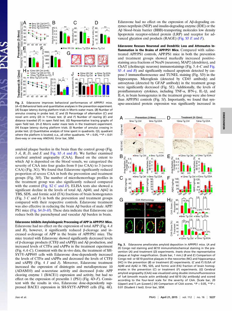
amyloid plaque burden in the brain than the control group (Fig.3 A, B, D, and E and Fig. S5 A and B). We further examinedcerebral amyloid angiopathy (CAA). Based on the extent towhich Aβ is deposited on the blood vessels, we categorized theseverity of CAA into four grades from 0 (no CAA) to 3 (severeCAA) (Fig. 3G). We found that Edaravone significantly reducedproportion of severe CAA in both the prevention and treatmentgroups (Fig. 3H). The number of microhemorrhage profiles inthe treatment group was also significantly reduced comparedwith the control (Fig. S2 C and D). ELISA tests also showed asignificant decline in the levels of total Aβ, Aβ40, and Aβ42 inTBS, SDS, and formic acid (FA) fractions of brain homogenates(Fig. 3 C and F) in both the prevention and treatment groupscompared with their respective controls. Edaravone treatmentwas also effective in reducing the brain Aβ burden of male APP/PS1 mice (Fig. S4 D–H). These data indicate that Edaravone canreduce both the parenchymal and vascular Aβ burden in brain.
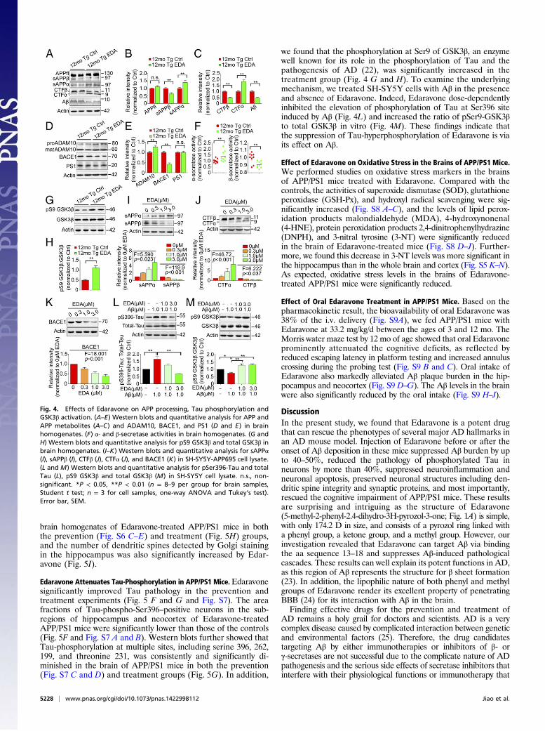
Edaravone Inhibits Amyloidogenic Processing of APP in APP/PS1 Mice.Edaravone had no effect on the expression of total APP (Fig. 4 Aand B), however, it significantly reduced β-cleavage and in-creased α-cleavage of APP in the brains of APP/PS1 mice. Themice treated with Edaravone showed significantly decreased levelsof β-cleavage products (CTFβ and sAPPβ) and Aβ production, andincreased levels of CTFα and sAPPα in the treatment experiment(Fig. 4 A–C). Consistent with the in vivo data, the treatment of SH-SY5Y-APP695 cells with Edaravone dose-dependently increasedthe levels of CTFα and sAPPα and decreased the levels of CTFβand sAPPβ (Fig. 4 I and J). Moveover, Edaravone treatmentincreased the expression of disintegrin and metalloprotease 10(ADAM10) and α-secretase activity and decreased β-site APPcleaving enzyme 1 (BACE1) expression and activity, but had noeffect on the expression of presenlin 1 (PS1) (Fig. 4D–F). Consis-tent with the results in vivo, Edaravone dose-dependently sup-pressed BACE1 expression in SH-SY5Y-APP695 cells (Fig. 4K).
Edaravone had no effect on the expression of Aβ-degrading en-zymes neprilysin (NEP) and insulin-degrading enzyme (IDE) or theAβ blood–brain barrier (BBB)-transporting molecules low densitylipoprotein receptor-related protein (LRP) and receptor for ad-vanced glycation end products (RAGE) (Fig. S5 E and F).
Edaravone Rescues Neuronal and Dendritic Loss and Attenuates In-flammation in the Brains of APP/PS1 Mice. Compared with saline-treated APP/PS1 controls, APP/PS1 mice in both the preventionand treatment groups showed markedly increased positive-staining area fractions of NeuN (neurons), MAP2 (dendrites), andChAT (cholinergic neurons) immunostainings (Fig. 5 A–C and Fig.S5 A and B) and significantly reduced apoptosis detected by cas-pase-3 immunofluorescence and TUNEL staining (Fig. 5D) in thehippocampus. Microgliosis (detected by CD45 antibody) andastrocytosis (detected by GFAP antibody) in the treatment groupwere significantly decreased (Fig. 5E). Additionally, the levels ofproinflammatory cytokines, including TNF-α, IFN-γ, IL-1β, andIL-6, in brain homogenates in the treatment group were also lowerthan APP/PS1 controls (Fig. 5J). Importantly, we found that syn-apse-associated protein expression was significantly increased in
Fig. 3. Edaravone ameliorates amyloid deposition in APP/PS1 mice. (A andD) Congo red staining and 6E10 immunohistochemical staining in the pre-vention (A) and treatment (D) experiments. Insets show the representativeplaque at higher magnification. (Scale bar, 1 mm.) (B and E) Comparison ofCongo red- or 6E10-positive plaques in the neocortex (NC) and hippocampus(HC) in the prevention (B) or treatment (E) experiments. (C and F) ELISA ofAβ40 and Aβ42 in TBS, SDS, and formic acid (FA) fraction of brain homog-enates in the prevention (C) or treatment (F) experiments. (G) Cerebralamyloid angiopathy (CAA) was visualized using double immunofluorescenceof 1a4 (smooth muscle actin antibody) and 6E10 (Aβ antibody) and scoredaccording to the four-level scale for the severity of CAA. [Scale bar, 20(Upper) and 5 μm (Lower).] (H) Comparison of CAA scores. *P < 0.05, **P <0.01 (Student t test). Error bar, SEM.
Fig. 2. Edaravone improves behavioral performances of APP/PS1 mice.(A–G) Behavioral tests and quantitative analyses in the prevention experiment.(A) Escape latency during platform trials in Morris water maze. (B) Number ofannulus crossing in probe test. (C and D) Percentage of alternation (C) andnovel arm entry (D) in Y-maze test. (E and F) Number of rearing (E) anddistance traveled (F) in open field test. (G) Representative tracing graphs ofopen field test. (H–J) Morris water maze tests in the treatment experiment.(H) Escape latency during platform trials. (I) Number of annulus crossing inprobe test. (J) Quantitative analysis of time spent in quadrants. Q3, quadrantwhere the platform is located; o.a., all other quadrants. *P < 0.05, **P < 0.01(two-way or one-way ANOVA). Error bar, SEM.
Jiao et al. PNAS | April 21, 2015 | vol. 112 | no. 16 | 5227
NEU
ROSC
IENCE
brain homogenates of Edaravone-treated APP/PS1 mice in boththe prevention (Fig. S6 C–E) and treatment (Fig. 5H) groups,and the number of dendritic spines detected by Golgi stainingin the hippocampus was also significantly increased by Edar-avone (Fig. 5I).
Edaravone Attenuates Tau-Phosphorylation in APP/PS1 Mice.Edaravonesignificantly improved Tau pathology in the prevention andtreatment experiments (Fig. 5 F and G and Fig. S7). The areafractions of Tau-phospho-Ser396–positive neurons in the sub-regions of hippocampus and neocortex of Edaravone-treatedAPP/PS1 mice were significantly lower than those of the controls(Fig. 5F and Fig. S7 A and B). Western blots further showed thatTau-phosphorylation at multiple sites, including serine 396, 262,199, and threonine 231, was consistently and significantly di-minished in the brain of APP/PS1 mice in both the prevention(Fig. S7 C and D) and treatment groups (Fig. 5G). In addition,
we found that the phosphorylation at Ser9 of GSK3β, an enzymewell known for its role in the phosphorylation of Tau and thepathogenesis of AD (22), was significantly increased in thetreatment group (Fig. 4 G and H). To examine the underlyingmechanism, we treated SH-SY5Y cells with Aβ in the presenceand absence of Edaravone. Indeed, Edaravone dose-dependentlyinhibited the elevation of phosphorylation of Tau at Ser396 siteinduced by Aβ (Fig. 4L) and increased the ratio of pSer9-GSK3βto total GSK3β in vitro (Fig. 4M). These findings indicate thatthe suppression of Tau-hyperphosphorylation of Edaravone is viaits effect on Aβ.
Effect of Edaravone on Oxidative Stress in the Brains of APP/PS1 Mice.We performed studies on oxidative stress markers in the brainsof APP/PS1 mice treated with Edaravone. Compared with thecontrols, the activities of superoxide dismutase (SOD), glutathioneperoxidase (GSH-Px), and hydroxyl radical scavenging were sig-nificantly increased (Fig. S8 A–C), and the levels of lipid perox-idation products malondialdehyde (MDA), 4-hydroxynonenal(4-HNE), protein peroxidation products 2,4-dinitrophenylhydrazine(DNPH), and 3-nitral tyrosine (3-NT) were significantly reducedin the brain of Edaravone-treated mice (Fig. S8 D–J). Further-more, we found this decrease in 3-NT levels was more significant inthe hippocampus than in the whole brain and cortex (Fig. S5 K–N).As expected, oxidative stress levels in the brains of Edaravone-treated APP/PS1 mice were significantly reduced.
Effect of Oral Edaravone Treatment in APP/PS1 Mice. Based on thepharmacokinetic result, the bioavailability of oral Edaravone was38% of the i.v. delivery (Fig. S9A), we fed APP/PS1 mice withEdaravone at 33.2 mg/kg/d between the ages of 3 and 12 mo. TheMorris water maze test by 12 mo of age showed that oral Edaravoneprominently attenuated the cognitive deficits, as reflected byreduced escaping latency in platform testing and increased annuluscrossing during the probing test (Fig. S9 B and C). Oral intake ofEdaravone also markedly alleviated Aβ plaque burden in the hip-pocampus and neocortex (Fig. S9 D–G). The Aβ levels in the brainwere also significantly reduced by the oral intake (Fig. S9 H–J).
DiscussionIn the present study, we found that Edaravone is a potent drugthat can rescue the phenotypes of several major AD hallmarks inan AD mouse model. Injection of Edaravone before or after theonset of Aβ deposition in these mice suppressed Aβ burden by upto 40–50%, reduced the pathology of phosphorylated Tau inneurons by more than 40%, suppressed neuroinflammation andneuronal apoptosis, preserved neuronal structures including den-dritic spine integrity and synaptic proteins, and most importantly,rescued the cognitive impairment of APP/PS1 mice. These resultsare surprising and intriguing as the structure of Edaravone(5-methyl-2-phenyl-2.4-dihydro-3H-pyrozol-3-one; Fig. 1A) is simple,with only 174.2 D in size, and consists of a pyrozol ring linked witha phenyl group, a ketone group, and a methyl group. However, ourinvestigation revealed that Edaravone can target Aβ via bindingthe aa sequence 13–18 and suppresses Aβ-induced pathologicalcascades. These results can well explain its potent functions in AD,as this region of Aβ represents the structure for β sheet formation(23). In addition, the lipophilic nature of both phenyl and methylgroups of Edaravone render its excellent property of penetratingBBB (24) for its interaction with Aβ in the brain.Finding effective drugs for the prevention and treatment of
AD remains a holy grail for doctors and scientists. AD is a verycomplex disease caused by complicated interaction between geneticand environmental factors (25). Therefore, the drug candidatestargeting Aβ by either immunotherapies or inhibitors of β- orγ-secretases are not successful due to the complicate nature of ADpathogenesis and the serious side effects of secretase inhibitors thatinterfere with their physiological functions or immunotherapy that
Fig. 4. Effects of Edaravone on APP processing, Tau phosphorylation andGSK3β activation. (A–E) Western blots and quantitative analysis for APP andAPP metabolites (A–C) and ADAM10, BACE1, and PS1 (D and E) in brainhomogenates. (F) α- and β-secretase activities in brain homogenates. (G andH) Western blots and quantitative analysis for pS9 GSK3β and total GSK3β inbrain homogenates. (I–K) Western blots and quantitative analysis for sAPPα(I), sAPPβ (I), CTFβ (J), CTFα (J), and BACE1 (K) in SH-SY5Y-APP695 cell lysate.(L and M) Western blots and quantitative analysis for pSer396-Tau and totalTau (L), pS9 GSK3β and total GSK3β (M) in SH-SY5Y cell lysate. n.s., non-significant. *P < 0.05, **P < 0.01 (n = 8–9 per group for brain samples,Student t test; n = 3 for cell samples, one-way ANOVA and Tukey’s test).Error bar, SEM.
5228 | www.pnas.org/cgi/doi/10.1073/pnas.1422998112 Jiao et al.

causes neural inflammation (5, 26, 27). We propose that an effec-tive intervention for AD should target multiple pathways that areable to break the multiple cascades of signals driving the patho-genesis of AD such as amyloidogenesis, oxidative stress, and GSK3β-Tau phosphorylation. We discovered that Edaravone appears tomeet this criteria of having multiple functions on AD pathogenesis.In the present study, we revealed an interaction of Edaravone
with Aβ with several classic techniques. First, we used TEM,ThT, and Western blot assays to determine the effect of Edaravoneon Aβ fibrillation. Edaravone indeed can efficiently suppress theformation of Aβ fibrils and dissolve the preformed Aβ fibrils, in-dicating that Edaravone acts on Aβ. Second, applying primaryneurons or cell lines as a model, we found that Edaravone cansuppress Aβ-induced neurotoxicity such as neurite collapse andcell death to a large extent. Third, multiple animal experimentsincluding preventive medication, therapeutic medication, and oraladministration showed that Edaravone significantly reduces amy-loid plaques in the hippocampus and neocortex, CAA, and the Aβlevels in the brain. More importantly, we also found that Edaravonesuppresses the expression of BACE1, sAPPβ, and CTFβ in vivoand in cultured human neuroblastoma-derived neurons, sug-gesting that the interaction between Edaravone and Aβ can breakthe positive feed-forward loop of Aβ-driven BACE1-mediatedamyloidogenesis (28–30). As both oxidative stress and Aβ arepositive regulators that drive the up-regulation of BACE1 via theactivation of JNK (31) and GSK3β (32), respectively, it is likelythat Edaravone suppresses BACE1 expression and amyloidogenesisby attenuation of oxidative stress and Aβ-induced GSK3βphosphorylation.
The intraneuronal NFT caused by hyperphosphorylation ofTau protein is another hallmark of AD (33) and consistentlycorrelated with the cognitive dysfunctions in patients (34, 35). Itis well recognized that the formation of NFT is a down-streamevent of Aβ toxicity that is caused by Tau hyperphosphorylationtriggered by Aβ-induced activation of several kinases such as Cdk5(36), GSK3β (37) and PKA (37). In the present study, we foundthat Edaravone can suppress elevated phosphorylation of Tau atseveral sites in vivo and in vitro in response to Aβ, as examinedby Western blot and by immunostaining methods. Consistently,Edaravone can increase the activation pS9-GSK3β which self-inactivates GSK3β kinase activities (38). These findings suggestthat Edaravone acts on Aβ and suppresses its toxicity of triggeringTau hyperphosphorylation by inhibiting the GSK3β pathway.As expected, we also found that Edaravone is a strong neu-
roprotective agent that protects neurons from neurite degeneration,dendritic spine shrinkage, down-regulation of synaptic proteins,apoptosis, and loss of ChAT in APP/PS1 mice and in neurons inresponse to Aβ. It is likely that the strong neuroprotection byEdaravone is through its dual functions in blocking Aβ aggregation/deposition and scavenging free radicals generated in the AD brain.This note is supported by our findings that Edaravone suppressesseveral oxidative stress markers such as oxidized lipids and proteinsand increases antioxidant markers such as SOD and GSH-Px inthe brain of AD mice and the discovery that Edaravone acts on Aβaggregation and fibrillation.CAA is a common disorder caused by the deposition of Aβ in
the vessel wall of small arterial vessels of the brain, leading tocerebral hemorrhage, ischemia, and infarction (39). No specificeffective drug for CAA is available at present. In both animal
Fig. 5. Edaravone attenuates neuronal loss and AD-type pathologies in APP/PS1 mice. APP/PS1 mice aged 9 mo were treated with Edaravone (12-mo Tg EDA,n = 9) or saline (12-mo Tg Ctrl, n = 7) for 3 mo. (A–C) MAP2, NeuN, and ChAT immunostaining and quantification in hippocampus and its subregions. [Scalebar, 400 (A) and 100 μm (B and C).] (D) Neuronal apoptosis detected by activated caspase-3 (cas-3) immunofluorescence (Top) in CA3 of hippocampus and byTUNEL staining (Middle) in neocortex. [Scale bar, 100 (Top) and 200 μm (Middle).] (E) Immunostaining and quantification of microgliosis and astrocytosis.(Scale bar, 1 mm.) (F) Quantification of Tau phosphorylation using pSer396-Tau immunohistochemistry. (G) Western blot and quantification for phosphor-ylated Tau at multiple sites including pS396-, pS262-, pS199-, pT231-Tau, and total Tau (T-tau) in brain homogenates. (H) Western blot and quantification forsynapse-associated proteins including SNAP25, PSD95, Synapsin I (Syn I), VAMP1, and synaptophysin (SYP) in brain homogenates. (I) Representative photo-micrograph and quantification of dendritic spines intensity of basal segment of CA1 hippocampal neuron (three mice per group and seven neurons permouse). (Scale bar, 10 μm.) (J) Quantification of IL-1β, IL-6, TNF-α, and IFN-γ in brain homogenates. *P < 0.05, **P < 0.01 (Student t test). Error bar, SEM.
Jiao et al. PNAS | April 21, 2015 | vol. 112 | no. 16 | 5229
NEU
ROSC
IENCE

and clinical studies of immunotherapies, CAA is aggravatedduring the clearance of brain Aβ with subsequent increase ofcerebral microhemorrhages (40, 41), which are related to for-mation of immune complex of antibody and Aβ (5). In the pre-sent study, we found that CAA is substantially reduced, with areduction in parenchymal Aβ deposition and without an increasein microhemorrhage. ROSs are a key contributor to CAA for-mation, CAA-induced vessel dysfunction, and CAA-relatedmicrohemorrhage, implying that the ROS scavenger is an im-portant therapeutic for CAA (42). These findings suggest thatEdaravone would also be a promising drug candidate for CAA.In summary, we uncovered an application of the ischemic stroke
drug Edaravone in the therapy for AD and CAA by targetingmultiple key AD pathways including Aβ, oxidative stress, andGSK3β-Tau phosphorylation. Edaravone would be effective inboth the prevention and treatment for AD, representing a futuredirection of drug discovery for AD by simultaneously blockingmultiple cascades leading to disease pathogenesis. BecauseEdaravone is currently used for stroke and proven safe in humans,the promising data from our current study warrant a large-scaleclinical trial to test its efficacy in sporadic and familial AD.
Materials and MethodsSee SI Materials and Methods for detailed descriptions.
The research protocol was approved by the institutional review boardsof both the Third Military Medical University and the University of South
Australia. The effects of Edaravone on Aβ aggregation and disaggregationwere examined using ThT, Western blot, and TEM methods (43). Edaravonebinding epitope mapping was performed by the competition assay using theThT-activated fluorescence method. The effects of Edaravone on neuronaltoxicity, neurite growth, APP processing, BACE1 expression, Tau phosphor-ylation, and GSK3β activation in response to Aβ were examined in SH-SY5Y(Chinese Academy of Sciences) and SH-SY5Y-APP695 cells and primary mousecortical neurons. APP/PS1 transgenic mice at either 3 or 9 mo of age wereused to test the effectiveness of Edaravone on AD as a prevention ortreatment, via either i.p. injection or oral administration. The behavioralperformance of mice treated with Edaravone was tested using the Morriswater maze, Y-maze, and open field protocols. The changes in brain Aβburden, CAA severity, amyloidogenic APP processing and Aβ metabolism, α-and β-secretase activities, oxidative stress, neuroinflammation, Tau phosphor-ylation, neurodegeneration, and neuronal loss were assessed with histologicaland biochemical methods. Unless otherwise stated, the results are presentedas mean ± SEM. Statistical comparisons between two groups were tested usingStudent t test, or Mann–Whitney u test, as applicable. The comparisons amonggroups were tested using one-way or two-way ANOVA, and the trend analysiswas performed when necessary. P < 0.05 was considered significant.
ACKNOWLEDGMENTS. This work was supported by National NaturalScience Foundation of China Grants 81270423, 30973144, 81200988, and81000549, National Health and Medical Research Council of AustraliaGrants 1020567 and 1021409, Ministry of Science and Technology ofChina Grant 2011CB944200, Chongqing Science and Technology CommitteeGrant CSTC2012JJA10111, CSTC2010BA5004, and China Postdoctoral ScienceFoundation Grants 2012M521861 and 2013T60955. X.-F.Z. is a visiting pro-fessor of Kunming Medical University.
1. Kalaria RN, et al.; World Federation of Neurology Dementia Research Group (2008)Alzheimer’s disease and vascular dementia in developing countries: Prevalence,management, and risk factors. Lancet Neurol 7(9):812–826.
2. US Burden of Disease Collaborators (2013) The state of US health, 1990-2010: Burdenof diseases, injuries, and risk factors. JAMA 310(6):591–608.
3. Korczyn AD (2008) The amyloid cascade hypothesis. Alzheimers Dement 4(3):176–178.4. Holtzman DM, Morris JC, Goate AM (2011) Alzheimer’s disease: The challenge of the
second century. Sci Transl Med 3(77):sr1.5. Liu YH, Giunta B, Zhou HD, Tan J, Wang YJ (2012) Immunotherapy for Alzheimer
disease: The challenge of adverse effects. Nat Rev Neurol 8(8):465–469.6. Wang YJ (2014) Alzheimer disease: Lessons from immunotherapy for Alzheimer dis-
ease. Nat Rev Neurol 10(4):188–189.7. Mucke L, Selkoe DJ (2012) Neurotoxicity of amyloid β-protein: Synaptic and network
dysfunction. Cold Spring Harb Perspect Med 2(7):a006338.8. Walsh DM, Selkoe DJ (2004) Deciphering the molecular basis of memory failure in
Alzheimer’s disease. Neuron 44(1):181–193.9. Zawia NH, Lahiri DK, Cardozo-Pelaez F (2009) Epigenetics, oxidative stress, and Alz-
heimer disease. Free Radic Biol Med 46(9):1241–1249.10. Guglielmotto M, Giliberto L, Tamagno E, Tabaton M (2010) Oxidative stress mediates
the pathogenic effect of different Alzheimer’s disease risk factors. Front AgingNeurosci 2:3.
11. Tamagno E, Guglielmotto M, Monteleone D, TabatonM (2012) Amyloid-β production:Major link between oxidative stress and BACE1. Neurotox Res 22(3):208–219.
12. Corbett A, et al. (2012) Drug repositioning for Alzheimer’s disease. Nat Rev DrugDiscov 11(11):833–846.
13. Yoshida H, et al. (2006) Neuroprotective effects of edaravone: A novel free radicalscavenger in cerebrovascular injury. CNS Drug Rev 12(1):9–20.
14. Feng S, et al. (2011) Edaravone for acute ischaemic stroke. Cochrane Database SystRev (12):CD007230.
15. Clark TA, et al. (2010) Oxidative stress and its implications for future treatments andmanagement of Alzheimer disease. Int J Biomed Sci 6(3):225–227.
16. De Felice FG, et al. (2007) Abeta oligomers induce neuronal oxidative stress throughan N-methyl-D-aspartate receptor-dependent mechanism that is blocked by theAlzheimer drug memantine. J Biol Chem 282(15):11590–11601.
17. Huang X, et al. (1999) The A beta peptide of Alzheimer’s disease directly produceshydrogen peroxide through metal ion reduction. Biochemistry 38(24):7609–7616.
18. Zhang GL, et al. (2013) Edaravone ameliorates oxidative damage associated withAβ25-35 treatment in PC12 cells. J Mol Neurosci 50(3):494–503.
19. He F, et al. (2014) Inhibitory effects of edaravone in β-amyloid-induced neurotoxicityin rats. Biomed Res Int 2014:370368.
20. Yang F, et al. (2005) Curcumin inhibits formation of amyloid beta oligomers and fi-brils, binds plaques, and reduces amyloid in vivo. J Biol Chem 280(7):5892–5901.
21. Wang J, et al. (2008) Grape-derived polyphenolics prevent Abeta oligomerization andattenuate cognitive deterioration in a mouse model of Alzheimer’s disease. J Neurosci28(25):6388–6392.
22. Flaherty DB, Soria JP, Tomasiewicz HG, Wood JG (2000) Phosphorylation of humantau protein by microtubule-associated kinases: GSK3beta and cdk5 are key partici-pants. J Neurosci Res 62(3):463–472.
23. Colletier JP, et al. (2011) Molecular basis for amyloid-beta polymorphism. Proc NatlAcad Sci USA 108(41):16938–16943.
24. Watanabe T, Tahara M, Todo S (2008) The novel antioxidant edaravone: From benchto bedside. Cardiovasc Ther 26(2):101–114.
25. Hampel H, et al.; German Task Force on Alzheimer’s Disease (GTF-AD) (2011) Thefuture of Alzheimer’s disease: The next 10 years. Prog Neurobiol 95(4):718–728.
26. Yan R, Vassar R (2014) Targeting the β secretase BACE1 for Alzheimer’s diseasetherapy. Lancet Neurol 13(3):319–329.
27. Golde TE, Koo EH, Felsenstein KM, Osborne BA, Miele L (2013) γ-Secretase inhibitorsand modulators. Biochim Biophys Acta 1828(12):2898–2907.
28. Buggia-Prevot V, Sevalle J, Rossner S, Checler F (2008) NFkappaB-dependent controlof BACE1 promoter transactivation by Abeta42. J Biol Chem 283(15):10037–10047.
29. Guglielmotto M, et al. (2011) Amyloid-β₄₂ activates the expression of BACE1 throughthe JNK pathway. J Alzheimers Dis 27(4):871–883.
30. Chami L, et al. (2012) Nuclear factor-κB regulates βAPP and β- and γ-secretases dif-ferently at physiological and supraphysiological Aβ concentrations. J Biol Chem287(29):24573–24584.
31. Chami L, Checler F (2012) BACE1 is at the crossroad of a toxic vicious cycle involvingcellular stress and β-amyloid production in Alzheimer’s disease. Mol Neurodegener7:52.
32. Ly PT, et al. (2013) Inhibition of GSK3β-mediated BACE1 expression reduces Alz-heimer-associated phenotypes. J Clin Invest 123(1):224–235.
33. Bloom GS (2014) Amyloid-β and tau: The trigger and bullet in Alzheimer diseasepathogenesis. JAMA Neurol 71(4):505–508.
34. Han SD, et al.; Alzheimer’s Disease Neuroimaging Initiative (2012) Beta amyloid, tau,neuroimaging, and cognition: Sequence modeling of biomarkers for Alzheimer’sDisease. Brain Imaging Behav 6(4):610–620.
35. Sunderland T, et al. (1999) Longitudinal stability of CSF tau levels in Alzheimer pa-tients. Biol Psychiatry 46(6):750–755.
36. Noble W, et al. (2003) Cdk5 is a key factor in tau aggregation and tangle formation invivo. Neuron 38(4):555–565.
37. Carlyle BC, et al. (2014) cAMP-PKA phosphorylation of tau confers risk for de-generation in aging association cortex. Proc Natl Acad Sci USA 111(13):5036–5041.
38. Wen Y, et al. (2008) Interplay between cyclin-dependent kinase 5 and glycogensynthase kinase 3 beta mediated by neuregulin signaling leads to differential effectson tau phosphorylation and amyloid precursor protein processing. J Neurosci 28(10):2624–2632.
39. Gahr M, Nowak DA, Connemann BJ, Schönfeldt-Lecuona C (2013) Cerebral AmyloidalAngiopathy—a disease with implications for neurology and psychiatry. Brain Res1519:19–30.
40. Racke MM, et al. (2005) Exacerbation of cerebral amyloid angiopathy-associatedmicrohemorrhage in amyloid precursor protein transgenic mice by immunotherapy isdependent on antibody recognition of deposited forms of amyloid beta. J Neurosci25(3):629–636.
41. Patton RL, et al. (2006) Amyloid-beta peptide remnants in AN-1792-immunized Alz-heimer’s disease patients: A biochemical analysis. Am J Pathol 169(3):1048–1063.
42. Han BH, et al. (2015) Contribution of reactive oxygen species to cerebral amyloidangiopathy, vasomotor dysfunction, and microhemorrhage in aged Tg2576 mice.Proc Natl Acad Sci USA 112(8):E881–E890.
43. Wang YJ, et al. (2011) p75NTR regulates Abeta deposition by increasing Abeta pro-duction but inhibiting Abeta aggregation with its extracellular domain. J Neurosci31(6):2292–2304.
5230 | www.pnas.org/cgi/doi/10.1073/pnas.1422998112 Jiao et al.