Embed Size (px)
Citation preview

Orthopedic Evaluation of the Ankle and Foot
Ed Mulligan, PT, DPT, OCS, SCS, ATCClinical Orthopedic Rehabilitation Education

Subjective history questions
• Patient's Chief Complaint and Rehab Goal(s)• Mechanism of Injury• Date of Injury‐Surgery• Length and type of immobilization• Weight bearing status and progression• Previous Treatment• Present Status
• better – worse –same• Orthotics/braces/sleeves, etc• Symptom Behavior• Past Medical ‐ Injury History

Remember S.I.N.S.?Considerable Influence on Intervention Strategy
Severity– How significantly this impairment affects the patient
Irritability– The reactivity or stability of the condition
• What does the patient have to do to set off the condition?• Once set off, how long and severe are the symptoms?• What does the patient have to do to calm the symptoms?
Nature– Numbness/tingling, Weakness, Popping, Locking, Giving way, Clicking,
Grinding, Skin changes Stage
– Has the condition stabilized (better), become stagnant (same), or deteriorated (worse)?

Foot-Ankle Specific Questions
• Does spinal motion or posture effect your leg‐ankle foot symptoms?
• Have you altered the IDF of your daily, occupational, or recreational activities?
• Do you have stiffness first thing in the morning that resolves in an hour or so?
• Does your heel hurt first thing in the morning or after prolonged sitting?
• Shoe preferences

Interclass Correlation CoefficientsReliability Interpretation
Degree ICCHigh .90 ‐ .99
Good .80 ‐ .89
Fair .70 ‐ .79
Poor < .69
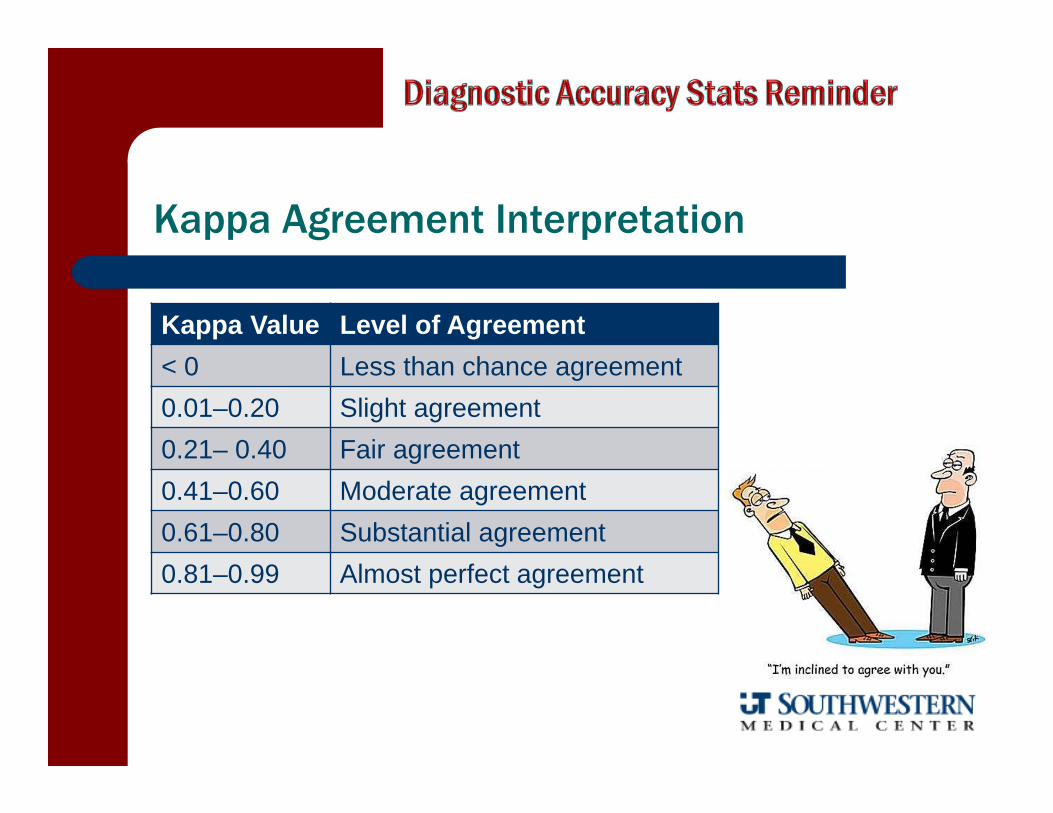
Kappa Agreement Interpretation
Kappa Value Level of Agreement< 0 Less than chance agreement0.01–0.20 Slight agreement0.21– 0.40 Fair agreement0.41–0.60 Moderate agreement0.61–0.80 Substantial agreement0.81–0.99 Almost perfect agreement

Sensitivity-Specificity-Likelihood Ratios
SN: % of true positive (SNOUT = rule out)
SP: % of true negative (SPIN = rule in)
LR: + or – (predicts post‐test probability based on prevalence)
is (+) is not (‐)
Positive Test(+)
true positive
a
false positive
b
total who test positive
a + b
Negative Test(‐)
false negative
c
true negative
d
total who test negative
c + d
TOTALS
total with condition
a + c
total without condition
b + d
total population
a + b + c + d
+ LR = sens/(1-spec)- LR = (1-sens)/spec

observation and general appearance
Posture Weight Bearing Status Symmetrical Appearance Soft Tissue swelling, effusion,
atrophy, etc. Plantar Lesions
check for callous patterns Shoe Wear and Type

Anthropometric Measurements • circumferential girth measurements may be recorded bilaterally at mid‐calf, mid foot, met heads, and Figure 8 or Heel Lock tape measurement
• High intra/intertester reliability (ICC = .99) with MDC of 10 mm
• volumetric displacement techniques may also be used to objectively quantify swelling
• High interrater reliability (ICC = .98)
Rohner‐Spengler M, et al, J Orthop Sports Phys Ther, 2007Petersen EJ, J Orthop Sports Phys Ther, 1999Mawdsley RH, et al, J Orthop Sports Phys Ther, 2000Tatro‐Adams D, et al, J Orthop Sports Phys Ther, 1995
observation and general appearance

Vascular Pulses
Dorsal Pedal Pulse Posterior Tibial Pulse
to rule out cardiovascular pathology

sagittal plane abnormalities
Feiss Line (medial longitudinal arch)– assessment of navicular tuberosity position
relative to bisection of medial malleolus apex and 1st metatarsal head
Longitudinal Arch Angle– angle formed between line from medial malleolus to navicular
tuberosity and 1st medial met head to navicular tuberosity – good interrater reliability, prognostic of
dynamic foot posture during ambulation, and may have some value in predicting the risk of lower extremity injuries
Jonson, et al, JOSPT, 1997McPoil, et al, JAMPA, 2004, 2007

Navicular Drop Test
Difference in navicular height from STJN position (with most of weight on contralateral extremity) to relaxed stance position
Intrarater ICC = .78 with S.E.M = 1.68 mm
Mueller MJ, et al, J Am Pod Med Assoc, 1993

Navicular Height and Drop
• The difference between navicular height in STJ neutral vs. STJ relaxed (or NWB vs. WBing)
• 6‐9 mm drop is normal
• > 10‐15 mm is indicative of compensatory pronation

Mulligan E, et al. Man Ther, 2013
ICC2,1 = 0.88Ave. ND = 12 mm

Arch Index
• AI = surface area of B/(A+B+C)
• The lower the arch – the higher the index
High Arch = < .21Normal = .21 ‐.26Flat Arch = > .26
Test‐Retest ICC = .99Length of truncated foot (excluding toes) is divided into equal thirds

Arch Height Index
Arch Height @ ½ Foot LengthTruncated Foot Length
Normal = 0.316High Arch = 0.356 (+1.5 SD)Low Arch = 0.275 (‐1.5 SD)
Williams DS, et al. Phys Ther 2000
My Foot Example8.7 cm ÷ 21.3 cm = .408 (high arch)
Mulligan E, et al. Man Ther 2013
ICC2,1 = 0.84Ave. AHI = 0.28

frontal plane abnormalities
Bony Landmark Symmetry Genu Varum/Valgus Tibial Varum STJ POSITION
– Relaxed vs. Neutral Haglund's Deformities Met Length Classification Leg Length Discrepancies Hallux Valgus

Tibial Varum
measured in unilateral stance with STJin neutral
extrinsic deformity in which the distal portion of the tibia is closer to the midline than the proximal portion
intratester reliability of 2‐3
Lohman, 1987

STJ Position - relaxed vs. neutral
Relaxed position indicates amount of compensation necessary in stance
interrater reliability of .75 in unilateral stance and .91 in bilateral stance
Smith‐Orricchio, 1990

STJ Neutral Reliability
Position Intratester ICC Intertester ICC Source
NWB.77.60
.06‐.27
.25 Elveru, PT, 1988
Smith-Orricchio, JOSPT, 1990
Picciano, JOSPT, 1995
WB.14‐.18.75‐.91.85
.15
.72
.79
Picciano, JOSPT, 1995
Smith-Orricchio, JOSPT, 1990
Sell, JOSPT, 1994

metatarsophalangeal hallux valgus angle (HVA) representing the lateral deviation of the phalanx ‐should be less than 15°
intermetatarsal angle (IMA) should be less than 9°
HVA
IMA
1st MTP Deformities
Hallux Valgus – “bunions”

foot types
Squared Foot Greek Foot Egyptian FootMorton’s Foot
9% 22% 69%
Index Plus Index Minus Index +/‐1>2>3>4>5 1<2>3>4>5 1=2>3>4>5

structural abnormalities of the toe
claw toe mallet toehammer toe
typically deficient intrinsics
typically long extensor contracture at MTP
DIP flexion typically secondary to poor fitting
shoes

Transverse plane abnormalities
• Tibial Torsion– 15° external tibial torsion
• Toe in/out– Femoral torsion– Hip ante/retroversion– Forefoot ab/adductus
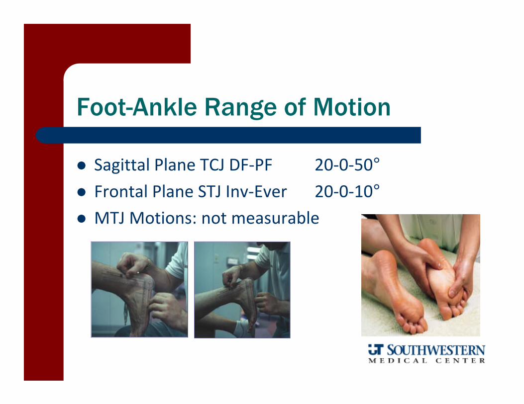
Foot-Ankle Range of Motion
Sagittal Plane TCJ DF‐PF 20‐0‐50° Frontal Plane STJ Inv‐Ever 20‐0‐10° MTJ Motions: not measurable

hallux dorsiflexion ROM
• 20‐30° with 1st ray stabilized• 60‐90° with 1st ray plantarflexion
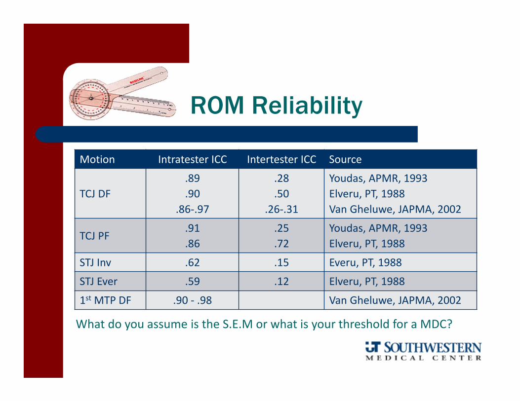
ROM Reliability
Motion Intratester ICC Intertester ICC Source
TCJ DF.89.90
.86‐.97
.28
.50.26‐.31
Youdas, APMR, 1993Elveru, PT, 1988Van Gheluwe, JAPMA, 2002
TCJ PF.91.86
.25
.72Youdas, APMR, 1993Elveru, PT, 1988
STJ Inv .62 .15 Everu, PT, 1988
STJ Ever .59 .12 Elveru, PT, 1988
1st MTP DF .90 ‐ .98 Van Gheluwe, JAPMA, 2002
What do you assume is the S.E.M or what is your threshold for a MDC?

MMT for the Foot-Ankle
• Anterior Tib• Extensor Hallicus Longus• Extensor Digitorum• Peroneals• Gastroc• Soleus• Posterior Tib
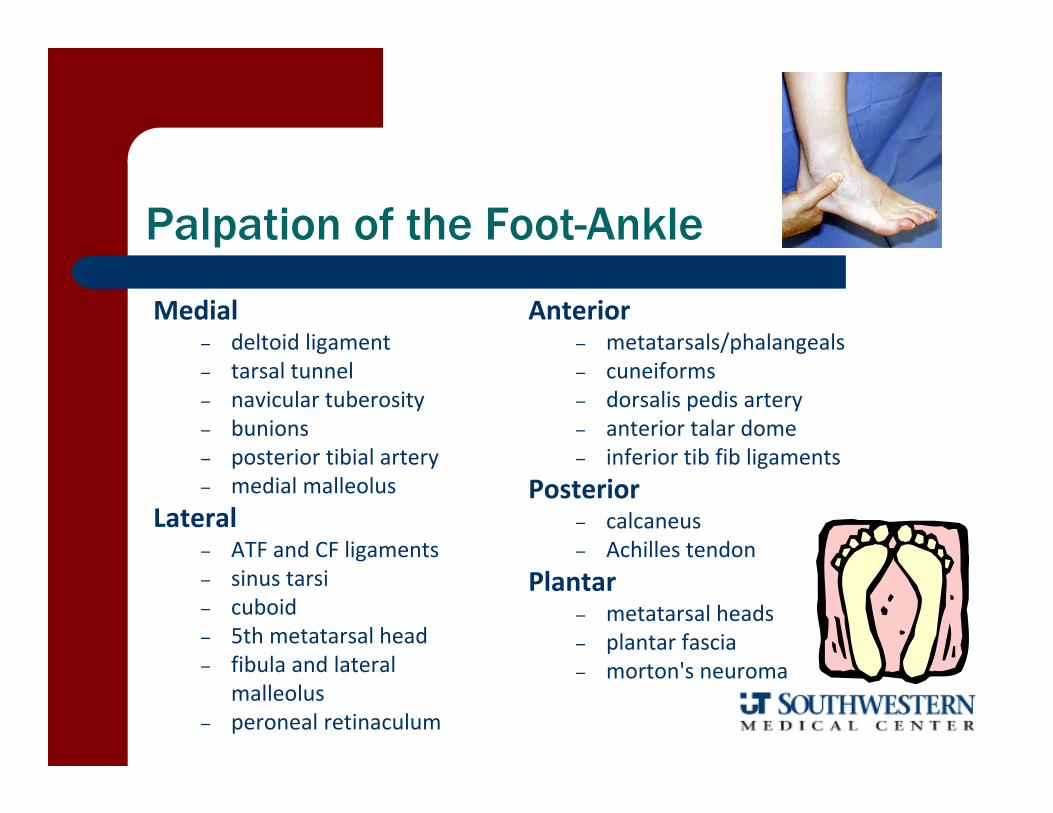
Palpation of the Foot-Ankle
Anterior– metatarsals/phalangeals– cuneiforms– dorsalis pedis artery– anterior talar dome– inferior tib fib ligaments
Posterior– calcaneus– Achilles tendon
Plantar– metatarsal heads– plantar fascia– morton's neuroma
Medial– deltoid ligament– tarsal tunnel– navicular tuberosity– bunions– posterior tibial artery– medial malleolus
Lateral– ATF and CF ligaments– sinus tarsi– cuboid– 5th metatarsal head– fibula and lateral
malleolus– peroneal retinaculum

Ankle/Foot Functional Outcome Tools
• Functional Ankle Activity Measure – FAAM• Functional Ankle Disability Index ‐ FADI• Foot Health Survey Questionnaire – FHSQ• Foot Function Index ‐ FFI

Special Tests
• Anterior Drawer – Talar Tilt• Cuboid Provocation• D.E.R.T./Kleiger Test• Valgus Stress• Thompson’s Test• Peroneal Subluxation• Neuroma Provocation• Windlass Test• Impingement Sign• Ottawa Fracture Rules• Homan Sign

• Ankle relaxed in 10° of plantarflexion and slight adduction
• Stabilize the tibia and draws the talus forward in the ankle mortise
• Inside hand stabilizes the talus and the opposite hand grasps the posterior calcaneus to draw anteriorly
• Lack of integrity of the ATF will allow an anterior subluxation and internal rotation of the talus out from under the mortise.
• Can also reverse the stress – stabilize calcaneus and translate tibia/fibula posteriorly
Anterior Drawer Test (of the Ankle)

• Grading is reported as 1+, 2+, or 3+
• 86% SN; 74% SP; 94% + PV − if skin dimple appears (occurs in about 50% of injuries)
Lateral Ankle Instability – ATFL InjuryIncreased Talar Anterior/IR Translation
71% SN; 33% SP; +LR = 1.06; ‐LR = 0.88 at less than 48 hours post injury
– van Dijk CN, et al, Acta Orthop Scand, 1996

improved diagnostic accuracy at 5 days post-injury if:
presence of:1. hematoma2. pain on palpation of ATFL3. positive anterior drawer test
+ LR = 6; ‐ LR = .05– van Dijk CN, et al, Acta Orthop Scand, 1996

Systematic ReviewDiagnostic Accuracy of Ankle/Foot Physical Exam
Table from Hertel J, et al, Med Sci Sports Exerc, 1999
SP = 100; SN = 58Schweiterman B, et al, Int J Sports Phys Ther, 2013

Anterior Drawer Test
66 subjects with lateral ankle sprain ADT with 30 lb. anterior translation Reference standard of 2 and 4 mm excessive translation
Croy T et al, J Orthop Sports Phys Ther, 2013
Graded as 2+ Graded as 3+> 2.3 mm > 3.7
mm> 2.3 mm > 3.7 mm
SP 0.38 0.40 0.67 0.73SN 0.74 0.83 0.26 0.33+ LR 1.21 1.40 0.79 1.09- LR 0.66 0.41 1.27 0.90

• Patient in sidelying or supine with the foot relaxed and the knee slightly flexed to relax the gastroc
• Foot held in neutral dorsiflexion to align the calcaneofibular ligament perpendicular to the long axis of the calcaneus and talus
• Varus (inversion) stress is produced through adduction and rotation of rearfoot
• Lack of integrity of the calcaneofibular and/or talofibular ligament will result in increased inversion, often with a clunk or bony end feel
• No research published on accuracy of clinical exam
Talar Tilt Test

Tilt and Drawer Tests
ATF/CF disruption• 20‐30 tilt or > 10 greater than uninjured side

intra-rater reliability of manual ankle instability tests
2 drawer tests (CKC and OKC) and 2 variations of the tilt test
ICC = ‐0.12 – 0.33 for 4 raters on 60 subjects r = ‐0.12 – 0.42 when examining relationship
to functional outcome tool (Cumberland Ankle Instability Tool
variability probably rooted in validity of tests, amount of force applied, and subjective judgment of translation
Wilken EJ, et al, Man Ther, 2012

cuboid syndrome provocationcalcaneocuboid ligament stress test
Midtarsal Supination Triplanar stress of cuboid
Midtarsal Adduction Test Transverse plane adduction stress
Keep TCJ in neutral so as to not confuse symptom reproduction with injuries to the ATFL

rotational mechanism for “high” ankle sprain

High Ankle Sprain MOI

Integrity of the tibiofibular ligaments can be assessed with passive abduc‐tion of foot on fixed leg (external rotation) with a dorsiflexed ankle
Kappa Reliability = .75Alonso A, J Orthop Sports Phys Ther, 1998
Generally considered low sensitivity and high specificity
Dorsiflexion External Rotation Test(Kleiger Test)

Crossed Leg Test
• Figure 4 sitting with mid‐fibula resting on thigh
• Gentle force to medial knee by the examiner
• Positive test if it reproduces pain in the area of the distal syndesmotic area

deltoid (medial collateral)ligament stress test
MOI – hyperpronation (particularly in position of dorsiflexion)
Test ‐ valgus (eversion of the talus/calcaneus) stress
May need to check for syndesmotic injuries or fractures if positive– Distal fibula– Avulsion of medial malleolus– Proximal fibula (Maisonneuve)

peroneal subluxation test
Resist active eversion with ankle dorsiflexed Can preformed in sitting or prone with knee flexed to 90
+ test is if peroneal tendon visibly sub‐luxes over the lateral malleolus

Subluxing Fibularis Tendon Video

Thompson’s Test
to detect Achilles tendon injury or integrity
SN = .96; SP = .93 + LR = 14; ‐ LR = 0.04
Mafulli N, et al, Am J Sports Med, 1998 and Clin J Sports Med, 2003

Achilles Tendon RuptureDiagnostic Triad
Positive and/or negative findings in all three parameters will yield essentially perfect accuracy
Mafulli N, et al, Am J Sports Med, 1998
Test Sensitivity Specificity
Thompson 96 93Matles 88 85Palpable Gap 73 89

Thompson Test Video

Posterior Tib Integrity
• First metatarsal rise sign– Passive ER in bilateral stance– If 1st metatarsal rises into extension it an indication of posterior tib insufficiency
– In the normal foot, supination of the RF raises the height of the medial arch and the forefoot will remain plantigrade due to tensioning of the intact plantar ligaments.
– With absent or lax plantar arch ligaments, inversion of the heel causes no arch raise and the forefoot simply inverts with the rearfoot as one unit.

Additional Tests
Dynamic Supination Test Arch Integrity− also used to assess the integrity of the PTT and plantar fascia – passive extension of the 1st MTPJ should cause a slight elevation of the medial arch
Too‐Many‐Toes Sign– 1 or 2 toes visible lateral to the heel is normal– 3 or more toes being visible is suggestive of hyperpronation or excessive forefoot abductus
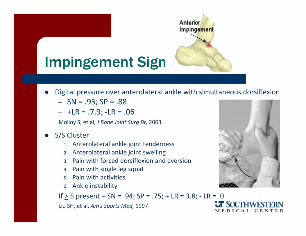
Impingement Sign
Digital pressure over anterolateral ankle with simultaneous dorsiflexion– SN = .95; SP = .88– +LR = .7.9; ‐LR = .06Molloy S, et al, J Bone Joint Surg Br, 2003
S/S Cluster1. Anterolateral ankle joint tenderness2. Anterolateral ankle joint swelling3. Pain with forced dorsiflexion and eversion4. Pain with single leg squat5. Pain with activities6. Ankle instability
If > 5 present – SN = .94; SP = .75; + LR = 3.8; ‐ LR = .0Liu SH, et al, Am J Sports Med, 1997

Interdigital (Morton’s) neuroma provocation
metatarsal head compression to reproduce chief complaint
A Mulder's click (painful reproduction of symptoms) may occur when the enlarged interdigital nerve subluxes between the met heads when they are compressed

Windlass Provocation Testfor Plantar Fasciitis
Forceful great‐toe extension in a standing position causing pain at the medial calcaneal tubercle
100% specificity 31% sensitivity
– just 13% sensitivity if performed in NWB
DeGarceau D, et al. Foot‐Ankle Int, 2003

Rule Out FracturesOttawa Fracture Rules
Excellent screening tool because of high sensitivityand very low negative likelihood ratio
Rule1. Inability to WB 4 steps2. Localized tenderness in any of 4 spots


R/0 Deep Vein Thrombosis

Suspect diagnostic value– Unreliable– Poor specificity (.56) and suspect sensitivity (.39)
+ LR = 1.40‐ LR = 0.87
– Individual clinical findings are inadequate to detect DVT– Wells CPR more useful in determining probability
Forcible ankle dorsiflexion with the knee flexed causing deep calf pain
Homan’s Sign

New Gold Standardbased on venography studies
Clinical Decision RuleWells, et al, 1997
9 medical history andphysical exam findingsthat categorize a patientas low, moderate, or high risk

On-line calculator at http://www.mdcalc.com/wells-criteria-for-dvt/
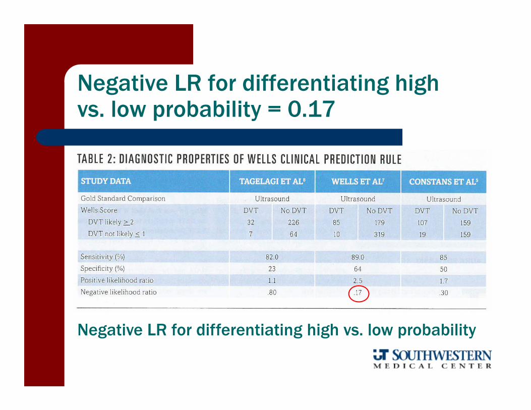
Negative LR for differentiating high vs. low probability = 0.17
Negative LR for differentiating high vs. low probability

Alternate Prediction Rule for DVTsimilar diagnostic utility as Wells CDR
Variables Assessed1. Male Gender2. Paralysis or Immobilization of Lower Limb3. Confinement to bed > 3 days4. Lower Limb Enlargement5. Unilateral Lower Limb Pain6. Other diagnosis at least as plausible
Predictive Ability• > 3 60‐80% possibility• 1‐2 30% possibility• < 0 5% possibility
Constans J, et al, Am J Med, 2003

Observational Gait AnalysisAnkle/Foot Focus
• Adequate sagittal plane talocrural motion in swing and stance phase,
• i.e., early heel off?, adequate toe clearance?• Controlled eccentric plantarflexion from heel
strike to foot flat• Rigid lever push off at heel raise• Adequate MTP/IP sagittal plane motion during
heel and toe off• Appropriate sequence, timing, and amount of
subtalar joint pronation/supination• Ability to accept weight on involved side• Appropriate angle and base of gait

Don’t forget to check for LLD

Functional Foot-Ankle Tests
Walk/Jog Walk on Toes/Heels Deep Squat or Step Down Heel Raises (bi/unilateral) Hopping (bi/unilateral) Stork Stand Dynamic Reach (SEBT)