Embed Size (px)
Citation preview
Efficient Isolation of Pseudomonas aeruginosa Type III SecretionTranslocators and Assembly of Heteromeric TransmembranePores in Model MembranesFabian B. Romano,∥,⊥ Kyle C. Rossi,∥ Christos G. Savva,† Andreas Holzenburg,†,‡,§
Eugenia M. Clerico,∥ and Alejandro P. Heuck*,∥,⊥
†Microscopy and Imaging Center, ‡Department of Biology, and §Department of Biochemistry and Biophysics, Texas A&MUniversity, College Station, Texas 77843, United States∥Department of Biochemistry and Molecular Biology and ⊥Program in Molecular and Cellular Biology, University of Massachusetts,Amherst, Massachusetts 01003, United States
*S Supporting Information
ABSTRACT: Translocation of bacterial toxins or effectorsinto host cells using the type III secretion (T3S) system is aconserved mechanism shared by many Gram-negativepathogens. Pseudomonas aeruginosa injects different proteinsacross the plasma membrane of target cells, altering the normalmetabolism of the host. Protein translocation presumablyoccurs through a proteinaceous transmembrane pore formedby two T3S secreted protein translocators, PopB and PopD.Unfolded translocators are secreted through the T3S needleprior to insertion into the target membrane. Purified PopB andPopD form pores in model membranes. However, theirtendency to form heterogeneous aggregates in solution had hampered the analysis of how these proteins undergo the transitionfrom a denatured state to a membrane-inserted state. Translocators were purified as stable complexes with the cognate chaperonePcrH and isolated from the chaperone using 6 M urea. We report here the assembly of stable transmembrane pores by dilution ofurea-denatured translocators in the presence of membranes. PopB and PopD spontaneously bound liposomes containing anionicphospholipids and cholesterol in a pH-dependent manner as observed by two independent assays, time-resolved Fo rsterresonance energy transfer and sucrose-step gradient ultracentrifugation. Using Bodipy-labeled proteins, we found that PopBinteracts with PopD on the membrane surface as determined by excitation energy migration and fluorescence quenching. Stabletransmembrane pores are more efficiently assembled at pH <5.0, suggesting that acidic residues might be involved in the initialmembrane binding and/or insertion. Altogether, the experimental setup described here represents an efficient method for thereconstitution and analysis of membrane-inserted translocators.
Transport of proteins across membranes is essential at manystages of pathogen infection and colonization of human
cells. This process is important for discharging the proteinsoutside the pathogenic organism (secretion) and for introduc-tion of these secreted toxins and effectors into the cytosol of thetarget cell (translocation). Many pathogens, including Shigella,Salmonella, Yersinia, and Pseudomonas species, exploit asophisticated and efficient mechanism of toxin secretion andtranslocation known as the type III secretion (T3S) system.1−3
It is well-known that Pseudomonas aeruginosa pathogenesisdepends on a vast arsenal of virulence factors, including the T3Ssystem, which is a key factor for acute infections.4,5 The T3Ssystem is a syringelike macromolecular secretion system formedby more than 20 different proteins organized into three majorstructures to span (i) the inner bacterial membrane, theperiplasmic space, and the outer bacterial membrane (secreton);(ii) the extracellular space (needle); and (iii) the host cellularmembrane (translocon). Great progress has been made in thestructural characterization of the secreton and the needle in
related organisms.6 However, virtually nothing is known abouthow T3S-secreted proteins are translocated across the plasmamembrane of the target cell.7 Some genetic and biochemicalevidence suggests that effector proteins are translocated acrossthe host plasma membrane through a proteinaceous pore ortranslocon formed by two bacterially secreted proteins (thetranslocon hypothesis).7−9
The translocon hypothesis states that the P. aeruginosa T3Stranslocators, PopB and PopD, insert into the target membrane,engage with the tip of the T3S needle (formed by PcrV10), andassist in the translocation of effector proteins into the host cell.The hypothesis is based on the following experimentalobservations. (i) PcrV, PopB, and PopD are not required foreffector secretion but are essential for translocation of theeffector into the target cell.11−13 (ii) Only PopB and PopD are
Received: June 11, 2011Revised: July 16, 2011
Article
pubs.acs.org/biochemistry
© XXXX American Chemical Society A dx.doi.org/10.1021/bi200905x |Biochemistry XXXX, XXX, XXX−XXX
found inserted into the target membrane after pathogen−hostcontact.13 (iii) PcrV, PopB, and PopD are necessary for theobservation of T3S-dependent cell lysis.11,13,14 (iv) PopB andPopD can form pores in model membranes either individuallyor in combination.15,16
Current models for the P. aeruginosa translocon complex arequite rudimentary, and they are based on the followingobservations. (i) Translocon proteins PopB and PopD arefound associated with cell membranes after the interaction ofP. aeruginosa with red blood cells.13 (ii) PopB co-immuno-precipitates with PopD after Triton X-100 solubilization ofmembrane-associated proteins.13 (iii) Ringlike structures areobserved using electron microscopy when the transloconproteins are incubated with model membranes.15 The topologyof the translocon proteins is based only on the bioinformaticanalysis of the primary structure for these proteins, whichsuggests that PopB possesses two potential transmembrane(TM) segments and PopD only one,8 but no experimental dataare available to corroborate such predictions. The nonpolarcharacter of the T3S translocators and their tendency toaggregate in solution have made the structural and functionalcharacterization of these proteins very difficult.17,18
We have purified and characterized the P. aeruginosa trans-locators individually as homogeneous complexes with thecognate chaperone PcrH. After isolation, PopB and PopD werequantitatively separated from PcrH by combining immobilizedmetal ion affinity chromatography (IMAC) and elution withthe chaotropic agent urea. The spectroscopic characterizationof the urea-isolated PopD showed little secondary structurecontent and the fact that the two Trp residues were exposed toa polar environment. In contrast, the urea-isolated PopBexhibited greater α-helical content than PopD, and its singleTrp residue was located in a nonpolar environment. The poreforming activity of the urea-isolated translocators was verysimilar to the activity of the translocators separated from thechaperone by acidification.16 Assembly and maximal poreformation occurred at pH <5, indicating that protonation ofacidic residues was critical for membrane insertion. Under theseconditions, PopD, PopB, and their mixtures formed discrete andstable pores in lipid vesicles. Cryo-electron microscopy (EM)and dynamic light scattering (DLS) revealed that no aggregationor disruption of the integrity of the vesicles occurred afterassembly of the transmembrane pores. Single Cys residues wereintroduced at specific locations in PopD and PopB, and thesederivatives purified and labeled with the fluorescent probeBodipy. Using both Bodipy excitation energy migration andself-quenching, we unambiguously showed that PopB interactswith PopD on lipid membranes. We have therefore establishedan efficient procedure for purifying, specifically labeling, andassembling the P. aeruginosa translocators and their derivativesinto model membranes.
■ EXPERIMENTAL PROCEDURES
Expression and Purification of Proteins. The expres-sion and purification of hisPcrH, the hisPcrH−PopD complex,the hisPcrH−PopB complex, and their derivatives wereconducted as described in the Supporting Information. Theprotein concentration was estimated using molar absorptivitiesof 18910 M−1 cm−1 for hisPcrH, 13980 M−1 cm−1 for PopD,and 6990 M−1 cm−1 for PopB,19 and assuming 1:1 chaperone−translocator complexes.
Isolation of PopB and PopD Using 6 M Urea. PurifiedhisPcrH-PopD [or hisPcrH-PopB (2−4 mg)] was loaded ontospinTrap IMAC columns (GE Healthcare) packed with 300 μLeach of Quelating Sepharose Fast Flow (GE Healthcare) resinslurry, previously loaded with Co2þ and equilibrated with bufferA [20 mM 2-amino-2-(hydroxymethyl)propane-1,3-diol (Tris-HCl) (pH 8.0) and 100 mM NaCl]. Dissociation of PopB orPopD from hisPcrH was achieved when the columns were spundry for 30 s at 2000g; 450 μL of buffer B [20 mM Tris-HCl(pH 8.0)] supplemented with 6 M urea and 20 mM Glywas added, and the columns were incubated for 30 min at 4 °Con a rocking platform. Dissociated PopD (or PopB) was elutedwhen the columns were spun for 30 s at 2000g. Samples werefractionated, frozen in liquid N2, and stored at −80 °C untilthey were used.Mass Spectrometry. Purified PopB and PopD were
analyzed using an Esquire Mass spectrometer (Bruker Daltonics,Billerica, MA) equipped with an electrospray ionization sourceand ion-trap mass detector; 0.1 mg of protein was dialyzedextensively against pure water at 4 °C. Then, protein was dilutedto 50% (v/v) methanol and 3% acetic acid and sprayed intro theionization source at a rate of 120 μL/h. Mass/charge data werecollected and averaged, and the protein molecular mass wascalculated from deconvolution of the average mass spectra.Fluorescent Protein Labeling. PopBS164C (introduced
mutations are indicated using a superscript in which thesubstituted amino acid and the introduced amino acid are givento the left and right of the number, respectively) and PopDF223C
were labeled using N-[(4,4-difluoro-5,7-dimethyl-4-bora-3a,4a-diaza-s-indacen-3-yl)methyl]iodoacetamide (Bodipy FL C1-IAor Bodipy, Invitrogen) as follows. Two milligrams of PopBS164C
or PopDF223C complexed with hisPcrH were first incubated inbuffer A supplemented with 5 mM DTT for 1 h and then runthrough a Sephadex G-25 column (1.5 cm inside diameter ×20 cm) pre-equilibrated with buffer C [50 mM Hepes (pH 8.0)and 100 mM NaCl]. Given the relatively low water solubility ofBodipy, dye dissolved in dimethyl sulfoxide was added in fourconsecutive steps to the protein solution with a 5:1 dye:proteinratio. The first two additions were followed by incubation for1 h at 20−23 °C in the dark with gentle shaking, while the lasttwo additions were followed by a 30 min incubation under thesame conditions. Then, any precipitated dye and protein werecleared out by centrifugation, and excess soluble fluorophore wasremoved by SEC using Sephadex G-25 resin pre-equilibrated with20 mM Tris (pH 7.5) and 100 mM NaCl. PopBS164C�Bodipy andPopDF223C�Bodipy were isolated from hisPcrH using 6 M urea asdescribed for wild-type translocators. The nonlytic, preporeformer Perfringolysin O derivative (PFOE167C=F181A=F318A=C459A
or PFO) was labeled with Bodipy as indicated above for thetranslocators. Labeling efficiencies were calculated to be 96%for PopDF223C�Bodipy, 68% for PopBS164C�Bodipy, and 100% forPFOBodipy using the molar absorptivities at 280 nm for PopB andPopD (see above), PFO,20 and Bodipy (55000 cm−1 M−1 at502 nm in 6 M urea). The absorbance of Bodipy at 280 nm was∼4% of the absorbance at 502 nm.Liposome Preparation. All non-sterol lipids were ob-
tained from Avanti Polar Lipids (Alabaster, AL). Cholesterolwas obtained from Steraloids (Newport, RI). Liposomes weregenerated using an Avanti (Alabaster, AL) Mini-Extruder andpolycarbonate filters with a 0.1 μm pore size (Whatman) asdescribed previously.21 Briefly, a mixture of 1-palmitoyl-2-oleoyl-sn-glycero-3-phosphocholine (POPC), cholesterol, and
Biochemistry Article
dx.doi.org/10.1021/bi200905x |Biochemistry XXXX, XXX, XXX−XXXB
1-palmitoyl-2-oleoyl-sn-glycero-3-phosphoserine (POPS) (in amolar ratio of 65:20:15) in chloroform was dried at 20−23 °Cunder N2 and then kept under vacuum for at least 3 h. Lipidswere hydrated via addition of buffer C to a final concentra-tion of total lipids of 10−30 mM and incubated for 30 min at20−23 °C with vortexing at 5 min intervals. The suspendedphospholipid/sterol mixture was frozen in liquid N2 andthawed at 37 °C a total of three cycles to reduce the number ofmultilamellar liposomes and to enhance the trapped volumes ofthe vesicles. Hydrated lipids were extruded 21 times through a0.1 μm pore size polycarbonate filter. All liposome preparationswere analyzed as monodisperse with an average particlediameter of ∼100 ± 5 nm using DLS. The resultant liposomeswere stored at 4 °C and used within 2 weeks of production.Liposomes used in Forster resonance energy transfer (FRET)experiments were prepared similarly, except that 0.5 mol %total lipid was replaced with rhodamine B 1,2-dihexadecanoyl-sn-glycero-3-phosphoethanolamine (Rh-PE), triethylammoniumsalt (Invitrogen). The pore forming activity of translocatorswas measured using liposomes containing Tb(DPA)33−. Theliposomes loaded with Tb(DPA)33− were prepared as describedpreviously by us.22
Liposome Flotation−Membrane Binding Assay. Bind-ing reaction mixtures (75 μL) containing liposomes (2 mMtotal lipids) and Bodipy-labeled PopB or PopD (400 nM totalprotein) were established in ultracentrifuge tubes and incubatedat 20−23 °C for 1 h. Binding reaction buffer was a mixture of30 mM sodium acetate and 30 mM 2-(N-morpholino)-ethanesulfonic acid regulated at pH 4.0 or 6.0. Liposomeswere equilibrated with the buffer prior to the addition ofprotein. Liposome-bound and unbound proteins were sepa-rated by flotation of proteoliposomes through a sucrosegradient as follows. A 225 μL aliquot of 67% sucrose wasthoroughly mixed with each binding reaction mixture, and thesamples were overlaid with 360 μL of 40% sucrose, followed by240 μL of 4% sucrose. Samples were centrifuged for 50 min at90000g and 4 °C.23 Three 300 μL fractions (upper fractioncontaining proteoliposomes, middle fraction empty, andbottom fraction containing free protein) were collected fromthe gradient. After trichloroacetic acid precipitation andresuspension in SDS denaturalization buffer, samples wereanalyzed by SDS−PAGE followed by a fluorescence scan usinga FLA-500 phosphorimager (Fujifilm Corp.). Protein bandscorresponding to liposome-bound protein were quantified bygel densitometry using Genetools version 4.01 (Syngene).Dynamic Light Scattering. Unless otherwise indicated,
the average size of the liposomes was determined at 20−23 °Cusing a PDDLS Coolbatch/PD2000DLS instrument (PrecisionDetectors, Inc., Franklin, MA) employing a 30 mW He−Nelaser source (658 nm) and a photodiode detector at an angleof 90°. Average autocorrelation functions were fit using thecumulant method and hydrodynamic radius derived from theobtained decay rates.24
Cryo-EM. Samples were prepared for cryo-EM viaapplication of 3 μL of the liposome/protein mixture to freshlyglow-discharged holey carbon films (C-Flat, Protochips Inc.)and plunge-frozen in liquid ethane using a FEI Vitrobot.Specimens were observed on an FEI Tecnai F20 transmissionelectron microscope operating at 200 kV. Images were acquiredunder low-dose and zero-loss imaging conditions on a GatanUltrascan 1000 CCD camera attached to the end of a GatanTridiem postcolumn energy filter.
Pore Formation Assay. Liposomes were suspended at afinal total lipid concentration of 0.15−0.30 mM in 300 μL ofbuffer D [50 mM sodium acetate (pH 4.0) and 5 mM EDTA].The net initial emission intensity (F0) was determined afterequilibration of the sample at 25 °C for 5 min. Aliquots ofPopB or PopD were added to the liposome suspension atconcentrations of 30−60 nM, and samples were incubated for15 min at ∼23 °C. After re-equilibration to 25 °C, the final netemission intensity (Ff ) of the sample was determined (i.e., afterblank subtraction and dilution correction) and the fraction ofTb(DPA)33− quenched was estimated using (F0 − Ff )/F0.
22
For the analysis of the pore forming activity at different pHvalues, an equimolar mixture of 30 mM sodium acetate and30 mM 2-(N-morpholino)ethanesulfonic acid was used.Analysis of Formed Pores. The presence of discrete size
membrane pores formed by PopB, PopD, or an additiveequimolar mixture of both was assessed by measuring theability of biocytin (∼1.5 nm size) or biotin-β-amylase (∼4 nmsize) to diffuse through the pores formed by the translocators.Liposomes encapsulating streptavidinBodipy were treated withthe translocator(s), and diffusion of the biotin markers throughthe pores was detected as an increase in streptavidinBodipy fluo-rescence as follows. Liposomes loaded with streptavidinBodipy
(100 μM total lipids) were suspended in 50 mM sodiumacetate buffer (pH 4.3) and 0.5 mM DTT containing 1 μMbiocytin or 100 nM biotin-β-amylase. The net initial emissionintensity (F0) was determined after equilibration of the sampleat 25 °C for 5 min. Translocators were added individually ortogether at a final concentration of 100 nM each, and sampleswere incubated at 20−23 °C for 15 min (protein:lipid ratio of1:1000 for individual proteins and 1:500 when both proteinswere added together). After re-equilibration at 25 °C, the finalnet emission intensity (F) of the sample was determined (i.e.,after blank subtraction and dilution correction) and theenhancement of streptavidinBodipy fluorescence emission wasestimated using F/F0, which is proportional to the amount ofbiotinylated marker that can diffuse through membrane poresand bind to streptavidinBodipy . As a control, samples containingbiotin-β-amylase were treated with Triton X-100 after F hadbeen recorded to disrupt membranes and corroborate thebinding activity between biotin-β-amylase and streptavidinBodipy
(not shown).Circular Dichroism (CD) Spectroscopy. Measurements
were taken at 25 °C on a Jasco J-715 spectropolarimeter (JascoCorp.) equipped with a Peltier effect device for temperaturecontrol. The scan speed was set to 20 nm/min with a 1 sresponse time, a 0.5 nm data pitch, and a 1 nm bandwidth.Far-UV spectra were recorded using 0.2 cm cells and proteinconcentrations of 2−3 μM in buffer E [10 mM sodiumphosphate (pH 7.5)]. Six spectra were recorded and averagedfor each sample.Steady-State Fluorescence Spectroscopy. Steady-state
fluorescence measurements were taken using a Fluorolog-3photon-counting spectrofluorimeter equipped with a doublemonochromator in the excitation light path, a single emissionmonochromator, cooled photomultiplier tube housing, a 450 Wxenon lamp, and a temperature-controlled sample holder.20
For pore formation activity assays employing Tb(DPA)33−
liposomes, excitation and emission wavelengths were set to 278and 544 nm, respectively, and a 385 nm long pass filter wasplaced in the emission channel to block second-order harmoniclight from passing through the emission monochromator. The
Biochemistry Article
dx.doi.org/10.1021/bi200905x |Biochemistry XXXX, XXX, XXX−XXXC

bandpass was typically 2 nm for excitation and 4 nm for emis-sion. For experiments using streptavidinBodipy , samples wereexcited at 492 nm and the emission intensity was measured at510 nm. Emission scans of intrinsic protein fluorescence weretaken at 1 nm intervals between 285 and 405 nm, with anexcitation wavelength of 278 nm. Emission scans for FRETexperiments were conducted at 1 nm intervals between 490 and560 nm, with an excitation wavelength of 485 nm.Time-Resolved Fluorescence Spectroscopy. Time-
resolved fluorescence measurements were taken in a Chronosmultifrequency cross-correlation phase and modulation fluor-ometer equipped with a three-chamber cuvette holder forbackground subtraction from ISS (Champaign, IL). Sampleswere excited with a 470 nm laser diode (HBW 4 nm) filteredthrough a 472 nm interference filter (transmittance % HBW 10nm) to eliminate spurious light. Emitted light was collectedthrough a Melles Griot GG 495 sharp cutoff glass filter toeliminate scattered light and a Melles Griot 03SWP608 dielectricshort-pass filter at 550 nm to minimize the contribution of directexcitation of Rh-PE. To avoid any polarization artifacts,measurements were taken under magic angle conditions usingGlan-Thompson Prism Polarizers (10 mm × 10 mm aperture forexcitation and 14 mm × 14 mm aperture for emission, set at 0°and 55° relative to the lab vertical axis, respectively).Fluorescence lifetimes were calculated by measuring the phasedelay and modulation ratio spectra of samples in the 10−200MHz frequency modulation range selecting 25 frequencies(25 °C). Blank subtraction was conducted using an equivalentsample without the fluorophore and using the algorithmdescribed by Reinhart et al.25 incorporated into the acquisitionsoftware. A solution of Fl (Invitrogen) in 0.1 M NaOH was usedas a reference lifetime with a value of 4.05 ns,21,26 and a totalintensity similar to that of the measured sample (±10%).27 Onesingle-exponential lifetime of 4.05 ± 0.1 ns was obtained for thisreference sample when measured against rhodamine B inmethanol (lifetime of 2.5 ns28). The lifetime data were analyzedassuming different models, including monoexponential, multi-exponential, or continuous lifetime distribution29 decay models.The goodness of fit was determined by using the reduced χ2
values. Uncertainties in the phase and modulation values were0.2 and 0.004, respectively.Membrane Binding FRET Measurements. The binding
of PopBS164C�Bodipy or PopDF223C�Bodipy to membranes underequilibrium conditions was measured by FRET between aBodipy-labeled translocator (donor or D) and Rh-PE as theacceptor (A), randomly distributed at the lipid bilayer. Fourbiochemically equivalent samples were prepared in parallel.Sample Do (D only) contained 120 nM total translocator (anequimolar mixture of Bodipy-labeled translocator mutant andwild-type translocator was used to minimize Bodipy self-quenching) and POPC/POPS/cholesterol membranes(65:15:20 molar ratio) lacking Rh-PE. Sample DA (D plusA) contained the same protein mixture as in Do andmembranes containing Rh-PE (0.5% of the total lipids).Sample Ao (A only) contained 120 nM wild-type translocatorand vesicles containing Rh-PE. The blank (B) sample contained120 nM wild-type translocator and vesicles lacking Rh-PE. In allfour samples, the total lipid concentration of the membraneswas 0.3 mM. All samples were incubated at 25 °C for 30 min topermit complete insertion of translocator derivatives into themodel membranes before spectral measurements were takenat 25 °C. FRET efficiency (E) was calculated as describe by
Wu and Brand30
where τDA and τD are the average amplitude-weighted lifetimesof D in the presence and absence of A, respectively. τDA and τDfor DA and Do samples were determined as described in Time-Resolved Fluorescence Spectroscopy using the Ao and Bsamples for blank subtraction, respectively. Phase delay andmodulation ratio data best fit to two-exponential componentmodels.
■ RESULTS
Purification of Homogeneous Chaperone−TranslocatorComplexes Containing Native PopB and PopD. Coex-pression of hisPcrH with native PopD (or PopB) was achievedby using the pETDuet-1 system (Merk4Biosciences) asdescribed in Experimental Procedures. The translocators weretherefore purified in their native state, without modifications(i.e., no affinity tags or amino acid additions or deletionsresulting from cloning into the expression vector). The absenceof poly-His tags or fusion proteins (e.g., GST) is critical whenelectrostatic interactions and oligomerization of proteins areinvolved in the mechanism being investigated. Water-solublehisPcrH−PopD and hisPcrH−PopB complexes were purifiedusing IMAC and AEC. The IMAC step rendered a mixture offree hisPcrH together with hisPcrH−PopD (or hisPcrH−PopB) complexes (not shown). The AEC step separated thefree hisPcrH chaperone and other minor contaminants fromthe hisPcrH−PopD (or hisPcrH−PopB) complex (Figure 1A,B).A major peak containing the hisPcrH−PopD complex elutedwhen the concentration of NaCl was 0.21 M. The free hisPcrHchaperone eluted later when the concentration of NaCl was0.35 M (Figure 1A).Size exclusion chromatography (SEC) analysis of the purified
hisPcrH−PopD complex revealed a single symmetric peak(Figure S1 of the Supporting Information). When compared tothe molecular mass standards, the hisPcrH−PopD complexeluted with an apparent molecular mass of 111 ± 1 kDa(expected value of 50.8 kDa), which suggested that thehisPcrH−PopD species may form 2:2 complexes. Similar SECresults have been described for Shigella f lexneri31 and Aeromonashydrophila32 translocators; however, analysis of these complexeshas shown they adopt a 1:1 stoichiometry. Therefore, it seemsthat the purified hisPcrH−PopD complex forms an elongated1:1 complex, as described for the P. aeruginosa CHAtranslocator and other related T3S proteins.15,31,32
In contrast to the hisPcrH−PopD complex, the hisPcrH−PopB complex eluted in two peaks during the AEC step (Figure1B). The first peak eluted at 0.19 M NaCl, and a second peakeluted at 0.25 M NaCl. A third peak corresponding to the freehisPcrH chaperone eluted at a higher NaCl concentration(∼0.30 M). SEC analysis of isolated fractions from the peaksrevealed that the first peak corresponded mainly to a 1:1hisPcrH−PopB complex (eluted at 13.9 mL), while the secondpeak contained a larger proportion of aggregates (Figure 1C).Interestingly, we noticed that the amount of the hisPcrH−PopB complex appearing as aggregates was affected by theconcentration of the proteins (Figure S2B of the SupportingInformation). The higher the protein concentration of thesample, the larger the amount of nonspecific hisPcrH−PopB
Biochemistry Article
dx.doi.org/10.1021/bi200905x |Biochemistry XXXX, XXX, XXX−XXXD

aggregates observed, confirming the intrinsic tendency of thecomplex to aggregate in aqueous solution.15,32
The hisPcrH−PopB peak corresponding to the 1:1 complexwas isolated and reanalyzed by SEC. A main symmetric peakeluted in the second SEC run (Figure S1 of the SupportingInformation), indicating that the hisPcrH−PopB complex wasstable in solution and ran with a hydrodynamic radiusequivalent to that of a 95 ± 3 kDa globular protein. Becausethe expected molecular mass of a 1:1 complex is 59.6 kDa, thisresult suggested that the hisPcrH−PopB complex adopted anelongated conformation rather than a globular shape, as shownpreviously for the homologue protein AopB.32
We have therefore optimized the procedures to obtain bothhisPcrH−PopD and hisPcrH−PopB complexes purified toapparent homogeneity (Figure 1D, lanes 2 and 3, and Figure S1of the Supporting Information).Spectroscopic and Functional Characterization of the
Purified Chaperone-Associated Proteins. The far-UV CDspectrum of the purified chaperone hisPcrH revealed a typicalall-α protein, with double minima around 222 and 209 nm(Figure 2A). These data correlated well with the recentlydetermined three-dimensional structure of the PcrH21−160fragment, which consists of α-helical tetratricopeptide re-peats.33,34 The far-UV CD spectrum of the hisPcrH−PopDcomplex suggested that PopD also contains a high α-helicalstructure content (Figure 2A and ref 33). The band with aminimum at 208 nm had a larger intensity than that at 222 nm,suggesting the presence of other secondary structural elementsin this complex.35 The far-UV CD spectrum of the hisPcrH−PopB complex was similar to that of the hisPcrH−PopDcomplex, but the intensity of the bands was lower, suggesting
Figure 1. Purification of the hisPcrH−translocator complexes. (A) The fractions containing the hisPcrH−PopD complex isolated after the firstIMAC purification step were dialyzed and loaded into a Q-Sepharose AEC column and eluted using a linear NaCl gradient. The peaks containinghisPcrH and the hisPcrH−PopD complex are indicated. (B) The hisPcrH−PopB complex was purified as described for the hisPcrH−PopD complex.The first two large peaks that eluted from the AEC column contained the hisPcrH−PopB complexes, and the shoulder that eluted around 400 mLcontained hisPcrH. (C) SEC analysis of aliquots corresponding to the 1st peak and the 2nd peak illustrated in panel B. (D) SDS−PAGE analysis ofpurified proteins: lane 1, molecular mass markers; lane 2, hisPcrH−PopB complex; lane 3, hisPcrH−PopD complex; lanes 4 and 5, urea-isolatedPopB and PopD, respectively.
Figure 2. Characterization of purified translocators. (A) Far-UVCD spectra of the SEC-isolated hisPcrH, hisPcrH−PopD complex,and hisPcrH−PopB complex recorded in buffer E, at a total proteinconcentration of 3.0 μM. (B) Normalized fluorescence emissionspectra of hisPcrH, the hisPcrH−PopD complex, and the hisPcrH−PopB complex recorded in buffer E. The excitation wavelengthwas 278 nm, and the total protein concentration was 2.4 μM.(C) Far-UV CD spectra of purified PopD and PopB recorded in20 mM phosphate buffer (pH 7.5) supplemented with 6 M urea,with a protein concentration of 2.4 μM. (D) Normalizedfluorescence emission spectra of PopD and PopB in 20 mMphosphate buffer (pH 7.5) supplemented with 6 M urea. Theexcitation wavelength was 278 nm, and the total proteinconcentration was 2.4 μM.
Biochemistry Article
dx.doi.org/10.1021/bi200905x |Biochemistry XXXX, XXX, XXX−XXXE

that PopB is less α-helical than PopD when bound to the PcrHchaperone.Additionally, we evaluated the local environment around the
aromatic residues of the P. aeruginosa PAO1 purified proteins.The hisPcrH chaperone contains one Trp and nine Tyrresidues, while PopD contains two Tyr and two Trp residues;PopB contains one Tyr and one Trp. The emission fluore-scence spectra of hisPcrH showed a peak with a maximum at349 nm (Figure 2B), indicating that the Trp residue waslocated in a polar environment, in agreement with the positionobserved in the structure determined via X-rays.34 The shoulderaround 303 nm corresponded to the emission of multiple Tyrresidues. The hisPcrH−PopD complex presented an emissionfluorescence spectrum similar to that of hisPcrH, with amaximum at 351 nm. This red-shifted maximum suggested thatboth the central and C-terminal Trp residues of PopD reside ina polar environment. In contrast, the emission fluorescencespectrum of the hisPcrH−PopB complex showed a maximumat 337 nm, suggesting that the Trp residue of PopB was locatedin a nonpolar environment (Figure 2B).The pore formation activity for both purified proteins was
analyzed using the fluorescence assay previously described byus.22 In this assay, a fluorescent marker was encapsulated intoliposomes and a quencher was added to the external buffersolution. A high-fluorescence intensity signal indicated that themembrane was intact and the quencher could not contact thefluorophore. If a transmembrane pore is formed upon additionof protein, the fluorophore becomes accessible to the quencherand the magnitude of the fluorescent signal decreases. BothPopB and PopD dissociate from PcrH in vitro when thesolution pH decreases to 5.3, forming heterogeneous proteinaggregates.15 These dissociated protein aggregates form poresin model membrane systems;16 however, the translocators arepresumably secreted as monomers in vivo and in the proximityof the target membrane. Under these circumstances, binding tothe membrane can precede any protein−protein association.We therefore reasoned that the dissociation of the translocatorsfrom the chaperone in the presence of liposomes would moreaccurately represent in vivo conditions.When separated from PcrH when the pH decreased from 8.0
to 5.1 in the presence of lipid membranes, PopB, PopD, andthe equimolar mixture of these proteins were able to form pores(Figure S3A of the Supporting Information). Interestingly, incontrast with the results observed when protein aggregates wereused, no synergy was observed between PopB and PopD.16 It istherefore clear that the history (i.e., aggregation) of the proteinsmay affect the mechanism by which the translocators form apore. Because the sequence of the events that leads to theassembly of a membrane-inserted translocon is far from under-stood, an experimental procedure that replicates the in vivoscenario encountered by the proteins after being secretedthrough the T3S needle is desirable.An Efficient Procedure for Isolating PopB and PopD
Derivatives. Insights into the mechanism of insertion ofprotein into lipid bilayers and the interaction of proteins withmembranes can be obtained by fluorescence spectroscopy andsite-directed fluorescence labeling.36 A Cys residue isintroduced by site-directed mutagenesis at a single site in theprotein, and the unique Cys is specifically labeled with thefluorophore of choice. Protein−membrane association andprotein−protein interactions can therefore be studied using
FRET, excitation energy migration (homo-FRET), or fluo-rescence quenching.37−39
P. aeruginosa PAO1 translocators do not contain Cysresidues, and therefore, they are optimal substrates for site-directed fluorescence labeling. However, the labeling of isolatedtranslocators in solution is hampered by their intrinsic tendencyto aggregate. Introduced Cys residues can be alternativelylabeled while the translocator is still bound to the chaperonePcrH. We noticed that in the recently determined X-raystructure of PcrH the three Cys residues are not exposed to thesolvent,34 and therefore, we reasoned that the specific labelingof the translocators may be possible even in the presence ofthe chaperone. However, our initial labeling reactionsdemonstrated that the chaperone was also efficiently labeledwith thiol-specific probes (not shown).Analysis of the solvent exposure for the native hisPcrH Cys
revealed that on average, 2.20 ± 0.03 Cys residues reacted withEllman’s reagent (DTNB). Interestingly, incubation of hisPcrHwith 6 M guanidinium chloride at 37 °C for 1 h did notincrease the reactivity of the hisPcrH Cys residues to DTNB,suggesting that a portion of those residues were forming intra-or intermolecular disulfide bonds. SEC analysis of purifiedhisPcrH samples revealed a first small peak eluted at 14.3 mLand a large second peak eluted at 15.3 mL (Figure S4A of theSupporting Information). Nonreducing SDS−PAGE analysis ofthe two peaks revealed that hisPcrH formed intermoleculardisulfide bonds in solution (Figure S4B of the SupportingInformation). Hence, it is clear from these data that thestructure of PcrH in solution is dynamic, and the side chains ofthe amino acids surrounding the Cys residues move and exposethe sulfhydryl groups to the solvent (see below).Any attempts to replace all three Cys residues in PcrH with
nonreactive residues (Ala or Ser) rendered a chaperone thatcan no longer bind PopD (not shown). Therefore, the inter-pretation of the fluorescence signal and observed spectralchanges derived from the single fluorescently labeled trans-locators would be veiled by the presence of a labeled chaperone.The spectroscopic characterization of the translocon assemblymechanism, therefore, requires an efficient separation of thelabeled translocators from the labeled hisPcrH chaperone priorto the analysis.We took advantage of the poly-His tag located at the
N-terminus of the chaperone to isolate labeled translocators.After being labeled, the translocators were separated fromhisPcrH by binding of the hisPcrH−PopD (or hisPcrH−PopB)complex to an IMAC column and a subsequent elution ofPopD (or PopB) with buffer containing 6 M urea. Urea diddissociate PopD or PopB from their cognate chaperone withoutaffecting the interaction of hisPcrH with the IMAC column(Figure 1D, lanes 4 and 5). Therefore, the single-Cys trans-locator mutants can be labeled while still bound to hisPcrH andseparated from the chaperone in a subsequent step. This is asimple and efficient procedure for obtaining the translocatorderivatives required for the structural and functional character-ization of the T3S translocon (e.g., single fluorescently labeledtranslocators).Spectroscopic and Functional Characterization of
Urea-Isolated PopB and PopD. We analyzed the structuraland functional properties of the urea-isolated translocatorsusing mass spectrometry, far-UV CD, intrinsic protein fluore-scence, and pore formation assays using model membranes.The molecular mass of urea-isolated PopB and PopD was
Biochemistry Article
dx.doi.org/10.1021/bi200905x |Biochemistry XXXX, XXX, XXX−XXXF

determined using ESI-ion trap mass spectrometry as detailed inExperimental Procedures. Only one species was detected foreach protein sample with a molecular mass of 40058 Da forPopB (expected value of 40061 Da) and 31308 Da for PopD(expected value of 31309 Da).The polarity around the Trp residues for the urea-isolated
PopD and PopB was similar to the polarity observed for theproteins complexed with the hisPcrH chaperone (Figure 2D).PopD and PopB in urea presented fluorescence emissionmaxima at 358 and 334 nm, respectively. The far-UV CDnegative band at 222 nm observed for PopB in 6 M ureasuggested that PopB conserved a high proportion of its α-helicalstructure even in the presence of the chaotropic agent (Figure2C). However, the far-UV CD spectrum of PopD in 6 M urearevealed that a sizable portion of the secondary structure of thistranslocator was lost in the presence of the chaotropic agent.Despite the differences observed in their secondary structure,
both urea-isolated PopB and PopD conserved their poreforming abilities at a mildly acidic pH (Figure S3B of theSupporting Information). The pore formation activity profiles ofurea-isolated translocators were very similar to those observedwhen the translocators were directly dissociated from thechaperone by reducing the pH of the medium (Figure S3 of theSupporting Information). Altogether, our data indicated that thepurification procedure described above constitutes a simple andefficient alternative for obtaining highly pure and active PopD,PopB, and their mutant derivatives.Optimization of a Model System for Analyzing the
Membrane-Inserted State of the Translocators. Unam-biguous interpretation of structural data requires that the com-ponents under investigation adopt a uniform conformationalstate. For the structural analysis of assembled T3S translocons,it is essential that the measured fluorescent signal come fromprobes located in the same conformation (i.e., membrane-inserted translocons). We therefore optimized our model systemto maximize the assembly and insertion of the T3S translocators.Three factors affect the assembly of active transmembrane poresin our experimental system: (i) the pH of the medium, (ii) thepresence of negatively charged phospholipids, and (iii) theconcentration of cholesterol in the target membrane.In membranes containing a mixture of the lipids commonly
present in mammalian plasma membranes (i.e., POPC, POPE,POPS, and sphingomyelin) and 15 mol % cholesterol, themaximal activity for PopB (∼60%) was observed at pH ≤5.1(Figure 3A). For PopD, the maximal activity (∼90%) wasobserved at pH <5. Interestingly, the activity of PopD waslower than the activity of PopB at pH >5.1, but it surpassed thatobserved for PopB at pH <4.8, suggesting that the insertion ofPopD into membranes is more dependent on acidic residueprotonation than the insertion of PopB.When a constant POPC:POPE:POPS:sphingomyelin molar
ratio was maintained in the absence or presence of a high levelof cholesterol (45 mol %), the pore forming activity of thetranslocators was less effective than that observed with 15 mol% cholesterol (not shown). Because the activities of PopD andPopB plateau below pH 4.5 at intermediate cholesterol con-centrations, we selected pH 4.0−4.3 and 20 mol % cholesterolfor our assays to maximize the formation of uniformmembrane-inserted translocons.Negatively charged phospholipids affect the degree of insertion
of T3S translocators.15,16 We therefore analyzed the effect ofPOPS on the activity of urea-isolated PopB and PopD in
membranes containing POPC and a fixed amount of cholesterol(20 mol %). Addition of 15 mol % POPS increased 2-fold theactivity of PopB and >3-fold the activity of PopD (Figure 3B).Doubling the amount of POPS to 30 mol % was not as effectiveas the addition of only 15 mol % POPS. Thus, to maximize theconformational homogeneity of membrane-inserted translocatorsand to minimize any effect caused by the variability of the lipidcomposition of the system, we chose the simplest membranemodel system that maximized the formation of transmembranepores, a 65:15:20 POPC:POPS:cholesterol molar ratio.The pore forming activity of urea-isolated PopD, PopB, and a
1:1 mixture of the translocators was studied using this modelsystem. Maximal activity for PopB and PopD was observedbelow pH 4.8 and 4.3, respectively (Figure 4A). Interestingly,no additive or synergic effect was detected at higher pH, wherethe individual proteins exhibited intermediate pore formingactivity. Maximal pore formation using these experimental condi-tions was observed when the protein:lipid ratio was ∼1:3000(Figure 4B).High Translocator:Lipid Ratios Produced Liposome
Aggregation. Our initial attempts to visualize the membrane-inserted PopD using transmission electron microscopy and2% uranyl acetate as a contrast agent revealed that even whenused at a 1:1000 protein:lipid ratio, PopD disrupted theliposomes forming tubular membrane structures (not shown).
Figure 3. Effect of pH and lipid composition on the pore formingactivity of the urea-isolated translocators. Pore formation wasdetermined as the percentage of the encapsulated Tb(DPA)33− thatwas quenched by EDTA as detailed in Experimental Procedures. (A)Urea-isolated PopB or PopD was directly diluted into a solution of 50mM sodium acetate buffered at the indicated pH, containingmembranes. The pore forming activity of the urea-isolated trans-locators increased at acidic pH. The total lipid concentration was0.1 mM, and the liposomes consisted of 35 mol % POPC, 15 mol %POPS, 20 mol % POPE, 15 mol % SM, and 15 mol % cholesterol. Theprotein:lipid ratio was 1:1000. (B) Effect of POPS on the poreformation activity of urea-isolated translocators. The activity wasmeasured as described for panel A; the pH was buffered at 4.3, and thelipid mixture consisted of POPC, 20 mol % cholesterol, and theindicated concentration of POPS.
Biochemistry Article
dx.doi.org/10.1021/bi200905x |Biochemistry XXXX, XXX, XXX−XXXG
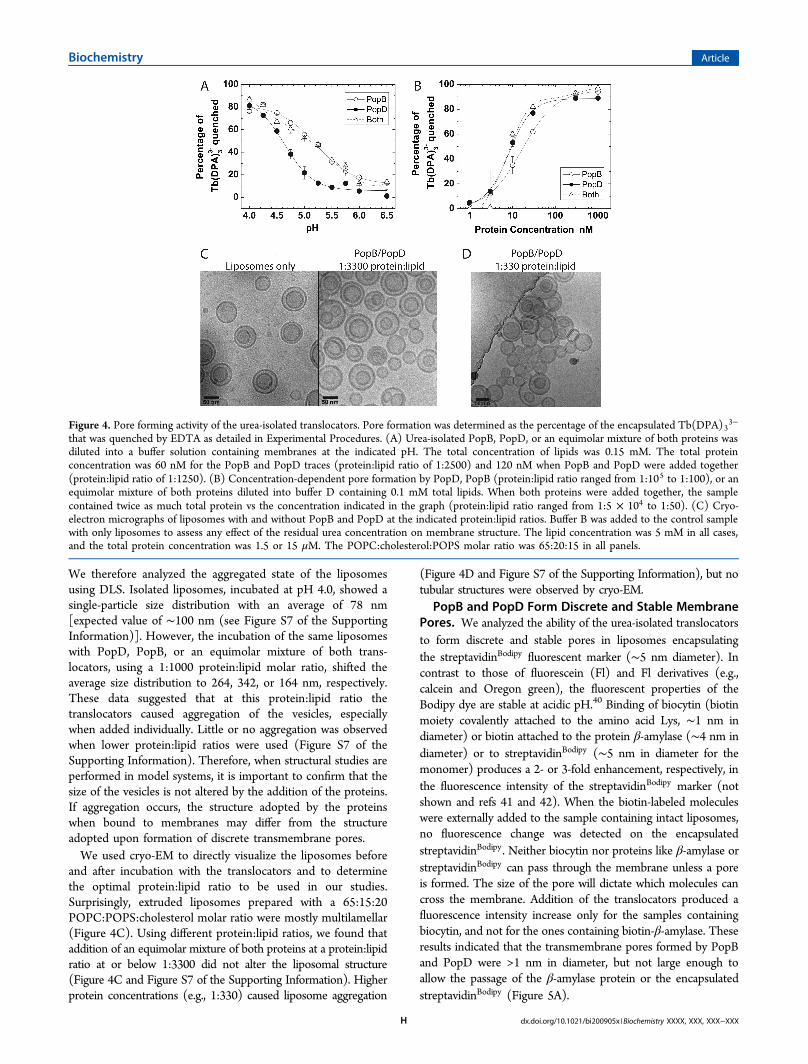
We therefore analyzed the aggregated state of the liposomesusing DLS. Isolated liposomes, incubated at pH 4.0, showed asingle-particle size distribution with an average of 78 nm[expected value of ∼100 nm (see Figure S7 of the SupportingInformation)]. However, the incubation of the same liposomeswith PopD, PopB, or an equimolar mixture of both trans-locators, using a 1:1000 protein:lipid molar ratio, shifted theaverage size distribution to 264, 342, or 164 nm, respectively.These data suggested that at this protein:lipid ratio thetranslocators caused aggregation of the vesicles, especiallywhen added individually. Little or no aggregation was observedwhen lower protein:lipid ratios were used (Figure S7 of theSupporting Information). Therefore, when structural studies areperformed in model systems, it is important to confirm that thesize of the vesicles is not altered by the addition of the proteins.If aggregation occurs, the structure adopted by the proteinswhen bound to membranes may differ from the structureadopted upon formation of discrete transmembrane pores.We used cryo-EM to directly visualize the liposomes before
and after incubation with the translocators and to determinethe optimal protein:lipid ratio to be used in our studies.Surprisingly, extruded liposomes prepared with a 65:15:20POPC:POPS:cholesterol molar ratio were mostly multilamellar(Figure 4C). Using different protein:lipid ratios, we found thataddition of an equimolar mixture of both proteins at a protein:lipidratio at or below 1:3300 did not alter the liposomal structure(Figure 4C and Figure S7 of the Supporting Information). Higherprotein concentrations (e.g., 1:330) caused liposome aggregation
(Figure 4D and Figure S7 of the Supporting Information), but notubular structures were observed by cryo-EM.PopB and PopD Form Discrete and Stable Membrane
Pores. We analyzed the ability of the urea-isolated translocatorsto form discrete and stable pores in liposomes encapsulatingthe streptavidinBodipy fluorescent marker (∼5 nm diameter). Incontrast to those of fluorescein (Fl) and Fl derivatives (e.g.,calcein and Oregon green), the fluorescent properties of theBodipy dye are stable at acidic pH.40 Binding of biocytin (biotinmoiety covalently attached to the amino acid Lys, ∼1 nm indiameter) or biotin attached to the protein β-amylase (∼4 nm indiameter) or to streptavidinBodipy (∼5 nm in diameter for themonomer) produces a 2- or 3-fold enhancement, respectively, inthe fluorescence intensity of the streptavidinBodipy marker (notshown and refs 41 and 42). When the biotin-labeled moleculeswere externally added to the sample containing intact liposomes,no fluorescence change was detected on the encapsulatedstreptavidinBodipy. Neither biocytin nor proteins like β-amylase orstreptavidinBodipy can pass through the membrane unless a poreis formed. The size of the pore will dictate which molecules cancross the membrane. Addition of the translocators produced afluorescence intensity increase only for the samples containingbiocytin, and not for the ones containing biotin-β-amylase. Theseresults indicated that the transmembrane pores formed by PopBand PopD were >1 nm in diameter, but not large enough toallow the passage of the β-amylase protein or the encapsulatedstreptavidinBodipy (Figure 5A).
Figure 4. Pore forming activity of the urea-isolated translocators. Pore formation was determined as the percentage of the encapsulated Tb(DPA)33−
that was quenched by EDTA as detailed in Experimental Procedures. (A) Urea-isolated PopB, PopD, or an equimolar mixture of both proteins wasdiluted into a buffer solution containing membranes at the indicated pH. The total concentration of lipids was 0.15 mM. The total proteinconcentration was 60 nM for the PopB and PopD traces (protein:lipid ratio of 1:2500) and 120 nM when PopB and PopD were added together(protein:lipid ratio of 1:1250). (B) Concentration-dependent pore formation by PopD, PopB (protein:lipid ratio ranged from 1:105 to 1:100), or anequimolar mixture of both proteins diluted into buffer D containing 0.1 mM total lipids. When both proteins were added together, the samplecontained twice as much total protein vs the concentration indicated in the graph (protein:lipid ratio ranged from 1:5 × 10⁴ to 1:50). (C) Cryo-electron micrographs of liposomes with and without PopB and PopD at the indicated protein:lipid ratios. Buffer B was added to the control samplewith only liposomes to assess any effect of the residual urea concentration on membrane structure. The lipid concentration was 5 mM in all cases,and the total protein concentration was 1.5 or 15 μM. The POPC:cholesterol:POPS molar ratio was 65:20:15 in all panels.
Biochemistry Article
dx.doi.org/10.1021/bi200905x |Biochemistry XXXX, XXX, XXX−XXXH
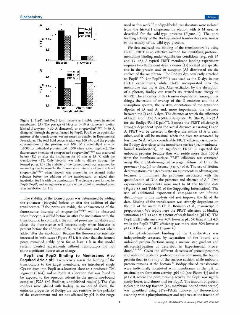
The stability of the formed pores was determined by addingthe enhancer (biocytin) before or after the addition of thetranslocators. If the pores are stable, the enhancement of thefluorescence intensity of streptavidinBodipy will be observedwhen biocytin is added before or after the incubation with thetranslocators. In contrast, if the formed pores are not stable andclose, the fluorescence will increase only when biocytin ispresent before the addition of the translocators, and not whenadded after the incubation. Because the fluorescence intensityincreased in both cases (Figure 5B), it is clear that the formedpores remained stably open for at least 1 h in this modelsystem. Control experiments without translocators did notshow significant fluorescence change.PopB and PopD Binding to Membranes Also
Required Acidic pH. To precisely assess the binding of thetranslocators to the target membrane, we introduced singleCys residues into PopB at a location close to a predicted TMsegment (S164), and in PopD at a location that was found tobe exposed to the aqueous solvent in the membrane-boundcomplex [F223 (M. Buckner, unpublished results)]. The Cysresidues were labeled with Bodipy. As mentioned above, theemission properties of Bodipy are not sensitive to the polarityof the environment and are not affected by pH in the range
used in this work.40 Bodipy-labeled translocators were isolatedfrom the hisPcrH chaperone by elution with 6 M urea asdescribed for the wild-type proteins (Figure 1). The poreforming activity of the Bodipy-labeled translocators was similarto the activity of the wild-type proteins.We first analyzed the binding of the translocators by using
FRET. FRET is an effective method for identifying protein−membrane binding under equilibrium conditions (e.g., refs 37and 43−46). A typical FRET membrane binding experimentrequires two fluorescent dyes, a donor (D) located at a specificsite in the protein and an acceptor (A) distributed on thesurface of the membrane. The Bodipy dye covalently attachedto PopBS164C (or PopDF223C) was used as the D dye in ourFRET experiments, while Rh-PE incorporated into themembrane was the A dye. After excitation by the absorptionof a photon, Bodipy can transfer its excited-state energy toRh-PE. The efficiency of this transfer depends on, among otherthings, the extent of overlap of the D emission and the Aabsorption spectra, the relative orientation of the transitiondipoles of D and A, and, more importantly, the distancebetween the D and A dyes. The distance at which the efficiencyof FRET from D to A is 50% is designated R0 (the R0 is ∼52 Åfor the Bodipy/Rh-PE pair44). Because the FRET efficiency isstrongly dependent upon the actual distance separating D andA, FRET will be detected if the dyes are within 95 Å of eachother, and it will be maximal when the dyes are separated byless than 24 Å. While considerable FRET efficiency is expectedfor Bodipy dyes close to the membrane surface (i.e., membrane-bound translocators), no significant FRET is expected forunbound proteins because they will reside more than 100 Åfrom the membrane surface. FRET efficiency was estimatedusing the amplitude-weighted average lifetime of D in thepresence (⟨τDA⟩a) or absence (⟨τD⟩a) of A. The use of lifetimedeterminations over steady-state measurements is advantageousbecause it minimizes the problems associated with thequantification of D in the presence and absence of A.30 Twoexponential components were used to fit the lifetime data(Figure S8 and Table S1 of the Supporting Information). Theuse of additional exponential components or lifetimedistributions in the analysis did not improve the fit of thedata. Binding of the translocators was strongly dependent onthe pH of the medium (F. B. Romano et al., manuscript inpreparation). We report here the FRET efficiency at bindingsaturation (pH 4) and at a point of weak binding (pH 6). ThePopB FRET efficiency was 40% lower at pH 6.0 than at pH 4.0,while the PopD FRET efficiency was more than 80% lower atpH 6.0 than at pH 4.0 (Figure 6).The pH-dependent binding of the translocators was
independently assessed by separation of the bound andunbound protein fractions using a sucrose step gradient andultracentrifugation as described in Experimental Proce-dures.15,23,47 Given the different densities of proteoliposomesand unbound proteins, proteoliposomes containing the boundprotein float to the top of the sucrose cushion while unboundprotein remains at the bottom.23 Bodipy-labeled translocatorswere individually incubated with membranes at the pH ofmaximal pore formation activity [pH 4.0 (see Figure 4)] and atpH 6.0, where the pore forming activity for PopB was signifi-cantly lower, and almost null for PopD. The amount of proteinisolated in the top fraction (i.e., membrane-bound translocator)was quantified using SDS−PAGE followed by fluorescencescanning with a phosphorimager and reported as the fraction of
Figure 5. PopD and PopB form discrete and stable pores in modelmembranes. (A) The passage of biocytin (∼10 Å diameter), biotin-labeled β-amylase (∼50 Å diameter), or streptavidinBodipy (∼50 Ådiameter) through the pores formed by PopD, PopB, or an equimolarmixture of the translocators was measured as detailed in ExperimentalProcedures. The total lipid concentration was 100 μM, and the proteinconcentration of the proteins was 100 nM (protein:lipid ratio of1:1000 for individual proteins and 1:500 when added together). Thefluorescence intensity of encapsulated streptavidinBodipy was measuredbefore (F0) or after the incubation for 60 min at 25 °C with thetranslocators (F). Only biocytin was able to diffuse through theformed pores. (B) The stability of the formed pores was examined bymeasuring the increase in the fluorescence intensity of encapsulatedstreptavidinBodipy when biocytin was present in the external buffersolution before the addition of the translocators, or added afterincubation for 1 h with the translocators. The discrete pores formed byPopB, PopD, and an equimolar mixture of the proteins remained openafter incubation for 1 h.
Biochemistry Article
dx.doi.org/10.1021/bi200905x |Biochemistry XXXX, XXX, XXX−XXXI
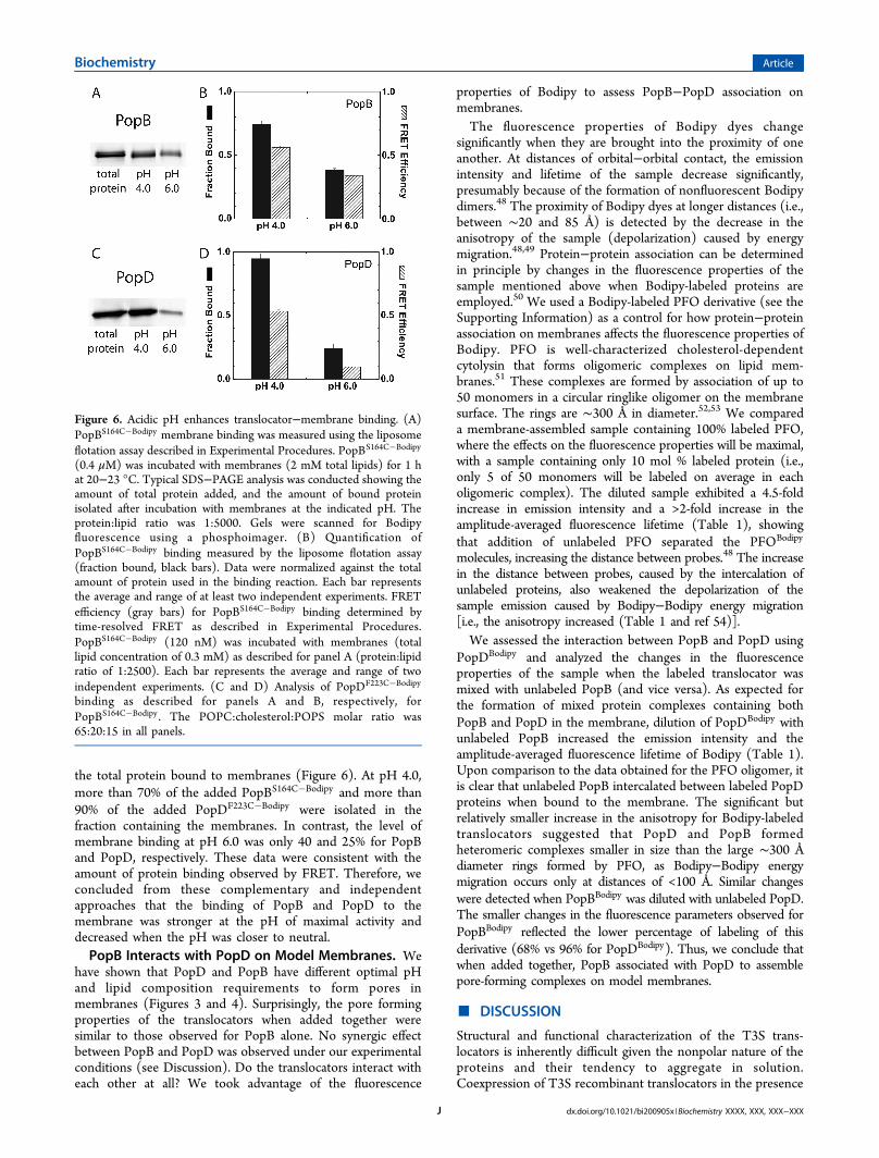
the total protein bound to membranes (Figure 6). At pH 4.0,more than 70% of the added PopBS164C�Bodipy and more than90% of the added PopDF223C�Bodipy were isolated in thefraction containing the membranes. In contrast, the level ofmembrane binding at pH 6.0 was only 40 and 25% for PopBand PopD, respectively. These data were consistent with theamount of protein binding observed by FRET. Therefore, weconcluded from these complementary and independentapproaches that the binding of PopB and PopD to themembrane was stronger at the pH of maximal activity anddecreased when the pH was closer to neutral.PopB Interacts with PopD on Model Membranes. We
have shown that PopD and PopB have different optimal pHand lipid composition requirements to form pores inmembranes (Figures 3 and 4). Surprisingly, the pore formingproperties of the translocators when added together weresimilar to those observed for PopB alone. No synergic effectbetween PopB and PopD was observed under our experimentalconditions (see Discussion). Do the translocators interact witheach other at all? We took advantage of the fluorescence
properties of Bodipy to assess PopB−PopD association onmembranes.The fluorescence properties of Bodipy dyes change
significantly when they are brought into the proximity of oneanother. At distances of orbital−orbital contact, the emissionintensity and lifetime of the sample decrease significantly,presumably because of the formation of nonfluorescent Bodipydimers.48 The proximity of Bodipy dyes at longer distances (i.e.,between ∼20 and 85 Å) is detected by the decrease in theanisotropy of the sample (depolarization) caused by energymigration.48,49 Protein−protein association can be determinedin principle by changes in the fluorescence properties of thesample mentioned above when Bodipy-labeled proteins areemployed.50 We used a Bodipy-labeled PFO derivative (see theSupporting Information) as a control for how protein−proteinassociation on membranes affects the fluorescence properties ofBodipy. PFO is well-characterized cholesterol-dependentcytolysin that forms oligomeric complexes on lipid mem-branes.51 These complexes are formed by association of up to50 monomers in a circular ringlike oligomer on the membranesurface. The rings are ∼300 Å in diameter.52,53 We compareda membrane-assembled sample containing 100% labeled PFO,where the effects on the fluorescence properties will be maximal,with a sample containing only 10 mol % labeled protein (i.e.,only 5 of 50 monomers will be labeled on average in eacholigomeric complex). The diluted sample exhibited a 4.5-foldincrease in emission intensity and a >2-fold increase in theamplitude-averaged fluorescence lifetime (Table 1), showingthat addition of unlabeled PFO separated the PFOBodipy
molecules, increasing the distance between probes.48 The increasein the distance between probes, caused by the intercalation ofunlabeled proteins, also weakened the depolarization of thesample emission caused by Bodipy−Bodipy energy migration[i.e., the anisotropy increased (Table 1 and ref 54)].We assessed the interaction between PopB and PopD using
PopDBodipy and analyzed the changes in the fluorescenceproperties of the sample when the labeled translocator wasmixed with unlabeled PopB (and vice versa). As expected forthe formation of mixed protein complexes containing bothPopB and PopD in the membrane, dilution of PopDBodipy withunlabeled PopB increased the emission intensity and theamplitude-averaged fluorescence lifetime of Bodipy (Table 1).Upon comparison to the data obtained for the PFO oligomer, itis clear that unlabeled PopB intercalated between labeled PopDproteins when bound to the membrane. The significant butrelatively smaller increase in the anisotropy for Bodipy-labeledtranslocators suggested that PopD and PopB formedheteromeric complexes smaller in size than the large ∼300 Ådiameter rings formed by PFO, as Bodipy−Bodipy energymigration occurs only at distances of <100 Å. Similar changeswere detected when PopBBodipy was diluted with unlabeled PopD.The smaller changes in the fluorescence parameters observed forPopBBodipy reflected the lower percentage of labeling of thisderivative (68% vs 96% for PopDBodipy). Thus, we conclude thatwhen added together, PopB associated with PopD to assemblepore-forming complexes on model membranes.
■ DISCUSSION
Structural and functional characterization of the T3S trans-locators is inherently difficult given the nonpolar nature of theproteins and their tendency to aggregate in solution.Coexpression of T3S recombinant translocators in the presence
Figure 6. Acidic pH enhances translocator−membrane binding. (A)PopBS164C�Bodipy membrane binding was measured using the liposomeflotation assay described in Experimental Procedures. PopBS164C�Bodipy
(0.4 μM) was incubated with membranes (2 mM total lipids) for 1 hat 20−23 °C. Typical SDS−PAGE analysis was conducted showing theamount of total protein added, and the amount of bound proteinisolated after incubation with membranes at the indicated pH. Theprotein:lipid ratio was 1:5000. Gels were scanned for Bodipyfluorescence using a phosphoimager. (B) Quantification ofPopBS164C�Bodipy binding measured by the liposome flotation assay(fraction bound, black bars). Data were normalized against the totalamount of protein used in the binding reaction. Each bar representsthe average and range of at least two independent experiments. FRETefficiency (gray bars) for PopBS164C�Bodipy binding determined bytime-resolved FRET as described in Experimental Procedures.PopBS164C�Bodipy (120 nM) was incubated with membranes (totallipid concentration of 0.3 mM) as described for panel A (protein:lipidratio of 1:2500). Each bar represents the average and range of twoindependent experiments. (C and D) Analysis of PopDF223C�Bodipy
binding as described for panels A and B, respectively, forPopBS164C�Bodipy . The POPC:cholesterol:POPS molar ratio was65:20:15 in all panels.
Biochemistry Article
dx.doi.org/10.1021/bi200905x |Biochemistry XXXX, XXX, XXX−XXXJ

of their cognate chaperone renders soluble protein complexesand allows their purification in large quantities.15,31−33
However, the analysis of individual T3S translocators requirestheir separation from the chaperone prior to their assembly intomembranes. These separations have been achieved by reducingthe pH of the medium15,31 or by the inclusion of milddetergents.31 Unfortunately, upon dissociation from the PcrHchaperone, both PopB and PopD form large and nonuniformprotein aggregates15 that make it difficult to characterize theprotein−membrane and protein−protein association processes.Furthermore, we observed that during the acidification of themedium to separate the translocators from the chaperone, PcrHprecipitates, carrying considerable amounts of the purifiedtranslocators (Figure S5 of the Supporting Information). Thus,the precise characterization of the assembly of the T3Stranslocon into membranes required a more efficientexperimental approach for isolation of active translocators.We have found in these studies that PopB (as well as PopD)
can be purified as a homogeneous complex with the cognatechaperone hisPcrH and be effectively dissociated and isolatedfrom hisPcrH using urea. In contrast to PopD, which lost mostof its secondary structure, PopB retained a high proportion ofits secondary structure after solubilization in 6 M urea. Theblue-shifted intrinsic fluorescence spectra of urea-solubilizedPopB suggested that the Trp located at the N-terminus of apredicted TM segment (residues 169−187) remained locatedin a nonpolar environment even in its denatured state. Asexpected for proteins that are proposed to travel in an unfoldedstate through the T3S needle and to be released in theproximity of the target membrane, both urea-isolated trans-locators formed discrete and stable TM pores upon dilution inthe presence of lipid bilayers. The pore forming properties ofurea-isolated PopB and PopD were consistent with thosedescribed previously for translocators isolated using low pH.16
Interestingly, quantitative assessment of binding of PopB andPopD to membranes showed that acidic pH was required notonly for membrane insertion but also for a stable protein−membrane interaction. In summary, we have established anefficient experimental procedure for isolating native andmodified forms of PopB and PopD and for specifically labelingthe translocators with fluorescent or other types of probes (e.g.,
cross-linkers). Isolated translocators were assembled on modelmembranes where they form discrete and stable pores.Moreover, our spectroscopic examination of Bodipy-labeledtranslocators showed that when added together, PopBintercalates with PopD on membrane-assembled complexes.Purification of isolated recombinant translocators has proven
to be a difficult task. PopD cloned in pET-based vector systemsproduced large amounts of protein, but mostly as inclusionbodies. The yield of water-soluble PopD can be improved byexpression at a low temperature (i.e., ∼18 °C), but water-soluble PopD forms aggregates (ref 15 and data not shown).PopB is toxic for Escherichia coli cells and cannot be stablyproduced (data not shown). Schoehn et al.15 elegantly solvedthese problems by coexpressing the translocators with theircognate chaperone, PcrH. The water-soluble 1:1 PcrH−PopDcomplex was obtained in large amounts, and the structuralcharacterization of PopD showed it adopts a molten globularconformation.33 Less is known about the structure of PopB,given that only minor amounts of a homogeneous PcrH−PopBcomplex were obtained using this procedure.15,32
We found that the purified hisPcrH−PopB complex canremain soluble and nonaggregated in aqueous solutions atrelatively low concentrations (Figure 1 and Figure S1 of theSupporting Information). The structure of the purifiedhisPcrH−PopB complex was analyzed using a combination ofCD and fluorescence spectroscopy (Figure 2). Far-UV CDanalysis revealed that PopB had less α-helical content thanPopD when bound to hisPcrH. However, in contrast to thelow secondary structure content observed in urea-isolatedPopD, urea-isolated PopB retained a considerable amount ofsecondary structure. Furthermore, the only Trp of PopB waslocated in a nonpolar environment when bound to thechaperone or when denatured in urea. These data suggestedthat PopB adopts a conformation more compact than thatobserved for molten globular PopD.33
PopB and PopD remain stably bound to PcrH in thebacterial cytoplasm in the absence of secretion, most likely toavoid protein aggregation and to maintain a secretion-competent conformation.7,9 After the bacterium has contactedthe target cell, PopB and PopD dissociate from PcrH and are
Table 1. Bodipy Detected Protein−Protein Interaction in Intact Membranesa
fraction of protein added
PFOBodipy PFO I (relative units) I10/I100 (relative units) r ⟨τ⟩αb (ns)
1 − 1.84 ± 0.09 0.050 ± 0.005 1.55 ± 0.011 9 8.27 ± 0.07 4.5 ± 0.2 0.123 ± 0.005 4.75 ± 0.01
PopDBodipy PopB
1 − 6.5 ± 0.2 0.161 ± 0.004 2.56 ± 0.031 9 14.4 ± 0.2 2.22 ± 0.08 0.186 ± 0.009 3.90 ± 0.03
PopBBodipy PopD
1 − 4.32 ± 0.04 0.185 ± 0.003 3.24 ± 0.021 9 7.1 ± 0.2 1.63 ± 0.02 0.226 ± 0.003 4.93 ± 0.04
aRelative fractions of proteins added are indicated. Labeled and unlabeled proteins were mixed in buffer B supplemented with 6 M urea and20 mM Gly solution prior to dilution in buffer D in the presence of membranes. The final fluorescence parameters measured for the differentsamples are indicated: I, fluorescence emission intensity; I10/I100, ratio of the fluorescence emissions of diluted to undiluted labeled proteinsamples; ⟨τ⟩α , amplitude-weighted average fluorescence lifetime. The fluorescence lifetime of Bodipy in 50 mM sodium acetate (pH 4.0) was5.85 ns. The fluorescence lifetime of monomeric PFOBodipy in buffer C was 5.51 ns. Labeling efficiencies were 100, 96, and 68% for PFOBodipy ,PopDF223C�Bodipy , and PopBS164C�Bodipy , respectively. The average and range of at least two determinations are shown. Protein:lipid ratioswere 1:2500 for fluorescence lifetimes and 1:10000 for steady-state determinations. bTime-resolved fluorescence data for 10% labeled PFObest fit to a single-exponential component. The remaining data best fit to a two-exponential component (see Table S1 of the SupportingInformation).
Biochemistry Article
dx.doi.org/10.1021/bi200905x |Biochemistry XXXX, XXX, XXX−XXXK
presumably secreted through a narrow channel formed by theT3S needle (2−2.5 nm in diameter). Unfolded translocatorsare proposed to emerge from the tip of the T3S needle (formedby PcrV), in the proximity of the target membrane. PcrV isrequired to engage the membrane-inserted translocon with theT3S needle;13,55,56 however, PcrV is not involved in theformation of the TM pore.15 Therefore, the use of urea-isolatedtranslocators to reconstitute membrane-inserted proteincomplexes is reasonable because the dilution of urea-isolatedPopB and PopD in the presence of membranes likely resemblesthe in vivo translocon assembly process.While negatively charged phospholipids are crucial for the
binding of translocators to membranes and formation of TMpores in model membranes, cholesterol seems not to play acentral role in vitro.16 We tested how different lipids affectedthe pore formation properties of PopB and PopD in ourexperimental system. The presence of cholesterol and POPSin the liposomal membranes facilitated the insertion of theurea-isolated PopB and PopD (Figure 3). Other lipids likesphingomyelin and PE did not significantly alter the poreformation efficiency observed with POPC/POPS/cholesterolliposomes [65:15:20 molar ratio (data not shown)]. Therefore,we chose this composition as a simple and efficient modelmembrane system to study the assembly of PopB and PopDinto lipid bilayers. Urea-isolated translocators formed discreteand stable pores in model membranes (Figures 4 and 5). Takentogether, our data showed that the treatment with 6 M urea didnot significantly alter the pore formation properties of thetranslocators.We found that urea-isolated PopB, PopD, or an equimolar
mixture of both translocators formed pores very similar to thepores formed when the translocators are dissociated from PcrHat acidic pH in the presence of membranes (Figure S3 of theSupporting Information). Interestingly, the comparison of bothexperimental systems suggested that acidic pH was requirednot only to dissociate the translocators from the chaperone(Figure S5 of the Supporting Information) but also to promotebinding and insertion of translocators into membranes (Figures4 and 6). We employed two independent experimentalapproaches, time-resolved FRET and sucrose-step gradientultracentrifugation, to analyze the binding of the translocatorsto membranes at different pH values (Figure 6). Both assaysclearly showed that binding at pH 4 was more efficient than atpH 6. Only a small fraction of the proteins was found associatedwith the membranes at pH 6. These results differ from thosereported for the case in which heterogeneous aggregates ofPopB and PopD were used.16 The kinetic parameters used as ameasure of membrane binding may represent the behavior of asmall fraction of protein in the experimental system, and notnecessarily the average behavior of all the protein in the sample(i.e., unbound protein does not generate a signal). Our datasuggested that the decrease in the activity of the translocators athigher pH was, at least in part, caused by the inability of theproteins to bind to the lipid membrane.A homogeneous sample is essential for the unambiguous
interpretation of the spectroscopic signal obtained during theanalysis of protein complexes assembled into membranes.39 Forthis purpose, negatively charged phospholipids and low pHhave been extensively used in the spectroscopic studies of pore-forming proteins like colicins, diphtheria toxin, Bcl-xL, etc.
57−61
The requirement of acidic pH for the insertion of protein intomembranes has been associated with the presence of acidic
molten globular states.62 While this pH-dependent conforma-tional change may be relevant to the more compact PopBconformation (see above), PopD was found to have a moltenglobule conformation even at neutral pH.33 Therefore, thespontaneous insertion of PopD (and/or PopB) segments intolipid bilayers may require the protonation of acidic residues, asshown before for bacteriorhodopsin fragments.63 In our modelsystem, the use of acidic pH maximized the assembly of discreteand stable TM pores. Such a low pH is presumably notencountered by the proteins when interacting with the plasmamembrane of the target cell in vivo. Clearly, other stillunidentified components present in the bacterium, or in thetarget cell, may facilitate the assembly of the translocon, butfurther studies are required in this area.It has been widely assumed that assembled translocons
contain both translocators forming a hetero-oligomer.9
However, the structural arrangement of the membrane-insertedtranslocon and the stoichiometry of the complex are notknown. By combining site-directed fluorescence labeling withboth excitation energy migration and fluorescence quenching,we have shown that fluorescently labeled PopB interacted withnative PopD in intact membrane-inserted complexes (Table 1).This interaction was confirmed by using the reverse combi-nation of labeled PopD and native PopB. In both experiments,the intercalation of unlabeled molecules among the labeledones produced the separation of the Bodipy dyes andconsequently an increase in the fluorescence intensity, lifetime,and anisotropy of the sample. These results also implied thatwhen added individually, both PopDF223C�Bodipy andPopBS164C�Bodipy formed homo-oligomers. Self-interaction ofthe translocators in intact membrane-inserted complexes wasconfirmed by the relative increase in the fluorescence intensity,lifetimes, and anisotropy of the sample when native PopD (orPopB) was mixed with PopDF223C�Bodipy (or PopBS164C�Bodipy)and added to the membranes (data not shown).In summary, we have established a cell-free system to analyze
the structural assembly of the P. aeruginosa T3S translocators.Using this system, we found that PopB interacts with PopDwhen forming discrete and stable pores in intact membranes.Cell-free systems have been extremely useful for examininginteractions among relevant protein components in membraneprotein complexes and the study of their assembly and theirstructure (e.g., refs 44, 46, 57, 61, and 64−66). The identifiedmechanism and structural information obtained using the cell-free system are very valuable for the design of in vivo experi-ments and evaluation of the contribution of other known orunknown factors to the mechanism of T3S effector translocation.
■ ASSOCIATED CONTENT
*S Supporting InformationDetails on cloning expression and purification of proteins,aggregation of the hisPcrH−PopB complex, PopB and PopDsite-directed mutagenesis, SEC analysis of homogeneouschaperone−translocator complexes, pH dissociation vs ureadilution activities at pH 5.1, intermolecular disulfide formationby hisPcrH, pH solubility of the hisPcrH−PopD complex,steady-state FRET for protein−membrane binding, particle sizeanalysis by DLS, and lifetime analysis for time-resolved FRETefficiencies. This material is available free of charge via theInternet at http://pubs.acs.org.
Biochemistry Article
dx.doi.org/10.1021/bi200905x |Biochemistry XXXX, XXX, XXX−XXXL
■ AUTHOR INFORMATION
Corresponding Author*Address: 710 N. Pleasant St., Lederle GRT, Rm 816,University of Massachusetts, Amherst, MA 01003. Phone:(413) 545-2497. Fax: (413) 545-3291. E-mail: [email protected].
FundingThis research was supported by a Biomedical Research Grantfrom the American Lung Association and the MassachusettsThoracic Society to A.P.H. F.B.R. was partially supported byNational Research Service Award T32 GM08515 from theNational Institutes of Health. K.C.R. was a recipient of theHoward Hughes Medical Institute Undergraduate ScienceProgram Summer Research Internship.
■ ACKNOWLEDGMENTS
We thank Prof. Carlos F. Gonzalez (Texas A&M University)for providing the P. aeruginosa PAO1 genomic DNA, Prof. LilaM. Gierasch for assistance with the CD measurements, Prof.Christopher Woodcock for his assistance with the initialtransmission EM experiments, William Boston-Howes for thepreliminary setup of the liposome flotation assay, MichaelBuckner for the preparation of the PopDF223C mutant, andSarah E. Kells for excellent technical assistance.
■ ABBREVIATIONS
Pop, Pseudomonas outer protein; Pcr, Pseudomonas low-calciumresponse; T3S, type III secretion; TM, transmembrane; IMAC,immobilized metal ion affinity chromatography; AEC, anionexchange chromatography; SEC, size exclusion chromatogra-phy; UV, ultraviolet; CD, circular dichroism; DLS, dynamiclight scattering; POPC, 1-palmitoyl-2-oleoyl-sn-glycero-3-phos-phocholine; POPE, 1-palmitoyl-2-oleoyl-sn-glycero-3-phos-phoethanolamine; POPS, 1-palmitoyl-2-oleoyl-sn-glycero-3-phospho-L-serine; Fl, fluorescein; DTT, (2S,3S)-1,4-bis-sulfa-nylbutane-2,3-diol; EDTA, ethylenedinitrilotetraacetic acid;DPA, dipicolinic acid; HEPES, 2-[4-(2-hydroxyethyl)-piperazin-1-yl]ethanesulfonic acid; LB, Luria-Bertani; IPTG,isopropyl α-D-thiogalactopyranoside; Tris, 2-amino-2-(hydroxymethyl)propane-1,3-diol; FRET, Forster resonanceenergy transfer; SDS−PAGE, sodium dodecyl sulfate−poly-acrylamide gel electrophoresis; Rh-PE, rhodamine B 1,2-dihexadecanoyl-sn-glycero-3-phosphoethanolamine; Bodipy,4,4-difluoro-5,7-dimethyl-4-bora-3a,4a-diaza-3-indacene; PFO,Perfringolysin O.
■ REFERENCES
(1) Hueck, C. J. (1998) Type III protein secretion systems inbacterial pathogens of animals and plants. Microbiol. Mol. Biol. Rev. 62,379−433.(2) Galan, J. E., and Wolf-Watz, H. (2006) Protein delivery into
eukaryotic cells by type III secretion machines. Nature 444, 567−573.(3) Cornelis, G. R. (2006) The type III secretion injectisome. Nat.
Rev. Microbiol. 4, 811−825.(4) Frank, D. (1997) The exoenzyme S regulon of Pseudomonas
aeruginosa. Mol. Microbiol. 26, 621−629.(5) Hauser, A. R., Cobb, E., Bodi, M., Mariscal, D., Valles, J., Engel, J.
N., and Rello, J. (2002) Type III protein secretion is associated withpoor clinical outcomes in patients with ventilator-associated pneumo-nia caused by Pseudomonas aeruginosa. Crit. Care Med. 30, 521−528.
(6) Moraes, T. F., Spreter, T., and Strynadka, N. C. J. (2008) Piecingtogether the type III injectisome of bacterial pathogens. Curr. Opin.Struct. Biol. 18, 258−266.(7) Mueller, C. A., Broz, P., and Cornelis, G. R. (2008) The type III
secretion system tip complex and translocon. Mol. Microbiol. 68,1085−1095.(8) Buttner, D., and Bonas, U. (2002) Port of entry: The type III
secretion translocon. Trends Microbiol. 10, 186−192.(9) Mattei, P.-J., Faudry, E., Job, V., Izore, T., Attree, I., and Dessen,
A. (2011) Membrane targeting and pore formation by the type IIIsecretion system translocon. FEBS J. 278, 414−426.(10) Mueller, C.A., Broz, P., Muller, S. A., Ringler, P., Erne-Brand, F.,
Sorg, I., Kuhn, M., Engel, A., and Cornelis, G. R. (2005) The V-antigenof Yersinia forms a distinct structure at the tip of injectisome needles.Science 310, 674−676.(11) Dacheux, D., Goure, J., Chabert, J., Usson, Y., and Attree, I.
(2001) Pore-forming activity of type III system-secreted proteins leadsto oncosis of Pseudomonas aeruginosa-infected macrophages. Mol.Microbiol. 40, 76−85.(12) Sundin, C., Thelaus, J., Broms, J. E., and Forsberg, A. (2004)
Polarisation of type III translocation by Pseudomonas aeruginosarequires PcrG, PcrV and PopN. Microb. Pathog. 37, 313−322.(13) Goure, J., Pastor, A., Faudry, E., Chabert, J., Dessen, A., and
Attree, I. (2004) The V antigen of Pseudomonas aeruginosa is requiredfor assembly of the functional PopB/PopD translocation pore in hostcell membranes. Infect. Immun. 72, 4741−4750.(14) Vance, R. E., Rietsch, A., and Mekalanos, J. J. (2005) Role of the
type III secreted exoenzymes S, T, and Y in systemic spread ofPseudomonas aeruginosa PAO1 in vivo. Infect. Immun. 73, 1706−1713.(15) Schoehn, G., Di Guilmi, A. M., Lemaire, D., Attree, I.,
Weissenhorn, W., and Dessen, A. (2003) Oligomerization of type IIIsecretion proteins PopB and PopD precedes pore formation inPseudomonas. EMBO J. 22, 4957−4967.(16) Faudry, E., Vernier, G., Neumann, E., Forge, V., and Attree, I.
(2006) Synergistic pore formation by type III toxin translocators ofPseudomonas aeruginosa. Biochemistry 45, 8117−8123.(17) Tardy, F., Homble, F., Neyt, C., Wattiez, R., Cornelis, G.,
Ruysschaert, J., and Cabiaux, V. (1999) Yersinia enterocolitica type IIIsecretion-translocation system: Channel formation by secreted Yops.EMBO J. 18, 6793−6799.(18) Hume, P. J., McGhie, E. J., Hayward, R. D., and Koronakis, V.
(2003) The purified Shigella IpaB and Salmonella SipB translocatorsshare biochemical properties and membrane topology. Mol. Microbiol.49, 425−439.(19) Pace, C. N., Vajdos, F., Fee, L., Grimsley, G., and Gray, T.
(1995) How to measure and predict the molar absorption coefficientof a protein. Protein Sci. 4, 2411−2423.(20) Moe, P. C., and Heuck, A. P. (2010) Phospholipid hydrolysis
caused by Clostridium perfringens α-toxin facilitates the targeting ofperfringolysin O to membrane bilayers. Biochemistry 49, 9498−9507.(21) Shepard, L. A., Heuck, A. P., Hamman, B. D., Rossjohn, J.,
Parker, M. W., Ryan, K. R., Johnson, A. E., and Tweten, R. K. (1998)Identification of a membrane-spanning domain of the thiol-activatedpore-forming toxin Clostridium perfringens perfringolysin O: Anα-helical to β-sheet transition identified by fluorescence spectroscopy.Biochemistry 37, 14563−14574.(22) Heuck, A. P., Tweten, R. K., and Johnson, A. E. (2003)
Assembly and topography of the prepore complex in cholesterol-dependent cytolysins. J. Biol. Chem. 278, 31218−31225.(23) Dalton, A. K., Murray, P. S., Murray, D., and Vogt, V. M. (2005)
Biochemical characterization of Rous sarcoma virus MA proteininteraction with membranes. J. Virol. 79, 6227−6238.(24) Koppel, D. E. (1972) Analysis of macromolecular polydispersity
in intensity correlation spectroscopy: The method of cumulants.J. Chem. Phys. 57, 4814−4820.
Biochemistry Article
dx.doi.org/10.1021/bi200905x |Biochemistry XXXX, XXX, XXX−XXXM

(25) Reinhart, G. D., Marzola, P., Jameson, D. M., and Gratton, E.(1991) A method for on-line background subtraction in frequencydomain fluorometry. J. Fluoresc. 1, 153−162.(26) Crowley, K. S., Liao, S., Worrell, V. E., Reinhart, G. D., and
Johnson, A. E. (1994) Secretory proteins move through theendoplasmic reticulum membrane via an aqueous, gated pore. Cell78, 461−471.(27) vandeVen, M., Ameloot, M., Valeur, B., and Boens, N. (2005)
Pitfalls and their remedies in time-resolved fluorescence spectroscopyand microscopy. J. Fluoresc. 15, 377−413.(28) Boens, N., Qin, W., Basaric, N., Hofkens, J., Ameloot, M.,
Pouget, J., Lefevre, J. P., Valeur, B., Gratton, E., vandeVen, M., Silva, N.D., Engelborghs, Y., Willaert, K., Sillen, A., Rumbles, G., Phillips, D.,Visser, A. J. W. G., vanHoek, A., Lakowicz, J. R., Malak, H., Gryczynski,I., Szabo, A. G., Krajcarski, D. T., Tamai, N., and Miura, A. (2007)Fluorescence lifetime standards for time and frequency domainfluorescence spectroscopy. Anal. Chem. 79, 2137−2149.(29) Alcala, J. R., Gratton, E., and Prendergast, F. G. (1987)
Resolvability of fluorescence lifetime distributions using phasefluorometry. Biophys. J. 51, 587−596.(30) Wu, P. G., and Brand, L. (1994) Resonance energy transfer:
Methods and applications. Anal. Biochem. 218, 1−13.(31) Birket, S. E., Harrington, A. T., Espina, M., Smith, N. D., Terry,
C. M., Darboe, N., Markham, A. P., Middaugh, C. R., Picking, W. L.,and Picking, W. D. (2007) Preparation and characterization oftranslocator/chaperone complexes and their component proteins fromShigella flexneri. Biochemistry 46, 8128−8137.(32) Tan, Y. W., Yu, H. B., Sivaraman, J., Leung, K. Y., and Mok, Y.-
K. (2009) Mapping of the chaperone AcrH binding regions oftranslocators AopB and AopD and characterization of oligomeric andmetastable AcrH-AopB-AopD complexes in the type III secretionsystem of Aeromonas hydrophila. Protein Sci. 18, 1724−1734.(33) Faudry, E., Job, V., Dessen, A., Attree, I., and Forge, V. (2007)
Type III secretion system translocator has a molten globuleconformation both in its free and chaperone-bound forms. FEBS J.274, 3601−3610.(34) Job, V., Mattei, P.-J., Lemaire, D., Attree, I., and Dessen, A.
(2010) Structural basis of chaperone recognition of type III secretionsystem minor translocator proteins. J. Biol. Chem. 285, 23224−23232.(35) Manavalan, P., and Johnson, W. C. (1983) Sensitivity of circular
dichroism to protein tertiary structure class. Nature 305, 831−832.(36) Heuck, A. P., and Johnson, A. E. (2002) Pore-forming protein
structure analysis in membranes using multiple independentfluorescence techniques. Cell Biochem. Biophys. 36, 89−101.(37) Mutucumarana, V. P., Duffy, E. J., Lollar, P., and Johnson, A. E.
(1992) The active site of factor IXa is located far above the membranesurface and its conformation is altered upon association with factorVIIIa. A fluorescence study. J. Biol. Chem. 267, 17012−17021.(38) Hamman, B.D., Oleinikov, A. V., Jokhadze, G. G., Traut, R. R.,
and Jameson, D. M. (1996) Dimer/monomer equilibrium and domainseparations of Escherichia coli ribosomal protein L7/L12. Biochemistry35, 16680−16686.(39) Johnson, A. E. (2005) Fluorescence approaches for determining
protein conformations, interactions and mechanisms at membranes.Traffic 6, 1078−1092.(40) Karolin, J., Johansson, L. B.-A., Strandberg, L., and Ny, T.
(1994) Fluorescence and absorption spectroscopic properties ofdipyrrometheneboron difluoride (Bodipy) derivatives in liquids, lipidmembranes, and proteins. J. Am. Chem. Soc. 116, 7801−7806.(41) Nicol, F., Nir, S., and Szoka, F. C. Jr. (1999) Orientation of the
pore-forming peptide gala in POPC vesicles determined by a Bodipy-avidin/biotin binding assay. Biophys. J. 76, 2121−2141.(42) Rosconi, M. P., Zhao, G., and London, E. (2004) Analyzing
topography of membrane-inserted diphtheria toxin T domain usingBodipy-streptavidin: At low pH, helices 8 and 9 form a transmembrane
hairpin but helices 5−7 form stable nonclassical inserted segments onthe cis side of the bilayer. Biochemistry 43, 9127−9139.(43) Bazzi, M. D., and Nelsestuen, G. L. (1987) Association of
protein kinase C with phospholipid vesicles. Biochemistry 26, 115−122.(44) Ramachandran, R., Tweten, R. K., and Johnson, A. E. (2005)
The domains of a cholesterol-dependent cytolysin undergo a majorFRET-detected rearrangement during pore formation. Proc. Natl. Acad.Sci. U.S.A. 102, 7139−7144.(45) Posokhov, Y. O., and Ladokhin, A. S. (2006) Lifetime
fluorescence method for determining membrane topology of proteins.Anal. Biochem. 348, 87−93.(46) Lovell, J. F., Billen, L. P., Bindner, S., Shamas-Din, A., Fradin,
C., Leber, B., and Andrews, D. W. (2008) Membrane binding by tBidinitiates an ordered series of events culminating in membranepermeabilization by Bax. Cell 135, 1074−1084.(47) Rivnay, B., and Metzger, H. (1983) Use of the airfuge for
analysis and preparation of receptors incorporated into liposomes:Studies with the receptor for immunoglobulin E. Anal. Biochem. 130,514−520.(48) Bergstrom, F., Mikhalyov, I., Hagglof, P., Wortmann, R., Ny, T.,
and Johansson, L. B. A. (2002) Dimers of dipyrrometheneborondifluoride (Bodipy) with light spectroscopic applications in chemistryand biology. J. Am. Chem. Soc. 124, 196−204.(49) Mikhalyov, I., Gretskaya, N., Bergstro m, F., and Johansson, L.
B.-Å. (2002) Electronic ground and excited state properties ofdipyrrometheneboron difluoride (Bodipy): Dimers with application tobiosciences. Phys. Chem. Chem. Phys. 4, 5663−5670.(50) Marushchak, D., Kalinin, S., Mikhalyov, I., Gretskaya, N., and
Johansson, L. B.-Å. (2006) Pyrromethene dyes (Bodipy) can formground state homo and hetero dimers: Photophysics and spectralproperties. Spectrochim. Acta, Part A 65, 113−122.(51) Heuck, A. P., Moe, P. C., and Johnson, B. B. (2010) The
cholesterol-dependent cytolysins family of Gram-positive bacterialtoxins. In Cholesterol binding proteins and cholesterol transport (Harris, J.R., Ed.) pp 551−577, Springer, New York.(52) Czajkowsky, D. M., Hotze, E. M., Shao, Z., and Tweten, R. K.
(2004) Vertical collapse of a cytolysin prepore moves its trans-membrane β-hairpins to the membrane. EMBO J. 23, 3206−3215.(53) Dang, T. X., Hotze, E. M., Rouiller, I., Tweten, R. K., and
Wilson-Kubalek, E. M. (2005) Prepore to pore transition of acholesterol-dependent cytolysin visualized by electron microscopy.J. Struct. Biol. 150, 100−108.(54) Jameson, D. M., Croney, J. C., and Moens, P. D. (2003)
Fluorescence: Basic concepts, practical aspects, and some anecdotes.Methods Enzymol. 360, 1−43.(55) Sawa, T., Yahr, T. L., Ohara, M., Kurahashi, K., Gropper, M. A.,
Wiener-Kronish, J. P., and Frank, D. W. (1999) Active and passiveimmunization with the Pseudomonas V antigen protects against type IIIintoxication and lung injury. Nat. Med. 5, 392−398.(56) Goure, J., Broz, P., Attree, O., Cornelis, G. R., and Attree, I.
(2005) Protective anti-V antibodies inhibit Pseudomonas and Yersiniatranslocon assembly within host membranes. J. Infect. Dis. 192, 218−225.(57) Wang, Y., Malenbaum, S. E., Kachel, K., Zhan, H., Collier, R. J.,
and London, E. (1997) Identification of shallow and deep membrane-penetrating forms of diphtheria toxin T domain that are regulated byprotein concentration and bilayer width. J. Biol. Chem. 272, 25091−25098.(58) Chenal, A., Savarin, P., Nizard, P., Guillain, F., Gillet, D., and
Forge, V. (2002) Membrane protein insertion regulated by bringingelectrostatic and hydrophobic interactions into play. A case study withthe translocation domain of the diphtheria toxin. J. Biol. Chem. 277,43425−43432.(59) Thuduppathy, G. R., Terrones, O., Craig, J. W., Basanez, G., and
Hill, R. B. (2006) The N-terminal domain of Bcl-xL reversibly binds
Biochemistry Article
dx.doi.org/10.1021/bi200905x |Biochemistry XXXX, XXX, XXX−XXXN

membranes in a pH-dependent manner. Biochemistry 45, 14533−14542.(60) Musse, A. A., Wang, J., deLeon, G. P., Prentice, G. A., London,
E., and Merrill, A. R. (2006) Scanning the membrane-boundconformation of helix 1 in the colicin E1 channel domain by site-directed fluorescence labeling. J. Biol. Chem. 281, 885−895.(61) Kyrychenko, A., Posokhov, Y. O., Rodnin, M. V., and Ladokhin,
A. S. (2009) Kinetic intermediate reveals staggered pH-dependenttransitions along the membrane insertion pathway of the diphtheriatoxin T-domain. Biochemistry 48, 7584−7594.(62) van der Goot, F. G., Gonzalez-Manas, J. M., Lakey, J. H., and
Pattus, F. (1991) A 'molten-globule' membrane-insertion intermediateof the pore-forming domain of colicin A. Nature 354, 408−410.(63) Hunt, J. F., Rath, P., Rothschild, K. J., and Engelman, D. M.
(1997) Spontaneous, pH-dependent membrane insertion of atransbilayer α-helix. Biochemistry 36, 15177−15192.(64) Shatursky, O., Heuck, A. P., Shepard, L. A., Rossjohn, J., Parker,
M. W., Johnson, A. E., and Tweten, R. K. (1999) The mechanism ofmembrane insertion for a cholesterol-dependent cytolysin: A novelparadigm for pore-forming toxins. Cell 99, 293−299.(65) Musse, A. A., and Merrill, A. R. (2003) The molecular basis for
the pH-activation mechanism in the channel-forming bacterial colicinE1. J. Biol. Chem. 278, 24491−24499.(66) Zakharov, S. D., Sharma, O., Zhalnina, M. V., and Cramer, W.
A. (2008) Primary events in the colicin translocon: FRET analysis ofcolicin unfolding initiated by binding to BtuB and OmpF. Biochemistry47, 12802−12809.
Biochemistry Article
dx.doi.org/10.1021/bi200905x |Biochemistry XXXX, XXX, XXX−XXXO

1
SUPPORTING INFORMATION
Supplemental Experimental procedures
Cloning, expression and purification of PcrH derivatives. The DNA sequence
coding PcrH was obtained by standard PCR procedures from P. aeruginosa PAO1
genomic DNA using the following primers: forward 5’- CCA TGA AGG ATC CGA
CCC CTT CCG ACA CCG ACC -3’, reverse 5’- GCA GTC CGA AGC TTT CAA GCG
TTA TCG GAT TCA TAT GC -3’. The PCR product was cloned into the multiple
cloning site 1 (MCS1) of the pETDuet1 vector (Novagen, Madison, WI) using the
BamHI and HindIII restriction sites. The first twenty N-terminal amino acids of the
cloned PcrH derivative (hereafter called hisPcrH) were MGSSHHHHHH
SQDP4TPSDTD, with the underlined native PcrH sequence starting at Pro number four.
The thirteen non-native amino acids included a poly-(His)6 tag used for affinity
purification. Single amino acid substitutions to the hisPcrH coding DNA sequence were
made using the Quikchange procedure (Stratagene, Santa Clara, CA).
The E. coli expression strain BL21(DE3) star (Invitrogen) was transformed with the
pETDuet1-hisPcrH plasmid by electroporation. Growth of the E. coli strain was initiated
by inoculating 1 L of sterile Luria-Bertani (LB) broth (containing 50 μg/mL ampicillin)
with 20 mL inoculum of an overnight culture grown at 37°C in LB broth (containing 100
μg/mL ampicillin). The 1 L culture was incubated at 37°C with constant agitation until
turbidity of the culture reached ~0.6-0.8 at 600 nm. The expression of hisPcrH was
induced by the addition of isopropyl α-D-thiogalactopyranoside (IPTG, Gold
Biochemicals, St. Louis, MO) to a final concentration of 1 mM. Cells were incubated
with constant agitation at this temperature for ~4 hrs and harvested by centrifugation

2
(4,400×g, 15 min, 4oC). Cell pellets were suspended in a total of 150 mL buffer A. The
protease inhibitors PMSF and Benzamidine were added to a final concentration of 40
μg/mL and 170 μg/mL, respectively. Cells were lysed using a Microfluidics M-110L
microfluidizer (Newton, MA) at 18 000 psi. The cell lysate was cleared by centrifugation
(20,000×g, 15 min, 4°C) and the hisPcrH containing supernatant was loaded onto a
Chelating Sepharose HP (GE Healthcare, Piscataway, NJ) IMAC column (1.6 cm I.D. × 6
cm) preloaded with Co2+ ions and equilibrated with buffer A at ~20-23°C. The column
was washed with 120 mL of buffer A (5 mL/min), followed by a step wash with 50 mL
of 5% buffer F (imidazole 0.5 M, pH 8.0) to remove additional contaminating proteins.
The bound hisPcrH was eluted with 50 mL of 60% buffer F. The fractions containing the
bulk of the protein were pooled, and dialyzed overnight at 4oC against 4 L of buffer B
supplemented with 5 mM EDTA and 5 mM 2-hydroxy-1-ethanethiol, and then loaded
onto a Q-Sepharose fast flow (GE Healthcare, Piscataway, NJ) AEC column (1.6 cm I.D.
× 13 cm L) previously equilibrated with buffer B. The column was washed with 60 mL of
buffer B (5 mL/min), and eluted with a 400 mL linear gradient (7 mL/min) from 0.1 to
0.5 M NaCl in buffer B. The pooled fractions containing the hisPcrH (single peak
centered around 290 mM NaCl) were concentrated using a Millipore Ultracel-10K
centrifugal filter unit and then dialyzed against 4L of buffer A. Protein was made 10%
(v/v) in glycerol, fractionated, quick-frozen in liquid nitrogen, and stored at -80oC until
use.
Cloning of PopD into the pETDuet1-PcrH vector. The DNA sequence coding for
PopD was obtained by standard PCR procedures from P. aeruginosa PAO1 genomic
DNA using the following primers: forward 5’- CGA TAA CAA TGG TAT CGA CAC
3
GCA ATA TTC CCT GGC GGC-3’, reverse 5’- GCT TCC TCG AGT CAG ACC ACT
CCG GCC GCC GCA CGC C -3’. The PCR product was cloned into the MCS2 of the
pETDuet1-hisPcrH vector (Novagen) using the MfeI and XhoI restriction sites and the
resulting plasmid named pETDuet1-hisPcrH/NcysPopD. The first ten N-terminal amino
acids of the expressed NcysPopD protein were MADLNCI2DTQ, with the underlined
native PopD sequence starting at Ile number two. Subsequently, the DNA coding for
NcysPopD was mutated to code wild type PopD by using the Quick Change procedure
(Stratagene). The resulting plasmid was named pETDuet1-hisPcrHPopD.
Cloning of PopB into the pETDuet1-PcrH vector. The DNA coding for PopB was
obtained by standard PCR procedures from P. aeruginosa PAO1 genomic DNA using the
following primers: forward 5’- CGA CGA TCA ATT GTA AGA ATC CGA TAA CGC
TTG AAC GCG -3’, reverse 5’- GCT TCT CGA GCT CAG ATC GCT GCC GGT CGG
CTG G -3’. The PCR product was cloned into the MCS2 of the pETDuet1-hisPcrH
vector (Novagen) using the MfeI and XhoI restriction sites and the resulting plasmid
named pETDuet1-hisPcrH/NcysPopB. The first ten N-terminal amino acids of the
expressed NcysPopB protein were MADLNCKN2PI, with the underlined native PopB
sequence starting at Asn number two. Subsequently, the DNA coding for NcysPopB was
mutated to code wild type PopB by using the Quick Change procedure (Stratagene). The
resulting plasmid was named pETDuet1-hisPcrHPopB.
Expression and Purification of hisPcrH-PopD and derivatives. The expression
and purification of the hisPcrH-PopD and derivatives were done as described for the
hisPcrH protein with the following modifications. A 1 L culture was incubated at 37°C
with constant agitation until turbidity of the culture at 600 nm reached ~1.2. The culture
4
was equilibrated to 18°C and the expression of hisPcrH-PopD was induced for 10 hrs by
the addition of IPTG to a final concentration of 1 mM. Two well-resolved peaks were
obtained during the elution of the AEC column. The first peak contained the hisPcrH-
PopD complex and eluted when the gradient was ~0.19 M NaCl, and the second peak
contained the free hisPcrH (Fig. 1A). Pooled fractions containing the hisPcrH-PopD were
concentrated, made 10% (v/v) in glycerol, separated into small aliquots, quick-frozen in
liquid nitrogen, and stored at -80oC.
Expression and Purification of hisPcrH-PopB derivatives. The expression and
purification of the hisPcrH-PopB derivatives were done as described above for hisPcrH
with the following modifications. The expression of hisPcrH-PopB was induced by the
addition of IPTG to a final concentration of 1 mM when the culture turbidity at 600 nm
reached 1.8, and induction continued at 37oC for an additional 6 hrs. Two peaks and a
shoulder were observed during the elution of the AEC column. The first peak contained
the hisPcrH-PopB complex, the second peak contained hisPcrH-PopB aggregates, and the
shoulder after the second peak contained the free hisPcrH (Fig. 1B and C). Pooled
fractions containing the hisPcrH-PopB complex were concentrated, made 10% (v/v) in
glycerol, separated into small aliquots, quick-frozen in liquid nitrogen, and stored at -
80oC.
PopB and PopD derivatives. The mutants PopBS164C, PopDF223C, and PFO(E167C-
F181A-F318A-C459A) were obtained using the Quickchange reverse PCR method (Stratagene)
and using the template expression plasmids pETDuet1-hisPcrHPopB, pETDuet1-
hisPcrHPopD, and pAH21 (1). PopBS164C and PopDF223C were expressed and purified as
5
described for wild type PopB and PopD. PopBS164C-Bodipy and PopDF223C-Bodipy showed
pore forming activities similar to wild type proteins (not shown).
Size exclusion chromatography analysis of translocators. Purified proteins were
analyzed using an ÄKTA FPLC system (GE Healthcare) equipped with a Superdex 200
10/300GL analytical size exclusion chromatography column (GE Healthcare). Elution
took place using 0.5 mL/min flow rate at ~20-23oC using buffer A. For column
calibration purposes, protein standards were analyzed using the same experimental
conditions. Bovine Thyroglobulin (669 kDa); Horse Spleen Apoferritin (443 kDa); Sweet
Potato β-Amylase (200 kDa); Yeast Alcohol Dehydrogenase (150 kDa), Bovine Serum
Albumin (66 kDa); Ovalbumin (45 kDa), and Bovine Carbonic Anhydrase (29 kDa).
Void volume of the column was determined by using Blue Dextran.
Preparation of liposomes containing streptavidin-Bodipy. Streptavidin
(GenScript) was dissolved in buffer G (sodium bicarbonate 100 mM pH 8.3) at 1.3
mg/mL final concentration. The amine reactive dye Bodipy Fl-SE (4,4-difluoro-5,7-
dimethyl-4-bora-3a,4a-diaza-s-indacene-3-propionic acid, succinimidyl ester)
(Invitrogen) was dissolved in dimethyl sulfoxide and added to the streptavidin solution up
to a final concetration of 150 µM (4:1 dye:protein ratio). Sample was incubated for 1 hr
at ~20-23oC while shaking gently. Labeled protein was buffer exchanged and purified
from un-reacted Bodipy by using a Sephadex G-25 SEC in buffer B. Labeling efficiency
was 65% as estimated using the BodipyFl-SE molar absorptivity of 68,000 cm-1M-1 at
504 nm, and the streptavidin molar absorptivity of 41,326 cm-1M-1 at 280 nm. Liposomes
encapsulating strepavidinBodipy were made as described previously for other proteins (2)
with some modifications. The dried lipid film was hydrated with buffer B containing 5
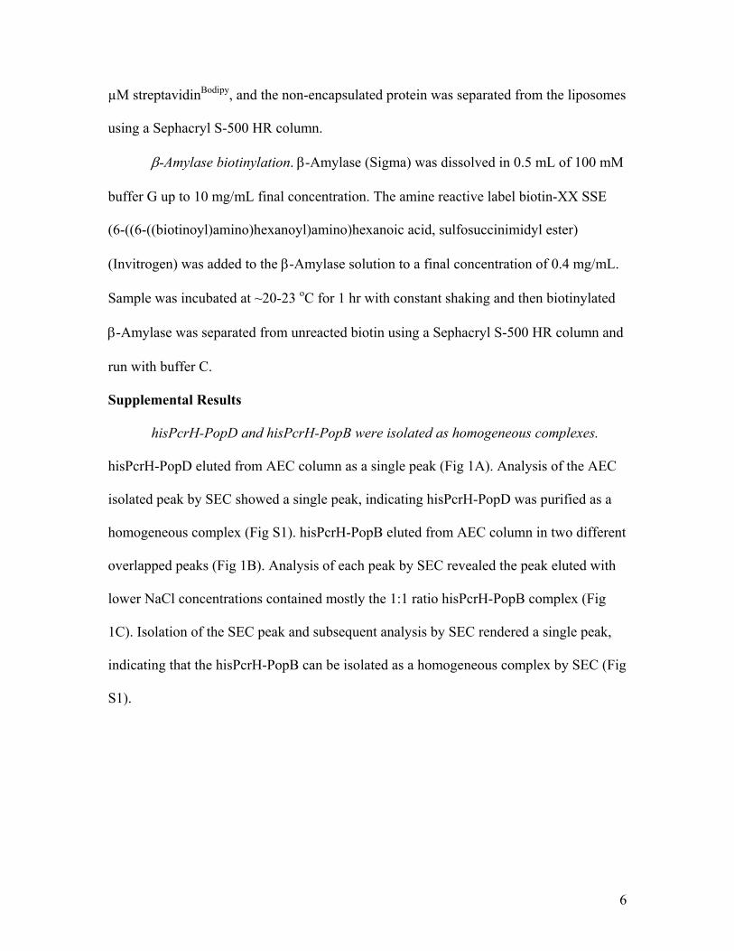
6
µM streptavidinBodipy, and the non-encapsulated protein was separated from the liposomes
using a Sephacryl S-500 HR column.
β-Amylase biotinylation. β-Amylase (Sigma) was dissolved in 0.5 mL of 100 mM
buffer G up to 10 mg/mL final concentration. The amine reactive label biotin-XX SSE
(6-((6-((biotinoyl)amino)hexanoyl)amino)hexanoic acid, sulfosuccinimidyl ester)
(Invitrogen) was added to the β-Amylase solution to a final concentration of 0.4 mg/mL.
Sample was incubated at ~20-23 oC for 1 hr with constant shaking and then biotinylated
β-Amylase was separated from unreacted biotin using a Sephacryl S-500 HR column and
run with buffer C.
Supplemental Results
hisPcrH-PopD and hisPcrH-PopB were isolated as homogeneous complexes.
hisPcrH-PopD eluted from AEC column as a single peak (Fig 1A). Analysis of the AEC
isolated peak by SEC showed a single peak, indicating hisPcrH-PopD was purified as a
homogeneous complex (Fig S1). hisPcrH-PopB eluted from AEC column in two different
overlapped peaks (Fig 1B). Analysis of each peak by SEC revealed the peak eluted with
lower NaCl concentrations contained mostly the 1:1 ratio hisPcrH-PopB complex (Fig
1C). Isolation of the SEC peak and subsequent analysis by SEC rendered a single peak,
indicating that the hisPcrH-PopB can be isolated as a homogeneous complex by SEC (Fig
S1).

7
Figure S1: SEC analysis of purified hisPcrH-PopD (solid line) and hisPcrH-PopB
(dashed line) complexes. Proteins were run on a Superdex-200 GL 10/30 column using
buffer A at a flow rate of 0.5 mL/min. hisPcrH-PopD load was a 400 µl aliquot the AEC
elution peak containing hisPcrH-PopD, while hisPcrH-PopB load was a 400 µL aliquot of
the first AEC elution peak containing hisPcrH-PopB (see Fig. 1).
hisPcrH-PopB aggregates at high concentration. hisPcrH-PopB eluted from AEC
column in two overlapped peaks (typical profiles are shown in Fig S2A and Fig 1B). SEC
analysis of samples isolated from these AEC eluted peaks revealed that the earlier eluting
peak, which was the most abundant peak, contained mostly the 1:1 hisPcrH-PopB
complex while the later eluting peak contained mostly aggregates of hisPcrH-PopB, as
well as free hisPcrH (Fig 1C). A significant increase in the amount of large hisPcrH-
PopB aggregates was observed by SEC analysis, when the two AEC eluted fractions
containing hisPcrH-PopB were pooled and concentrated to 10 mg/mL final total protein,
(Fig S2B). While the formation of large heterogeneous hisPcrH-PopB aggregates is
favored at high protein concentrations, homogeneous hisPcrH-PopB complexes can be
isolated and stored at lower protein concentration (Fig. S1).

8
Figure S2: hisPcrH-PopB aggregates when concentrated. A) Elution of hisPcrH-PopB
from Q-Sepharose AEC by linear NaCl gradient resulted in three main peaks, the first
two contained hisPcrH-PopB and the last one containing hisPcrH. Fractions indicated
(Pool) were combined and concentrated to 10 mg/mL. B) SEC analysis of hisPcrH-PopB
concentrated to 10 mg/mL before SEC run. hisPcrH-PopB eluted mostly as an
asymmetrical peak with elution volume corresponding to high molecular weight
aggregates. SEC conditions were as described in the methods section.
Pore forming activities at pH 5.1 of urea-isolated translocators and acid
dissociated translocators are very similar. hisPcrH-PopB, hisPcrH-PopD or a 1:1 mixture
of both complexes stored in buffer A were added at the indicated concentrations into
quartz cuvettes containing liposomes loaded with Tb(DPA)33- (0.1 mM total lipids),
buffer sodium acetate 30 mM, pH 5.1, and EDTA 5 mM. Pore formation was determined

9
as indicated in experimental procedures. Urea-isolated PopB, PopD or an equimolar and
additive mixture of both translocators (final concentration for each translocator is
indicated) were assayed for pore forming activity (Fig S3B). The experimental conditions
were similar to the conditions employed by Faudry et al (3). The activity profiles
obtained at pH 5.1 did not show apparent saturation at high protein concentration. These
results suggested that only a fraction of the added translocators were forming pores at this
pH when compared to the profiles obtained at pH 4.1 (Fig. 4). Interestingly, no synergism
was observed between PopB and PopD at any of the analyzed concentrations.
Figure S3: The pore forming activities of translocators dissociated from hisPcrH due to
pH drop in the presence of membranes is very similar to the activity of the urea-isolated
translocators diluted in the presence of membranes. A) Concentration dependent pore
formation by hisPcrH-PopD, hisPcrH-PopB or an additive and equimolar mixture of both

10
complexes was determined as described in the text. Lipid composition was 80 mol %
POPC, 20 mol % POPS. B) Concentration dependent pore formation by urea-isolated
PopB, PopD, or an additive equimolar mixture of both proteins measured as indicated in
the text.
hisPcrH can form intermolecular disulfide bonds. hisPcrH stored for more than
24 hr at 4oC in buffer A, was analyzed by SEC as described in the experimental
procedures section. hisPcrH was distributed in two overlapping peaks centered at 14.3
mL and 15.3 mL elution volumes (Fig. S4A). SDS-PAGE analysis of the collected
fractions using reducing and non-reducing conditions indicated the presence of
intermolecular disulfide-bonds (Fig. S4B).
Figure S4: PcrH form intermolecular
disulphide bonds. A) SEC analysis of
hisPcrH stored under non reducing
conditions. B) SDS-PAGE analysis of
SEC eluted fractions (fractions of 1 mL
were collected). Samples were treated 50
mM DTT (reducing) or without DTT
(non-reducing) before loading in the gel.
The molecular weights for the protein
standards are indicated. The location of
the hisPcrH monomer and the dimer are
indicated.
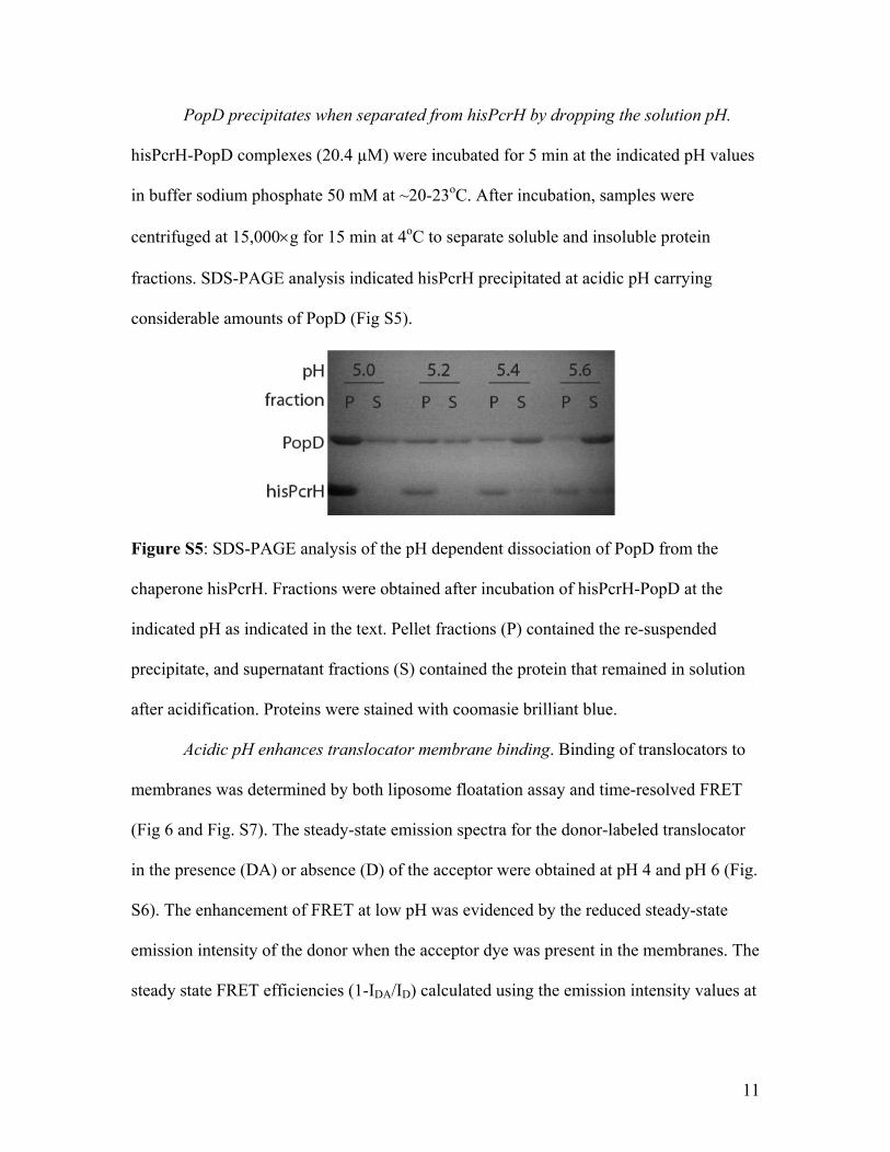
11
PopD precipitates when separated from hisPcrH by dropping the solution pH.
hisPcrH-PopD complexes (20.4 µM) were incubated for 5 min at the indicated pH values
in buffer sodium phosphate 50 mM at ~20-23oC. After incubation, samples were
centrifuged at 15,000×g for 15 min at 4oC to separate soluble and insoluble protein
fractions. SDS-PAGE analysis indicated hisPcrH precipitated at acidic pH carrying
considerable amounts of PopD (Fig S5).
Figure S5: SDS-PAGE analysis of the pH dependent dissociation of PopD from the
chaperone hisPcrH. Fractions were obtained after incubation of hisPcrH-PopD at the
indicated pH as indicated in the text. Pellet fractions (P) contained the re-suspended
precipitate, and supernatant fractions (S) contained the protein that remained in solution
after acidification. Proteins were stained with coomasie brilliant blue.
Acidic pH enhances translocator membrane binding. Binding of translocators to
membranes was determined by both liposome floatation assay and time-resolved FRET
(Fig 6 and Fig. S7). The steady-state emission spectra for the donor-labeled translocator
in the presence (DA) or absence (D) of the acceptor were obtained at pH 4 and pH 6 (Fig.
S6). The enhancement of FRET at low pH was evidenced by the reduced steady-state
emission intensity of the donor when the acceptor dye was present in the membranes. The
steady state FRET efficiencies (1-IDA/ID) calculated using the emission intensity values at

12
513 nm were 0.76 and 0.17 for PopD, and 0.70and 0.43 for PopB at pH 4 and pH 6,
respectively.
Figure S6: (A) PopBS164C-Bodipy (120 nM) was incubated at pH 4 with liposomes (300 µM
total lipids) containing 0.5 mol % of the acceptor dye Rh-PE (DA, dashed lines) or
without the acceptor (D, solid lines) at ~20-23oC for 30 min. Protein:lipid ratio was
1/2500. (B) Same as in A) but at pH 6. (C) Same as in A) but using PopDF223C-Bodipy. (D)
Same as in B) but using PopDF223C-Bodipy. Emission spectra were acquired at 25oC using a
Fluorolog 3-21 spectrofluorimeter. Excitation wavelength was 485 nm, and the excitation
and emission bandpass was 2 nm and 4 nm, respectively. The POPC:cholesterol:POPS
molar ratio was 65:20:15.
High translocator-lipid ratios produced liposome aggregation. The liposome size
distribution before and after incubation with the translocators was analyzed for different

13
protein:lipid ratios using DLS. No significant aggregation was observed at protein:lipid
ratios of up to 1:3300 (Fig S7 top and center). In contrast, when a protein:lipid ratio of
1:330 was used large aggregates with average diameter of about 1.3 µm were observed
(Fig S7 bottom). These data correlated well with the cryo-EM observations (Fig. 4C and
4D).
Figure S7: Particle size distribution analysis of liposomes before and after incubation
with translocators. Distribution averages are indicated. Error bars correspond to ±
standard deviations of 3 measurements. Urea-isolated PopB/PopD at 1:1 ratio was
incubated with liposomes (200 µM total lipids) at the indicated protein:lipid ratios in
buffer sodium acetate 50 mM pH 4 for 30 min at 21-23oC. The POPC:cholesterol:POPS
molar ratio was 65:20:15. DLS measurements were carried out at 25oC using a Zen 3600
instrument (Malvern Instruments, UK), equipped with 633 nm laser diode and avalanche
14
photodiode detector at 90o angle. Autocorrelation data was fit using Dispersion
Technologies Software® version 5.1 and using the cumulant method.
Time-resolved life time measurements for protein-membrane FRET. Bodipy-
labeled translocators were incubated with liposomes with or without Rh-PE at pH 4 or pH
6 as detailed in experimental procedures. Phase and modulation values for the samples
were determined using a Chronos multifrequency cross-correlation phase and modulation
fluorometer at 25 different frequencies as detailed in experimental procedures.
Figure S8: (A) Phase (red) and modulation (blue) values as a function of frequency for
PopDF223C-Bodipy incubated with membranes at pH 4 containing (filled symbols) or not
(open symbols) the Rh-PE acceptor. Solid lines correspond to a two-exponential
component best fit of the data with lifetimes and fractional amplitudes reported in table
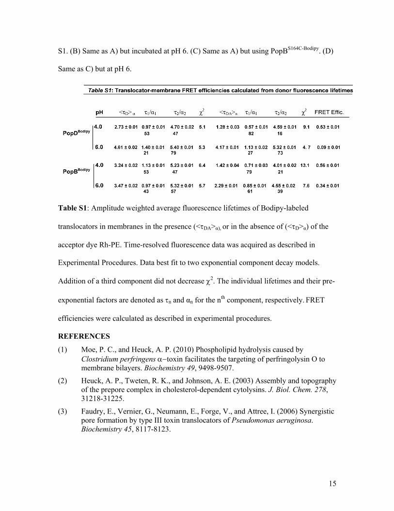
15
S1. (B) Same as A) but incubated at pH 6. (C) Same as A) but using PopBS164C-Bodipy. (D)
Same as C) but at pH 6.
Table S1: Amplitude weighted average fluorescence lifetimes of Bodipy-labeled
translocators in membranes in the presence (<τDA>α), or in the absence of (<τD>α) of the
acceptor dye Rh-PE. Time-resolved fluorescence data was acquired as described in
Experimental Procedures. Data best fit to two exponential component decay models.
Addition of a third component did not decrease χ2. The individual lifetimes and their pre-
exponential factors are denoted as τn and αn for the nth component, respectively. FRET
efficiencies were calculated as described in experimental procedures.
REFERENCES
(1) Moe, P. C., and Heuck, A. P. (2010) Phospholipid hydrolysis caused by Clostridium perfringens α−toxin facilitates the targeting of perfringolysin O to membrane bilayers. Biochemistry 49, 9498-9507.
(2) Heuck, A. P., Tweten, R. K., and Johnson, A. E. (2003) Assembly and topography of the prepore complex in cholesterol-dependent cytolysins. J. Biol. Chem. 278, 31218-31225.
(3) Faudry, E., Vernier, G., Neumann, E., Forge, V., and Attree, I. (2006) Synergistic pore formation by type III toxin translocators of Pseudomonas aeruginosa. Biochemistry 45, 8117-8123.