Embed Size (px)
Citation preview
Submitted 2 May 2016Accepted 13 August 2016Published 7 September 2016
Corresponding authorGiuseppe Di Gioia, [email protected]
Academic editorAlessandra Lo Presti
Additional Information andDeclarations can be found onpage 11
DOI 10.7717/peerj.2439
Copyright2016 Di Gioia et al.
Distributed underCreative Commons CC-BY 4.0
OPEN ACCESS
ECG is an inefficient screening-toolfor left ventricular hypertrophy innormotensive African children populationGiuseppe Di Gioia1, Antonio Creta1, Cosimo Marco Campanale1,Mario Fittipaldi2, Riccardo Giorgino1, Fabio Quintarelli3, Umberto Satriano1,Alessandro Cruciani1, Vincenzo Antinolfi4, Stefano Di Berardino5,Davide Costanzo1, Ranieri Bettini6, Giuseppe Mangiameli5, Marco Caricato5 andGiovanni Mottini7
1Department of Medicine and Surgery, Unit of Cardiology, Campus Bio-Medico University of Rome,Rome, Italy
2Paediatric Cardiothoracic Surgery, Starship Greenlane Paediatric and Congenital Heart Service, Auckland,New Zealand
3Department of Medicine and Surgery, Service of Pediatrics, Campus Bio-Medico University of Rome,Rome, Italy
4Heart and Great Vessels ‘‘Attilio Reale’’, University of Roma ‘‘La Sapienza’’, Rome, Italy5Department of Medicine and Surgery, Geriatric Surgery Unit, Campus Bio-Medico University of Rome,Rome, Italy
6Cardiology Department, University of Pisa, Pisa, Italy7 Institute of Philosophy of Scientific and Technological Practise (FAST), Campus Bio-Medico University ofRome, Rome, Italy
ABSTRACTBackground. Left ventricular hypertrophy (LVH) is amarker of pediatric hypertensionand predicts development of cardiovascular events. Electrocardiography (ECG) screen-ing is used in pediatrics to detect LVH thanks to major accessibility, reproducibilityand easy to use compared to transthoracic echocardiography (TTE), that remains thestandard technique. Several diseases were previously investigated, but no data existsregarding our study population. The aim of our study was to evaluate the relationshipbetween electrocardiographic and echocardiographic criteria of LVH in normotensiveAfrican children.Methods. We studied 313 children (mean age 7,8 ± 3 yo), in north-Madagascar. Theyunderwent ECG and TTE. Sokolow-Lyon index was calculated to identify ECG-LVH(>35mm). Left ventricle mass (LVM)with TTEwas calculated and indexed by height2.7
(LVMI2.7) and weight (LVMIw). We report the prevalence of TTE-LVH using threemethods: (1) calculating percentiles age- and sex- specific with values >95th percentileidentifying LVH; (2) LVMI2.7 >51 g/m2.7; (3) LVMIw >3.4 g/weight.Results. 40 (13%) children showed LVMI values >95th percentile, 24 children (8%)an LVMI2.7 >51 g/m2.7 while 19 children (6%) an LVMIw >3.4 g/kg. LVH-ECG bySokolow-Lyon index was present in five, three and three children respectively, withpoor values of sensitivity (ranging from 13 to 16%), positive predictive value (from11 to 18%) and high values of specificity (up to 92%). The effects of anthropometricsparameters on Sokolow-Lyon were analyzed and showed poor correlation.Conclusion. ECG is a poor screening test for detecting LVH in children. In clinicalpractice, TTE remains the only tool to be used to exclude LVH.
How to cite this article Di Gioia et al. (2016), ECG is an inefficient screening-tool for left ventricular hypertrophy in normotensiveAfrican children population. PeerJ 4:e2439; DOI 10.7717/peerj.2439

Subjects Cardiology, Epidemiology, PediatricsKeywords ECG, Ventricular hypertrophy, Screening, African children
INTRODUCTIONLeft ventricular hypertrophy (LVH) in adults has receivedmuch attention since its detectionis correlated to long-term clinical outcome, predicting cardiovascular events as myocardialinfarction, stroke and death (Devereux et al., 2001; Koren et al., 1991; Brown, Giles & Croft,2000; Levy et al., 1990). LVH results from adaptation of the heart to increased hemodynamicburden, therefore early diagnosis is important, especially in children. In the pediatricpopulation, LVH can be used as a marker to identify hypertensive children and predictdevelopment of future cardiovascular events (Hanevold et al., 2004). Electrocardiographic(ECG) screening is widely used in pediatrics to detect and diagnose LVH and is considereda possible screening tool for hypertrophic cardiomyopathy (Gersh et al., 2011), whichis responsible for almost half of sudden cardiac death cases in developed countries(Maron et al., 1995). Transthoracic echocardiography (TTE) is generally considered asthe standard technique to diagnose LVH, but ECG seems to be more attractive as ascreening tool, especially in developing countries with less resources available, thanks toits lower costs, major accessibility and good reproducibility. ECG is also easily readableby non-specialist users compared to TTE. The validity of ECG criteria for diagnosingLVH has been previously studied in several diseases, such as pediatric hypertension(Ramaswamy et al., 2009), rheumatic heart disease (Sastroasmoro, Madiyono & Oesman,1991), hypertrophic cardiomyopathy (Panza & Maron, 1989), HIV infection (Rivenes etal., 2003), aortic stenosis and ventricular septal defects (Fogel, Lieb & Seliem, 1995). Thiscorrelation remains to be determined in the population of this study, which is composedby African normotensive children population.
The aim of our study was to evaluate the role of ECG as screening tool of LVHthrough the relationship between electrocardiographic and echocardiographic criteria in anormotensive African children population.
MATERIAL AND METHODSWe performed a clinical, electrocardiographic and echocardiographic evaluation of 313consecutive African children (ranging from four to 16 years old) describing the correlationbetween electrocardiographic and echocardiographic criteria for LVH. This study wasconducted during a medical workcamp, coordinated by the Cardiovascular Department ofCampus Bio-Medico of Rome, with the participation of cardiologists, pediatrics, pediatriccardiothoracic surgeons, surgeons and medical students. We had our main base at the‘‘Clinique Médico-Surgicale St. Damien’’ of Ambanja, and we have investigated the regionof Antsiranana, in the north of Madagascar, visiting the catholic schools of Sekoly VenanceManifatra ‘‘SE.VE.MA’’ and Foyer Mangafaly in Ambanja and the college Sainte Theresede l’Enfant Jesus in Maromandia, during the month of October 2015. In primary and highschools, we randomly selected a class from each group of age to enroll individuals fromfour to 16 years. At the ‘‘Clinique St. Damien’’ we enrolled children who came for routine
Di Gioia et al. (2016), PeerJ, DOI 10.7717/peerj.2439 2/15

Figure 1 Flow-chart of children’s evaluation.
visits or screening, or in-patients. All individuals underwent four steps (see Fig. 1). Thefirst step consisted in registration into our database with name, surname (when available)and date-of-birth. The second step consisted in annotation of weight, height, body massindex (BMI) calculation, blood pressure, heart rate and cardiac auscultation. Auscultationwas made by a pediatrician using a standard approach. The clinical questionnaire wasperfomed by a visiting medical student aided and—when necessary—by a local interpreterwho has been educated in advance. Teachers or their coworkers helped the youngerchildren understanding and answering the questions. The third step consisted in a12-leads ECG execution and the fourth step in consisted of a complete transthoracicechocardiogram for all the study-included subjects. Medical students—supervised bycardiologists—performed ECG according to international guidelines, by using a portableelectro-cardiographer General Electric,MarquetteMAC5000 (GE,Milwaukee,WI, USA) ata sampling rate of 150 Hz, at standard paper speed (25 mm/sec) and voltages (10 mm/mV).ECG voltages, representing left ventricular forces, were calculated by hand for each ECG.Non-voltage based anomalies, including T waves abnormalities were noted. Sokolow-Lyonindex (SV1+RV5/RV6, mm) was calculated to identify children with electrocardiographicdiagnosis of LVH (>35 mm). To calculate the left ventricular voltages, each patient’s BMIwas indexed to the age-based population-derived 50% normative data for age, developingthe formula: Indexed ECG voltage = [ECG voltage × (BMIpatient/BMI50%)] (Czosek et al.,2014). The QTc interval was calculated with Bazett formula as follows: QT/
√RR. The
Di Gioia et al. (2016), PeerJ, DOI 10.7717/peerj.2439 3/15

upper normal limit was defined as 450 ms for boys and 460 ms for girls (Pearl, 1996). Ateam of cardiologists, with a portable echocardiograph (Esaote Mylab Five, Genoa, Italy)equipped with a S5-1 transducer probe, performed TTE. All the exams were stored onappropriate supports. Subjects were studied in the left lateral recumbent position and allstandard echocardiographic views were acquired. The LV inner dimensions were measuredat end-diastole and end-systole using M-Mode echocardiography in the parasternal longaxis view. End-diastole was defined as the frame following mitral valve closure; whileend-systole as the frame following mitral valve opening. Left ventricular mass (LVM) wascalculated based on Devereux’s formula (Devereux et al., 1986) and indexed (National HighBlood Pressure Education ProgramWorking Group on High Blood Pressure in Children andAdolescents, 2004) by height2,7 (LVMI2.7) or weight (LVMIw). In particular, LVM resultsfrom the formula: LVM = 1/4 0.8 × (1.04 [LVIDd + PWTd + SWTd)3−(LVIDd)3]) +0.6 g, where LVIDd is the left ventricular internal dimension at the end diastole, PWTdis the posterior wall thickness at the end diastole, and SWTd is the septal wall thicknessat the end diastole (Lang et al., 2015). We report the prevalence of TTE-LVH by usingthree different methods (De Simone et al., 1992; Daniels et al., 1995; De Simone et al., 1995;Rijnbeek et al., 2008; Overbeek et al., 2006): (1) calculating the population percentiles age-and sex-specific with values above the 95th percentile identifying children with LVH; (2)LVMI value > 51 g/height2.7 (Hanevold et al., 2004); (3) LVMI > 3.4 g/weight. Because ofwidespread illiteracy in the population, written informed consent was difficult to obtain.Indeed, a verbal informed consent—with a teacher’s help to translate the language—wasobtained from the children’s parents, who gave study approval to the school’s teachersand our research group. The study was reviewed and approved before it began by theethics committees of the University Campus Bio-Medico of Rome (approval number 21.15TS) and the project started in collaboration with the doctors of the hospital ‘‘CliniqueMédico-Surgicale St. Damien’’ of Ambanja that approved the study and approved thesubmission to the ethics committees of the University Campus Bio-Medico of Rome (since‘‘Clinique Médico-Surgicale St. Damien’’ of Ambanja’’ lacked an ethics committee).
Statistical analysisCategorical variables are expressed as frequencies and percentages in parentheses, and arecompared by using Fisher’s exact test or Chi-square test, as appropriate. Normality criteriawere checked andmet for any continuous variable, which is presented asmean and standarddeviation and compared using Student t -test for independent data. Correlations betweencontinuous variables were calculated using Pearson’s test. Considering the correlationbetween Sokolow-Lyon Indexed and LVM as the primary end-point, we expected acorrelation coefficient of 0.20 with an alpha error 0.05 and 90% power; this led us tocalculate a sample size of 258 patients. The recruitment target was increased of 20% due tounexpected variability and final sample size was of 313 children. The sensitivity, specificity,positive predictive value (PPV) and negative predicting value (NPV) were calculated using2×2 contingency tables. A P value less than 0.05 was considered statistically significant.Statistical analysis was performed with STATA Statistics for Windows (SE, version 13).
Di Gioia et al. (2016), PeerJ, DOI 10.7717/peerj.2439 4/15

RESULTSA total of 313 children of African race were studied, with a slight prevalence of femalesex (53%). Mean age was 7,8 ± 3 years, ranging from four to 16 years. Clinical,electrocardiographic and echocardiographic features of population study are listed inTable 1. All children had normal arterial blood pressure. In 36 children (12%), a cardiacmurmur was detected at physical examination.
At ECG evaluation, in 19% of all children sinus rhythm was found with physiologicalsinus arrhythmia and sinus tachycardia (mean value > 100 beats/minute) typical of theinvestigated range of age. Seven (2%) children with short PR segment (<120 ms) wereidentified. None of them had evident signs of pre-excitation or history of cardiac arrest.Abnormalities of T wave (prevalently flat T wave) were identified in 67 children (21,4%).There was no correlation between clinical evaluation and evidence of ECG abnormalities:only 14 children had both a cardiacmurmur and an ECG abnormality with a 21% sensitivityof clinical evaluation to predict ECG abnormalities, a 39% PPV and a 91% specificity. TheQT interval was in the normal range in all children under study. 28 children (9%) presenteda Sokolow-Lyon index >35 mm, having an ECG diagnosis of LVH. I degree AV block waspresent in only 2 children, so extra beats. Positional Q waves were identified in 12 childrenwhile mild IV conduction delay (between 100 and 110 mesc) were present in 33 children.No bundle brunch blocks were identified.
At TTE evaluation, a mild mitral regurgitation (MR) was identified in 23 (7%) children,while three children showed moderate MR. No severe MR were identified. Five cases ofmild aortic regurgitation (AR) were diagnosed with only one young boy having moderateAR. Only two children showed mitral valve prolapse of anterior mitral leaflet with mildregurgitation. Five congenital heart defects (1.6% of children) were diagnosed: two inter-atrial defects, one child with an inter-ventricular defect, one child with bicuspid aortic valveand one young girl with cor triatriatum sinister. Only four children with cardiac murmurpresented also an echocardiographic evidence of valve regurgitation.
Following the work of Khoury et al. (2009), which gave age-specific reference values forchildren’s LVMI, the 313 children in our study were divided according to age, sex and LVMas follows: 86 children (27%) were in the ≤10th percentile, 32 (10%) were in the 25th, 57(18%) in the 50th; 55 (18%) in the 75th, 30 (10%) in the 90th, 13 children (4%) in the95th and 40 (13%) with values above 95th percentile.
The mean values of LVMI g/m2.7 were 31,9± 11,8 g/m2.7 in the overall population, withonly a slight increase in children with an ECG-LVH diagnosis (32,6 ± 45,9 g/m2.7), whilethe mean values for LVMI in g/kg were 2,3± 3 g/kg for the overall population and 2,4± 3,4g/kg for the children with an ECG-LVH diagnosis. 24 children (8%) had an LVMI2.7 > 51g/m2.7 while 19 children (6%) showed an LVMIw (>3.4 g/kg). Sensitivity, specificity, PPVand NPV of three investigated methods to diagnose LVH are listed in Table 2.
The capability of Sokolow-Lyon index to identify children with TTE-LVH appears to bevery poor. Only five children with ECG-LVH showed also an echocardiographic diagnosisof LVH (>95◦ percentile); only three children were identified with other two methods. Thedistribution of children having an ECG-LVH according to LMVI percentiles is showed
Di Gioia et al. (2016), PeerJ, DOI 10.7717/peerj.2439 5/15

Table 1 Characteristics of study population.
No. of children 313Male, n (%) 146 (47)Age, years 7,8± 3Height, cm 120± 20Weight, kg 23,2± 8,2BMI, kg/m2 15,5± 1,9BSA 0,6± 0,3SP, mmHg 113± 8DP, mmHg 70± 7Heart murmur, n (%) 36 (12)
ECGSinus arrhythmia, n (%) 59 (19)HR, beats 103± 18PR segment, ms 140± 25Short PR segment, n (%) 7 (2)QRS complex, ms 85± 13Axis, degrees 56± 31Axial deviation, n (%) 35 (11)T wave abnormalities, n (%) 67 (21)LVH, n (%) 28 (9)Sokolow-Lyon, mm 23,9± 9,2Sokolow-Lyon Indexed, mm 24,1± 9,8I degree AV block 2 (1)IV conduction delay, n (%) 33 (11)Early repolarization, n (%) 8 (3)Ventricular extrasystoles, n (%) 1 (0,3)Supra ventricular extrasystoles, n (%) 1 (0,3)QT interval, msec 364± 41QTc, msec 411± 48
EchocardiographyLVEDD, mm 35± 4IVS, mm 6,3± 1,3PW, mm 5,3± 0,9LVM, g 50,5± 16
Notes.The numbers are expressed as numerical values (%) or mean± standard deviation.Abbreviations: AV, atrio-ventricular; BMI, body mass index; BSA, body surface area; DP, diastolic pressure; HR, heartrate; IV, intra-ventricular; IVS, inter-ventricular septum; LVEDD, left ventricular end-diastolic diameter; LVH, left ven-tricular hypertrophy; LVM, left ventricular mass; PW, posterior wall; SP, systolic pressure.
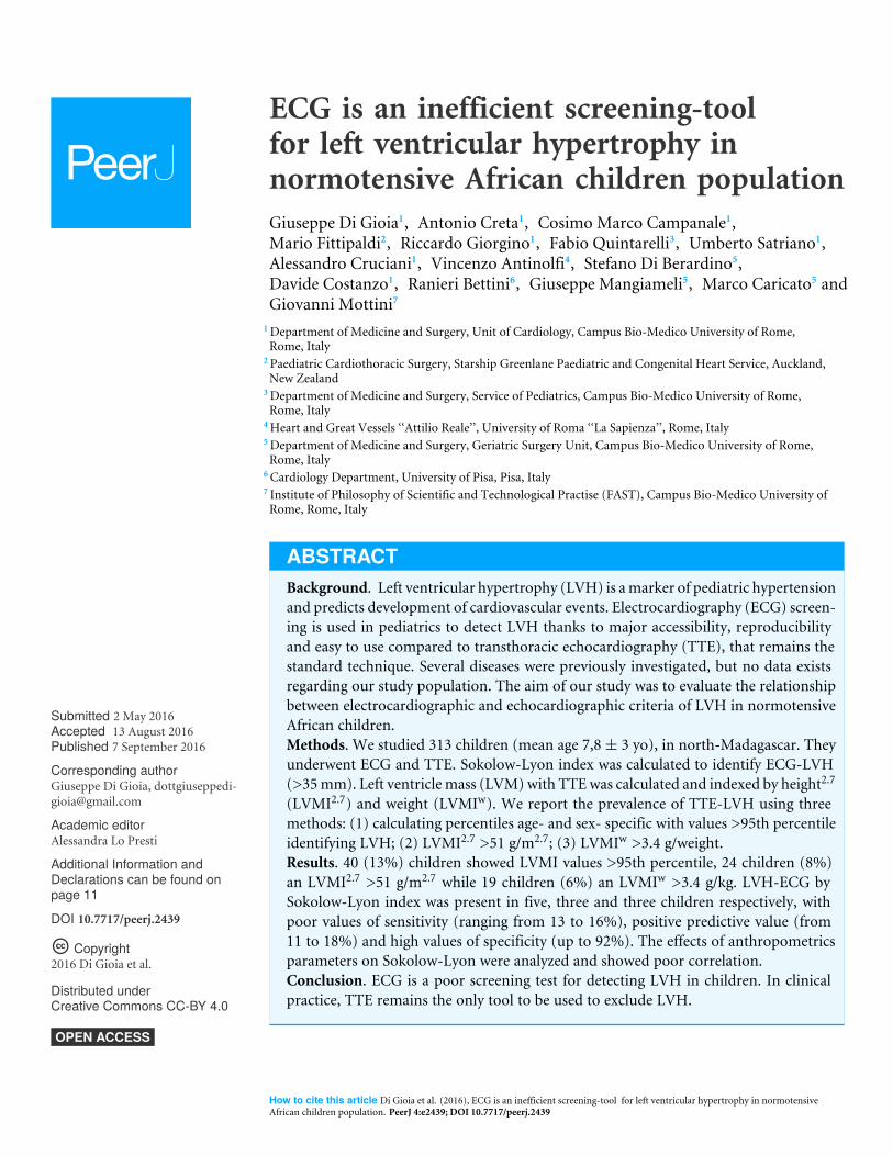
in Table 3. Values of Sokolow-Lyon Index did not change according to LVMI percentiles(Fig. 2).
Effects of anthropometrics parameters, including body surface area (BSA), BMI, height,weight, and LVM (also indexed) on Sokolow-Lyon formula were analyzed (Fig. 3) andshowed poor correlation. In the same way, when ECG voltages were indexed to patient’s
Di Gioia et al. (2016), PeerJ, DOI 10.7717/peerj.2439 6/15
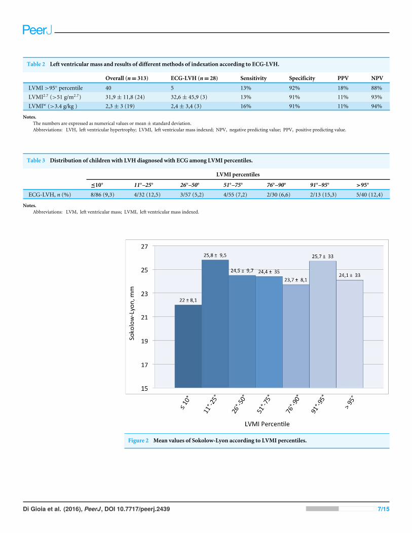
Table 2 Left ventricular mass and results of different methods of indexation according to ECG-LVH.
Overall (n= 313) ECG-LVH (n= 28) Sensitivity Specificity PPV NPV
LVMI >95◦ percentile 40 5 13% 92% 18% 88%LVMI2.7 (>51 g/m2.7) 31,9± 11,8 (24) 32,6± 45,9 (3) 13% 91% 11% 93%LVMIw (>3.4 g/kg ) 2,3± 3 (19) 2,4± 3,4 (3) 16% 91% 11% 94%
Notes.The numbers are expressed as numerical values or mean± standard deviation.Abbreviations: LVH, left ventricular hypertrophy; LVMI, left ventricular mass indexed; NPV, negative predicting value; PPV, positive predicting value.
Table 3 Distribution of children with LVH diagnosed with ECG among LVMI percentiles.
LVMI percentiles
≤10◦ 11◦–25◦ 26◦–50◦ 51◦–75◦ 76◦–90◦ 91◦–95◦ >95◦
ECG-LVH, n (%) 8/86 (9,3) 4/32 (12,5) 3/57 (5,2) 4/55 (7,2) 2/30 (6,6) 2/13 (15,3) 5/40 (12,4)
Notes.Abbreviations: LVM, left ventricular mass; LVMI, left ventricular mass indexed.
Figure 2 Mean values of Sokolow-Lyon according to LVMI percentiles.
Di Gioia et al. (2016), PeerJ, DOI 10.7717/peerj.2439 7/15

Figure 3 Correlation between ECG Sokolow-Lyon formula with echocardiographic LVmass indices and anthropometric parameters.
BMI (Sokolow-Lyon Indexed), direct correlation with LVM and LVMI values (Fig. 4) werenot statistically significant.
Physical parameters, such as weight, height, BMI and BSA had no independent effect inthe value of ECG parameters and the accuracy of the detection of LVH.
Authors acknowledge the fact that the reference value of Sokolow-Lyon > 35 mm todefine LVH is used in adults. We were not able to find a better cut-off in our youngpopulation. Considering LVMI2.7 > 51 g/m2.7 to define echocardiographic LVH, the areaunder the ROC curve (Fig. 5) for the electrocardiographic Sokolow-Lyon Indexed was 0.496(95% CI [0.374–0.618], P value = 0.949). Therefore, a precise cutoff in our populationcould not be calculated accurately.
DISCUSSIONThis study has examined the correlation and the accuracy of ECG criteria to detectTTE-LVH in a not hypertensive Malagasy children population.
Cardiac magnetic resonance imaging and 3-dimensional echocardiography have provedto be very accurate tools to diagnose and quantify LVH, but they are very expensive, theyrequire operator expertise and their use is unachievable in less developed countries where,most of the time, it is difficult to submit population to an echocardiogram screening.
Di Gioia et al. (2016), PeerJ, DOI 10.7717/peerj.2439 8/15
Figure 4 Correlation between ECG Sokolow-Lyon indexed to BMI and echocardiographic LVmass in-dices.
Figure 5 ROC curve between LVMI g/m2.7 and LVH evaluated through Sokolow-Lyon indexed.
Di Gioia et al. (2016), PeerJ, DOI 10.7717/peerj.2439 9/15

The ECG tool has been used since a long time to investigate heart anomalies. To date,there are no clearly established benefits of a widespread, universal ECG screening in theyoung population (Sharma et al., 2013; Fuller, 2000). It is inexpensive compared to TTE orother imaging modalities, requires less specialist skills and takes considerably less time.
In contrast, measuring LVH by TTE M-mode technique is relatively easy and quiteavailable, and actually the first method to be validated and currently the standard clinicaldiagnostic method for detecting LVH (Alfakih et al., 2006).
Our study used different methods for the calculation of LVM.In a population of not hypertensive children, ECG Sokolow-Lyon criteria analysis for
the detection of TTE-LVH suggests that ECG is a poor screening method for LVH.More than 30 different ECG criteria exists for the detection of LVH on standard 12
leads ECG. In a review (Pewsner et al., 2007) of 21 studies including more than 5,000hypertensive patients, using six different ECG criteria, the authors concluded that ECGcriteria cannot be used to exclude LVH in adult hypertensive patients. However, many ofthe criteria used in adult subjects cannot be applied to a children population of differentage, gender and body surface area.
Previously several studies have examined a variety of ECG parameters in children, butour type of population was not investigated before.
The discordance between ECG criteria and TTE-LVH has been described in severalpediatric disease states, such as rheumatic heart disease (Sastroasmoro, Madiyono &Oesman, 1991), myocarditis (Oda, Hamamoto & Morinaga, 1982) and hypertrophiccardiomyopathy (Panza & Maron, 1989; Louie & Maron, 1986;Maron et al., 1983).
In children with rheumatic heart disease (Sastroasmoro, Madiyono & Oesman, 1991),sensitivity and specificity of ECG were 68% and 76%, respectively. Sensitivity inhypertrophic cardiomyopathy reached 76% (Louie & Maron, 1986).
Rivenes et al. (2003) investigated children with human immunodeficiency virus (HIV)infection; prevalence of ECG-LVH was 7.4% with sensitivity less than 20% and 90%specificity but used ECG-LVH criteria by Davignon (Davignon, Rautaharju & Boisselle1979), which are not gender specific.
Rijnbeek et al. (2008) reported a study of 832 unselected pediatric hospital populationusing wide types of parameters for ECG-LVH detection, reporting a less than 25%sensitivity.
In diseases with pressure or volume ventricular overload as aortic valve stenosisor ventricular septal defect, Fogel, Lieb & Seliem (1995) found that, regardless of age,Sokolow-Lyon criteria were statistically higher compared to normal children, with thehighest sensitivity in aortic stenosis patients (67%).
The study by Morganroth et al. (1975) found excessive values of sensitivity of ECGcriteria with false-positive diagnosis in an adolescent cohort with no TTE-LVH, but LVMwas not indexed to BSA.
An interesting data that emerged from our study was the high prevalence of childrenshowing LVH, revealed both from the value of LVMI2.7, adjusted by age and sex (24children, 8%), from LVMI > 95th percentile (40 children, 13%) and LVMIw (19 children,6%). In fact, the prevalence of LVH in pediatric hypertensive population has been reported
Di Gioia et al. (2016), PeerJ, DOI 10.7717/peerj.2439 10/15

to vary from 8%–41% depending on the criteria used for determining hypertension andLVMI (Brady et al., 2008; Daniels et al., 1998; Daniels, Meyer & Loggie, 1990; Sorof et al.,2002; Laird & Fixler, 1981; Niederle et al., 1982).
ECG is an easily obtainable, low cost, rapid test but with several limitations that do notallow to substitute a TTE evaluation, even in developing countries, where less resourcesare available.
Nowadays, ECG screening is used inmany preparticipation sports screening programs todetect cardiac abnormalities. ECG is a poor screeningmethod for LVH, with very low valuesof sensitivity in general population, as demonstrated by our study in normotensive children.Other studied showed similar values for hypertensive children and, at best, modest values ofspecificity and sensitivity where reached, in diseases affecting directly left ventricular massor pressure-loading conditions like aortic stenosis or hypertrophic cardiomyopathy,but non-obtaining optimal value to consider ECG as a valid screening method.Echocardiography remains the best clinical tool for LVH screening in pediatric population.
CONCLUSIONIn a normotensive African population, ECG is a poor screening test for the detection ofLVH in children. In clinical practice, TTE remains the only tool to be used to excludeLVH.
ADDITIONAL INFORMATION AND DECLARATIONS
FundingThe authors received no funding for this work.
Competing InterestsThe authors declare there are no competing interests.
Author Contributions• Giuseppe Di Gioia conceived and designed the experiments, analyzed the data, wrotethe paper, prepared figures and/or tables, reviewed drafts of the paper.• Antonio Creta conceived and designed the experiments, wrote the paper, reviewed draftsof the paper.• Cosimo Marco Campanale analyzed the data, reviewed drafts of the paper.• Mario Fittipaldi, Fabio Quintarelli and Ranieri Bettini contributed reagents/materials/-analysis tools.• Riccardo Giorgino, Umberto Satriano, Alessandro Cruciani, Vincenzo Antinolfi, StefanoDi Berardino and Davide Costanzo performed the experiments.• Giuseppe Mangiameli analyzed the data.• Marco Caricato analyzed the data, wrote the paper, prepared figures and/or tables,reviewed drafts of the paper.• Giovanni Mottini conceived and designed the experiments, wrote the paper.
Di Gioia et al. (2016), PeerJ, DOI 10.7717/peerj.2439 11/15

Human EthicsThe following information was supplied relating to ethical approvals (i.e., approving bodyand any reference numbers):
Campus Bio Medico University of Rome Ethics Committee.Approval number: 21.15 TSDate of approval: 27/09/2015.
Data AvailabilityThe following information was supplied regarding data availability:
The raw data has been supplied as a Supplemental Information.
Supplemental InformationSupplemental information for this article can be found online at http://dx.doi.org/10.7717/peerj.2439#supplemental-information.
REFERENCESAlfakih K, Reid S, Hall A, SivananthanMU. 2006. The assessment of left ven-
tricular hypertrophy in hypertension. Journal of Hypertension 24:1223–1230DOI 10.1097/01.hjh.0000234097.47379.fd.
Brady TM, Fivush B, Flynn JT, Parekh R. 2008. Ability of blood pressure to predict leftventricular hypertrophy in children with primary hypertension. Journal of Pediatrics152:73–78 DOI 10.1016/j.jpeds.2007.05.053.
Brown DW, GilesWH, Croft JB. 2000. Left ventricular hypertrophy as a predictor ofcoronary heart disease mortality and the effect of hypertension. American HeartJournal 140:848–856 DOI 10.1067/mhj.2000.111112.
Czosek RJ, Cnota JF, Knilans TK, Pratt J, Guerrier K, Anderson JB. 2014. Relationshipbetween echocardiographic LV mass and ECG based left ventricular voltages in anadolescent population: related or random? Pacing and Clinical Electrophysiology37(9):1133–1140 DOI 10.1111/pace.12416.
Daniels SR, Kimball TR, Morrison JA, Khoury P, Meyer RA. 1995. Indexing leftventricular mass to account for differences in body size in children and adolescentswithout cardiovascular disease. American Journal of Cardiology 76:699–701DOI 10.1016/S0002-9149(99)80200-8.
Daniels SR, Loggie JMH, Khourt P, Kimball TR. 1998. Left ventricular geometryand severe left ventricular hypertrophy in children and adolescents with essentialhypertension. Circulation 97:1907–1911 DOI 10.1161/01.CIR.97.19.1907.
Daniels SD, Meyer RA, Loggie JMH. 1990. Determinants of cardiac involvement inchildren and adolescents with essential hypertension. Circulation 82:1243–1248DOI 10.1161/01.CIR.82.4.1243.
Davignon A, Rautaharju P, Boisselle E. 1979. Normal ECG standards for infants andchildren. Pediatric Cardiology 1:123–131 DOI 10.1007/BF02083144.
De Simone G, Daniels SR, Devereux RB, Meyer RA, RomanMJ, De Divitiis O, Alder-manMH. 1992. Left ventricular mass and body size in normotensive children and
Di Gioia et al. (2016), PeerJ, DOI 10.7717/peerj.2439 12/15

adults: assessment of allometric relations and impact of overweight. Journal of theAmerican College of Cardiology 20:1251–1260 DOI 10.1016/0735-1097(92)90385-Z.
De Simone G, Devereux RB, Daniels SR, KorenMJ, Meyer RA, Laragh JH. 1995. Effectof growth on variability of left ventricular mass: assessment of allometric signals inadults and children and their capacity to predict cardiovascular risk. Journal of theAmerican College of Cardiology 25:1056–1062 DOI 10.1016/0735-1097(94)00540-7.
Devereux RB, Alonso DR, Lutas EM, Gottlieb GJ, Campo E, Sachs I, Reichek N.1986. Echocardiographic assessment of left ventricular hypertrophy: com-parison to necropsy findings. American Journal of Cardiology 57:450–458DOI 10.1016/0002-9149(86)90771-X.
Devereux RB, Bella J, Boman K, Gerdts E, NieminenMS, Rokkedal J, PapademetriouV,Wachtell K, Wright J, Paranicas M, Okin PM, RomanMJ, Smith G, Dahlöf B.2001. Echocardiographic left ventricular geometry in hypertensive patients withelectrocardiographic left ventricular hypertrophy: the LIFE Study. Blood Pressure10:74–82 DOI 10.1080/08037050152112050.
Fogel MA, Lieb DR, SeliemMA. 1995. Validity of electrocardiographic criteria for leftventricular hypertrophy in children with pressure- or volume-loaded ventricles:comparison with echocardiographic left ventricular muscle mass. Pediatric Cardi-ology 16:261–269 DOI 10.1007/BF00798059.
Fuller CM. 2000. Cost effectiveness for screening high school athletes for risk of suddencardiac death.Medicine and Science in Sports and Exercise 32:887–890.
Gersh BJ, Maron BJ, Bonow RO, Dearani JA, Fifer MA, LinkMS, Naidu SS, NishimuraRA, Ommen SR, Rakowski H, Seidman CE, Towbin JA, Udelson JE, Yancy CW,American College of Cardiology Foundation/American Heart Association TaskForce on Practice Guidelines, American Association for Thoracic Surgery, Ameri-can Society of Echocardiography, American Society of Nuclear Cardiology, HeartFailure Society of America, Heart Rhythm Society, Society for CardiovascularAngiography and Interventions, Society of Thoracic Surgeons. 2011. 2011 ACCF/AHA guidelines for the diagnosis and treatment of hypertrophic cardiomyopathy:a report of the American College of Cardiology Foundation/American HeartAssociation Task Force on Practice Guidelines. Circulation 124:e783–e831DOI 10.1161/CIR.0b013e318223e2bd.
Hanevold C,Waller J, Daniels S, Portman R, Sorof J, International Pediatric Hyper-tension Association. 2004. The effects of obesity, gender and ethnic group on leftventricular hypertrophy and geometry in hypertensive children: a collaborative studyof the International Pediatric Hypertension Association. Pediatrics 113:328–333DOI 10.1542/peds.113.2.328.
Khoury PR, Mitsnefes M, Daniels SR, Kimball TR. 2009. Age-specific reference intervalsfor indexed left ventricular mass in children. Journal of the American Society ofEchocardiography 22(6):709–714 DOI 10.1016/j.echo.2009.03.003.
KorenMJ, Devereux RB, Casale PN, Savage DD, Laragh JH. 1991. Relation of leftventricular mass and geometry to morbidity and mortality in uncomplicated
Di Gioia et al. (2016), PeerJ, DOI 10.7717/peerj.2439 13/15

essential hypertension. Annals of Internal Medicine 114:345–352DOI 10.7326/0003-4819-114-5-345.
LairdWP, Fixler DE. 1981. Left ventricular hypertrophy in adolescents with elevatedblood pressure: assessment by chest roentgenography, electrocardiography andechocardiography. Pediatrics 67:255–259.
Lang RM, Badano LP, Mor-Avi V, Afilalo J, Armstrong A, Ernande L, FlachskampfFA, Foster E, Goldstein SA, Kuznetsova T, Lancellotti P, Muraru D, PicardMH,Rietzschel ER, Rudski L, Spencer KT, TsangW, Voigt J. 2015. Recommendationsfor cardiac chamber quantification by echocardiography in adults: an updatefrom the American Society of Echocardiography and the European Associationof Cardiovascular Imaging. Journal of the American Society of Echocardiography28(1):1–39 DOI 10.1016/j.echo.2014.10.003.
Levy D, Garrison RJ, Savage DD, KannelWB, Castelli WP. 1990. Prognostic implica-tions of echocardiographically determined left ventricular mass in the FraminghamHeart Study. New England Journal of Medicine 322:1561–1566DOI 10.1056/NEJM199005313222203.
Louie EK, Maron BJ. 1986.Hypertrophic cardiomyopathy with extreme increase inleft ventricular wall thickness: functional and morphologic features and clinicalsignificance. Journal of the American College of Cardiology 8:57–65DOI 10.1016/S0735-1097(86)80092-4.
Maron BJ, Gardin JM, Flack JM, Gidding SS, Kurosaki TT, Bild DE. 1995. Assessmentof the prevalence of hypertrophic cardiomyopathy in a general population of youngadults: echocardiographic analysis of 4111 subjects in the CARDIA Study. Circulation92:785–789 DOI 10.1161/01.CIR.92.4.785.
Maron BJ, Wolfson JK, Ciro E, Spirito P. 1983. Relation of electrocardiographic abnor-malities and patterns of left ventricular hypertrophy identified by 2-dimensionalechocardiography in patients with hypertrophic cardiomyopathy. American Journalof Cardiology 51:189–194 DOI 10.1016/S0002-9149(83)80034-4.
Morganroth J, Maron BJ, Krovetz LJ, HenryWL, Epstein SE. 1975. Electrocardio-graphic evidence of left ventricular hypertrophy in otherwise normal children:clarification by echocardiography. American Journal of Cardiology 135:278–281DOI 10.1016/0002-9149(75)90013-2.
National High Blood Pressure Education ProgramWorking Group on High BloodPressure in Children and Adolescents. 2004. The fourth report on the diagnosis,evaluation, and treatment of high blood pressure in children and adolescents.Pediatrics 114:555–576.
Niederle P, Widimsky J, Jandova R, Ressl J, Grospic A. 1982. Echocardiographicassessment of the left ventricle in juvenile hypertension. International Journal ofCardiology 2:91–101 DOI 10.1016/0167-5273(82)90014-6.
Oda T, Hamamoto K, Morinaga H. 1982. Left ventricular hypertrophy in non-rheumatic myocarditis in children. Japanese Circulation Journal 46:1235–1238DOI 10.1253/jcj.46.1235.
Di Gioia et al. (2016), PeerJ, DOI 10.7717/peerj.2439 14/15

Overbeek LI, Kapusta L, Peer PG, De Korte CL, Thijssen J, Daniels O. 2006. Newreference values for echocardiographic dimensions of healthy Dutch children.European Journal of Echocardiography 7:113–121 DOI 10.1016/j.euje.2005.03.012.
Panza JA, Maron BJ. 1989. Relation of electrocardiographic abnormalities to evolvingleft ventricular hypertrophy in hypertrophic cardiomyopathy during childhood.American Journal of Cardiology 63:1258–1265 DOI 10.1016/0002-9149(89)90187-2.
Pearl W. 1996. Effects of gender, age, and heart rate on QT intervals in children. PediatricCardiology 17(3):135–136 DOI 10.1007/BF02505201.
Pewsner D, Jüni P, Egger M, Battaglia M, Sundström J, Bachmann LM. 2007. Accuracyof electrocardiography in diagnosis of left ventricular hypertrophy in arterial hyper-tension: systematic review. BMJ 335(7622):711 DOI 10.1136/bmj.39276.636354.AE.
Ramaswamy P, Patel E, FaheyM,Mahgerefteh J, Lytrivi ID, Kupferman JC. 2009. Elec-trocardiographic predictors of left ventricular hypertrophy in pediatric hypertension.Journal of Pediatrics 154(1):106–110 DOI 10.1016/j.jpeds.2008.07.005.
Rijnbeek PR, Van Herpen G, Kapusta L, Ten Harkel AD,WitsenburgM, Kors JA. 2008.Electrocardiographic criteria for left ventricular hypertrophy in children. PediatricCardiology 29:923–928 DOI 10.1007/s00246-008-9235-y.
Rivenes SM, Colan SD, Easley KA, Kaplan S, Jenkins KJ, KhanMN, LaiWW, LipshultzSE, Moodie DS, Starc TJ, Sopko G, ZhangW, Bricker JT. 2003. Usefulness of thepediatric electrocardiogram in detecting left ventricular hypertrophy: results fromthe prospective pediatric pulmonary and cardiovascular complications of verticallytransmitted HIV infection multicenter study. American Heart Journal 145:716–723DOI 10.1067/mhj.2003.15.
Sastroasmoro S, Madiyono B, Oesman I. 1991. Sensitivity and specificity of electrocar-diographic criteria for left ventricular hypertrophy in children with rheumatic heartdisease. Paediatrica Indonesiana 31:233–244.
Sharma S, Estes 3rd NA, Vetter VL, Corrado D. 2013. Clinical decisions: cardiac screen-ing before participation in sports. New England Journal of Medicine 369:2049–2053DOI 10.1056/NEJMclde1311642.
Sorof J, Cardwell G, Franco K, Portman RJ. 2002. Ambulatory blood pressure andleft ventricular mass index in hypertensive children. Hypertension 39:903–908DOI 10.1161/01.HYP.0000013266.40320.3B.
Di Gioia et al. (2016), PeerJ, DOI 10.7717/peerj.2439 15/15