Embed Size (px)
Citation preview

B. Blok (Co-chair), J. Pannek (Co-chair) D. Castro-Diaz, G. del Popolo, J. Groen, R. Hamid, G. Karsenty, T.M. KesslerGuidelines Associates: H. Ecclestone, B. Padilla-Fernández,
L. ‘t Hoen, S. Musco, V. Phé, S. Reuvers, M.P. Schneider
© European Association of Urology 2017
Neuro-UrologyEAU Guidelines on

NEURO-UROLOGY - LIMITED UPDATE MARCH 20172
TABLE OF CONTENTS PAGE1. INTRODUCTION 4 1.1 Aim and objectives 4 1.2 Panel composition 4 1.3 Available publications 4 1.4 Publication history 4 1.5 Background 4
2. METHODS 4 2.1 Introduction 4 2.2 Review 5
3. THE GUIDELINE 5 3.1 Epidemiology, aetiology and pathophysiology 5 3.1.1 Introduction 5 3.2 Classification systems 7 3.2.1 Introduction 7 3.2.2 Definitions 7 3.3 Diagnostic evaluation 11 3.3.1 Introduction 11 3.3.2 Classification systems 12 3.3.3 Timing of diagnosis and treatment 12 3.3.4 Patient history 12 3.3.4.1 Bladder diaries 13 3.3.5 Patient quality of life questionnaires 14 3.3.5.1 Questions 14 3.3.5.2 Evidence 14 3.3.6 Physical examination 15 3.3.6.1 Autonomic dysreflexia 15 3.3.6.2 Recommendations for history taking and physical examination 16 3.3.7 Urodynamics 16 3.3.7.1 Introduction 16 3.3.7.2 Urodynamic tests 16 3.3.7.3 Specialist uro-neurophysiological tests 17 3.3.7.4 Recommendations for urodynamics and uro-neurophysiology 18 3.3.8 Renal function 18 3.4 Disease management 18 3.4.1 Introduction 18 3.4.2 Non-invasive conservative treatment 18 3.4.2.1 Assisted bladder emptying - Credé manoeuvre, Valsalva manoeuvre, triggered reflex voiding 18 3.4.2.2 Neuro-urological rehabilitation 19 3.4.2.2.1 Bladder rehabilitation including electrical stimulation 19 3.4.2.3 Drug treatment 19 3.4.2.3.1 Drugs for storage symptoms 19 3.4.2.3.2 Drugs for voiding symptoms 20 3.4.2.4 Recommendations for drug treatments 20 3.4.2.5 Minimally invasive treatment 21 3.4.2.5.1 Catheterisation 21 3.4.2.5.2 Recommendations for catheterisation 21 3.4.2.5.3 Intravesical drug treatment 21 3.4.2.5.4 Botulinum toxin injections in the bladder 21 3.4.2.5.5 Bladder neck and urethral procedures 21 3.4.2.5.6 Recommendations for minimal invasive treatment* 22 3.4.3 Surgical treatment 22 3.4.3.1 Bladder neck and urethral procedures 22 3.4.3.2 Denervation, deafferentation, sacral neuromodulation 23 3.4.3.3 Bladder covering by striated muscle 23 3.4.3.4 Bladder augmentation 23

3NEURO-UROLOGY - LIMITED UPDATE MARCH 2017
3.4.3.5 Urinary diversion 23 3.4.3.6 Recommendations for surgical treatment 24 3.5 Urinary tract infection in neuro-urological patients 24 3.5.1 Epidemiology, aetiology and pathophysiology 24 3.5.2 Diagnostic evaluation 24 3.5.3 Disease management 24 3.5.3.1 Recurrent UTI 25 3.5.3.2 Prevention 25 3.5.4 Recommendations for the treatment of UTI 25 3.6 Sexual (dys)function and fertility 25 3.6.1 Erectile dysfunction (ED) 26 3.6.1.1 Phosphodiesterase type 5 inhibitors (PDE5Is) 26 3.6.1.2 Drug therapy other than PDE5Is 26 3.6.1.3 Mechanical devices 26 3.6.1.4 Intracavernous injections and intraurethral application 26 3.6.1.5 Sacral neuromodulation 26 3.6.1.6 Penile prostheses 26 3.6.1.7 Recommendations for erectile dysfunction 27 3.6.2 Male fertility 27 3.6.2.1 Sperm quality and motility 27 3.6.2.2 Recommendations for male fertility 27 3.6.3 Female sexuality 28 3.6.3.1 Recommendation for female sexuality 28 3.6.4 Female fertility 28 3.6.4.1 Recommendation for female fertility 29 3.7 Follow-up 29 3.7.1 Introduction 29 3.7.2 Recommendations for follow-up 29 3.8 Conclusions 29
4. REFERENCES 29
5. CONFLICT OF INTEREST 52

NEURO-UROLOGY - LIMITED UPDATE MARCH 20174
1. INTRODUCTION1.1 Aim and objectivesThe European Association of Urology (EAU) Neuro-Urology Guidelines aim to provide information for clinical practitioners on the incidence, definitions, diagnosis, therapy, and follow-up of neuro-urological disorders. These Guidelines reflect the current opinion of experts in this specific pathology and represent a state-of-the-art reference for all clinicians, as of the publication date.
The terminology used and the diagnostic procedures advised throughout these Guidelines follow the recommendations for investigations of the lower urinary tract (LUT) as published by the International Continence Society (ICS) [1-3]. Readers are advised to consult other EAU Guidelines that may address different aspects of the topics discussed in this document.
It must be emphasised that clinical guidelines present the best evidence available to the experts but following guideline recommendations will not necessarily result in the best outcome. Guidelines can never replace clinical expertise when making treatment decisions for individual patients, but rather help to focus decisions - also taking personal values and preferences/individual circumstances of patients into account. Guidelines are not mandates and do not purport to be a legal standard of care.
1.2 Panel compositionThe EAU Neuro-Urology Guidelines Panel consists of an international multidisciplinary group of neuro-urological experts. All experts involved in the production of this document have submitted potential conflict of interest statements which can be viewed on the EAU website: http://www.uroweb.org/guideline/neuro-urology/.
1.3 Available publicationsA quick reference document, the Pocket Guidelines, is also available, both in print and as a mobile application. These are abridged versions which may require consultation with the full text version. A guideline summary has also been published in European Urology [4]. All are available through the EAU website: http://www.uroweb.org/guideline/neurourology/.
1.4 Publication historyThe EAU published the first Neuro-Urology Guidelines in 2003 with updates in 2008, 2014, and 2016. This 2017 document represents a limited update of the 2016 publication. The literature was assessed for all chapters.
1.5 BackgroundThe function of the LUT is mainly storage and voiding of urine, which is regulated by the nervous system that co-ordinates the activity of the urinary bladder and bladder outlet. The part of the nervous system that regulates LUT function is disseminated from the peripheral nerves in the pelvis to highly specialised cortical areas. Any disturbance of the nervous system involved, can result in neuro-urological symptoms. The extent and location of the disturbance will determine the type of LUT dysfunction, which can be symptomatic or asymptomatic. Neuro-urological symptoms can cause a variety of long-term complications; the most significant being deterioration of renal function. Since symptoms and long-term complications do not correlate [5], it is important to identify patients with neuro-urological symptoms, and establish if they have a low or high risk of subsequent complications. The risk of developing upper urinary tract (UUT) damage and renal failure is much lower in patients with slowly progressive non-traumatic neurological disorders than in those with spinal cord injury or spina bifida [6]. In summary, treatment and intensity of follow-up examinations are based on the type of neuro-urological disorder and the underlying cause.
2. METHODS2.1 IntroductionFor the 2017 Neuro-Urology Guidelines, new and relevant evidence has been identified, collated and appraised through a structured assessment of the literature. A broad and comprehensive literature search, covering all sections of the Neuro-Urology Guidelines was performed. Databases searched included Medline, EMBASE, and the Cochrane Libraries, covering a time frame between January 1st 2013 and June 30th 2016. A total of 2,221 unique records were identified, retrieved and screened for relevance. A detailed search strategy is available online: http://uroweb.org/guideline/neuro-urology/?type=appendices-publications Specific sections were updated by way of systematic reviews based on topics or questions prioritised by the Guideline Panel. These reviews were performed using standard Cochrane systematic review methodology; http://www.cochranelibrary.com/about/about-cochrane-systematicreviews.html.
5NEURO-UROLOGY - LIMITED UPDATE MARCH 2017
Systematic review results included in the 2017 Neuro-Urology Guidelines are:1. Continent catheterisable tubes/stomas in neuro-urological patients: A systematic review [7].2. What is the long-term effectiveness and complication rate for bladder augmentation in
patients with neurogenic bladder dysfunction [8]?
References used in this text are graded according to their level of evidence (LE) and Guidelines are given a grade of recommendation (GR), according to a classification system modified from the Oxford Centre for Evidence-Based Medicine Levels of Evidence [9]. Additional information can be found in the general Methodology section of this print, and online at the EAU website; http://www.uroweb.org/guideline/. A list of associations endorsing the EAU Guidelines can also be viewed online at the above address.
2.2 ReviewPublications ensuing from the systematic reviews have all been peer-reviewed. The 2015 Neuro-Urology Guidelines were subject to peer review prior to publication.
3. THE GUIDELINE3.1 Epidemiology, aetiology and pathophysiology3.1.1 IntroductionNeuro-urological symptoms may be caused by a variety of diseases and events affecting the nervous system controlling the LUT. The resulting neuro-urological symptoms depend predominantly on the location and the extent of the neurological lesion. There are no exact figures on the overall prevalence of neuro-urological disorders in the general population, but data are available on the prevalence of the underlying conditions and the relative risk of these for the development of neuro-urological symptoms. It is important to note that the majority of the data shows a very wide range of prevalence/incidence. This reflects the variability in the cohort (e.g. early or late stage disease) and the frequently small sample sizes, resulting in a low level of evidence in most published data (summarised in Table 1).
Table 1: Epidemiology of Neuro-Urological Disorders
Suprapontine and pontine lesions and diseasesNeurological Disease Frequency in General Population Type and Frequency of Neuro-
Urological SymptomsCerebrovascular accident (Strokes)
450 cases/100,000/yr (Europe) [10], 10% of cardiovascular mortality.
Nocturia - overactive bladder (OAB)-urgency urinary incontinence (UUI) - detrusor overactivity (DO), other patterns less frequent [11]. 57-83% of neuro-urological symptoms at 1 month post stroke, 71-80% spontaneous recovery at 6 months [12]. Persistence of urinary incontinence (UI) correlates with poor prognosis [13].
Dementias: Alzheimer’s disease (80%) Vascular (10%) Other (10%)
6.4% of adults > 65 yrs [14]. OAB - UUI - DO 25% of incontinence in Alzheimer’s disease, > 25% in other dementias: Lewy body, NPH, Binswanger, Nasu-Hakola, Pick Disease [15]. Incontinence 3 times more frequent in geriatric patients with dementia than without [16].

NEURO-UROLOGY - LIMITED UPDATE MARCH 20176
Parkinsonian syndrome (PS) Idiopathic Parkinson’s disease (IPD): 75-80% of PS
Non-IPD: Parkinson’s-plus (18%): - Multiple system atrophy (MSA); - Progressive supranuclear palsy; - Corticobasal degeneration; - Dementia with Lewy bodies. Secondary Parkinson’s (2%)
2nd most prevalent neurodegenerative disease after Alzheimer’s disease. Rising prevalence of IPD with age [17].
MSA is the most frequent non-IPD PS.
LUTS frequency 30% at onset, 70% after 5 yrs. Storage phase symptoms: Nocturia (78%) OAB - UUI - DO [18].
OAB and DO at the initial phase, intrinsic sphincter deficiency and impaired contractility appear as the disease progress. Complications of neuro-urological symptoms (infections) account for a major cause of mortality in MSA [19].
Impaired detrusor contractility seems to be the urodynamic finding distinguishing MSA from IPD [20, 21].
Brain tumours 26.8/100,000/yr in adults (> 19 yrs), (17.9 benign, 8.9 malignant) [22].
Incontinence occurs mainly in frontal location (part of frontal syndrome or isolated in frontal location) [23].
Cerebral palsy Cerebral palsy: 3.1-3.6/1,000 in children aged 8 yrs [24].
62% of women and 58% of men with cerebral palsy suffer from UI [25] 70% detrusor overactivity. Recurrent urinary tract infection (UTI) and radiologic abnormalities in > 10% of cases [24, 25].
Traumatic brain injury 235/100,000/yr [26] 44% storage dysfunction. 38% voiding dysfunction, 60% urodynamic abnormalities [27].
Lesions and diseases between caudal brainstem and sacral spinal cordSpinal cord injury (SCI) Prevalence of traumatic SCI in
developed countries ranges from 280 to 906/million [28].
Neurogenic detrusor overactivity (NDO) and detrusor spincter dyssynergia (DSD) (up to 95%) and detrusor underactivity (up to 83%) depending on the level of the lesion [29].
Spina bifida (SB) Spina bifida 3-4/10,000 Lumbar and lumbosacral form are the most common (60%) [30].
Bladder function is impaired in up to 96% of SB patients [31].
Lesions and diseases of the peripheral nervous systemLumbar spine
Degenerative disease
Disk prolapse
Lumbar canal stenosis
Male (5%) and female (3%) > 35 yr have had a lumboscitic episode related to disc prolapse.
Incidence: approx. 5/100,000/yrMore common in females > 45 yr.
26% difficulty to void and acontractile detrusor [32].
Detrusor underactivity (up to 83%) [29].
Iatrogenic pelvic nerve lesions Rectal cancer.
Cervical cancer (multimodal therapy, radiotherapy and surgery). Endometriosis surgery.
After abdomino-perineal resection (APR): 50% urinary retention. After total mesorectal excision (TME): 10-30% voiding dysfunction [33].
Peripheral neuropathy Diabetes Other causes of peripheral neuropathy causing neuro-urological symptoms: alcohol abuse; lumbosacral zona and genital herpes; Guillain Barré syndrome; porphyria; sarcoidosis
Worldwide, prevalence of pharmacologically treated diabetes 8.3% [34].
Urgency/frequency +/-incontinence [35].
Hyposensitive and detrusor underactivity at later phase [35].

7NEURO-UROLOGY - LIMITED UPDATE MARCH 2017
Disseminated central diseasesMultiple sclerosis (MS) Prevalence: 83/100,000 in Europe
[36].10% of MS patients present with voiding dysfunction at disease onset, 75% of patients will develop it after 10 yrs of MS [37].
DO: 86% [37].
DSD: 35% [37].
Detrusor underactivity: 25% [37].
3.2 Classification systems3.2.1 IntroductionRelevant definitions are found in the general ICS standardisation report [1, 2]. Section 3.2.2 lists the definitions from these references, partly adapted, and other definitions considered useful for clinical practice (Tables 2 and 3).
3.2.2 Definitions
Table 2: Definitions useful in clinical practice
Autonomic dysreflexia (AD) Autonomic dysreflexia is a sudden and exaggerated autonomic response to various stimuli in patients with SCI or spinal dysfunction at or above level Th 6. It is defined as an increase in SBP > 20 mmHg from baseline [38]. Autonomic dysreflexia may be symptomatic (headache, blurred vision, stuffy nose, piloerection, flushing, sweating above the lesion level (vasodilatation), pale and cold skin (vasoconstriction) below the lesion level or asymptomatic (silent).
Bladder expression Various manoeuvres aimed at increasing intravesical pressure in order to facilitate bladder emptying (abdominal straining, Valsalva’s manoeuvre and Crede´s manoeuvre) [3].
Bladder reflex triggering Various manoeuvres performed by the patient or the therapist in order to elicit reflex detrusor contraction by exteroceptive stimuli (suprapubic tapping, thigh scratching and anal/rectal manipulation) [3].
Bladder sensation, absent During history taking, the patient reports no sensation of bladder filling or desire to void [3]. During filling cystometry, the patient has no bladder sensation [3].
Bladder sensation, normal During history taking, the patient is aware of bladder filling and increasing sensation up to a strong desire to void [3].
First sensation of bladder filling The feeling, during filling cystometry, when the patient first becomes aware of the bladder filling [3]. During filling cystometry, can further be judged by the two following defined points and evaluated in relation to the bladder volume at that moment and in relation to the patient’s symptomatic complaints [3].
First desire to void The feeling, during filling cystometry, that would lead the patient to pass urine at the next convenient moment, but voiding can be delayed if necessary [3].
Strong desire to void Persistent desire to void, during filling cystometry, without the fear of leakage [3].
Bladder sensation, increased During history taking, the patient feels an early and persistent desire to void [3]. During filling cystometry, an early first sensation of bladder filling (or an early desire to void) and/or an early strong desire to void, which occurs at low bladder volume and which persists. It is a subjective assessment, not possible to quantify [3].
NEURO-UROLOGY - LIMITED UPDATE MARCH 20178
Bladder sensation, non-specific During history taking, the patient reports no specific bladder sensation but may perceive bladder filling as abdominal fullness, vegetative symptoms, or spasticity [3]. During filling cystometry, may make the patient aware of bladder filling, for example, abdominal fullness or vegetative symptoms [3].
Bladder sensation, reduced During history taking, the patient is aware of bladder filling but does not feel a definite desire to void [3]. During filling cystometry, a diminished sensation throughout bladder filling [3].
Catheterisation Technique for bladder emptying employing a catheter to drain the bladder or a urinary reservoir [3].
Catheterisation, indwelling An indwelling catheter remains in the bladder, urinary reservoir or urinary conduit for a period of time longer than one emptying [3].
Catheterisation, intermittent (IC) Drainage or aspiration of the bladder or a urinary reservoir with subsequent removal of the catheter [3]. When not specified “self”, it is performed by an attendant (e.g. doctor, nurse or relative).
Aseptic IC Use of a sterile technique. This implies genital disinfection and the use of sterile catheters and instruments/gloves [3].
Clean IC Use of a clean technique. This implies ordinary washing techniques and use of disposable or cleansed reusable catheters [3].
Intermittent self-catheterisation Performed by the patient him/herself [3].Daytime frequency, increased Complaint by the patient who considers that he/she voids too often by
day. This term is equivalent to pollakiuria used in many countries [3]. Many population-based studies of OAB have defined frequency as either eight or more voids/day, or eight or more voids/24 hours [39].
Diary, bladder Records the times of micturitions and voided volumes, incontinence episodes, pad usage and other information such as fluid intake, the degree of urgency and the degree of incontinence [3].
Frequency volume chart (FVC) Records the volumes voided as well as the time of each micturition, day and night, for at least 24 hours [3].
Micturition time chart Records only the times of micturitions, day and night, for at least 24 hours [3].
Enuresis Any involuntary loss of urine. If it is used to denote incontinence during sleep, it should always be qualified with the adjective “nocturnal” [3].
Hesitancy Difficulty in initiating micturition resulting in a delay in the onset of voiding after the individual is ready to pass urine [3].
Intermittent stream (Intermittency) Urine flow which stops and starts, on one or more occasions, during micturition [3].
Motor neuron lesion, lower (LMNL)
Lesion resulting from damage to motor neurons of the ventral horns or motor neuron of the cranial nerve nuclei, or resulting from interruption of the final common pathway connecting the neuron via its axon with the muscle fibres it innervates (the motor unit) [3].
Motor neuron lesion, upper (UMNL)
Lesion resulting from damage to cortical neurons that give rise to corticospinal and corticobulbar tracts. It may occur at all levels of the neuraxis from the cerebral cortex to the spinal cord. When rostral to the pyramidal decussation of the caudal medulla, they result in deficits below the lesion, on the contralateral side. When caudal to the pyramidal decussation, they result in deficits below the lesion, on the ipsilateral side [40].
Neurogenic shock Loss of vascular tone in part of the body deprived of supraspinal control. It commonly occurs during the acute period following spinal cord injury (SCI) and is associated with failure of the sympathetic nervous system. In this condition, systolic blood pressure < 90 mmHg in the supine posture is not the result of low intravascular volume (e.g. blood loss, dehydration, sepsis, cardiac disorders) [38].
Spinal shock Characterised by marked reductions in spinal reflex activity below the level of injury [38].
Nocturia The complaint that the individual has to wake at night one or more times to void [3]. Each void is preceded and followed by sleep.

9NEURO-UROLOGY - LIMITED UPDATE MARCH 2017
Nocturnal polyuria It is present when an increased proportion of the 24-hour output occurs at night (normally during the 8 hours whilst the patient is in bed). The night time urine output excludes the last void before sleep but includes the first void of the morning [3].
Neurogenic lower urinary tract dysfunction (NLUTD)
Lower urinary tract dysfunction (LUTD) secondary to confirmed pathology of the nervous supply.
Orthostatic hypotension Symptomatic (dizziness, headache or neck ache, fatigue) or asymptomatic decrease in blood pressure defined as a drop of at least 20 mmHg systolic or 10 mmHg diastolic within 3 minutes of moving from the supine to an upright position [2, 39].
Overactive bladder syndrome (also urge syndrome or urgency-frequency syndrome)
Urgency, with or without urge incontinence, usually with frequency and nocturia [3].
Pain, genital and lower urinary tract
Abnormal sensations felt by the individual as pain, discomfort and pressure. Should be characterised by type, frequency, duration, precipitating and relieving factors and by location [3].
Bladder pain During history taking, pain that is felt suprapubically or retropubically, and usually increases with bladder filling, it may persist after voiding [3]. During filling cystometry, is an abnormal finding [3].
Pelvic pain Is less well defined than, for example, bladder, urethral or perineal pain and is less clearly related to the micturition cycle or to bowel function and is not localised to any single pelvic organ [3].
Perineal pain In females, between the posterior fourchette (posterior lip of the introitus) and the anus. In males, between the scrotum and the anus [3].
Scrotal pain May or may not be localised, for example to the testis, epididymis, cord structures or scrotal skin [3].
Urethral pain Pain that is felt in the urethra and the individual indicates the urethra as the site [3].
Vaginal pain Is felt internally, above the introitus [3].Vulvar pain Is felt in and around the external genitalia [3].Pelvic organ prolapse Descent of one or more of the anterior vaginal wall, the posterior vaginal
wall, and the apex of the vagina (cervix/uterus) or vault (cuff) after hysterectomy. Absence of prolapse is defined as stage 0 support; prolapse can be staged from stage I to stage IV [3].
Slow stream Perception of reduced urine flow, usually compared to previous performance or in comparison to others [3].
Spinal cord injury Incomplete: if partial preservation of sensory and/or motor functions is found below the neurological level and includes the lowest sacral segment. Complete: when there is an absence of sensory and motor function in the lowest sacral segment [41].
Cauda equina Injuries affecting the cauda equina and generally causing an acontractile or lower motor neuron picture affecting the LUT, distal bowel and sexual function [38].
Conal Injuries affecting the conus medullaris of the spinal cord and often causing a mixed lesion to the LUT, distal bowel and sexual functions with a resultant either overactive or acontractile picture [38].
Supraconal Injuries occurring above the conus medullaris. In general, supraconal injuries cause an overactive or upper motor neuron pattern of damage affecting the LUT, distal bowel and sexual functions [38].
Straining to void Muscular effort used to either initiate, maintain or improve the urinary stream [3].
Terminal dribble Prolonged final part of micturition, when the flow has slowed to a trickle/dribble [3].
Urgency The complaint of a sudden compelling desire to pass urine which is difficult to defer [3].
Urinary incontinence (UI) Complaint of any involuntary leakage of urine [3].

NEURO-UROLOGY - LIMITED UPDATE MARCH 201710
Stress urinary incontinence (SUI) Complaint of involuntary leakage on effort or exertion, or on sneezing or coughing [3].
Urge urinary incontinence (UUI) Complaint of involuntary leakage accompanied by or immediately preceded by urgency [3].
Mixed urinary incontinence Complaint of involuntary leakage associated with urgency and also with exertion, effort, sneezing or coughing [3].
Continuous urinary incontinence Complaint of continuous leakage [3].Voided volume, maximum The largest volume of urine voided during a single micturition which is
determined either from the frequency/volume chart or bladder diary [3].
Table 3: Definitions useful when interpreting urodynamic studies.
Bladder compliance Relationship between change in bladder volume and change in detrusor pressure. Compliance is calculated by dividing the volume change (ΔV) by the associated change in detrusor pressure (Δpdet) during the change in bladder volume (C=ΔV/Δpdet). It is expressed in mL/cm H2O [3].
Bladder filling, artificial Filling the bladder, via a catheter, with a specified liquid at a specified rate [3].
Bladder filling, natural The bladder is filled by the production of urine rather than by an artificial medium [3].
Bladder outlet obstruction Generic term for obstruction during voiding, characterised by increased detrusor pressure and reduced urine flow rate. It is usually diagnosed by studying the synchronous values of flow rate and detrusor pressure [40].
Cystometric capacity The bladder volume at the end of the filling cystometrogram, when “permission to void” is usually given. The volume voided together with any residual urine [3].
Maximum anaesthetic bladder capacity The volume to which the bladder can be filled under deep general or spinal anaesthetic and should be qualified according to the type of anaesthesia used, the speed, the length of time, and the pressure at which the bladder is filled [3].
Maximum cystometric capacity In patients with normal sensation, the volume at which the patient feels they can no longer delay micturition (has a strong desire to void) [3].
Detrusor function, normal Allows bladder filling with little or no change in pressure. No involuntary phasic contractions occur despite provocation [40]. Normal voiding is achieved by a voluntarily initiated continuous detrusor contraction that leads to complete bladder emptying within a normal time span, and in the absence of obstruction. For a given detrusor contraction, the magnitude of the recorded pressure rise will depend on the degree of outlet resistance [3].
Detrusor overactivity Urodynamic observation characterised by involuntary detrusor contractions during the filling phase which may be spontaneous or provoked [3].
Detrusor overactivity incontinence Incontinence due to an involuntary detrusor contraction [3].Idiopathic detrusor overactivity When there is no defined cause [3].Phasic detrusor overactivity Is defined by a characteristic wave form and may or may not lead to
UI [3].Neurogenic detrusor overactivity When there is a relevant neurological condition present [3].Terminal detrusor overactivity A single, involuntary detrusor contraction, occurring at cystometric
capacity, which cannot be suppressed and results in incontinence usually resulting in bladder emptying (voiding) [3].
Detrusor sphincter dyssynergia (DSD) A detrusor contraction concurrent with an involuntary contraction of the urethral and/or periurethral striated muscle. Occasionally, flow may be prevented altogether [3]. This term is specific to patients with a neurological diagnosis.
11NEURO-UROLOGY - LIMITED UPDATE MARCH 2017
Detrusor underactivity Contraction of reduced strength and/or duration, resulting in prolonged bladder emptying and/or a failure to achieve complete bladder emptying within a normal time span [3].
Acontractile detrusor Detrusor that cannot be demonstrated to contract during urodynamic studies [3].
Dysfunctional voiding Intermittent and/or fluctuating flow rate due to involuntary intermittent contractions of the peri-urethral striated muscle during voiding in neurologically normal individuals [3].
Filling cystometry Method by which the pressure/volume relationship of the bladder is measured during bladder filling [3].
Filling rate, physiological Filling rate less than the predicted maximum - body weight (kg) /4 in mL/min [3, 42].
Filling rate, non-physiological Filling rate greater than the predicted maximum filling rate [3, 42].Leak point pressure, abdominal (ALPP) The intravesical pressure at which urine leakage occurs due
to increased abdominal pressure in the absence of a detrusor contraction [3].
Leak point pressure, detrusor (DLPP) The lowest detrusor pressure at which urine leakage occurs in the absence of either a detrusor contraction or increased abdominal pressure [3].
Non-relaxing urethral sphincter obstruction
Characterised by a non-relaxing, obstructing urethra resulting in reduced urine flow. Usually occurs in individuals with a neurological lesion [3].
Post void residual (PVR) The volume of urine left in the bladder at the end of micturition [3].Pressure flow study Method by which the relationship between pressure in the bladder
and urine flow rate is measured during bladder emptying [3].Provocative manoeuvres Techniques used during urodynamics in an effort to provoke detrusor
overactivity, for example, rapid filling, use of cooled or acid medium, postural changes and hand washing [3].
Urethral closure mechanism, incompetent
Allows leakage of urine in the absence of a detrusor contraction [3].
Urethral relaxation incontinence Leakage due to urethral relaxation in the absence of raised abdominal pressure or detrusor overactivity [3].
Urethral closure mechanism, normal Maintains a positive urethral closure pressure during bladder filling even in the presence of increased abdominal pressure, although it may be overcome by detrusor overactivity.
Urethral pressure The fluid pressure needed to just open a closed urethra [3].Urethral pressure, maximum The maximum pressure of the measured profile [3].Urethral pressure profile A graph indicating the intraluminal pressure along the length of the
urethra [3].Urethral closure pressure profile Is given by the subtraction of intravesical pressure from urethral
pressure [3].Urethral closure pressure, maximum (MUCP)
The maximum difference between the urethral pressure and the intravesical pressure [3].
Urethral functional profile length The length of the urethra along which the urethral pressure exceeds intravesical pressure in women [3].
Urethral pressure “transmission” ratio The increment in urethral pressure on stress as a percentage of the simultaneously recorded increment in intravesical pressure [3].
Urodynamic stress incontinence The involuntary leakage of urine during increased abdominal pressure, in the absence of a detrusor contraction [3].
Urodynamic study, ambulatory Functional test of the lower urinary tract, utilising natural filling, and reproducing the subject’s every day activities [3].
Urodynamic study, conventional Normally takes place in the urodynamic laboratory and usually involve artificial bladder filling [3].
3.3 Diagnostic evaluation3.3.1 IntroductionThe normal physiological function of the LUT depends on an intricate interplay between the sensory and motor nervous systems. When diagnosing neuro-urological symptoms, the aim is to describe the type of dysfunction
NEURO-UROLOGY - LIMITED UPDATE MARCH 201712
involved. A thorough medical history, physical examination and bladder diary are mandatory before any additional diagnostic investigations can be planned. Results of the initial evaluation are used to decide the patient’s long-term treatment and follow-up.
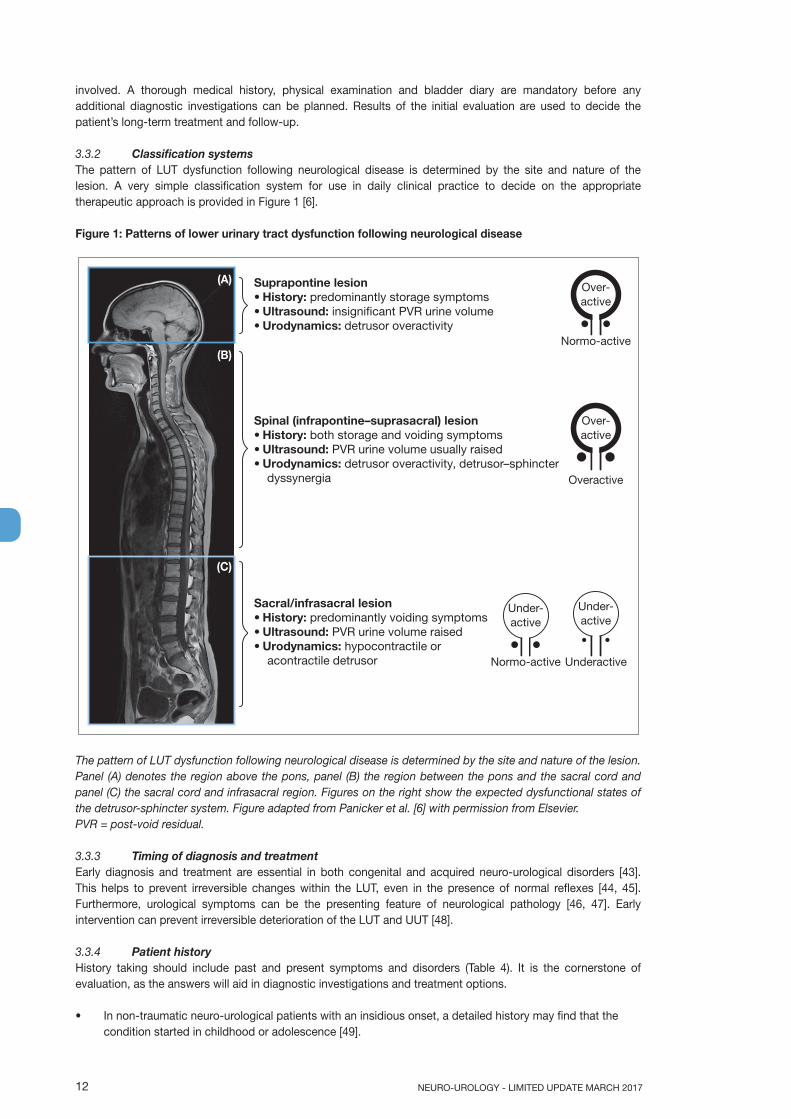
3.3.2 Classification systemsThe pattern of LUT dysfunction following neurological disease is determined by the site and nature of the lesion. A very simple classification system for use in daily clinical practice to decide on the appropriate therapeutic approach is provided in Figure 1 [6].
Figure 1: Patterns of lower urinary tract dysfunction following neurological disease
The pattern of LUT dysfunction following neurological disease is determined by the site and nature of the lesion. Panel (A) denotes the region above the pons, panel (B) the region between the pons and the sacral cord and panel (C) the sacral cord and infrasacral region. Figures on the right show the expected dysfunctional states of the detrusor-sphincter system. Figure adapted from Panicker et al. [6] with permission from Elsevier. PVR = post-void residual.
3.3.3 Timing of diagnosis and treatmentEarly diagnosis and treatment are essential in both congenital and acquired neuro-urological disorders [43]. This helps to prevent irreversible changes within the LUT, even in the presence of normal reflexes [44, 45]. Furthermore, urological symptoms can be the presenting feature of neurological pathology [46, 47]. Early intervention can prevent irreversible deterioration of the LUT and UUT [48].
3.3.4 Patient historyHistory taking should include past and present symptoms and disorders (Table 4). It is the cornerstone of evaluation, as the answers will aid in diagnostic investigations and treatment options.
• In non-traumatic neuro-urological patients with an insidious onset, a detailed history may find that the condition started in childhood or adolescence [49].
Suprapontine lesion• History: predominantly storage symptoms• Ultrasound: insignificant PVR urine volume• Urodynamics: detrusor overactivity
Spinal (infrapontine–suprasacral) lesion• History: both storage and voiding symptoms• Ultrasound: PVR urine volume usually raised• Urodynamics: detrusor overactivity, detrusor–sphincter dyssynergia
Sacral/infrasacral lesion• History: predominantly voiding symptoms• Ultrasound: PVR urine volume raised• Urodynamics: hypocontractile or acontractile detrusor
Normo-active
Over-active
Overactive
Over-active
Underactive
Under-active
Normo-active
Under-active
(A)
(B)
(C)

13NEURO-UROLOGY - LIMITED UPDATE MARCH 2017
• Urinary history consists of symptoms associated with both urine storage and emptying.• Bowel history is important because patients with neuro-urological symptoms may also have related
neurogenic bowel dysfunction [50].• Sexual function may be impaired because of the neuro-urological condition [51].• Special attention should be paid to possible warning signs and symptoms (e.g. pain, infection, haematuria
and fever) requiring further investigation.• Patients with SCI usually find it difficult to report UTI-related symptoms accurately [52, 53].• The presence of urinary, bowel and sexual symptoms without neurological symptoms could be suggestive
of an underlying neurological disease or condition.• Ambulatory status after acute SCI does not predict presence or absence of unfavourable urodynamic
parameters [54].
Table 4: History taking in patients with suspected neuro-urological disorder
Past historyChildhood through to adolescence and into adulthoodHereditary or familial risk factorsSpecific female: Menarche (age); this may suggest a metabolic disorderObstetric historyHistory of diabetesDiseases, e.g. multiple sclerosis, parkinsonism, encephalitis, syphilisAccidents and operations, especially those involving the spine and central nervous systemPresent historyPresent medicationLifestyle (smoking, alcohol and drugs); may influence urinary, sexual and bowel functionQuality of lifeSpecific urinary historyOnset of urological historyRelief after voiding; to detect the extent of a neurological lesion in the absence of obstructive uropathyBladder sensationInitiation of micturition (normal, precipitate, reflex, strain, Credé)Interruption of micturition (normal, paradoxical, passive)EnuresisMode and type of voiding (catheterisation)Frequency, voided volume, incontinence, urgency episodesSexual historyGenital or sexual dysfunction symptomsSensation in genital areaSpecific male: erection, (lack of) orgasm, ejaculationSpecific female: dyspareunia, (lack of) orgasmBowel historyFrequency and faecal incontinenceDesire to defecateDefecation patternRectal sensationInitiation of defecation (digitation)Neurological historyAcquired or congenital neurological conditionMental status and comprehensionNeurological symptoms (somatic and sensory), with onset, evolution and any treatmentSpasticity or autonomic dysreflexia (especially in lesions at or above level Th 6)Mobility and hand function
3.3.4.1 Bladder diariesBladder diaries provide data on the number of voids, voided volume, pad weight and incontinence and urgency episodes. Although a 24 hour bladder diary (recording should be done for three consecutive days) is reliable in women with UI [55, 56], no research has been done on bladder diaries in neuro-urological patients.
(B)
(C)

NEURO-UROLOGY - LIMITED UPDATE MARCH 201714
Nevertheless, bladder diaries are considered a valuable diagnostic tool.
3.3.5 Patient quality of life questionnairesAn assessment of the patient’s present and expected future quality of life (QoL) is important to evaluate the effect of any therapy. Quality of life is an essential aspect of the overall management of neuro-urological patients, for example when evaluating treatment related changes on a patient’s QoL [57]. The type of bladder management has been shown to affect health-related QoL (HRQoL) in patients with SCI [58] and MS [59]. Other research has also highlighted the importance of urological treatment and its impact on the urodynamic functionality of the neuro-urological patient in determining patient QoL [60].
In recent years a proliferation in the number of questionnaires to evaluate symptoms and QoL has been seen. Condition-specific questionnaires can be used to assess symptom severity and the impact of symptoms on QoL. A patient’s overall QoL can be assessed using generic questionnaires. It is important that the questionnaire of choice has been validated in the neuro-urological population, and that it is available in the language that it is to be used in.
3.3.5.1 Questions• Which validated patient questionnaires are available for neuro-urological patients?• Which questionnaires are the most appropriate for use in neuro-urological patients?
3.3.5.2 EvidenceThree condition-specific questionnaires for urinary or bowel dysfunction and QoL have been developed specifically for adult neuro-urological patients [61]. In MS and SCI patients the Qualiveen [62, 63] is validated and can be used for urinary symptoms. A short form of the Qualiveen is available [62, 63] and it has been translated into various languages [64-67]. The Neurogenic Bladder Symptom Score (NBSS) has been validated in neurological patients to measure urinary symptoms and their consequences [68]. The QoL scoring tool related to Bowel Management (QoL-BM) [69] can be used to assess bowel dysfunction in MS and SCI patients.
In addition, sixteen validated questionnaires that evaluate QoL and asses urinary symptoms as a subscale or question in neuro-urological patients have been identified [70] (Table 5). The condition-specific Incontinence-Quality of Life (I-QoL) questionnaire which was initially developed for the non-neurological population has now also been validated for neuro-urological patients [71].
A patient’s overall QoL can be assessed by generic HRQoL questionnaires, the most commonly used being the Incontinence Quality of Life Instrument (I-QOL), King’s Health Questionnaire (KHQ), or the Short Form 36-item and 12-item Health Survey Questionnaires (SF-36, SF-12) [61]. In addition, the quality-adjusted life year (QALY), quantifies outcomes, by weighing years of life spent in a specified health state, adjusted by a factor representing the value placed by society or patients on their specific health state [72].
No evidence was found for which validated questionnaires are the most appropriate for use, since no quality criteria for validated questionnaires have been assessed [73].
Table 5: Patient questionnaires
Questionnaire Underlying neurological disorder
Bladder Bowel Sexual function
FAMS [74] MS X XFILMS [75] MS X XHAQUAMS [76] MS X X XIQOL [71] MS, SCI X XMDS [77] MS X XMSISQ-15 / MSISQ-19 [78, 79] MS X X XMSQLI [80] MS X X XMSQoL-54 [81] MS X X XMSWDQ [82] MS X XNBSS [83] MS, SCI, Congenital
neurogenic bladderX
QoL-BM [69] SCI XQualiveen/SF-Qualiveen [63, 84] MS, SCI X XRAYS [85] MS X X RHSCIR [86] SCI X X XFransceschini [85] SCI X X X
15NEURO-UROLOGY - LIMITED UPDATE MARCH 2017
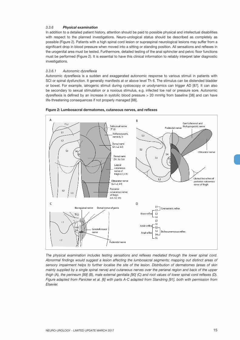
3.3.6 Physical examinationIn addition to a detailed patient history, attention should be paid to possible physical and intellectual disabilities with respect to the planned investigations. Neuro-urological status should be described as completely as possible (Figure 2). Patients with a high spinal cord lesion or supraspinal neurological lesions may suffer from a significant drop in blood pressure when moved into a sitting or standing position. All sensations and reflexes in the urogenital area must be tested. Furthermore, detailed testing of the anal sphincter and pelvic floor functions must be performed (Figure 2). It is essential to have this clinical information to reliably interpret later diagnostic investigations.
3.3.6.1 Autonomic dysreflexiaAutonomic dysreflexia is a sudden and exaggerated autonomic response to various stimuli in patients with SCI or spinal dysfunction. It generally manifests at or above level Th 6. The stimulus can be distended bladder or bowel. For example, iatrogenic stimuli during cystoscopy or urodynamics can trigger AD [87]. It can also be secondary to sexual stimulation or a noxious stimulus, e.g. infected toe nail or pressure sore. Autonomic dysreflexia is defined by an increase in systolic blood pressure > 20 mmHg from baseline [38] and can have life-threatening consequences if not properly managed [88].
Figure 2: Lumbosacral dermatomes, cutaneous nerves, and reflexes
The physical examination includes testing sensations and reflexes mediated through the lower spinal cord. Abnormal findings would suggest a lesion affecting the lumbosacral segments; mapping out distinct areas of sensory impairment helps to further localise the site of the lesion. Distribution of dermatomes (areas of skin mainly supplied by a single spinal nerve) and cutaneous nerves over the perianal region and back of the upper thigh (A), the perineum [89] (B), male external genitalia [90] (C) and root values of lower spinal cord reflexes (D). Figure adapted from Panicker et al. [6] with parts A-C adapted from Standring [91], both with permission from Elsevier.
NEURO-UROLOGY - LIMITED UPDATE MARCH 201716
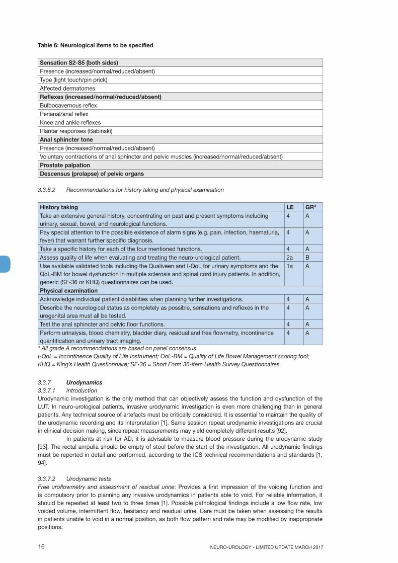
Table 6: Neurological items to be specified
Sensation S2-S5 (both sides)Presence (increased/normal/reduced/absent)Type (light touch/pin prick)Affected dermatomesReflexes (increased/normal/reduced/absent)Bulbocavernous reflexPerianal/anal reflexKnee and ankle reflexesPlantar responses (Babinski)Anal sphincter tonePresence (increased/normal/reduced/absent)Voluntary contractions of anal sphincter and pelvic muscles (increased/normal/reduced/absent)Prostate palpationDescensus (prolapse) of pelvic organs
3.3.6.2 Recommendations for history taking and physical examination
History taking LE GR*Take an extensive general history, concentrating on past and present symptoms including urinary, sexual, bowel, and neurological functions.
4 A
Pay special attention to the possible existence of alarm signs (e.g. pain, infection, haematuria, fever) that warrant further specific diagnosis.
4 A
Take a specific history for each of the four mentioned functions. 4 AAssess quality of life when evaluating and treating the neuro-urological patient. 2a BUse available validated tools including the Qualiveen and I-QoL for urinary symptoms and the QoL-BM for bowel dysfunction in multiple sclerosis and spinal cord injury patients. In addition, generic (SF-36 or KHQ) questionnaires can be used.
1a A
Physical examinationAcknowledge individual patient disabilities when planning further investigations. 4 ADescribe the neurological status as completely as possible, sensations and reflexes in the urogenital area must all be tested.
4 A
Test the anal sphincter and pelvic floor functions. 4 APerform urinalysis, blood chemistry, bladder diary, residual and free flowmetry, incontinence quantification and urinary tract imaging.
4 A
* All grade A recommendations are based on panel consensus. I-QoL = Incontinence Quality of Life Instrument; OoL-BM = Quality of Life Bowel Management scoring tool; KHQ = King’s Health Questionnaire; SF-36 = Short Form 36-item Health Survey Questionnaires.
3.3.7 Urodynamics3.3.7.1 IntroductionUrodynamic investigation is the only method that can objectively assess the function and dysfunction of the LUT. In neuro-urological patients, invasive urodynamic investigation is even more challenging than in general patients. Any technical source of artefacts must be critically considered. It is essential to maintain the quality of the urodynamic recording and its interpretation [1]. Same session repeat urodynamic investigations are crucial in clinical decision making, since repeat measurements may yield completely different results [92].
In patients at risk for AD, it is advisable to measure blood pressure during the urodynamic study [93]. The rectal ampulla should be empty of stool before the start of the investigation. All urodynamic findings must be reported in detail and performed, according to the ICS technical recommendations and standards [1, 94].
3.3.7.2 Urodynamic testsFree uroflowmetry and assessment of residual urine: Provides a first impression of the voiding function and is compulsory prior to planning any invasive urodynamics in patients able to void. For reliable information, it should be repeated at least two to three times [1]. Possible pathological findings include a low flow rate, low voided volume, intermittent flow, hesitancy and residual urine. Care must be taken when assessing the results in patients unable to void in a normal position, as both flow pattern and rate may be modified by inappropriate positions.

17NEURO-UROLOGY - LIMITED UPDATE MARCH 2017
Filling cystometry: This test is the only method for quantifying the patient’s filling function. The status of LUT function must be documented during the filling phase. However, this technique has limited use as a solitary procedure. It is much more effective combined with bladder pressure measurement during micturition and is even more effective in video-urodynamics.
The bladder should be empty at the start of filling. A physiological filling rate should be used with body-warm saline. Possible pathological findings include DO, low bladder compliance, abnormal bladder sensations, incontinence, and an incompetent or relaxing urethra.
Detrusor leak point pressure (DLPP) [95]: Appears to have no use as a diagnostic tool. Some positive findings have been reported [96, 97], but sensitivity is too low to estimate the risk to the UUT or for secondary bladder damage [98, 99].
Pressure flow study: Reflects the co-ordination between detrusor and urethra or pelvic floor during the voiding phase. It is even more powerful if combined with filling cystometry and video-urodynamics. Lower urinary tract function must be recorded during the voiding phase. Possible pathological findings include detrusor underactivity, bladder outlet obstruction (BOO), DSD, a high urethral resistance, and residual urine.
Most types of obstruction caused by neuro-urological disorders are due to DSD [100, 101], non-relaxing urethra, or non-relaxing bladder neck [102, 103]. Pressure-flow analysis mainly assesses the amount of mechanical obstruction caused by the urethra’s inherent mechanical and anatomical properties and has limited value in patients with neuro-urological disorders.
Electromyography (EMG): Reflects the activity of the external urethral sphincter, the peri-urethral striated musculature, the anal sphincter and the striated pelvic floor muscles. Correct interpretation may be difficult due to artefacts introduced by other equipment. In the urodynamic setting, an EMG is useful as a gross indication of the patient’s ability to control the pelvic floor. Possible pathological findings include inadequate recruitment upon specific stimuli (e.g. bladder filling, involuntary detrusor contractions, onset of voiding, coughing, Valsalva manoeuvre) suggesting a diagnosis of DSD [104].
Urethral pressure measurement: Has a very limited role in neuro-urological disorders. There is noconsensus on parameters indicating pathological findings [105].
Video-urodynamics: Is the combination of filling cystometry and pressure flow studys with imaging. It is the optimum procedure for urodynamic investigation in neuro-urological disorders. Possible pathological findings include all those described in the cystometry and the pressure flow study sections, and any morphological pathology of the LUT and reflux to the UUT [106].
Ambulatory urodynamics: This is the functional investigation of the urinary tract, which predominantly uses the natural filling of the urinary tract to reproduce the patient’s normal activity. Although this type of study might be considered when conventional urodynamics does not reproduce the patient’s symptoms, its role in the neuro-urological patient still needs to be determined [107, 108].
Triggered tests during urodynamics: Lower urinary tract function can be provoked by coughing, triggered voiding, or anal stretch. Fast-filling cystometry with cooled saline (the ‘ice water test’) will discriminate between upper and lower motor neuron lesions [109, 110]. Patients with UMNL develop a detrusor contraction if the detrusor is intact, while patients with LMNL do not. However, the test does not seem to be fully discriminative in other types of patients [111].
Previously, a positive bethanechol test [112] (detrusor contraction > 25 cm H2O) was thought to indicate detrusor denervation hypersensitivity and the muscular integrity of an acontractile detrusor. However, in practice, the test has given equivocal results. A variation of this method was reported using intravesical electromotive administration of the bethanechol [113], but there was no published follow-up. Currently, there is no indication for this test.
3.3.7.3 Specialist uro-neurophysiological testsThe following tests are advised as part of the neurological work-up [114]:• electromyography (in a neurophysiological setting) of pelvic floor muscles, urethral sphincter and/or anal
sphincter;• nerve conduction studies of pudendal nerve;• reflex latency measurements of bulbocavernosus and anal reflex arcs;• evoked responses from clitoris or glans penis;• sensory testing on bladder and urethra.

NEURO-UROLOGY - LIMITED UPDATE MARCH 201718
Other elective tests, for specific conditions, may become obvious during the work-up and urodynamic investigations.
3.3.7.4 Recommendations for urodynamics and uro-neurophysiology
Recommendations LE GRRecord a bladder diary. 3 ANon-invasive testing is mandatory before invasive urodynamics is planned. 4 A*Perform a urodynamic investigation to detect and specify lower urinary tract (dys-)function, use same session repeat measurement as it is crucial in clinical decision making.
1b A
Use video-urodynamics for invasive urodynamics in neuro-urological patients. If this is not available, then perform a filling cystometry continuing into a pressure flow study.
4 A*
Use a physiological filling rate and body-warm saline. 4 A*Specific uro-neurophysiological tests are elective procedures and should only be carried out in specialised settings.
4 C
*Upgraded based on panel consensus.
3.3.8 Renal functionIn many patients with neuro-urological disorders, the UUT is at risk, particularly in patients who develop high detrusor pressure during the filling phase. Although effective treatment can reduce this risk, there is still a relatively high incidence of renal morbidity [115]. Patients with SCI or SB have a substantially higher risk of developing renal failure compared with patients with slowly progressive non-traumatic neurological disorders, such as MS and Parkinson’s disease (PD) [116].
Caregivers must be informed of this risk and instructed to watch carefully for any signs or symptoms of a possible deterioration in the patient’s renal function. There are no high level evidence publications available which show the optimal management to preserve renal function in these patients [117].
3.4 Disease management3.4.1 IntroductionThe primary aims for treatment of neuro-urological symptoms, and their priorities, are [118, 119]:• protection of the UUT;• achievement (or maintenance) of urinary continence;• restoration of the LUT function;• improvement of the patient’s QoL.
Further considerations are the patient’s disability, cost-effectiveness, technical complexity and possible complications [119].
Renal failure is the main mortality factor in SCI patients who survive the trauma [120, 121]. Keeping the detrusor pressure during both the filling and voiding phases within safe limits significantly reduces the mortality from urological causes in these patients [122-124] and has consequently become the top priority in the treatment of patients with neuro-urological symptoms [118, 119].
In patients with high detrusor pressure during the filling phase (DO, low bladder compliance), treatment is aimed primarily at conversion of an overactive, high-pressure bladder into a low-pressure reservoir despite the resulting residual urine [118]. Reduction of the detrusor pressure contributes to urinary continence, and consequently to social rehabilitation and QoL. It is also pivotal in preventing UTIs [125, 126]. Complete continence, however, cannot always be obtained.
3.4.2 Non-invasive conservative treatment3.4.2.1 Assisted bladder emptying - Credé manoeuvre, Valsalva manoeuvre, triggered reflex voidingIncomplete bladder emptying is a serious risk factor for UTI, high intravesical pressure during the filling phase, and incontinence. Methods to improve the voiding process are therefore practiced.
Bladder expression (Credé manoeuvre) and voiding by abdominal straining (Valsalva manoeuvre): The downwards movement of the lower abdomen by suprapubic compression (Credé) or by abdominal straining (Valsalva) leads to an increase in intravesical pressure, and generally also causes a reflex sphincter contraction [127, 128]. The latter may increase bladder outlet resistance and lead to inefficient emptying. The high pressures created during these procedures are hazardous for the urinary tract [129, 130]. Therefore, their use should be discouraged unless urodynamics show that the intravesical pressure remains within safe limits [119].

19NEURO-UROLOGY - LIMITED UPDATE MARCH 2017
Long-term complications are unavoidable for both methods of bladder emptying [128]. The already weak pelvic floor function may be further impaired, thus introducing or exacerbating already existing stress urinary incontinence (SUI) [130].
Triggered reflex voiding: Stimulation of the sacral or lumbar dermatomes in patients with UMNL can elicit a reflex detrusor contraction [130]. The risk of high pressure voiding is present and interventions to decrease outlet resistance may be necessary [131]. Triggering can induce AD, especially in patients with high level SCI (at or above Th 6) [132]. All assisted bladder emptying techniques require low outlet resistance. Even then, high detrusor pressures may still be present. Hence, patients need dedicated education and close urodynamic and urological surveillance [130, 133-135].
Note: In the literature, including some of the references cited here, the concept “reflex voiding” is sometimes used to cover all three assisted voiding techniques described in this section.
External appliances: Social continence may be achieved by collecting urine during incontinence, for instance using pads [119]. Condom catheters with urine collection devices are a practical method for men [119]. The infection risk must be closely observed [119]. The penile clamp is absolutely contraindicated in case of DO or low bladder compliance because of the risk of developing high intravesical pressure and pressure sores/necrosis in cases of altered/absent sensations.
3.4.2.2 Neuro-urological rehabilitation3.4.2.2.1 Bladder rehabilitation including electrical stimulationThe term bladder rehabilitation summarises treatment options that aim to re-establish bladder function in patients with neuro-urological symptoms. Strong contraction of the urethral sphincter and/or pelvic floor, as well as anal dilatation, manipulation of the genital region, and physical activity inhibit micturition in a reflex manner [119, 136]. The first mechanism is affected by activation of efferent nerve fibres, and the latter ones are produced by activation of afferent fibres [98]. Electrical stimulation of the pudendal nerve afferents strongly inhibits the micturition reflex and detrusor contraction [137]. This stimulation might then support the restoration of the balance between excitatory and inhibitory inputs at the spinal or supraspinal level [119, 138]. Evidence for bladder rehabilitation using electrical stimulation in neurological patients is mainly based on small non-comparative studies with high risk of bias.
Peripheral temporary electrostimulation: Tibial nerve stimulation and transcutaneous electrical nerve stimulation might be effective and safe for treating neurogenic lower urinary tract dysfunction, but more reliable evidence from well-designed RCTs is required to reach definitive conclusions [138, 139].
Peripheral temporary electrostimulation combined with pelvic floor muscle training and biofeedback: In MS patients, combining active neuromuscular electrical stimulation with pelvic floor muscle training and EMG biofeedback can achieve a substantial reduction of neuro-urological symptoms [140]. This treatment combination seems to be more effective than either therapy alone [141, 142].
Intravesical electrostimulation: Intravesical electrostimulation can increase bladder capacity and improve bladder compliance and bladder filling sensation in patients with incomplete SCI or myelomeningocele (MMC) [143]. In patients with neurogenic detrusor underactivity, intravesical electrostimulation may also improve voiding and reduce residual volume [144, 145].
Repetitive transcranial magnetic stimulation: Although improvement of neuro-urological symptoms has been described in PD and MS patients, this technique is still under investigation [146, 147].
Summary: To date, bladder rehabilitation techniques are mainly based on electrical or magnetic stimulation. However, there is a lack of well-designed studies.
3.4.2.3 Drug treatmentA single, optimal, medical therapy for neuro-urological symptoms is not yet available. Commonly, a combination of different therapies (e.g. intermittent catheterisation and antimuscarinic drugs) is advised to prevent urinary tract damage and improve long-term outcomes, particularly in patients with a suprasacral SCI or MS [130, 148-150].
3.4.2.3.1 Drugs for storage symptomsAntimuscarinic drugs: They are the first-line choice for treating NDO, increasing bladder capacity and

NEURO-UROLOGY - LIMITED UPDATE MARCH 201720
reducing episodes of UI secondary to NDO by the inhibition of parasympathetic pathways [119, 151-157]. Antimuscarinic drugs have been used for many years to treat patients with NDO [154, 155, 158], and the responses of individual patients to antimuscarinic treatment are variable. Despite a meta-analysis confirming the clinical and urodynamic efficacy of antimuscarinic therapy compared to placebo in adult NDO, a more recent integrative review has indicated that the information provided is still too limited for clinicians to be able to match trial data to the needs of individual patients with SCI mainly because of the lack of standardised clinical evaluation tools such as the ASIA, bladder diary and validated symptoms score. [155, 159]. Higher doses or a combination of antimuscarinic agents may be an option to maximise outcomes in neurological patients [156, 157, 160-163]. However, these drugs have a high incidence of adverse events, which may lead to early discontinuation of therapy [155, 156].
Choice of antimuscarinic agent: Oxybutynin [119, 154-157, 164], trospium [155, 162, 165], tolterodine [166] and propiverine [155, 167] are established, effective and well tolerated treatments even in long-term use [154, 155, 168, 169]. Darifenacin [170, 171] and solifenacin [169, 172] have been evaluated in NDO secondary to SCI and MS [155, 170, 171, 173] with results similar to other antimuscarinic drugs. A pilot study using solifenacin in NDO due to PD showed an improvement in UI [174]. The relatively new drug, fesoterodine, an active metabolite of tolterodine, has also been introduced, even though to date there has been no published clinical evidence of its use in the treatment of neuro-urological disorders. Favourable results with the new drug imidafenacin have been reported [175].
Side effects: Controlled-release antimuscarinics have some minor side effects, e.g. dry mouth [176, 177]. It has been suggested that different ways of administration may help to reduce side effects. Moreover, imidafenacine has been safely used in neurological patients with no worsening of cognitive function [175].
Other agentsBeta-3-adrenergic receptor agonists have recently been introduced and evaluated in OAB, but clinical experience in neuro-urological patients is limited [178]. Studies on safety and effectiveness in NDO are ongoing [179]. Depending on the results of these studies, combined therapy with antimuscarinics may be an attractive option [180].
3.4.2.3.2 Drugs for voiding symptomsDetrusor underactivity: Cholinergic drugs, such as bethanechol and distigmine, have been considered to enhance detrusor contractility and promote bladder emptying, but are not frequently used in clinical practice [181]. Only preclinical studies have documented the potential benefits of cannabinoid agonists on improving detrusor contractility when administered intravesically [182, 183]. Conversely, RCTs on the use of nabixinols, D-9-tetrahydrocannabinol or oral cannabis extract did not report any significant reduction of incontinence episodes in MS patients [184].
Decreasing bladder outlet resistance: α-blockers (e.g. tamsulosin, naftopidil and silodosin) seem to be effective for decreasing bladder outlet resistance, post-void residual and AD [185-187].
Increasing bladder outlet resistance: Several drugs have shown efficacy in selected cases of mild SUI, but there are no high-level evidence studies in neurological patients [119].
3.4.2.4 Recommendations for drug treatments
Recommendations LE GRUse antimuscarinic therapy as the first-line medical treatment for neurogenic detrusor overactivity.
1a A
Alternative routes of administration (i.e., transdermal or intravesical) of antimuscarinic agents may be used.
2 A
Maximise outcomes for neurogenic detrusor overactivity by considering a combination of antimuscarinic agents.
3 B
Prescribe α-blockers to decrease bladder outlet resistance. 1b ADo not prescribe parasympathomimetics for underactive detrusor. 1a ADo not prescribe drug treatment in neurogenic stress urinary incontinence. 4 A*
*Upgraded based on panel consensus.

21NEURO-UROLOGY - LIMITED UPDATE MARCH 2017
3.4.2.5 Minimally invasive treatment3.4.2.5.1 CatheterisationIntermittent self- or third-party catheterisation [188, 189] is the preferred management for neuro-urological patients who cannot effectively empty their bladders [119]. Sterile IC, as originally proposed by Guttmann and Frankel [188], significantly reduces the risk of UTI and bacteriuria [119, 190, 191], compared with clean IC introduced by Lapides et al. [189]. However, it has not yet been established whether or not the incidence of UTI, other complications and user satisfaction are affected by either sterile or clean IC, coated or uncoated catheters or by any other strategy.
Sterile IC cannot be considered a routine procedure [119, 191]. Aseptic IC is an alternative to sterile IC [192].
Contributing factors to contamination are insufficient patient education and the inherently greater risk of UTI in neuro-urological patients [119, 193-197]. The average frequency of catheterisations per day is four to six times [198] and the catheter size most often used is between 12-16 Fr. In aseptic IC, an optimum frequency of five times showed a reduction of UTI [198]. Ideally, bladder volume at catheterisation should, as a rule, not exceed 400-500 mL.
Indwelling transurethral catheterisation and, to a lesser extent, suprapubic cystostomy are associated with a range of complications as well as an enhanced risk for UTI [119, 199-207]. Therefore, both procedures should be avoided, when possible. Silicone catheters are preferred as they are less susceptible to encrustation and because of the high incidence of latex allergy in the neuro-urological patient population [208].
3.4.2.5.2 Recommendations for catheterisation
Recommendations LE GRUse intermittent catheterisation, whenever possible aseptic technique, as a standard treatment for patients who are unable to empty their bladder.
3 A
Thoroughly instruct patients in the technique and risks of intermittent catheterisation. 3 AUse a catheter size between 12-16 Fr. 4 B*Avoid indwelling transurethral and suprapubic catheterisation whenever possible. 3 A
*Upgraded based on panel consensus.
3.4.2.5.3 Intravesical drug treatmentTo reduce DO, antimuscarinics can also be administered intravesically [209-213]. The efficacy, safety and tolerability of intravesical administration of 0.1% oxybutynin hydrochloride compared to its oral administration for treatment of NDO has been demonstrated in a recent randomised controlled study [213]. This approach may reduce adverse effects due to the fact that the antimuscarinic drug is metabolised differently [210] and a greater amount is sequestered in the bladder, even more than with electromotive administration [209].
The vanilloids, capsaicin and resiniferatoxin, desensitise the C-fibres and thereby decrease DO, for a period of a few months, until the sensation of these fibres has been restored [214-216]. The dosage is 1-2 mMol capsaicin in 100 mL 30% alcohol, or 10-100 nMol resiniferatoxin in 100 mL 10% alcohol for 30 minutes. Resiniferatoxin has about a 1,000-fold potency compared to capsaicin, with less pain during the instillation, and is effective in a patient refractory to capsaicin. Clinical studies have shown that resiniferatoxin has limited clinical efficacy compared to botulinum toxin A (BTX-A) injections in the detrusor [215]. Currently, there is no indication for the use of these substances, which are not licensed for intravesical treatment.
3.4.2.5.4 Botulinum toxin injections in the bladderBotulinum toxin A causes a long-lasting but reversible chemical denervation that lasts for about nine months [217, 218]. The toxin injections are mapped over the detrusor in a dosage that depends on the preparation used. Botulinum toxin A has been proven effective in patients with neuro-urological disorders due to MS or SCI in phase III RCTs [219, 220] and systematic reviews [221, 222]. Repeated injections seem to be possible without loss of efficacy [217, 223, 224]. The most frequent side effects are UTIs and elevated PVR [220, 223]. Intermittent catheterisation may become necessary. Rare but severe adverse events include AD and respiratory problems. Generalised muscular weakness may occur [217, 220, 224].
3.4.2.5.5 Bladder neck and urethral proceduresReduction of the bladder outlet resistance may be necessary to protect the UUT. This can be achieved by chemical denervation of the sphincter or by surgical interventions (bladder neck or sphincter incision or urethral stent). Incontinence may result and can be managed by external devices (see Section 3.4.2.1).

NEURO-UROLOGY - LIMITED UPDATE MARCH 201722
Botulinum toxin A: This can be used to treat DSD effectively by injection at a dose that depends on the preparation used. The dyssynergia is abolished for a few months, necessitating repeat injections. The efficacy of this treatment has been reported to be high and with few adverse effects [225-227]. However, a recent Cochrane report concluded that because of limited evidence future RCTs assessing the effectiveness of BTX-A injections also need to address the uncertainty about the optimal dose and mode of injection [228]. In addition, this therapy is not licensed.
Balloon dilatation: Favourable immediate results were reported [229], but there have been no further reports since 1994 therefore, this method is no longer recommended.
Sphincterotomy: By staged incision, bladder outlet resistance can be reduced without completely losing the closure function of the urethra [118, 119, 219]. Different techniques are used, and laser treatment appears to be advantageous [230, 231]. Sphincterotomy needs to be repeated at regular intervals in many patients [232], but it is efficient and does not cause severe adverse effects [118, 229]. Secondary narrowing of the bladder neck may occur, for which combined bladder neck incision might be considered [233].
Bladder neck incision: This is indicated only for secondary changes at the bladder neck (fibrosis) [118, 230]. This procedure is not recommended in patients with detrusor hypertrophy, which causes thickening of the bladder neck [118].
Stents: Implantation of urethral stents results in continence being dependent on adequate closure of the bladder neck [119]. The results are comparable with sphincterotomy and the stenting procedure has a shorter duration of surgery and hospital stay [234, 235]. However, the costs [118], possible complications and re-interventions [236, 237] are limiting factors in their use [238-241].
Increasing bladder outlet resistance: This can improve the continence condition. Despite early positive results with urethral bulking agents, a relative early loss of continence is reported in patients with neuro-urological disorders [119, 242, 243].
Urethral inserts: Urethral plugs or valves for the management of (female) stress incontinence have not been applied in neuro-urological patients. The experience with active pumping urethral prosthesis for treatment of the underactive or acontractile detrusor were disappointing [244].
3.4.2.5.6 Recommendations for minimal invasive treatment*
Recommendations LE GRUse botulinum toxin injection in the detrusor to reduce neurogenic detrusor overactivity in multiple sclerosis or spinal cord injury patients if antimuscarinic therapy is ineffective.
1a A
Bladder neck incision is effective in a fibrotic bladder neck. 4 B*Recommendations for catheterisation are listed separately under Section 3.4.2.5.2.
3.4.3 Surgical treatment3.4.3.1 Bladder neck and urethral proceduresIncreasing the bladder outlet resistance has the inherent risk of causing high intravesical pressure. Procedures to treat sphincteric incontinence are therefore suitable only when the detrusor activity can be controlled and when no significant reflux is present. A simultaneous bladder augmentation and IC may be necessary [119].
Urethral sling: Various materials have been used for this procedure with enduring positive results. The procedure is established in women with the ability to self-catheterise [119, 245-250]. There is growing evidence that synthetic slings can be used effectively with acceptable medium to long-term results and minimal morbidity in neurological patients [251, 252]. Besides the pubovaginal sling, which has been considered the procedure of choice in this subgroup of patients, recent reports suggest that both the transobturator and the retropubic approaches may also be considered, with similar failure rates and a reduction in the need for IC. However, for both approaches a higher incidence of de novo urgency was reported [252, 253]. In men, both autologous and synthetic slings may also be an alternative [251, 252, 254-256].
Artificial urinary sphincter: This device was introduced by Light and Scott [257] for patients with neuro-urological disorders [119]. It has stood the test of time and acceptable long-term outcomes can be obtained [258-263].
23NEURO-UROLOGY - LIMITED UPDATE MARCH 2017
Functional sphincter augmentation: Transposing the gracilis muscle to the bladder neck [264] or proximal urethra [265], can enable the possible creation of a functional autologous sphincter by electrical stimulation [264-266]. Therefore, raising the prospect of restoring control over the urethral closure.
Bladder neck and urethra reconstruction: The classical Young-Dees-Leadbetter procedure [267] for bladder neck reconstruction in children with bladder exstrophy, and Kropp urethra lengthening [268] improved by Salle [269], are established methods to restore continence provided that IC is practiced and/or bladder augmentation is performed [119, 270].
Urethral inserts: See section 3.4.2.5.5.
3.4.3.2 Denervation, deafferentation, sacral neuromodulationSacral rhizotomy, also known as sacral deafferentation, has achieved some success in reducing DO [271-273], but nowadays, it is used mostly as an adjuvant to sacral anterior root stimulation (SARS) [274-278]. Alternatives to rhizotomy are sought in this treatment combination [279-281].
Sacral anterior root stimulation is aimed at producing detrusor contraction. The technique was developed by Brindley [282] and is only applicable to complete lesions above the implant location, as its stimulation amplitude is over the pain threshold. The urethral sphincter efferents are also stimulated, but because the striated muscle relaxes faster than the smooth muscle of the detrusor, so-called “post-stimulus voiding” occurs. This approach has been successful in highly selected patients [275, 283, 284]. By changing the stimulation parameters, this method can also induce defecation or erection. A recent study reports that Charcot spinal arthropathy should be considered as a potential long-term complication of SARS, leading to spinal instability and to SARS dysfunction [285].
Sacral neuromodulation [286] might be effective and safe for treating neuro-urological symptoms, but there is a lack of RCTs and it is unclear which neurological patients are most suitable [287-290].
3.4.3.3 Bladder covering by striated muscleWhen the bladder is covered by striated muscle that can be stimulated electrically, or ideally that can be contracted voluntarily, voiding function can be restored to an acontractile bladder. The rectus abdominis [291] and latissimus dorsi [292] have been used successfully in patients with neuro-urological symptoms [293, 294].
3.4.3.4 Bladder augmentationThe aim of auto-augmentation (detrusor myectomy) is to reduce DO or improve low bladder compliance. The advantages are: low surgical burden, low rate of long-term adverse effects, positive effect on patient QoL, and it does not preclude further interventions [118, 119, 295-301].
Replacing or expanding the bladder by intestine or other passive expandable coverage will improve bladder compliance and at least reduce the pressure effect of DO [302, 303]. Inherent complications associated with these procedures are: recurrent infection, stone formation, perforation or diverticula, possible malignant changes, and for the intestine, metabolic abnormality, mucus production and impaired bowel function [119, 304-306]. The procedure should be used with caution in patients with neuro-urological symptoms, but may become necessary if all less-invasive treatment methods have failed. Special attention should be paid to patients with pre-operative renal scars since metabolic acidosis can develop [307].
Bladder augmentation is a valid option to decrease detrusor pressure and increase bladder capacity, whenever more conservative approaches have failed. Several different techniques have been published, with comparable and satisfactory results [297, 308-316]. Bladder substitution, even by performing a supratrigonal cystectomy [317], to create a low-pressure reservoir is indicated in patients with a severely thick and fibrotic bladder wall [119]. Intermittent catheterisation may become necessary after this procedure. A significant improvement in QoL has been reported, probably related to the perception of better health and the resolution/improvement of urinary incontinence [318].
3.4.3.5 Urinary diversionWhen no other therapy is successful, urinary diversion must be considered for the protection of the UUT and for the patient’s QoL [119].
Continent diversion: This should be the first choice for urinary diversion. Patients with limited dexterity may prefer a stoma instead of using the urethra for catheterisation. A continent stoma can be created using various techniques. However, all of them have frequent complications, including leakage or stenosis. The short-term continence rates are > 80% and good protection of the UUT is achieved [119, 319-331]. For cosmetic reasons, the umbilicus is often used for the stoma site [326, 329, 330, 332-334].

NEURO-UROLOGY - LIMITED UPDATE MARCH 201724
Incontinent diversion: If catheterisation is impossible, incontinent diversion with a urine-collecting device is indicated. Ultimately, it could be considered in patients who are wheelchair bound or bed-ridden with intractable and untreatable incontinence, in patients with LUT destruction, when the UUT is severely compromised, and in patients who refuse other therapy [119]. An ileal segment is used for the deviation in most cases [119, 335-338]. Patients gain better functional status and QoL after surgery [339].
Undiversion: Long-standing diversions may be successfully undiverted or an incontinent diversion changed to a continent one with the emergence of new and better techniques for control of detrusor pressure and incontinence [119]. The patient must be carefully counselled and must comply meticulously with the instructions [119]. Successful undiversion can then be performed [340].
3.4.3.6 Recommendations for surgical treatment
Recommendations LE GRPerform bladder augmentation in order to treat refractory neurogenic detrusor overactivity. 3 APlace an autologous urethral sling in female patients with neurogenic stress urinary incontinence who are able to self-catheterise.
4 B*
Insert an artificial urinary sphincter in male patients with neurogenic stress urinary incontinence.
3 A
*Upgraded bases on panel consensus.
3.5 Urinary tract infection in neuro-urological patients3.5.1 Epidemiology, aetiology and pathophysiologyUrinary tract infection is the onset of signs and/or symptoms accompanied by laboratory findings of a UTI (bacteriuria, leukocyturia and positive urine culture) [332]. There are no evidence-based cut-off values for the quantification of these findings. The published consensus is that a significant bacteriuria in persons performing IC is present with > 102 cfu/mL, > 104 cfu/mL in clean-void specimens and any detectable concentration in suprapubic aspirates. Regarding leukocyturia, 10 or more leukocytes in centrifuged urine samples per microscopic field (400x) are regarded as significant [332].
The pathogenesis of UTI in neuro-urological patients is multifactorial. Male gender seems to be a risk factor for febrile UTI [341]. Several etiological factors have been described: altered intrinsic defence mechanisms, impaired washout and catheterisation [342]. The exact working mechanisms, however, still remain unknown. The presence of asymptomatic bacteriuria in SCI patients is higher than in the general population, and varies depending on bladder management. Prevalence of bacteriuria in those performing clean IC varies from 23-89% [343]. Sphincterotomy and condom catheter drainage has a 57% prevalence [344]. Asymptomatic bacteria should not be routinely screened for in this population [345].
Individuals with neuro-urological symptoms, especially those with SCI, may have other signs and symptoms in addition to or instead of traditional signs and symptoms of a UTI in able-bodied individuals. Other problems, such as AD, may develop or worsen due to a UTI [346]. The most common signs and symptoms suspicious of a UTI in those with neuro-urological disorders are fever, new onset or increase in incontinence, including leaking around an indwelling catheter, increased spasticity, malaise, lethargy or sense of unease, cloudy urine with increased urine odour, discomfort or pain over the kidney or bladder, dysuria, or AD [346, 347].
3.5.2 Diagnostic evaluationThe gold standard for diagnosis is urine culture and urinalysis. A dipstick test may be more useful to exclude than to prove UTI [348, 349]. As bacterial strains and resistance patterns in persons with neuro-urological disorders may differ from those of able-bodied patients, microbiologic testing is mandatory [350].
3.5.3 Disease managementBacteriuria in patients with neuro-urological disorders should not be treated. Treatment of asymptomatic bacteriuria results in significantly more resistant bacterial strains without improving the outcome [351]. Urinary tract infections in persons with neuro-urological disorders are by definition a complicated UTI. Therefore, single-dose treatment is not advised. There is no consensus in the literature about the duration of treatment, it depends on the severity of the UTI and the involvement of kidneys and the prostate. Generally, a five to seven day course of antibiotic treatment is advised, which can be extended up to fourteen days according to the extent of the infection [351]. The choice of antibiotic therapy should be based on the results of the microbiologic testing. If immediate treatment is mandatory (e.g. fever, septicaemia, intolerable clinical

25NEURO-UROLOGY - LIMITED UPDATE MARCH 2017
symptoms, extensive AD), the choice of treatment should be based on local and individual resistance profiles [352].
3.5.3.1 Recurrent UTIRecurrent UTI in patients with neuro-urological disorders may indicate suboptimal management of the underlying functional problem, e.g. high bladder pressure during storage and voiding, incomplete voiding or bladder stones. The improvement of bladder function, by treating DO by BTX-A injection in the detrusor [353], and the removal of bladder stones or other direct supporting factors, especially indwelling catheters, as early as possible, are mandatory [350].
3.5.3.2 PreventionIf the improvement of bladder function and removal of foreign bodies/stones is not successful, additional UTI prevention strategies should be utilised. The use of hydrophilic catheters was associated with a lower rate of UTI in a recent meta-analysis [354]. Bladder irrigation has not been proven effective [355].
Various medical approaches have been tested for UTI prophylaxis in patients with neuro-urological disorders. The benefit of cranberry juice for the prevention of UTI could not be demonstrated in RCTs [356]. Methenamine hippurate is not effective in individuals with neuro-urological symptoms [357]. There is no sufficient evidence to support the use of L-methionine for urine acidification to prevent recurrent UTI [358]. There is only weak evidence that oral immunotherapy reduces bacteriuria in patients with SCI, and no evidence that recurrent UTIs are reduced [359]. Low-dose, long-term, antibiotic prophylaxis cannot reduce UTI frequency, but increases bacterial resistance and is therefore not recommended [351].
An application scheme of antibiotic substances for antibiotic prophylaxis provided long-term positive results, but the results of this trial need to be confirmed in further studies [360]. Another possible future option, the inoculation of apathogenic Escherichia coli strains into the bladder, has provided positive results in initial studies, but because of the paucity of data [361], cannot be recommended as a treatment option.
In summary, based on the criteria of evidence-based medicine, there is currently no preventive measure for recurrent UTI in patients with neuro-urological disorders that can be recommended without limitations. Therefore, individualised concepts should be taken into consideration, including immunostimulation, phytotherapy and complementary medicine [362]. Prophylaxis in patients with neuro-urological disorders is important to pursue, but since there are no data favouring one approach over another, prophylaxis is essentially a trial and error approach.
3.5.4 Recommendations for the treatment of UTI
Recommendations LE GRDo not screen for or treat asymptomatic bacteriuria in patients with neuro-urological disorders. 4 A*Avoid the use of long-term antibiotics for recurrent urinary tract infections (UTIs). 2a AIn patients with recurrent UTI, optimise treatment of neuro-urological symptoms and remove foreign bodies (e.g. stones, indwelling catheters) from the urinary tract.
3 A
In patients with neuro-urological disorders, UTI prophylaxis must be individualised since there is no optimal prophylactic measure available.
4 C
*Upgraded based on panel consensus.
3.6 Sexual (dys)function and fertilityThese Guidelines specifically focus on sexual dysfunction and infertility in patients with a neurological disease [363, 364]. Non-neurogenic, male sexual dysfunction and infertility are covered in separate EAU Guidelines [365, 366]. In neuro-urological patients sexual problems can be identified at three levels: primary (direct neurological damage), secondary (general physical disabilities) and tertiary (psychosocial and emotional issues) sexual dysfunction [367]. Adopting a systematic approach, such as the PLISSIT model (Permission, Limited Information, Specific Suggestions and Intensive Therapy) [368], provides a framework for counselling and treatment involving a stepwise approach to the management of neurogenic sexual dysfunction. Sexual dysfunction is associated with neurogenic lower urinary tract dysfunction in patients with MS [369] and spina bifida [370]. Although various patient-reported outcome measures (PROMs) are available to evaluate sexual function, the evidence for good PROMs is limited and studies with high methodological quality are needed [371].

NEURO-UROLOGY - LIMITED UPDATE MARCH 201726
3.6.1 Erectile dysfunction (ED)3.6.1.1 Phosphodiesterase type 5 inhibitors (PDE5Is)Questions:• What is the effectiveness of the various PDE5Is in the different neuro-urological patient groups?• What common side-effects are described?
Evidence:Phosphodiesterase type 5 inhibitors (PDE5Is) are recommended as first-line treatment in neurogenic erectile dysfunction (ED) [363, 372]. In SCI patients, tadalafil, vardenafil and sildenafil have all improved retrograde ejaculation and improved erectile function and satisfaction on IIEF-15. Tadalafil 10 mg was shown to be more effective than sildenafil 50 mg. All currently available PDE5Is appear to be effective and safe, although there are no high level evidence studies in neuro-urological patients investigating the efficacy and side effects across different PDE5Is, dosages and formulations [373].
For MS patients two studies reported significant improvement in ED when using sildenafil and tadalafil. One study, however, showed no improvement in ED with sildenafil.
In PD normal erectile function was described in over half of the patients using sildenafil 100 mg and a significant improvement in IIEF-15 score was found compared to placebo. While most neuro-urological patients require long-term therapy for ED some have a low compliance rate or stop therapy because of side effects [374, 375], most commonly headache and flushing [372]. In addition, PDE5Is may induce relevant hypotension in patients with tetraplegia/high-level paraplegia and multiple system atrophy [374, 375]. As a prerequisite for successful PDE5I-therapy, some residual nerve function is required to induce erection. Since many patients with SCI use on-demand nitrates for the treatment of AD, they must be counselled that PDE5Is are contraindicated when using nitrate medication.
3.6.1.2 Drug therapy other than PDE5IsFampridine to treat neurogenic spasticity has been shown to be beneficial in improving ED in two domains of the IIEF-15 in SCI and MS patients, however, with a significant discontinuation rate due to severe adverse events [376]. Sublingual apomorfine was shown to have poor results on ED in SCI patients and side-effects in half of the patients [377]. In PD pergolide mesylate showed a significant improvement in IIEF-15 scores up to twelve months follow-up [378].
3.6.1.3 Mechanical devicesMechanical devices (vacuum tumescence devices and penile rings) may be effective but are less popular [379-383].
3.6.1.4 Intracavernous injections and intraurethral applicationPatients not responding to oral drugs may be offered intracavernous injections (alprostadil, papaverine and phentolamine) that have been shown to be effective in a number of neurological conditions, including SCI, MS, and diabetes mellitus [384-390] but their use requires careful dose titration and some precautions. Complications of intracavernous drugs include pain, priapism and corpora cavernosa fibrosis. Intracavernous vasoactive drug injection is the first-line therapeutic option in patients taking nitrate medications, as well as those with concerns about drug interactions with PDE5Is, or in whom PDE5Is are ineffective. The impact of intracavernous injections on ejaculation and orgasmic function, their early use for increasing the recovery rate of a spontaneous erection, and their effectiveness and tolerability in the long-term are unclear [374]. Intraurethral alprostadil application is an alternative but a less effective route of administration [390, 391].
3.6.1.5 Sacral neuromodulationSacral neuromodulation for LUT dysfunction may improve sexual function but high level evidence studies are lacking [372].
3.6.1.6 Penile prosthesesPenile prostheses may be considered for treatment of neurogenic ED when all conservative treatments have failed. At a mean follow-up of seven years 83.7% of patients with SCI were able to have sexual intercourse [372]. Serious complications, including infection and prosthesis perforation, may occur in about 10% of patients, depending on implant type [392-394].

27NEURO-UROLOGY - LIMITED UPDATE MARCH 2017
3.6.1.7 Recommendations for erectile dysfunction
Recommendations LE GRPrescribe oral phosphodiesterase type 5 inhibitors as first-line medical treatment in neurogenic erectile dysfunction.
1b A
Give intracavernous injections of vasoactive drugs (alone or in combination) as second-line medical treatment in neurogenic erectile dysfunction.
3 A
Offer mechanical devices such as vacuum devices and rings to patients with neurogenic erectile dysfunction.
3 B
Reserve penile prostheses for selected patients with neurogenic erectile dysfunction. 4 B**Upgraded based on panel consensus.
3.6.2 Male fertilityMale fertility can be compromised in the neurological patient by ED, ejaculation disorder, impaired sperm quality or various combinations of these three disorders. Among the major conditions contributing to neurogenic infertility are pelvic and retroperitoneal surgery, diabetes mellitus, SB, MS and SCI [395]. Erectile dysfunction is managed as described previously. Retrograde ejaculation may be reversed by sympathomimetic agents contracting the bladder neck, including imipramine, ephedrine, pseudoephedrine, and phenylpropanolamine [395]. The use of a balloon catheter to obstruct the bladder neck may be effective in obtaining antegrade ejaculation [396]. If antegrade ejaculation is not achieved, the harvest of semen from the urine may be considered [397].
Prostatic massage is safe and easy to use for obtaining semen in men with lesions above Th 10 [398]. In several patients, vibrostimulation or transrectal electroejaculation are needed for sperm retrieval [390, 395, 399, 400]. Semen retrieval is more likely with vibrostimulation in men with lesions above Th 10 [401-403]. In men with SCI, especially at or above Th 6, AD might occur during sexual activity and ejaculation [404, 405]; patients at risk and fertility clinics must be informed and aware of this potentially life-threatening condition. In SCI patients the use of oral midodrine can improve sperm retrieval at vibrostimulation [406].
In men with MS, use of disease modifying drugs during the conception phase, has not been associated with altered pregnancy outcomes [407]. Surgical procedures, such as, microsurgical epididymal sperm aspiration (MESA) or testicular sperm extraction (TESE), may be used if vibrostimulation and electroejaculation are not successful [408, 409]. Pregnancy rates in patients with SCI are lower than in the general population, but since the introduction of intracytoplasmic sperm injection (ICSI), men with SCI now have a good chance of becoming biological fathers [410-412].
3.6.2.1 Sperm quality and motilityThe following has been reported on sperm quality and motility:• bladder management with clean IC may improve semen quality compared to indwelling catheterisation,
reflex voiding or bladder expression [413];• in SCI patients sperm quality decreases at the early post traumatic phase demonstrating lower
spermatozoid vitality (necrospermia), reduced motility (asthenospermia) and leucospermia [408];• long-term valproate treatment for epilepsy negatively influences sperm count and motility [414];• vibrostimulation produces samples with better sperm motility than electrostimulation [415, 416];• electroejaculation with interrupted current produces better sperm motility than continuous current [417];• freezing of sperm is unlikely to improve fertility rates in men with SCI [418].
3.6.2.2 Recommendations for male fertility
Recommendations LE GRPerform vibrostimulation and transrectal electroejaculation for sperm retrieval in men with spinal cord injury.
3 B
Perform microsurgical epididymal sperm aspiration, testicular sperm extraction and intracytoplasmic sperm injection after failed vibrostimulation and/or transrectal electroejaculation in men with spinal cord injury.
3 B
Counsel men with spinal cord injury at or above Th 6 and fertility clinics about the potentially life-threatening condition of autonomic dysreflexia.
3 A*
*Upgraded based on panel consensus.
NEURO-UROLOGY - LIMITED UPDATE MARCH 201728
3.6.3 Female sexualityThe most relevant publications on neurogenic female sexual dysfunction are in women with SCI and MS. After SCI, about 65-80% of women continue to be sexually active, but to a much lesser extent than before the injury, and about 25% report a decreased satisfaction with their sexual life [419-421]. Although sexual dysfunction is very common in women with MS, it is still often overlooked by medical professionals [422, 423].
The greatest physical barrier to sexual activity is UI. A correlation has been found between the urodynamic outcomes of low bladder capacity, compliance and high maximum detrusor pressure and sexual dysfunction in MS patients. Problems with positioning and spasticity affect mainly tetraplegic patients. Peer support may help to optimise the sexual adjustment of women with SCI in achieving a more positive self-image, self-esteem and feelings of being attractive to themselves and others [419, 424-426].
The use of specific drugs for sexual dysfunction is indicated to treat inadequate lubrication. Data on sildenafil for treating female sexual dysfunction are poor and controversial [427]. Although good evidence exists that psychological interventions are effective in the treatment of female hypoactive sexual desire disorder and female orgasmic disorder [428], there is a lack of high-evidence level studies in the neurological population.
Neurophysiological studies have shown that women with the ability to perceive Th 11-L2 pin-prick sensations may have psychogenic genital vasocongestion. Reflex lubrication and orgasm is more prevalent in women with SCI who have preserved the sacral reflex arc (S2-S5), even when it has not been shown in an individual woman that a specific level and degree of lesion is the cause of a particular sexual dysfunction. In SCI women with a complete lesion of the sacral reflex, arousal and orgasm may be evoked through stimulation of other erogenous zones above the level of lesions [429-431].
Sacral neuromodulation for LUT dysfunction may improve sexual function but high-evidence studies are lacking [427].
Women with SCI reported dissatisfaction with the quality and quantity of sexuality-related rehabilitation services and were less likely to receive sexual information than men [429, 432, 433].
3.6.3.1 Recommendation for female sexuality
Recommendation LE GRDo not offer medical therapy for the treatment of neurogenic sexual dysfunction in women. 4 A*
*Upgraded based on panel consensus.
3.6.4 Female fertilityThere are few studies on female fertility in neurological patients. More than a third (38%) of women with epilepsy had infertility and the relevant predictors were exposure to multiple (three or more) antiepileptic drugs, older age and lower education [434].
Although it seems that the reproductive capacity of women with SCI is only temporarily affected by SCI with cessation of menstruation for approximately six months after SCI [435], there are no high-evidence level studies. About 70% of sexually active women use some form of contraception after injury, but fewer women use the birth control pill compared to before their injury [436].
Women with SCI are more likely to suffer complications during pregnancy, labour and delivery compared to able-bodied women. Complications of labour and delivery include bladder problems, spasticity, pressure sores, anaemia, and AD [437, 438]. Obstetric outcomes include higher rates of Caesarean sections and an increased incidence of low birth-weight babies [436].
Epidural anaesthesia is chosen and effective for most patients with AD during labour and delivery [439, 440].
There is very little published data on women’s experience of the menopause following SCI [441]. Women with MS who plan a pregnancy should evaluate their current drug treatment with their treating physician [442]. Clinical management should be individualised to optimise both the mother’s reproductive outcomes and MS course [443].

29NEURO-UROLOGY - LIMITED UPDATE MARCH 2017
3.6.4.1 Recommendation for female fertility
Recommendation LE GRTake a multidisciplinary approach, tailored to individual patient’s needs and preferences, in the management of fertility, pregnancy and delivery in women with neurological diseases.
4 A*
*Upgraded based on panel consensus.
3.7 Follow-up3.7.1 IntroductionNeuro-urological disorders are often unstable and the symptoms may vary considerably, even within a relatively short period. Regular follow-up is therefore necessary [117].
Depending on the type of the underlying neurological pathology and the current stability of the neuro-urological symptoms, the interval between initial investigations and control diagnostics may vary and in many cases should not exceed one to two years. In high-risk neuro-urological patients this interval should be much shorter. Urinalysis should be performed regularly; the frequency to be guided by patient symptoms. The UUT should be checked by ultrasonography at regular intervals in high-risk patients; about once every six months. In these patients, physical examination and urine laboratory should take place every year. Any significant clinical change warrants further, specialised, investigation. However, there is a complete lack of high level evidence studies on this topic and every recommendation must be viewed critically in each individual neuro-urological patient [117].
In addition, bladder wall thickness can be measured on ultrasonography as an additional risk assessment for upper tract damage [444], although a ‘safe’ cut-off threshold for this has not been agreed [445]. The utility of DMSA for follow-up of neuro-urological patients has not been fully evaluated [446].
3.7.2 Recommendations for follow-up
Recommendations LE GRAssess the upper urinary tract at regular intervals in high risk patients. 4 A*Preform a physical examination and urine laboratory every year in high risk patients. 4 A*Any significant clinical changes should instigate further, specialised, investigation. 4 A*Preform urodynamic investigation as a mandatory baseline diagnostic intervention in high-risk patients at regular intervals.
3 A
*Upgraded based on panel consensus.
3.8 ConclusionsNeuro-urological disorders have a multi-faceted pathology. They require an extensive and specific diagnosis before one can embark on an individualised therapy, which takes into account the medical and physical condition of the patient and the patient’s expectations about their future. The urologist can select from a wealth of therapeutic options, each with its own pros and cons. Notwithstanding the success of any therapy embarked upon, close surveillance is necessary for the patient’s entire life.
These Guidelines offer you expert advice on how to define the patient’s neuro-urological symptoms as precisely as possible and how to select, together with the patient, the appropriate therapy. This last choice, as always, is governed by the golden rule: as effective as needed, as non-invasive as possible.
4. REFERENCES1. Schafer, W., et al. Good urodynamic practices: uroflowmetry, filling cystometry, and pressure-flow
studies. Neurourol Urodyn, 2002. 21: 261.https://www.ncbi.nlm.nih.gov/pubmed/11948720
2. Abrams, P., et al. Reviewing the ICS 2002 terminology report: the ongoing debate. Neurourol Urodyn, 2009. 28: 287.https://www.ncbi.nlm.nih.gov/pubmed/19350662

NEURO-UROLOGY - LIMITED UPDATE MARCH 201730
3. Abrams, P., et al. The standardisation of terminology of lower urinary tract function: report from the Standardisation Sub-committee of the International Continence Society. Neurourol Urodyn, 2002. 21: 167.https://www.ncbi.nlm.nih.gov/pubmed/11857671
4. Groen, J., et al. Summary of European Association of Urology (EAU) Guidelines on Neuro-Urology. Eur Urol, 2015.https://www.ncbi.nlm.nih.gov/pubmed/26304502
5. Nosseir, M., et al. Clinical usefulness of urodynamic assessment for maintenance of bladder function in patients with spinal cord injury. Neurourol Urodyn, 2007. 26: 228.https://www.ncbi.nlm.nih.gov/pubmed/16998859
6. Panicker, J.N., et al. Lower urinary tract dysfunction in the neurological patient: clinical assessment and management. Lancet Neurol, 2015. 14: 720.https://www.ncbi.nlm.nih.gov/pubmed/26067125
7. Phe, V., et al. Continent catheterizable tubes/stomas in adult neuro-urological patients: A systematic review. Neurourol Urodyn, 2017. DOI: 10.1002/nau.23213https://www.ncbi.nlm.nih.gov/pubmed/28139848
8. Ecclestone, H., et al. Long term effectiveness and complication rate of bladder reconstruction and urinary diversions in patients with neuropathic bladder dysfunction. PROSPERO 2015. CRD42015015762.http://www.crd.york.ac.uk/PROSPERO/display_record.asp?ID=CRD42015015762
9. Phillips B, et al. Oxford Centre for Evidence-based Medicine Levels of Evidence. Updated by Jeremy Howick March 2009. 1998.http://www.cebm.net/oxford-centre-evidence-based-medicine-levels-evidence-march-2009/
10. Townsend, N., et al. Cardiovascular disease in Europe - epidemiological update 2015. Eur Heart J, 2015.https://www.ncbi.nlm.nih.gov/pubmed/26306399
11. Tibaek, S., et al. Prevalence of lower urinary tract symptoms (LUTS) in stroke patients: a cross-sectional, clinical survey. Neurourol Urodyn, 2008. 27: 763.https://www.ncbi.nlm.nih.gov/pubmed/18551565
12. Marinkovic, S.P., et al. Voiding and sexual dysfunction after cerebrovascular accidents. J Urol, 2001. 165: 359.https://www.ncbi.nlm.nih.gov/pubmed/11176374
13. Rotar, M., et al. Stroke patients who regain urinary continence in the first week after acute first-ever stroke have better prognosis than patients with persistent lower urinary tract dysfunction. Neurourol Urodyn, 2011. 30: 1315.https://www.ncbi.nlm.nih.gov/pubmed/21488096
14. Lobo, A., et al. Prevalence of dementia and major subtypes in Europe: A collaborative study of population-based cohorts. Neurologic Diseases in the Elderly Research Group. Neurology, 2000. 54: S4.https://www.ncbi.nlm.nih.gov/pubmed/10854354
15. Na, H.R., et al. Urinary incontinence in Alzheimer’s disease is associated with Clinical Dementia Rating-Sum of Boxes and Barthel Activities of Daily Living. Asia Pac Psychiatry, 2015. 7: 113.https://www.ncbi.nlm.nih.gov/pubmed/23857871
16. Grant, R.L., et al. First diagnosis and management of incontinence in older people with and without dementia in primary care: a cohort study using The Health Improvement Network primary care database. PLoS Med, 2013. 10: 1001.https://www.ncbi.nlm.nih.gov/pubmed/24015113
17. Pringsheim, T., et al. The prevalence of Parkinson’s disease: a systematic review and meta-analysis. Mov Disord, 2014. 29: 1583.https://www.ncbi.nlm.nih.gov/pubmed/24976103
18. Ragab, M.M., et al. Idiopathic Parkinson’s disease patients at the urologic clinic. Neurourol Urodyn, 2011. 30: 1258.https://www.ncbi.nlm.nih.gov/pubmed/21404318
19. Papatsoris, A.G., et al. Urinary and erectile dysfunction in multiple system atrophy (MSA). Neurourol Urodyn, 2008. 27: 22.https://www.ncbi.nlm.nih.gov/pubmed/17563111
20. Kim, M., et al. Impaired detrusor contractility is the pathognomonic urodynamic finding of multiple system atrophy compared to idiopathic Parkinson’s disease. Parkinsonism Relat Disord, 2015. 21: 205.https://www.ncbi.nlm.nih.gov/pubmed/25534084
31NEURO-UROLOGY - LIMITED UPDATE MARCH 2017
21. Sakakibara, R., et al. A guideline for the management of bladder dysfunction in Parkinson’s disease and other gait disorders. Neurourol Urodyn, 2015.https://www.ncbi.nlm.nih.gov/pubmed/25810035
22. Dolecek, T.A., et al. CBTRUS statistical report: primary brain and central nervous system tumors diagnosed in the United States in 2005-2009. Neuro Oncol, 2012. 14 Suppl 5: v1.https://www.ncbi.nlm.nih.gov/pubmed/23095881
23. Maurice-Williams, R.S. Micturition symptoms in frontal tumours. J Neurol Neurosurg Psychiatry, 1974. 37: 431.https://www.ncbi.nlm.nih.gov/pubmed/4365244
24. Christensen, D., et al. Prevalence of cerebral palsy, co-occurring autism spectrum disorders, and motor functioning - Autism and Developmental Disabilities Monitoring Network, USA, 2008. Dev Med Child Neurol, 2014. 56: 59.https://www.ncbi.nlm.nih.gov/pubmed/24117446
25. Marciniak, C., et al. Urinary incontinence in adults with cerebral palsy: prevalence, type, and effects on participation. PM R, 2014. 6: 110.https://www.ncbi.nlm.nih.gov/pubmed/23978464
26. Tagliaferri, F., et al. A systematic review of brain injury epidemiology in Europe. Acta Neurochir (Wien), 2006. 148: 255.https://www.ncbi.nlm.nih.gov/pubmed/16311842
27. Kulakli, F., et al. Relationship between urinary dysfunction and clinical factors in patients with traumatic brain injury. Brain Inj, 2014. 28: 323.https://www.ncbi.nlm.nih.gov/pubmed/24377376
28. Singh, A., et al. Global prevalence and incidence of traumatic spinal cord injury. Clin Epidemiol, 2014. 6: 309.https://www.ncbi.nlm.nih.gov/pubmed/25278785
29. Weld, K.J., et al. Association of level of injury and bladder behavior in patients with post-traumatic spinal cord injury. Urology, 2000. 55: 490.https://www.ncbi.nlm.nih.gov/pubmed/10736489
30. Kondo, A., et al. Neural tube defects: prevalence, etiology and prevention. Int J Urol, 2009. 16: 49.https://www.ncbi.nlm.nih.gov/pubmed/19120526
31. Sawin, K.J., et al. The National Spina Bifida Patient Registry: profile of a large cohort of participants from the first 10 clinics. J Pediatr, 2015. 166: 444.https://www.ncbi.nlm.nih.gov/pubmed/25444012
32. Bartolin, Z., et al. Relationship between clinical data and urodynamic findings in patients with lumbar intervertebral disk protrusion. Urol Res, 2002. 30: 219.https://www.ncbi.nlm.nih.gov/pubmed/12202938
33. Lange, M.M., et al. Urinary and sexual dysfunction after rectal cancer treatment. Nat Rev Urol, 2011. 8: 51.https://www.ncbi.nlm.nih.gov/pubmed/21135876
34. IDF Diabetes Atlas, 7th edn. 2015, International Diabetes Federation: Brussels, Belgium. http://www.diabetesatlas.org/
35. Yuan, Z., et al. Diabetic cystopathy: A review. J Diabetes, 2015. 7: 442.https://www.ncbi.nlm.nih.gov/pubmed/25619174
36. Pugliatti, M., et al. The epidemiology of multiple sclerosis in Europe. Eur J Neurol, 2006. 13: 700.https://www.ncbi.nlm.nih.gov/pubmed/16834700
37. de Seze, M., et al. The neurogenic bladder in multiple sclerosis: review of the literature and proposal of management guidelines. Mult Scler, 2007. 13: 915.https://www.ncbi.nlm.nih.gov/pubmed/17881401
38. Krassioukov, A., et al. International standards to document remaining autonomic function after spinal cord injury. J Spinal Cord Med, 2012. 35: 201.https://www.ncbi.nlm.nih.gov/pubmed/22925746
39. Irwin, D.E., et al. Understanding the elements of overactive bladder: questions raised by the EPIC study. BJU Int, 2008. 101: 1381.https://www.ncbi.nlm.nih.gov/pubmed/18336602
40. Fix, J.D., Neuroanatomy. 4th ed. 2008, Philadelphia, Pennsylvania, USA.41. Maynard, F.M., Jr., et al. International Standards for Neurological and Functional Classification of
Spinal Cord Injury. American Spinal Injury Association. Spinal Cord, 1997. 35: 266.https://www.ncbi.nlm.nih.gov/pubmed/9160449

NEURO-UROLOGY - LIMITED UPDATE MARCH 201732
42. Klevmark, B. Natural pressure-volume curves and conventional cystometry. Scand J Urol Nephrol Suppl, 1999. 201: 1.https://www.ncbi.nlm.nih.gov/pubmed/10573769
43. Del Popolo, G., et al. Diagnosis and therapy for neurogenic bladder dysfunctions in multiple sclerosis patients. Neurol Sci, 2008. 29 Suppl 4: S352.https://www.ncbi.nlm.nih.gov/pubmed/19089675
44. Satar, N., et al. The effects of delayed diagnosis and treatment in patients with an occult spinal dysraphism. J Urol, 1995. 154: 754.https://www.ncbi.nlm.nih.gov/pubmed/7609171
45. Watanabe, T., et al. High incidence of occult neurogenic bladder dysfunction in neurologically intact patients with thoracolumbar spinal injuries. J Urol, 1998. 159: 965.https://www.ncbi.nlm.nih.gov/pubmed/9474194
46. Ahlberg, J., et al. Neurological signs are common in patients with urodynamically verified “idiopathic” bladder overactivity. Neurourol Urodyn, 2002. 21: 65.https://www.ncbi.nlm.nih.gov/pubmed/11835426
47. Bemelmans, B.L., et al. Evidence for early lower urinary tract dysfunction in clinically silent multiple sclerosis. J Urol, 1991. 145: 1219.https://www.ncbi.nlm.nih.gov/pubmed/2033697
48. Klausner, A.P., et al. The neurogenic bladder: an update with management strategies for primary care physicians. Med Clin North Am, 2011. 95: 111.https://www.ncbi.nlm.nih.gov/pubmed/21095415
49. Bors, E., et al. History and physical examination in neurological urology. J Urol, 1960. 83: 759.https://www.ncbi.nlm.nih.gov/pubmed/13802958
50. Cameron, A.P., et al. The Severity of Bowel Dysfunction in Patients with Neurogenic Bladder. J Urol, 2015.https://www.ncbi.nlm.nih.gov/pubmed/25956470
51. Vodusek, D.B. Lower urinary tract and sexual dysfunction in neurological patients. Eur Neurol, 2014. 72: 109.https://www.ncbi.nlm.nih.gov/pubmed/24993182
52. Linsenmeyer, T.A., et al. Accuracy of individuals with spinal cord injury at predicting urinary tract infections based on their symptoms. J Spinal Cord Med, 2003. 26: 352.https://www.ncbi.nlm.nih.gov/pubmed/14992336
53. Massa, L.M., et al. Validity, accuracy, and predictive value of urinary tract infection signs and symptoms in individuals with spinal cord injury on intermittent catheterization. J Spinal Cord Med, 2009. 32: 568.https://www.ncbi.nlm.nih.gov/pubmed/20025153
54. Bellucci, C.H.S., et al. Acute spinal cord injury - Do ambulatory patients need urodynamic investigations? J Urol, 2013. 189: 1369.https://www.ncbi.nlm.nih.gov/pubmed/23069382
55. Honjo, H., et al. Impact of convenience void in a bladder diary with urinary perception grade to assess overactive bladder symptoms: a community-based study. Neurourol Urodyn, 2010. 29: 1286.https://www.ncbi.nlm.nih.gov/pubmed/20878998
56. Naoemova, I., et al. Reliability of the 24-h sensation-related bladder diary in women with urinary incontinence. Int Urogynecol J Pelvic Floor Dysfunct, 2008. 19: 955.https://www.ncbi.nlm.nih.gov/pubmed/18235981
57. Henze, T. Managing specific symptoms in people with multiple sclerosis. Int MS J, 2005. 12: 60.https://www.ncbi.nlm.nih.gov/pubmed/16417816
58. Liu, C.W., et al. The relationship between bladder management and health-related quality of life in patients with spinal cord injury in the UK. Spinal Cord, 2010. 48: 319.https://www.ncbi.nlm.nih.gov/pubmed/19841636
59. Khalaf, K.M., et al. The impact of lower urinary tract symptoms on health-related quality of life among patients with multiple sclerosis. Neurourol Urodyn, 2016. 35: 48.https://www.ncbi.nlm.nih.gov/pubmed/25327401
60. Pannek, J., et al. Does optimizing bladder management equal optimizing quality of life? Correlation between health-related quality of life and urodynamic parameters in patients with spinal cord lesions. Urology, 2009. 74: 263.https://www.ncbi.nlm.nih.gov/pubmed/19428089
61. Patel, D.P., et al. Patient reported outcomes measures in neurogenic bladder and bowel: A systematic review of the current literature. Neurourol Urodyn, 2014.https://www.ncbi.nlm.nih.gov/pubmed/25327455

33NEURO-UROLOGY - LIMITED UPDATE MARCH 2017
62. Bonniaud, V., et al. Qualiveen, a urinary-disorder specific instrument: 0.5 corresponds to the minimal important difference. J Clin Epidemiol, 2008. 61: 505.https://www.ncbi.nlm.nih.gov/pubmed/18394545
63. Bonniaud, V., et al. Development and validation of the short form of a urinary quality of life questionnaire: SF-Qualiveen. J Urol, 2008. 180: 2592.https://www.ncbi.nlm.nih.gov/pubmed/18950816
64. Bonniaud, V., et al. Italian version of Qualiveen-30: cultural adaptation of a neurogenic urinary disorder-specific instrument. Neurourol Urodyn, 2011. 30: 354.https://www.ncbi.nlm.nih.gov/pubmed/21305589
65. Ciudin, A., et al. Quality of life of multiple sclerosis patients: translation and validation of the Spanish version of Qualiveen. Neurourol Urodyn, 2012. 31: 517.https://www.ncbi.nlm.nih.gov/pubmed/22396437
66. D’Ancona, C.A., et al. Quality of life of neurogenic patients: translation and validation of the Portuguese version of Qualiveen. Int Urol Nephrol, 2009. 41: 29.https://www.ncbi.nlm.nih.gov/pubmed/18528780
67. Pannek, J., et al. [Quality of life in German-speaking patients with spinal cord injuries and bladder dysfunctions. Validation of the German version of the Qualiveen questionnaire]. Urologe A, 2007. 46: 1416.https://www.ncbi.nlm.nih.gov/pubmed/17605119
68. Welk, B., et al. The conceptualization and development of a patient-reported neurogenic bladder symptom score. Res Rep Urol, 2013. 5: 129.https://www.ncbi.nlm.nih.gov/pubmed/24400244
69. Gulick, E.E. Bowel management related quality of life in people with multiple sclerosis: psychometric evaluation of the QoL-BM measure. Int J Nurs Stud, 2011. 48: 1066.https://www.ncbi.nlm.nih.gov/pubmed/21377677
70. Tsang, B., et al. A systematic review and comparison of questionnaires in the management of spinal cord injury, multiple sclerosis and the neurogenic bladder. Neurourol Urodyn, 2015.https://www.ncbi.nlm.nih.gov/pubmed/25620137
71. Schurch, B., et al. Reliability and validity of the Incontinence Quality of Life questionnaire in patients with neurogenic urinary incontinence. Arch Phys Med Rehabil, 2007. 88: 646.https://www.ncbi.nlm.nih.gov/pubmed/17466735
72. Hollingworth, W., et al. Exploring the impact of changes in neurogenic urinary incontinence frequency and condition-specific quality of life on preference-based outcomes. Qual Life Res, 2010. 19: 323.https://www.ncbi.nlm.nih.gov/pubmed/20094804
73. Patel, D.P., et al. Patient reported outcomes measures in neurogenic bladder and bowel: A systematic review of the current literature. Neurourol Urodyn, 2016. 35: 8.https://www.ncbi.nlm.nih.gov/pubmed/25327455
74. Cella, D.F., et al. Validation of the functional assessment of multiple sclerosis quality of life instrument. Neurology, 1996. 47: 129.https://www.ncbi.nlm.nih.gov/pubmed/8710066
75. Wesson, J.M., et al. The functional index for living with multiple sclerosis: development and validation of a new quality of life questionnaire. Mult Scler, 2009. 15: 1239.https://www.ncbi.nlm.nih.gov/pubmed/19737850
76. Gold, S.M., et al. Disease specific quality of life instruments in multiple sclerosis: validation of the Hamburg Quality of Life Questionnaire in Multiple Sclerosis (HAQUAMS). Mult Scler, 2001. 7: 119.https://www.ncbi.nlm.nih.gov/pubmed/11424632
77. Goodin, D.S. A questionnaire to assess neurological impairment in multiple sclerosis. Mult Scler, 1998. 4: 444.https://www.ncbi.nlm.nih.gov/pubmed/9839306
78. Foley, F.W., et al. The Multiple Sclerosis Intimacy and Sexuality Questionnaire -- re-validation and development of a 15-item version with a large US sample. Mult Scler, 2013. 19: 1197.https://www.ncbi.nlm.nih.gov/pubmed/
79. Sanders, A.S., et al. The Multiple Sclerosis Intimacy and Sexuality Questionnaire-19 (MSISQ-19). Sexuality and Disability, 2000. 18: 3.https://www.ncbi.nlm.nih.gov/pubmed/23369892
80. Marrie, R.A., et al. Validity and reliability of the MSQLI in cognitively impaired patients with multiple sclerosis. Mult Scler, 2003. 9: 621.https://www.ncbi.nlm.nih.gov/pubmed/14664477

NEURO-UROLOGY - LIMITED UPDATE MARCH 201734
81. Vickrey, B.G., et al. A health-related quality of life measure for multiple sclerosis. Qual Life Res, 1995. 4: 187.https://www.ncbi.nlm.nih.gov/pubmed/7613530
82. Honan, C.A., et al. The multiple sclerosis work difficulties questionnaire (MSWDQ): development of a shortened scale. Disabil Rehabil, 2014. 36: 635.https://www.ncbi.nlm.nih.gov/pubmed/23786346
83. Welk, B., et al. The validity and reliability of the neurogenic bladder symptom score. J Urol, 2014. 192: 452.https://www.ncbi.nlm.nih.gov/pubmed/24518764
84. Bonniaud, V., et al. Measuring quality of life in multiple sclerosis patients with urinary disorders using the Qualiveen questionnaire. Arch Phys Med Rehabil, 2004. 85: 1317.https://www.ncbi.nlm.nih.gov/pubmed/15295759
85. Franceschini, M., et al. Follow-up in persons with traumatic spinal cord injury: questionnaire reliability. Eura Medicophys, 2006. 42: 211.https://www.ncbi.nlm.nih.gov/pubmed/17039217
86. Noreau, L., et al. Development and assessment of a community follow-up questionnaire for the Rick Hansen spinal cord injury registry. Arch Phys Med Rehabil, 2013. 94: 1753.https://www.ncbi.nlm.nih.gov/pubmed/23529142
87. Liu, N., et al. Autonomic dysreflexia severity during urodynamics and cystoscopy in individuals with spinal cord injury. Spinal Cord, 2013. 51: 863.https://www.ncbi.nlm.nih.gov/pubmed/24060768
88. Liu, N., et al. Iatrogenic urological triggers of autonomic dysreflexia: a systematic review. Spinal Cord, 2015. 53: 500.https://www.ncbi.nlm.nih.gov/pubmed/25800696
89. Labat, J.J., et al. Diagnostic criteria for pudendal neuralgia by pudendal nerve entrapment (Nantes criteria). Neurourol Urodyn, 2008. 27: 306.https://www.ncbi.nlm.nih.gov/pubmed/17828787
90. Brown, D., Atlas of regional anesthesia. 3rd. ed. 2006, Philadelphia91. Standring, S., Gray’s anatomy . 40th ed. 2008.92. Bellucci, C.H., et al. Neurogenic lower urinary tract dysfunction--do we need same session repeat
urodynamic investigations? J Urol, 2012. 187: 1318.https://www.ncbi.nlm.nih.gov/pubmed/22341264
93. Walter, M., et al. Autonomic dysreflexia and repeatability of cardiovascular changes during same session repeat urodynamic investigation in women with spinal cord injury. World J Urol, 2015.https://www.ncbi.nlm.nih.gov/pubmed/26055644
94. Gammie, A., et al. International Continence Society guidelines on urodynamic equipment performance. Neurourol Urodyn, 2014. 33: 370.https://www.ncbi.nlm.nih.gov/pubmed/24390971
95. McGuire, E.J., et al. Leak-point pressures. Urol Clin North Am, 1996. 23: 253.https://www.ncbi.nlm.nih.gov/pubmed/8659025
96. Ozkan, B., et al. Which factors predict upper urinary tract deterioration in overactive neurogenic bladder dysfunction? Urology, 2005. 66: 99.https://www.ncbi.nlm.nih.gov/pubmed/15992868
97. Wang, Q.W., et al. Is it possible to use urodynamic variables to predict upper urinary tract dilatation in children with neurogenic bladder-sphincter dysfunction? BJU Int, 2006. 98: 1295.https://www.ncbi.nlm.nih.gov/pubmed/17034510
98. Linsenmeyer, T.A., et al. The impact of urodynamic parameters on the upper tracts of spinal cord injured men who void reflexly. J Spinal Cord Med, 1998. 21: 15.https://www.ncbi.nlm.nih.gov/pubmed/9541882
99. McGuire, E.J., et al. Prognostic value of urodynamic testing in myelodysplastic patients. J Urol, 1981. 126: 205.https://www.ncbi.nlm.nih.gov/pubmed/7196460
100. Krongrad, A., et al. Bladder neck dysynergia in spinal cord injury. Am J Phys Med Rehabil, 1996. 75: 204.https://www.ncbi.nlm.nih.gov/pubmed/8663928
101. Weld, K.J., et al. Clinical significance of detrusor sphincter dyssynergia type in patients with post-traumatic spinal cord injury. Urology, 2000. 56: 565.https://www.ncbi.nlm.nih.gov/pubmed/11018603

35NEURO-UROLOGY - LIMITED UPDATE MARCH 2017
102. Rossier, A.B., et al. 5-microtransducer catheter in evaluation of neurogenic bladder function. Urology, 1986. 27: 371.https://www.ncbi.nlm.nih.gov/pubmed/3962062
103. Al-Ali, M., et al. A 10 year review of the endoscopic treatment of 125 spinal cord injured patients with vesical outlet obstruction: does bladder neck dyssynergia exist? Paraplegia, 1996. 34: 34.https://www.ncbi.nlm.nih.gov/pubmed/8848321
104. Bacsu, C.D., et al. Diagnosing detrusor sphincter dyssynergia in the neurological patient. BJU Int, 2012. 109 Suppl 3: 31.https://www.ncbi.nlm.nih.gov/pubmed/22458490
105. Lose, G., et al. Standardisation of urethral pressure measurement: report from the Standardisation Sub-Committee of the International Continence Society. Neurourol Urodyn, 2002. 21: 258.https://www.ncbi.nlm.nih.gov/pubmed/11948719
106. Marks, B.K., et al. Videourodynamics: indications and technique. Urol Clin North Am, 2014. 41: 383.https://www.ncbi.nlm.nih.gov/pubmed/25063594
107. Virseda, M., et al. Reliability of ambulatory urodynamics in patients with spinal cord injuries. Neurourology and Urodynamics, 2013. 32: 387.https://www.ncbi.nlm.nih.gov/pubmed/23002043
108. Virseda-Chamorro, M., et al. Comparison of ambulatory versus video urodynamics in patients with spinal cord injury. Spinal Cord, 2014. 52: 551.https://www.ncbi.nlm.nih.gov/pubmed/24663000
109. Geirsson, G., et al. The ice-water test--a simple and valuable supplement to routine cystometry. Br J Urol, 1993. 71: 681.https://www.ncbi.nlm.nih.gov/pubmed/8343894
110. Geirsson, G., et al. Pressure, volume and infusion speed criteria for the ice-water test. Br J Urol, 1994. 73: 498.https://www.ncbi.nlm.nih.gov/pubmed/8012770
111. Al-Hayek, S., et al. The 50-year history of the ice water test in urology. J Urol, 2010. 183: 1686.https://www.ncbi.nlm.nih.gov/pubmed/20299050
112. Lapides, J. Neurogenic bladder. Principles of treatment. Urol Clin North Am, 1974. 1: 81.https://www.ncbi.nlm.nih.gov/pubmed/4428540
113. Riedl, C.R., et al. Electromotive administration of intravesical bethanechol and the clinical impact on acontractile detrusor management: introduction of a new test. J Urol, 2000. 164: 2108.https://www.ncbi.nlm.nih.gov/pubmed/11061937
114. Podnar, S., et al. Lower urinary tract dysfunction in patients with peripheral nervous system lesions. Handb Clin Neurol, 2015. 130: 203.https://www.ncbi.nlm.nih.gov/pubmed/26003246
115. Ouyang, L., et al. Characteristics and survival of patients with end stage renal disease and spina bifida in the United States renal data system. J Urol, 2015. 193: 558.https://www.ncbi.nlm.nih.gov/pubmed/25167993
116. Lawrenson, R., et al. Renal failure in patients with neurogenic lower urinary tract dysfunction. Neuroepidemiology, 2001. 20: 138.https://www.ncbi.nlm.nih.gov/pubmed/11359083
117. Averbeck, M.A., et al. Follow-up of the neuro-urological patient: a systematic review. BJU Int, 2015. 115 Suppl 6: 39.https://www.ncbi.nlm.nih.gov/pubmed/
118. Stöhrer, M., et al. Diagnosis and treatment of bladder dysfunction in spinal cord injury patients. Eur Urol Update Series 1994. 3: 170. [No abstract available]
119. Drake, M., et al., Conservative management in neuropathic urinary incontinence, in Incontinence, P. Abrams, L. Cardozo, S. Khoury & A. Wein, Editors. 2013, Health Publication: Plymouth: , 2013; pp. 827-1000.
120. Chamberlain, J.D., et al. Mortality and longevity after a spinal cord injury: systematic review and meta-analysis. Neuroepidemiology, 2015. 44: 182.https://www.ncbi.nlm.nih.gov/pubmed/25997873
121. Game, X., et al. Botulinum toxin A detrusor injections in patients with neurogenic detrusor overactivity significantly decrease the incidence of symptomatic urinary tract infections. Eur Urol, 2008. 53: 613.https://www.ncbi.nlm.nih.gov/pubmed/17804150
122. Frankel, H.L., et al. Long-term survival in spinal cord injury: a fifty year investigation. Spinal Cord, 1998. 36: 266.https://www.ncbi.nlm.nih.gov/pubmed/9589527

NEURO-UROLOGY - LIMITED UPDATE MARCH 201736
123. Jamil, F. Towards a catheter free status in neurogenic bladder dysfunction: a review of bladder management options in spinal cord injury (SCI). Spinal Cord, 2001. 39: 355.https://www.ncbi.nlm.nih.gov/pubmed/11464308
124. Thietje, R., et al. Mortality in patients with traumatic spinal cord injury: descriptive analysis of 62 deceased subjects. J Spinal Cord Med, 2011. 34: 482.https://www.ncbi.nlm.nih.gov/pubmed/22118255
125. Hackler, R.H. A 25-year prospective mortality study in the spinal cord injured patient: comparison with the long-term living paraplegic. J Urol, 1977. 117: 486.https://www.ncbi.nlm.nih.gov/pubmed/850323
126. Rodrigues, P., et al. Involuntary detrusor contraction is a frequent finding in patients with recurrent urinary tract infections. Urol Int, 2014. 93: 67.https://www.ncbi.nlm.nih.gov/pubmed/
127. Bauer, S.B. Neurogenic bladder: etiology and assessment. Pediatr Nephrol, 2008. 23: 541.https://www.ncbi.nlm.nih.gov/pubmed/18270749
128. Barbalias, G.A., et al. Critical evaluation of the Crede maneuver: a urodynamic study of 207 patients. J Urol, 1983. 130: 720.https://www.ncbi.nlm.nih.gov/pubmed/6887405
129. Reinberg, Y., et al. Renal rupture after the Crede maneuver. J Pediatr, 1994. 124: 279.https://www.ncbi.nlm.nih.gov/pubmed/8301439
130. Wyndaele, J.J., et al. Neurologic urinary incontinence. Neurourol Urodyn, 2010. 29: 159.https://www.ncbi.nlm.nih.gov/pubmed/20025021
131. Menon, E.B., et al. Bladder training in patients with spinal cord injury. Urology, 1992. 40: 425.https://www.ncbi.nlm.nih.gov/pubmed/1441039
132. Furusawa, K., et al. Incidence of symptomatic autonomic dysreflexia varies according to the bowel and bladder management techniques in patients with spinal cord injury. Spinal Cord, 2011. 49: 49.https://www.ncbi.nlm.nih.gov/pubmed/20697419
133. Outcomes following traumatic spinal cord injury: clinical practice guidelines for health-care professionals. J Spinal Cord Med, 2000. 23: 289.https://www.ncbi.nlm.nih.gov/pubmed/17536300
134. El-Masri, W.S., et al. Long-term follow-up study of outcomes of bladder management in spinal cord injury patients under the care of the Midlands Centre for Spinal Injuries in Oswestry. Spinal Cord, 2012. 50: 14.https://www.ncbi.nlm.nih.gov/pubmed/21808256
135. Singh, R., et al. Bladder management methods and urological complications in spinal cord injury patients. Indian J Orthop, 2011. 45: 141.https://www.ncbi.nlm.nih.gov/pubmed/21430869
136. Fall, M., et al. Electrical stimulation. A physiologic approach to the treatment of urinary incontinence. Urol Clin North Am, 1991. 18: 393.https://www.ncbi.nlm.nih.gov/pubmed/2017820
137. Vodusek, D.B., et al. Detrusor inhibition induced by stimulation of pudendal nerve afferents. Neurourol Urodyn, 1986. 5: 381.http://onlinelibrary.wiley.com/doi/10.1002/nau.1930050404/abstract
138. Gross, T., et al. Transcutaneous Electrical Nerve Stimulation for Treating Neurogenic Lower Urinary Tract Dysfunction: A Systematic Review. Eur Urol, 2016. 69: 1102.http://www.europeanurology.com/article/S0302-2838(16)00061-0/abstract/
139. Schneider, M.P., et al. Tibial Nerve Stimulation for Treating Neurogenic Lower Urinary Tract Dysfunction: A Systematic Review. Eur Urol, 2015.https://www.ncbi.nlm.nih.gov/pubmed/26194043
140. McClurg, D., et al. Neuromuscular electrical stimulation and the treatment of lower urinary tract dysfunction in multiple sclerosis--a double blind, placebo controlled, randomised clinical trial. Neurourol Urodyn, 2008. 27: 231.https://www.ncbi.nlm.nih.gov/pubmed17705160
141. McClurg, D., et al. Comparison of pelvic floor muscle training, electromyography biofeedback, and neuromuscular electrical stimulation for bladder dysfunction in people with multiple sclerosis: a randomized pilot study. Neurourol Urodyn, 2006. 25: 337.https://www.ncbi.nlm.nih.gov/pubmed/16637070
142. Ferreira, A.P., et al. Impact of a Pelvic Floor Training Program Among Women with Multiple Sclerosis: A Controlled Clinical Trial. Am J Phys Med Rehabil. 2016. 95: 1.https://www.ncbi.nlm.nih.gov/pubmed/25888662
37NEURO-UROLOGY - LIMITED UPDATE MARCH 2017
143. Hagerty, J.A., et al. Intravesical electrotherapy for neurogenic bladder dysfunction: a 22-year experience. J Urol, 2007. 178: 1680.https://www.ncbi.nlm.nih.gov/pubmed/17707024
144. Primus, G., et al. Restoration of micturition in patients with acontractile and hypocontractile detrusor by transurethral electrical bladder stimulation. Neurourol Urodyn, 1996. 15: 489.https://www.ncbi.nlm.nih.gov/pubmed/8857617
145. Lombardi, G., et al. Clinical efficacy of intravesical electrostimulation on incomplete spinal cord patients suffering from chronic neurogenic non-obstructive retention: a 15-year single centre retrospective study. Spinal Cord, 2013. 51: 232.https://www.ncbi.nlm.nih.gov/pubmed/23147136
146. Brusa, L., et al. Effects of inhibitory rTMS on bladder function in Parkinson’s disease patients. Mov Disord, 2009. 24: 445.https://www.ncbi.nlm.nih.gov/pubmed/19133657
147. Centonze, D., et al. Effects of motor cortex rTMS on lower urinary tract dysfunction in multiple sclerosis. Mult Scler, 2007. 13: 269.https://www.ncbi.nlm.nih.gov/pubmed/17439897
148. Thomas, L.H., et al. Treatment of urinary incontinence after stroke in adults. Cochrane Database Syst Rev, 2008: Cd004462.https://www.ncbi.nlm.nih.gov/pubmed/18254050
149. Yeo, L., et al. Urinary tract dysfunction in Parkinson’s disease: a review. Int Urol Nephrol, 2012. 44: 415.https://www.ncbi.nlm.nih.gov/pubmed/21553114
150. Phe, V., et al. Management of neurogenic bladder in patients with multiple sclerosis. Nat Rev Urol, 2016. 13: 275.https://www.ncbi.nlm.nih.gov/pubmed/27030526
151. Stohrer, M., et al. EAU guidelines on neurogenic lower urinary tract dysfunction. Eur Urol, 2009. 56: 81.https://www.ncbi.nlm.nih.gov/pubmed/19403235
152. Andersson, K.E. Antimuscarinic mechanisms and the overactive detrusor: an update. Eur Urol, 2011. 59: 377.https://www.ncbi.nlm.nih.gov/pubmed/21168951
153. Kennelly, M.J., et al. Overactive bladder: pharmacologic treatments in the neurogenic population. Rev Urol, 2008. 10: 182.https://www.ncbi.nlm.nih.gov/pubmed/18836537
154. Madersbacher, H., et al. Neurogenic detrusor overactivity in adults: a review on efficacy, tolerability and safety of oral antimuscarinics. Spinal Cord, 2013. 51: 432.https://www.ncbi.nlm.nih.gov/pubmed/23743498
155. Madhuvrata, P., et al. Anticholinergic drugs for adult neurogenic detrusor overactivity: a systematic review and meta-analysis. Eur Urol, 2012. 62: 816.https://www.ncbi.nlm.nih.gov/pubmed/22397851
156. Bennett, N., et al. Can higher doses of oxybutynin improve efficacy in neurogenic bladder? J Urol, 2004. 171: 749.https://www.ncbi.nlm.nih.gov/pubmed/14713802
157. Horstmann, M., et al. Neurogenic bladder treatment by doubling the recommended antimuscarinic dosage. Neurourol Urodyn, 2006. 25: 441.https://www.ncbi.nlm.nih.gov/pubmed/16847942
158. Mehnert, U., et al. The management of urinary incontinence in the male neurological patient. Curr Opin Urol, 2014. 24: 586.https://www.ncbi.nlm.nih.gov/pubmed/25389549
159. Stothers, L., et al. An integrative review of standardized clinical evaluation tool utilization in anticholinergic drug trials for neurogenic lower urinary tract dysfunction. Spinal Cord, 2016. 31: 31.https://www.ncbi.nlm.nih.gov/pubmed/27241452
160. Amend, B., et al. Effective treatment of neurogenic detrusor dysfunction by combined high-dosed antimuscarinics without increased side-effects. Eur Urol, 2008. 53: 1021.https://www.ncbi.nlm.nih.gov/pubmed/18243516
161. Cameron, A.P. Pharmacologic therapy for the neurogenic bladder. Urol Clin North Am, 2010. 37: 495.https://www.ncbi.nlm.nih.gov/pubmed/20955901

NEURO-UROLOGY - LIMITED UPDATE MARCH 201738
162. Menarini, M., et al. Trospium chloride in patients with neurogenic detrusor overactivity: is dose titration of benefit to the patients? Int J Clin Pharmacol Ther, 2006. 44: 623.https://www.ncbi.nlm.nih.gov/pubmed/17190372
163. Nardulli, R., et al. Combined antimuscarinics for treatment of neurogenic overactive bladder. Int J Immunopathol Pharmacol, 2012. 25: 35s.https://www.ncbi.nlm.nih.gov/pubmed/22652160
164. Cameron, A.P., et al. Combination drug therapy improves compliance of the neurogenic bladder. J Urol, 2009. 182: 1062.https://www.ncbi.nlm.nih.gov/pubmed/19616807
165. Isik, A.T., et al. Trospium and cognition in patients with late onset Alzheimer disease. J Nutr Health Aging, 2009. 13: 672.https://www.ncbi.nlm.nih.gov/pubmed/19657549
166. Ethans, K.D., et al. Efficacy and safety of tolterodine in people with neurogenic detrusor overactivity. J Spinal Cord Med, 2004. 27: 214.https://www.ncbi.nlm.nih.gov/pubmed/15478523
167. McKeage, K. Propiverine: A review of its use in the treatment of adults and children with overactive bladder associated with idiopathic or neurogenic detrusor overactivity, and in men with lower urinary tract symptoms. Clin Drug Investig, 2013. 33: 71.https://www.ncbi.nlm.nih.gov/pubmed/23288694
168. Nicholas, R.S., et al. Anticholinergics for urinary symptoms in multiple sclerosis. Cochrane Database Syst Rev, 2009: Cd004193.https://www.ncbi.nlm.nih.gov/pubmed/19160231
169. van Rey, F., et al. Solifenacin in multiple sclerosis patients with overactive bladder: a prospective study. Adv Urol, 2011. 2011: 834753.https://www.ncbi.nlm.nih.gov/pubmed/21687581
170. Bycroft, J., et al. The effect of darifenacin on neurogenic detrusor overactivity in patients with spinal cord injury. Neurourol Urodyn 2003. 22: A190.https://www.ics.org/Abstracts/Publish/41/000190.pdf
171. Carl, S., et al. Darifenacin is also effective in neurogenic bladder dysfunction (multiple sclerosis). Urology, 2006. 68 250. [No abstract available]
172. Krebs, J., et al. Effects of solifenacin in patients with neurogenic detrusor overactivity as a result of spinal cord lesion. Spinal Cord, 2013. 51: 306.https://www.ncbi.nlm.nih.gov/pubmed/23247012
173. Amarenco, G., et al. Solifenacin is effective and well tolerated in patients with neurogenic detrusor overactivity: Results from the double-blind, randomized, active- and placebo-controlled SONIC urodynamic study. Neurourol UrodyN, 2015. 29: 29.https://www.ncbi.nlm.nih.gov/pubmed/26714009
174. Zesiewicz, T.A., et al. Randomized, controlled pilot trial of solifenacin succinate for overactive bladder in Parkinson’s disease. Parkinsonism Relat Disord, 2015. 21: 514.https://www.ncbi.nlm.nih.gov/pubmed/25814050
175. Sakakibara, R., et al. Imidafenacin on bladder and cognitive function in neurologic OAB patients. Clin Auton Res, 2013. 23: 189.https://www.ncbi.nlm.nih.gov/pubmed/23820664
176. Stohrer, M., et al. Efficacy and tolerability of propiverine hydrochloride extended-release compared with immediate-release in patients with neurogenic detrusor overactivity. Spinal Cord, 2013. 51: 419.https://www.ncbi.nlm.nih.gov/pubmed/23338657
177. Caremel, R., et al. [Drug therapy of bladder dysfunction]. Prog Urol. 2013 Nov;23(15):1271-86.https://www.ncbi.nlm.nih.gov/pubmed/24183086
178. Wollner, J., et al. Initial experience with the treatment of neurogenic detrusor overactivity with a new beta-3 agonist (mirabegron) in patients with spinal cord injury. Spinal Cord, 2015.https://www.ncbi.nlm.nih.gov/pubmed/26503222
179. Welk, B. Urodynamic and Clinical Efficacy of Mirabegron for Neurogenic Bladder Patients, Ongoing study: Clinical Trials. Gov Identifier NCT02044510.https://clinicaltrials.gov/ct2/show/NCT02044510
180. Abrams, P., et al. Combination treatment with mirabegron and solifenacin in patients with overactive bladder: efficacy and safety results from a randomised, double-blind, dose-ranging, phase 2 study (Symphony). Eur Urol, 2015. 67: 577.https://www.ncbi.nlm.nih.gov/pubmed/24612659

39NEURO-UROLOGY - LIMITED UPDATE MARCH 2017
181. Barendrecht, M.M., et al. Is the use of parasympathomimetics for treating an underactive urinary bladder evidence-based? BJU Int, 2007. 99: 749.https://www.ncbi.nlm.nih.gov/pubmed/17233798
182. Apostolidis, A. Taming the cannabinoids: new potential in the pharmacologic control of lower urinary tract dysfunction. Eur Urol, 2012. 61: 107.https://www.ncbi.nlm.nih.gov/pubmed/21996529
183. Gratzke, C., et al. Effects of cannabinor, a novel selective cannabinoid 2 receptor agonist, on bladder function in normal rats. Eur Urol, 2010. 57: 1093.https://www.ncbi.nlm.nih.gov/pubmed/20207474
184. Koppel, B.S., et al. Systematic review: Efficacy and safety of medical marijuana in selected neurologic disorders: Report of the Guideline Development Subcommittee of the American Academy of Neurology. Neurology, 2014. 82: 1556.https://www.ncbi.nlm.nih.gov/pubmed/24778283
185. Abrams, P., et al. Tamsulosin: efficacy and safety in patients with neurogenic lower urinary tract dysfunction due to suprasacral spinal cord injury. J Urol, 2003. 170: 1242.https://www.ncbi.nlm.nih.gov/pubmed/14501734
186. Gomes, C.M., et al. Neurological status predicts response to alpha-blockers in men with voiding dysfunction and Parkinson’s disease. Clinics, 2014. 69: 817.https://www.ncbi.nlm.nih.gov/pubmed/25627993
187. Moon, K.H., et al. A 12-week, open label, multi-center study to evaluate the clinical efficacy and safety of silodosin on voiding dysfunction in patients with neurogenic bladder. Low Urin Tract Symptoms, 2015. 7: 27.https://www.ncbi.nlm.nih.gov/pubmed/26663648
188. Guttmann, L., et al. The value of intermittent catheterisation in the early management of traumatic paraplegia and tetraplegia. Paraplegia, 1966. 4: 63.https://www.ncbi.nlm.nih.gov/pubmed/5969402
189. Lapides, J., et al. Clean, intermittent self-catheterization in the treatment of urinary tract disease. J Urol, 1972. 107: 458.https://www.ncbi.nlm.nih.gov/pubmed/5010715
190. Wyndaele, J.J. Intermittent catheterization: which is the optimal technique? Spinal Cord, 2002. 40: 432.https://www.ncbi.nlm.nih.gov/pubmed/12185603
191. Prieto-Fingerhut, T., et al. A study comparing sterile and nonsterile urethral catheterization in patients with spinal cord injury. Rehabil Nurs, 1997. 22: 299.https://www.ncbi.nlm.nih.gov/pubmed/9416190
192. Kiddoo, D., et al. Randomized Crossover Trial of Single Use Hydrophilic Coated vs Multiple Use Polyvinylchloride Catheters for Intermittent Catheterization to Determine Incidence of Urinary Infection. J Urol, 2015. 194: 174.https://www.ncbi.nlm.nih.gov/pubmed/25584995
193. Günther, M., et al. Auswirkungen des aseptischen intermittierenden Katheterismus auf die männliche Harnröhre. Der Urologe B, 2001. 41: 359.http://link.springer.com/article/10.1007/s001310170044
194. Bakke, A., et al. Physical predictors of infection in patients treated with clean intermittent catheterization: a prospective 7-year study. Br J Urol, 1997. 79: 85.https://www.ncbi.nlm.nih.gov/pubmed/9043503
195. Waller, L., et al. Clean intermittent catheterization in spinal cord injury patients: long-term followup of a hydrophilic low friction technique. J Urol, 1995. 153: 345.https://www.ncbi.nlm.nih.gov/pubmed/7815579
196. Wyndaele, J.J. Complications of intermittent catheterization: their prevention and treatment. Spinal Cord, 2002. 40: 536.https://www.ncbi.nlm.nih.gov/pubmed/12235537
197. Kurze, I., et al. Intermittent Catheterisation and Prevention of Urinary Tract Infections in Patients with Neurogenic Lower Urinary Tract Dysfunction - Best PracticeAn Overview. [German]. Aktuelle Neurologie, 2015. 42: 515. [No abstract available]
198. Woodbury, M.G., et al. Intermittent catheterization practices following spinal cord injury: a national survey. Can J Urol, 2008. 15: 4065.https://www.ncbi.nlm.nih.gov/pubmed/18570710
199. Weld, K.J., et al. Effect of bladder management on urological complications in spinal cord injured patients. J Urol, 2000. 163: 768.https://www.ncbi.nlm.nih.gov/pubmed/10687973

NEURO-UROLOGY - LIMITED UPDATE MARCH 201740
200. Bennett, C.J., et al. Comparison of bladder management complication outcomes in female spinal cord injury patients. J Urol, 1995. 153: 1458.https://www.ncbi.nlm.nih.gov/pubmed/7714965
201. Chancellor, M.B., et al. Functional urethral closure with pubovaginal sling for destroyed female urethra after long-term urethral catheterization. Urology, 1994. 43: 499.https://www.ncbi.nlm.nih.gov/pubmed/8154071
202. Chao, R., et al. Fate of upper urinary tracts in patients with indwelling catheters after spinal cord injury. Urology, 1993. 42: 259.https://www.ncbi.nlm.nih.gov/pubmed/
203. Larsen, L.D., et al. Retrospective analysis of urologic complications in male patients with spinal cord injury managed with and without indwelling urinary catheters. Urology, 1997. 50: 418.https://www.ncbi.nlm.nih.gov/pubmed/9301708
204. Mitsui, T., et al. Is suprapubic cystostomy an optimal urinary management in high quadriplegics?. A comparative study of suprapubic cystostomy and clean intermittent catheterization. Eur Urol, 2000. 38: 434.https://www.ncbi.nlm.nih.gov/pubmed/11025382
205. Park, Y.I., et al. A method to minimize indwelling catheter calcification and bladder stones in individuals with spinal cord injury. J Spinal Cord Med, 2001. 24: 105.https://www.ncbi.nlm.nih.gov/pubmed/11587416
206. Weld, K.J., et al. Influences on renal function in chronic spinal cord injured patients. J Urol, 2000. 164: 1490.https://www.ncbi.nlm.nih.gov/pubmed/11025689
207. West, D.A., et al. Role of chronic catheterization in the development of bladder cancer in patients with spinal cord injury. Urology, 1999. 53: 292.https://www.ncbi.nlm.nih.gov/pubmed/9933042
208. Hollingsworth, J.M., et al. Determining the noninfectious complications of indwelling urethral catheters: a systematic review and meta-analysis. Ann Intern Med, 2013. 159: 401.https://www.ncbi.nlm.nih.gov/pubmed/24042368
209. Di Stasi, S.M., et al. Intravesical oxybutynin: mode of action assessed by passive diffusion and electromotive administration with pharmacokinetics of oxybutynin and N-desethyl oxybutynin. J Urol, 2001. 166: 2232.https://www.ncbi.nlm.nih.gov/pubmed/11696741
210. Buyse, G., et al. Intravesical oxybutynin for neurogenic bladder dysfunction: less systemic side effects due to reduced first pass metabolism. J Urol, 1998. 160: 892.https://www.ncbi.nlm.nih.gov/pubmed/9720583
211. Haferkamp, A., et al. Dosage escalation of intravesical oxybutynin in the treatment of neurogenic bladder patients. Spinal Cord, 2000. 38: 250.https://www.ncbi.nlm.nih.gov/pubmed/10822396
212. Pannek, J., et al. Combined intravesical and oral oxybutynin chloride in adult patients with spinal cord injury. Urology, 2000. 55: 358.https://www.ncbi.nlm.nih.gov/pubmed/10699610
213. Schroder, A., et al. Efficacy, safety, and tolerability of intravesically administered 0.1% oxybutynin hydrochloride solution in adult patients with neurogenic bladder: A randomized, prospective, controlled multi-center trial. Neurourol Urodyn, 2015.https://www.ncbi.nlm.nih.gov/pubmed/25754454
214. Geirsson, G., et al. Clinical and urodynamic effects of intravesical capsaicin treatment in patients with chronic traumatic spinal detrusor hyperreflexia. J Urol, 1995. 154: 1825.https://www.ncbi.nlm.nih.gov/pubmed/7563356
215. Giannantoni, A., et al. Intravesical resiniferatoxin versus botulinum-A toxin injections for neurogenic detrusor overactivity: a prospective randomized study. J Urol, 2004. 172: 240.https://www.ncbi.nlm.nih.gov/pubmed/15201783
216. Kim, J.H., et al. Intravesical resiniferatoxin for refractory detrusor hyperreflexia: a multicenter, blinded, randomized, placebo-controlled trial. J Spinal Cord Med, 2003. 26: 358.https://www.ncbi.nlm.nih.gov/pubmed/14992337
217. Del Popolo, G., et al. Neurogenic detrusor overactivity treated with english botulinum toxin a: 8-year experience of one single centre. Eur Urol, 2008. 53: 1013.https://www.ncbi.nlm.nih.gov/pubmed/17950989

41NEURO-UROLOGY - LIMITED UPDATE MARCH 2017
218. Reitz, A., et al. European experience of 200 cases treated with botulinum-A toxin injections into the detrusor muscle for urinary incontinence due to neurogenic detrusor overactivity. Eur Urol, 2004. 45: 510.https://www.ncbi.nlm.nih.gov/pubmed/15041117
219. Schurch, B., et al. Botulinum toxin type a is a safe and effective treatment for neurogenic urinary incontinence: results of a single treatment, randomized, placebo controlled 6-month study. J Urol, 2005. 174: 196.https://www.ncbi.nlm.nih.gov/pubmed/15947626
220. Cruz, F., et al. Efficacy and safety of onabotulinumtoxinA in patients with urinary incontinence due to neurogenic detrusor overactivity: a randomised, double-blind, placebo-controlled trial. Eur Urol, 2011. 60: 742.https://www.ncbi.nlm.nih.gov/pubmed/21798658
221. Mehta, S., et al. Meta-analysis of botulinum toxin A detrusor injections in the treatment of neurogenic detrusor overactivity after spinal cord injury. Arch Phys Med Rehabil, 2013. 94: 1473.https://www.ncbi.nlm.nih.gov/pubmed/23632286
222. Mangera, A., et al. An updated systematic review and statistical comparison of standardised mean outcomes for the use of botulinum toxin in the management of lower urinary tract disorders. Eur Urol, 2014. 65: 981.https://www.ncbi.nlm.nih.gov/pubmed/24239446
223. Ginsberg, D., et al. Phase 3 efficacy and tolerability study of onabotulinumtoxinA for urinary incontinence from neurogenic detrusor overactivity. J Urol, 2012. 187: 2131.https://www.ncbi.nlm.nih.gov/pubmed/22503020
224. Grosse, J., et al. Success of repeat detrusor injections of botulinum a toxin in patients with severe neurogenic detrusor overactivity and incontinence. Eur Urol, 2005. 47: 653.https://www.ncbi.nlm.nih.gov/pubmed/15826758
225. Dykstra, D.D., et al. Treatment of detrusor-sphincter dyssynergia with botulinum A toxin: a double-blind study. Arch Phys Med Rehabil, 1990. 71: 24.https://www.ncbi.nlm.nih.gov/pubmed/2297305
226. Petit, H., et al. Botulinum A toxin treatment for detrusor-sphincter dyssynergia in spinal cord disease. Spinal Cord, 1998. 36: 91.https://www.ncbi.nlm.nih.gov/pubmed/9494997
227. Schurch, B., et al. Botulinum-A toxin as a treatment of detrusor-sphincter dyssynergia: a prospective study in 24 spinal cord injury patients. J Urol, 1996. 155: 1023.https://www.ncbi.nlm.nih.gov/pubmed/8583552
228. Utomo, E., et al. Surgical management of functional bladder outlet obstruction in adults with neurogenic bladder dysfunction. Cochrane Database Syst Rev, 2014. 5: Cd004927.https://www.ncbi.nlm.nih.gov/pubmed/24859260
229. Chancellor, M.B., et al. Prospective comparison of external sphincter balloon dilatation and prosthesis placement with external sphincterotomy in spinal cord injured men. Arch Phys Med Rehabil, 1994. 75: 297.https://www.ncbi.nlm.nih.gov/pubmed/8129583
230. Perkash, I. Use of contact laser crystal tip firing Nd:YAG to relieve urinary outflow obstruction in male neurogenic bladder patients. J Clin Laser Med Surg, 1998. 16: 33.https://www.ncbi.nlm.nih.gov/pubmed/9728128
231. Reynard, J.M., et al. Sphincterotomy and the treatment of detrusor-sphincter dyssynergia: current status, future prospects. Spinal Cord, 2003. 41: 1.https://www.ncbi.nlm.nih.gov/pubmed/12494314
232. Noll, F., et al. Transurethral sphincterotomy in quadriplegic patients: long-term-follow-up. Neurourol Urodyn, 1995. 14: 351.https://www.ncbi.nlm.nih.gov/pubmed/7581471
233. Derry, F., et al. Audit of bladder neck resection in spinal cord injured patients. Spinal Cord, 1998. 36: 345.https://www.ncbi.nlm.nih.gov/pubmed/9601115
234. Chancellor, M.B., et al. Long-term followup of the North American multicenter UroLume trial for the treatment of external detrusor-sphincter dyssynergia. J Urol, 1999. 161: 1545.https://www.ncbi.nlm.nih.gov/pubmed/10210393
235. Seoane-Rodriguez, S., et al. Long-term follow-up study of intraurethral stents in spinal cord injured patients with detrusor-sphincter dyssynergia. Spinal Cord, 2007. 45: 621.https://www.ncbi.nlm.nih.gov/pubmed/17211463

NEURO-UROLOGY - LIMITED UPDATE MARCH 201742
236. Gajewski, J.B., et al. Removal of UroLume endoprosthesis: experience of the North American Study Group for detrusor-sphincter dyssynergia application. J Urol, 2000. 163: 773.https://www.ncbi.nlm.nih.gov/pubmed/10687974
237. Wilson, T.S., et al. UroLume stents: lessons learned. J Urol, 2002. 167: 2477.https://www.ncbi.nlm.nih.gov/pubmed/11992061
238. Polguer, T., et al. [Treatment of detrusor-striated sphincter dyssynergia with permanent nitinol urethral stent: results after a minimum follow-up of 2 years]. Prog Urol, 2012. 22: 1058.https://www.ncbi.nlm.nih.gov/pubmed/23182120
239. van der Merwe, A., et al. Outcome of dual flange metallic urethral stents in the treatment of neuropathic bladder dysfunction after spinal cord injury. J Endourol, 2012. 26: 1210.https://www.ncbi.nlm.nih.gov/pubmed/22519741
240. Pannek, J., et al. Clinical usefulness of the memokath stent as a second-line procedure after sphincterotomy failure. J Endourol, 2011. 25: 335.https://www.ncbi.nlm.nih.gov/pubmed/20977372
241. Abdul-Rahman, A., et al. A 20-year follow-up of the mesh wallstent in the treatment of detrusor external sphincter dyssynergia in patients with spinal cord injury. BJU Int, 2010. 106: 1510.https://www.ncbi.nlm.nih.gov/pubmed/20500511
242. Bennett, J.K., et al. Collagen injections for intrinsic sphincter deficiency in the neuropathic urethra. Paraplegia, 1995. 33: 697.https://www.ncbi.nlm.nih.gov/pubmed/8927407
243. Block, C.A., et al. Long-term efficacy of periurethral collagen injection for the treatment of urinary incontinence secondary to myelomeningocele. J Urol, 2003. 169: 327.https://www.ncbi.nlm.nih.gov/pubmed/12478183
244. Schurch, B., et al. Intraurethral sphincter prosthesis to treat hyporeflexic bladders in women: does it work? BJU Int, 1999. 84: 789.https://www.ncbi.nlm.nih.gov/pubmed/10532973
245. Barthold, J.S., et al. Results of the rectus fascial sling and wrap procedures for the treatment of neurogenic sphincteric incontinence. J Urol, 1999. 161: 272.https://www.ncbi.nlm.nih.gov/pubmed/10037423
246. Daneshmand, S., et al. Puboprostatic sling repair for treatment of urethral incompetence in adult neurogenic incontinence. J Urol, 2003. 169: 199.https://www.ncbi.nlm.nih.gov/pubmed/12478135
247. Gormley, E.A., et al. Pubovaginal slings for the management of urinary incontinence in female adolescents. J Urol, 1994. 152: 822.https://www.ncbi.nlm.nih.gov/pubmed/8022024
248. Herschorn, S., et al. Fascial slings and bladder neck tapering in the treatment of male neurogenic incontinence. J Urol, 1992. 147: 1073.https://www.ncbi.nlm.nih.gov/pubmed/1552586
249. Kakizaki, H., et al. Fascial sling for the management of urinary incontinence due to sphincter incompetence. J Urol, 1995. 153: 644.https://www.ncbi.nlm.nih.gov/pubmed/7861504
250. Mingin, G.C., et al. The rectus myofascial wrap in the management of urethral sphincter incompetence. BJU Int, 2002. 90: 550.https://www.ncbi.nlm.nih.gov/pubmed/12230615
251. Abdul-Rahman, A., et al. Long-term outcome of tension-free vaginal tape for treating stress incontinence in women with neuropathic bladders. BJU Int, 2010. 106: 827.https://www.ncbi.nlm.nih.gov/pubmed/20132201
252. Losco, G.S., et al. Long-term outcome of transobturator tape (TOT) for treatment of stress urinary incontinence in females with neuropathic bladders. Spinal Cord, 2015. 53: 544.https://www.ncbi.nlm.nih.gov/pubmed/25917951
253. El-Azab, A.S., et al. Midurethral slings versus the standard pubovaginal slings for women with neurogenic stress urinary incontinence. Int Urogynecol J, 2015. 26: 427.https://www.ncbi.nlm.nih.gov/pubmed/25315169
254. Athanasopoulos, A., et al. Treating stress urinary incontinence in female patients with neuropathic bladder: the value of the autologous fascia rectus sling. Int Urol Nephrol, 2012. 44: 1363.https://www.ncbi.nlm.nih.gov/pubmed/22821050
255. Groen, L.A., et al. The AdVance male sling as a minimally invasive treatment for intrinsic sphincter deficiency in patients with neurogenic bladder sphincter dysfunction: a pilot study. Neurourol Urodyn, 2012. 31: 1284.https://www.ncbi.nlm.nih.gov/pubmed/22847896

43NEURO-UROLOGY - LIMITED UPDATE MARCH 2017
256. Mehnert, U., et al. Treatment of neurogenic stress urinary incontinence using an adjustable continence device: 4-year followup. J Urol, 2012. 188: 2274.https://www.ncbi.nlm.nih.gov/pubmed/23083648
257. Light, J.K., et al. Use of the artificial urinary sphincter in spinal cord injury patients. J Urol, 1983. 130: 1127.https://www.ncbi.nlm.nih.gov/pubmed/6644893
258. Costa, P., et al. Long-term results of artificial urinary sphincter for women with type III stress urinary incontinence. Eur Urol, 2013. 63: 753.https://www.ncbi.nlm.nih.gov/pubmed/22445222
259. Thomas, K., et al. Outcome of the artificial urinary sphincter in female patients. J Urol, 2002. 167: 1720.https://www.ncbi.nlm.nih.gov/pubmed/11912395
260. Chartier Kastler, E., et al. Treatment of neurogenic male urinary incontinence related to intrinsic sphincter insufficiency with an artificial urinary sphincter: a French retrospective multicentre study. BJU Int, 2011. 107: 426.https://www.ncbi.nlm.nih.gov/pubmed/20633005
261. Elliott, D.S., et al. Mayo Clinic long-term analysis of the functional durability of the AMS 800 artificial urinary sphincter: a review of 323 cases. J Urol, 1998. 159: 1206.https://www.ncbi.nlm.nih.gov/pubmed/9507835
262. Fulford, S.C., et al. The fate of the ‘modern’ artificial urinary sphincter with a follow-up of more than 10 years. Br J Urol, 1997. 79: 713.https://www.ncbi.nlm.nih.gov/pubmed/9158507
263. Viers, B.R., et al. Simultaneous augmentation cystoplasty and cuff only artificial urinary sphincter in children and young adults with neurogenic urinary incontinence. J Urol, 2014. 191: 1104.https://www.ncbi.nlm.nih.gov/pubmed/24060640
264. Janknegt, R.A., et al. Electrically stimulated gracilis sphincter for treatment of bladder sphincter incontinence. Lancet, 1992. 340: 1129.https://www.ncbi.nlm.nih.gov/pubmed/1359213
265. Chancellor, M.B., et al. Gracilis muscle transposition with electrical stimulation for sphincteric incontinence: a new approach. World J Urol, 1997. 15: 320.https://www.ncbi.nlm.nih.gov/pubmed/9372585
266. Chancellor, M.B., et al. Gracilis urethromyoplasty--an autologous urinary sphincter for neurologically impaired patients with stress incontinence. Spinal Cord, 1997. 35: 546.https://www.ncbi.nlm.nih.gov/pubmed/9267922
267. Donnahoo, K.K., et al. The Young-Dees-Leadbetter bladder neck repair for neurogenic incontinence. J Urol, 1999. 161: 1946.https://www.ncbi.nlm.nih.gov/pubmed/10332478
268. Kropp, K.A., et al. Urethral lengthening and reimplantation for neurogenic incontinence in children. J Urol, 1986. 135: 533.https://www.ncbi.nlm.nih.gov/pubmed/3944902
269. Salle, J.L., et al. Urethral lengthening with anterior bladder wall flap (Pippi Salle procedure): modifications and extended indications of the technique. J Urol, 1997. 158: 585.https://www.ncbi.nlm.nih.gov/pubmed/9224369
270. Rawashdeh, Y.F., et al. International Children’s Continence Society’s recommendations for therapeutic intervention in congenital neuropathic bladder and bowel dysfunction in children. Neurourol Urodyn, 2012. 31: 615.https://www.ncbi.nlm.nih.gov/pubmed/22532368
271. Nagib, A., et al. Successful control of selective anterior sacral rhizotomy for treatment of spastic bladder and ureteric reflux in paraplegics. Med Serv J Can, 1966. 22: 576.https://www.ncbi.nlm.nih.gov/pubmed/5966992
272. Schneidau, T., et al. Selective sacral rhizotomy for the management of neurogenic bladders in spina bifida patients: long-term followup. J Urol, 1995. 154: 766.https://www.ncbi.nlm.nih.gov/pubmed/7609174
273. Young, B., et al. Percutaneous sacral rhizotomy for neurogenic detrusor hyperreflexia. J Neurosurg, 1980. 53: 85.https://www.ncbi.nlm.nih.gov/pubmed/7411212
274. Koldewijn, E.L., et al. Bladder compliance after posterior sacral root rhizotomies and anterior sacral root stimulation. J Urol, 1994. 151: 955.https://www.ncbi.nlm.nih.gov/pubmed/8126835

NEURO-UROLOGY - LIMITED UPDATE MARCH 201744
275. Krasmik, D., et al. Urodynamic results, clinical efficacy, and complication rates of sacral intradural deafferentation and sacral anterior root stimulation in patients with neurogenic lower urinary tract dysfunction resulting from complete spinal cord injury. Neurourol Urodyn, 2014. 33: 1202.https://www.ncbi.nlm.nih.gov/pubmed/24038405
276. Singh, G., et al. Intravesical oxybutynin in patients with posterior rhizotomies and sacral anterior root stimulators. Neurourol Urodyn, 1995. 14: 65.https://www.ncbi.nlm.nih.gov/pubmed/7742851
277. Van Kerrebroeck, P.E., et al. Results of the treatment of neurogenic bladder dysfunction in spinal cord injury by sacral posterior root rhizotomy and anterior sacral root stimulation. J Urol, 1996. 155: 1378.https://www.ncbi.nlm.nih.gov/pubmed/8632580
278. Kutzenberger, J.S. Surgical therapy of neurogenic detrusor overactivity (hyperreflexia) in paraplegic patients by sacral deafferentation and implant driven micturition by sacral anterior root stimulation: methods, indications, results, complications, and future prospects. Acta Neurochir Suppl, 2007. 97: 333.https://www.ncbi.nlm.nih.gov/pubmed/17691394
279. Bhadra, N., et al. Selective suppression of sphincter activation during sacral anterior nerve root stimulation. Neurourol Urodyn, 2002. 21: 55.https://www.ncbi.nlm.nih.gov/pubmed/11835425
280. Kirkham, A.P., et al. Neuromodulation through sacral nerve roots 2 to 4 with a Finetech-Brindley sacral posterior and anterior root stimulator. Spinal Cord, 2002. 40: 272.https://www.ncbi.nlm.nih.gov/pubmed/12037708
281. Schumacher, S., et al. Extradural cold block for selective neurostimulation of the bladder: development of a new technique. J Urol, 1999. 161: 950.https://www.ncbi.nlm.nih.gov/pubmed/10022732
282. Brindley, G.S. An implant to empty the bladder or close the urethra. J Neurol Neurosurg Psychiatry, 1977. 40: 358.https://www.ncbi.nlm.nih.gov/pubmed/406364
283. Benard, A., et al. Comparative cost-effectiveness analysis of sacral anterior root stimulation for rehabilitation of bladder dysfunction in spinal cord injured patients. Neurosurgery, 2013. 73: 600.https://www.ncbi.nlm.nih.gov/pubmed/23787880
284. Martens, F.M., et al. Quality of life in complete spinal cord injury patients with a Brindley bladder stimulator compared to a matched control group. Neurourol Urodyn, 2011. 30: 551.https://www.ncbi.nlm.nih.gov/pubmed/21328472
285. Krebs, J., et al. Charcot arthropathy of the spine in spinal cord injured individuals with sacral deafferentation and anterior root stimulator implantation. Neurourol Urodyn, 2016. 35: 241.https://www.ncbi.nlm.nih.gov/pubmed/25524388
286. Wollner, J., et al. Surgery Illustrated - surgical atlas sacral neuromodulation. BJU Int, 2012. 110: 146.https://www.ncbi.nlm.nih.gov/pubmed/22691023
287. Kessler, T.M., et al. Sacral neuromodulation for neurogenic lower urinary tract dysfunction: systematic review and meta-analysis. Eur Urol, 2010. 58: 865.https://www.ncbi.nlm.nih.gov/pubmed/20934242
288. Lombardi, G., et al. Sacral neuromodulation for neurogenic non-obstructive urinary retention in incomplete spinal cord patients: a ten-year follow-up single-centre experience. Spinal Cord, 2014. 52: 241.https://www.ncbi.nlm.nih.gov/pubmed/24394604
289. Lay, A.H., et al. The role of neuromodulation in patients with neurogenic overactive bladder. Curr Urol Rep, 2012. 13: 343.https://www.ncbi.nlm.nih.gov/pubmed/22865208
290. Puccini, F., et al. Sacral neuromodulation: an effective treatment for lower urinary tract symptoms in multiple sclerosis. Int Urogynecol J. 2016. 27: 347.https://www.ncbi.nlm.nih.gov/pubmed/26156206
291. Zhang, Y.H., et al. Enveloping the bladder with displacement of flap of the rectus abdominis muscle for the treatment of neurogenic bladder. J Urol, 1990. 144: 1194.https://www.ncbi.nlm.nih.gov/pubmed/2146404
292. Stenzl, A., et al. Restoration of voluntary emptying of the bladder by transplantation of innervated free skeletal muscle. Lancet, 1998. 351: 1483.https://www.ncbi.nlm.nih.gov/pubmed/9605805
45NEURO-UROLOGY - LIMITED UPDATE MARCH 2017
293. Gakis, G., et al. Functional detrusor myoplasty for bladder acontractility: long-term results. J Urol, 2011. 185: 593.https://www.ncbi.nlm.nih.gov/pubmed/21168866
294. Ninkovic, M., et al. The latissimus dorsi detrusor myoplasty for functional treatment of bladder acontractility. Clin Plast Surg, 2012. 39: 507.https://www.ncbi.nlm.nih.gov/pubmed/23036300
295. Braren, V., et al. Laparoscopic bladder autoaugmentation in children. Urol Clin North Am, 1998. 25: 533.https://www.ncbi.nlm.nih.gov/pubmed/9728222
296. Cartwright, P.C., et al. Bladder autoaugmentation: early clinical experience. J Urol, 1989. 142: 505.https://www.ncbi.nlm.nih.gov/pubmed/2746767
297. Duel, B.P., et al. Alternative techniques for augmentation cystoplasty. J Urol, 1998. 159: 998.https://www.ncbi.nlm.nih.gov/pubmed/9474216
298. Poppas, D.P., et al. Laparoscopic laser assisted auto-augmentation of the pediatric neurogenic bladder: early experience with urodynamic followup. J Urol, 1996. 155: 1057.https://www.ncbi.nlm.nih.gov/pubmed/8583564
299. Snow, B.W., et al. Bladder autoaugmentation. Urol Clin North Am, 1996. 23: 323.https://www.ncbi.nlm.nih.gov/pubmed/8659030
300. Stohrer, M., et al. Bladder auto-augmentation--an alternative for enterocystoplasty: preliminary results. Neurourol Urodyn, 1995. 14: 11.https://www.ncbi.nlm.nih.gov/pubmed/7742844
301. Stohrer, M., et al. Bladder autoaugmentation in adult patients with neurogenic voiding dysfunction. Spinal Cord, 1997. 35: 456.https://www.ncbi.nlm.nih.gov/pubmed/9232751
302. Vainrib, M., et al. Differences in urodynamic study variables in adult patients with neurogenic bladder and myelomeningocele before and after augmentation enterocystoplasty. Neurourol Urodyn, 2013. 32: 250.https://www.ncbi.nlm.nih.gov/pubmed/22965686
303. Krebs, J., et al. Functional outcome of supratrigonal cystectomy and augmentation ileocystoplasty in adult patients with refractory neurogenic lower urinary tract dysfunction. Neurourol Urodyn, 2014.https://www.ncbi.nlm.nih.gov/pubmed/25524480
304. Gough, D.C. Enterocystoplasty. BJU Int, 2001. 88: 739.https://www.ncbi.nlm.nih.gov/pubmed/11890246
305. Greenwell, T.J., et al. Augmentation cystoplasty. BJU Int, 2001. 88: 511.https://www.ncbi.nlm.nih.gov/pubmed/11678743
306. Vajda, P., et al. Histological findings after colocystoplasty and gastrocystoplasty. J Urol, 2002. 168: 698.https://www.ncbi.nlm.nih.gov/pubmed/12131353
307. Mitsui, T., et al. Preoperative renal scar as a risk factor of postoperative metabolic acidosis following ileocystoplasty in patients with neurogenic bladder. Spinal Cord, 2014. 52: 292.https://www.ncbi.nlm.nih.gov/pubmed/24469144
308. Chapple, C.R., et al. Surgery for detrusor overactivity. World J Urol, 1998. 16: 268.https://www.ncbi.nlm.nih.gov/pubmed/9775426
309. Comer, M.T., et al. Reconstruction of the urinary bladder by auto-augmentation, enterocystoplasty, and composite enterocystoplasty. Adv Exp Med Biol, 1999. 462: 43.https://www.ncbi.nlm.nih.gov/pubmed/10599412
310. Cranidis, A., et al. Bladder augmentation. Int Urogynecol J Pelvic Floor Dysfunct, 2000. 11: 33.https://www.ncbi.nlm.nih.gov/pubmed/10738932
311. Leng, W.W., et al. Enterocystoplasty or detrusor myectomy? Comparison of indications and outcomes for bladder augmentation. J Urol, 1999. 161: 758.https://www.ncbi.nlm.nih.gov/pubmed/10022679
312. Niknejad, K.G., et al. Bladder augmentation techniques in women. Int Urogynecol J Pelvic Floor Dysfunct, 2000. 11: 156.https://www.ncbi.nlm.nih.gov/pubmed/11484743
313. Oge, O., et al. Urothelium-preserving augmentation cystoplasty covered with a peritoneal flap. BJU Int, 2000. 85: 802.https://www.ncbi.nlm.nih.gov/pubmed/10792156
314. Siracusano, S., et al. Laparoscopic bladder auto-augmentation in an incomplete traumatic spinal cord injury. Spinal Cord, 2000. 38: 59.https://www.ncbi.nlm.nih.gov/pubmed/10762200

NEURO-UROLOGY - LIMITED UPDATE MARCH 201746
315. Westney, O.L., et al. Surgical procedures for the treatment of urge incontinence. Tech Urol, 2001. 7: 126.https://www.ncbi.nlm.nih.gov/pubmed/11383990
316. Zhang, F., et al. Sigmoidocolocystoplasty with ureteral reimplantation for treatment of neurogenic bladder. Urology, 2012. 80: 440.https://www.ncbi.nlm.nih.gov/pubmed/22857763
317. Krebs, J., et al. Functional outcome of supratrigonal cystectomy and augmentation ileocystoplasty in adult patients with refractory neurogenic lower urinary tract dysfunction. Neurourol Urodyn, 2016. 35: 260.https://www.ncbi.nlm.nih.gov/pubmed/25524480
318. Lima, D.X., et al. Quality of life evaluation of patients with neurogenic bladder submitted to reconstructive urological surgeries preserving the bladder. Int Braz J Urol, 2015. 41: 542.https://www.ncbi.nlm.nih.gov/pubmed/26200548
319. Khavari, R., et al. A modification to augmentation cystoplasty with catheterizable stoma for neurogenic patients: technique and long-term results. Urology, 2012. 80: 460.https://www.ncbi.nlm.nih.gov/pubmed/22704181
320. Hadley, D., et al. Creation of a continent urinary channel in adults with neurogenic bladder: long-term results with the Monti and Casale (Spiral Monti) procedures. Urology, 2014. 83: 1176.https://www.ncbi.nlm.nih.gov/pubmed/24612618
321. Leslie, B., et al. Long-term followup and time to event outcome analysis of continent catheterizable channels. J Urol, 2011. 185: 2298.https://www.ncbi.nlm.nih.gov/pubmed/21511280
322. Duckett, J.W., et al. Appendicovesicostomy (and variations) in bladder reconstruction. J Urol, 1993. 149: 567.https://www.ncbi.nlm.nih.gov/pubmed/8437267
323. Kajbafzadeh, A.M., et al. Simultaneous Malone antegrade continent enema and Mitrofanoff principle using the divided appendix: report of a new technique for prevention of stoma complications. J Urol, 2001. 165: 2404.https://www.ncbi.nlm.nih.gov/pubmed/11371987
324. Kawai, K., et al. Tissue-engineered artificial urothelium. World J Surg, 2000. 24: 1160.https://www.ncbi.nlm.nih.gov/pubmed/11071451
325. Liard, A., et al. The Mitrofanoff procedure: 20 years later. J Urol, 2001. 165: 2394.https://www.ncbi.nlm.nih.gov/pubmed/11371985
326. Moreno, J.G., et al. Improved quality of life and sexuality with continent urinary diversion in quadriplegic women with umbilical stoma. Arch Phys Med Rehabil, 1995. 76: 758.https://www.ncbi.nlm.nih.gov/pubmed/7632132
327. Sekar, P., et al. Comparison of long-term renal function after spinal cord injury using different urinary management methods. Arch Phys Med Rehabil, 1997. 78: 992.https://www.ncbi.nlm.nih.gov/pubmed/9305274
328. Stein, R., et al. Urinary diversion and orthotopic bladder substitution in children and young adults with neurogenic bladder: a safe option for treatment? J Urol, 2000. 163: 568.https://www.ncbi.nlm.nih.gov/pubmed/10647686
329. Sylora, J.A., et al. Intermittent self-catheterization by quadriplegic patients via a catheterizable Mitrofanoff channel. J Urol, 1997. 157: 48.https://www.ncbi.nlm.nih.gov/pubmed/8976213
330. Van Savage, J.G., et al. Transverse retubularized sigmoidovesicostomy continent urinary diversion to the umbilicus. J Urol, 2001. 166: 644.https://www.ncbi.nlm.nih.gov/pubmed/11458110
331. Karsenty, G., et al. A novel technique to achieve cutaneous continent urinary diversion in spinal cord-injured patients unable to catheterize through native urethra. Spinal Cord, 2008. 46: 305.https://www.ncbi.nlm.nih.gov/pubmed/17700513
332. Peterson, A.C., et al. Urinary diversion in patients with spinal cord injury in the United States. Urology, 2012. 80: 1247.https://www.ncbi.nlm.nih.gov/pubmed/23206770
333. Vanni, A.J., et al. Ileovesicostomy for the neurogenic bladder patient: outcome and cost comparison of open and robotic assisted techniques. Urology, 2011. 77: 1375.https://www.ncbi.nlm.nih.gov/pubmed/21146864
334. Wiener, J.S., et al. Bladder augmentation versus urinary diversion in patients with spina bifida in the United States. J Urol, 2011. 186: 161.https://www.ncbi.nlm.nih.gov/pubmed/21575969
47NEURO-UROLOGY - LIMITED UPDATE MARCH 2017
335. Atan, A., et al. Advantages and risks of ileovesicostomy for the management of neuropathic bladder. Urology, 1999. 54: 636.https://www.ncbi.nlm.nih.gov/pubmed/10510920
336. Cass, A.S., et al. A 22-year followup of ileal conduits in children with a neurogenic bladder. J Urol, 1984. 132: 529.https://www.ncbi.nlm.nih.gov/pubmed/6471190
337. Hald, T., et al. Vesicostomy--an alternative urine diversion operation. Long term results. Scand J Urol Nephrol, 1978. 12: 227.https://www.ncbi.nlm.nih.gov/pubmed/725543
338. Schwartz, S.L., et al. Incontinent ileo-vesicostomy urinary diversion in the treatment of lower urinary tract dysfunction. J Urol, 1994. 152: 99.https://www.ncbi.nlm.nih.gov/pubmed/8201699
339. Sakhri, R., et al. [Laparoscopic cystectomy and ileal conduit urinary diversion for neurogenic bladders and related conditions. Morbidity and better quality of life]. Prog Urol, 2015. 25: 342.https://www.ncbi.nlm.nih.gov/pubmed/25726693
340. Herschorn, S., et al. Urinary undiversion in adults with myelodysplasia: long-term followup. J Urol, 1994. 152: 329.https://www.ncbi.nlm.nih.gov/pubmed/8015064
341. Mukai, S., et al. Retrospective study for risk factors for febrile UTI in spinal cord injury patients with routine concomitant intermittent catheterization in outpatient settings. Spinal Cord, 2016. 54: 69.https://www.ncbi.nlm.nih.gov/pubmed/26458969
342. Vasudeva, P., et al. Factors implicated in pathogenesis of urinary tract infections in neurogenic bladders: some revered, few forgotten, others ignored. Neurourol Urodyn, 2014. 33: 95.https://www.ncbi.nlm.nih.gov/pubmed/23460489
343. Bakke, A., et al. Bacteriuria in patients treated with clean intermittent catheterization. Scand J Infect Dis, 1991. 23: 577.https://www.ncbi.nlm.nih.gov/pubmed/1767253
344. Waites, K.B., et al. Epidemiology and risk factors for urinary tract infection following spinal cord injury. Arch Phys Med Rehabil, 1993. 74: 691.https://www.ncbi.nlm.nih.gov/pubmed/8328888
345. Nicolle, L.E., et al. Infectious Diseases Society of America guidelines for the diagnosis and treatment of asymptomatic bacteriuria in adults. Clin Infect Dis, 2005. 40: 643.https://www.ncbi.nlm.nih.gov/pubmed/15714408
346. Goetz, L.L., et al. International Spinal Cord Injury Urinary Tract Infection Basic Data Set. Spinal Cord, 2013. 51: 700.https://www.ncbi.nlm.nih.gov/pubmed/23896666
347. Pannek, J. Treatment of urinary tract infection in persons with spinal cord injury: guidelines, evidence, and clinical practice. A questionnaire-based survey and review of the literature. J Spinal Cord Med, 2011. 34: 11.https://www.ncbi.nlm.nih.gov/pubmed/21528621
348. Deville, W.L., et al. The urine dipstick test useful to rule out infections. A meta-analysis of the accuracy. BMC Urol, 2004. 4: 4.https://www.ncbi.nlm.nih.gov/pubmed/15175113
349. Hoffman, J.M., et al. Nitrite and leukocyte dipstick testing for urinary tract infection in individuals with spinal cord injury. J Spinal Cord Med, 2004. 27: 128.https://www.ncbi.nlm.nih.gov/pubmed/15162883
350. Biering-Sorensen, F., et al. Urinary tract infections in patients with spinal cord lesions: treatment and prevention. Drugs, 2001. 61: 1275.https://www.ncbi.nlm.nih.gov/pubmed/11511022
351. Everaert, K., et al. Urinary tract infections in spinal cord injury: prevention and treatment guidelines. Acta Clin Belg, 2009. 64: 335.https://www.ncbi.nlm.nih.gov/pubmed/19810421
352. D’Hondt, F., et al. Urinary tract infections in patients with spinal cord injuries. Curr Infect Dis Rep, 2011. 13: 544.https://www.ncbi.nlm.nih.gov/pubmed/21853416
353. Jia, C., et al. Detrusor botulinum toxin A injection significantly decreased urinary tract infection in patients with traumatic spinal cord injury. Spinal Cord, 2013. 51: 487.https://www.ncbi.nlm.nih.gov/pubmed/23357928

NEURO-UROLOGY - LIMITED UPDATE MARCH 201748
354. Li, L., et al. Impact of hydrophilic catheters on urinary tract infections in people with spinal cord injury: systematic review and meta-analysis of randomized controlled trials. Arch Phys Med Rehabil, 2013. 94: 782.https://www.ncbi.nlm.nih.gov/pubmed/23168400
355. Waites, K.B., et al. Evaluation of 3 methods of bladder irrigation to treat bacteriuria in persons with neurogenic bladder. J Spinal Cord Med, 2006. 29: 217.https://www.ncbi.nlm.nih.gov/pubmed/16859225
356. Gallien, P., et al. Cranberry versus placebo in the prevention of urinary infections in multiple sclerosis: a multicenter, randomized, placebo-controlled, double-blind trial. Mult Scler, 2014. 20: 1252.https://www.ncbi.nlm.nih.gov/pubmed/24402038
357. Lee, B.S., et al. Methenamine hippurate for preventing urinary tract infections. Cochrane Database Syst Rev, 2012. 10: Cd003265.https://www.ncbi.nlm.nih.gov/pubmed/23076896
358. Günther, M., et al. Harnwegsinfektprophylaxe. Urinansäuerung mittels L-Methionin bei neurogener Blasenfunktionsstörung. Urologe B, 2002. 42: 218.http://link.springer.com/article/10.1007/s00131-002-0207-x
359. Hachen, H.J. Oral immunotherapy in paraplegic patients with chronic urinary tract infections: a double-blind, placebo-controlled trial. J Urol, 1990. 143: 759.https://www.ncbi.nlm.nih.gov/pubmed/2179584
360. Poirier, C., et al. Prevention of urinary tract infections by antibiotic cycling in spinal cord injury patients and low emergence of multidrug resistant bacteria. Medecine et Maladies Infectieuses, 2016. 16: 16.https://www.ncbi.nlm.nih.gov/pubmed/27321478
361. Darouiche, R.O., et al. Multicenter randomized controlled trial of bacterial interference for prevention of urinary tract infection in patients with neurogenic bladder. Urology, 2011. 78: 341.https://www.ncbi.nlm.nih.gov/pubmed/21683991
362. Pannek, J., et al. Usefulness of classical homoeopathy for the prevention of urinary tract infections in patients with neurogenic bladder dysfunction: A case series. Indian J Res Homoeopathy, 2014. 8: 31.http://www.ijrh.org/text.asp?2014/8/1/31/129675
363. Rees, P.M., et al. Sexual function in men and women with neurological disorders. Lancet, 2007. 369: 512.https://www.ncbi.nlm.nih.gov/pubmed/17292771
364. Lombardi, G., et al. Management of sexual dysfunction due to central nervous system disorders: A systematic review. BJU International, 2015. 115: 47.https://www.ncbi.nlm.nih.gov/pubmed/25599613
365. Jungwirth, A., et al., EAU Guidelines on Male Infertility. In: EAU Guidelines, 2017 edition presented at the annual EAU Congress London 2017. ISBN 978-90-79754-91-5.
366. Hatzimouratidis, K., et al., EAU guidelines on Male Sexual Dysfunction. In: EAU Guidelines, 2017 edition presented at the annual EAU Congress London 2017. ISBN 978-90-79754-91-5.
367. Foley, F.W., Sexuality. In: Multiple Sclerosis: A Guide for Families. Kalb, R.C., Editor. Demos Medical Publishing, 2006. New York, USA.
368. Annon, J.S., PLISSIT Therapy. In: Handbook of Innovative Psychotherapies. Corsini, R., Editor. Wiley & Sons, 1981. New York, USA.
369. Fragala, E., et al. Relationship between urodynamic findings and sexual function in multiple sclerosis patients with lower urinary tract dysfunction. Eur J Neurol, 2015. 22: 485.https://www.ncbi.nlm.nih.gov/pubmed/25410608
370. Game, X., et al. Sexual function of young women with myelomeningocele. J Pediatr Urol, 2014. 10: 418.https://www.ncbi.nlm.nih.gov/pubmed/23992838
371. ‘t Hoen, A., et al. A Quality Assessment of Patient-Reported Outcome Measures for Sexual Function in Neurologic Patients Using the Consensus-based Standards for the Selection of Health Measurement Instruments Checklist: A Systematic Review. European Urology Focus, 2016.http://www.europeanurology.com/article/S2405-4569(16)30069-4/abstract/
372. Lombardi, G., et al. Treatments for erectile dysfunction in spinal cord patients: alternatives to phosphodiesterase type 5 inhibitors? A review study. Spinal Cord, 2015.https://www.ncbi.nlm.nih.gov/pubmed/26193811

49NEURO-UROLOGY - LIMITED UPDATE MARCH 2017
373. Chen, L., et al. Phosphodiesterase 5 Inhibitors for the Treatment of Erectile Dysfunction: A Trade-off Network Meta-analysis. Eur Urol, 2015. 68: 674.https://www.ncbi.nlm.nih.gov/pubmed/25817916
374. Lombardi, G., et al. Ten years of phosphodiesterase type 5 inhibitors in spinal cord injured patients. J Sex Med, 2009. 6: 1248.https://www.ncbi.nlm.nih.gov/pubmed/19210710
375. Lombardi, G., et al. Treating erectile dysfunction and central neurological diseases with oral phosphodiesterase type 5 inhibitors. Review of the literature. J Sex Med, 2012. 9: 970.https://www.ncbi.nlm.nih.gov/pubmed/22304626
376. Cardenas, D.D., et al. Two phase 3, multicenter, randomized, placebo-controlled clinical trials of fampridine-SR for treatment of spasticity in chronic spinal cord injury. Spinal Cord, 2014. 52: 70.https://www.ncbi.nlm.nih.gov/pubmed/24216616
377. Strebel, R.T., et al. Apomorphine sublingual as primary or secondary treatment for erectile dysfunction in patients with spinal cord injury. BJU Int, 2004. 93: 100.https://www.ncbi.nlm.nih.gov/pubmed/14678378
378. Pohanka, M., et al. The long-lasting improvement of sexual dysfunction in patients with advanced, fluctuating Parkinson’s disease induced by pergolide: evidence from the results of an open, prospective, one-year trial. Parkinsonism Relat Disord, 2005. 11: 509.https://www.ncbi.nlm.nih.gov/pubmed/15994112
379. Chancellor, M.B., et al. Prospective comparison of topical minoxidil to vacuum constriction device and intracorporeal papaverine injection in treatment of erectile dysfunction due to spinal cord injury. Urology, 1994. 43: 365.https://www.ncbi.nlm.nih.gov/pubmed/8134992
380. Cookson, M.S., et al. Long-term results with vacuum constriction device. J Urol, 1993. 149: 290.https://www.ncbi.nlm.nih.gov/pubmed/8426404
381. Denil, J., et al. Vacuum erection device in spinal cord injured men: patient and partner satisfaction. Arch Phys Med Rehabil, 1996. 77: 750.https://www.ncbi.nlm.nih.gov/pubmed/8702367
382. Levine, L.A. External devices for treatment of erectile dysfunction. Endocrine, 2004. 23: 157.https://www.ncbi.nlm.nih.gov/pubmed/15146095
383. Levine, L.A., et al. Vacuum constriction and external erection devices in erectile dysfunction. Urol Clin North Am, 2001. 28: 335.https://www.ncbi.nlm.nih.gov/pubmed/11402585
384. Bella, A.J., et al. Intracavernous pharmacotherapy for erectile dysfunction. Endocrine, 2004. 23: 149.https://www.ncbi.nlm.nih.gov/pubmed/15146094
385. Bodner, D.R., et al. The application of intracavernous injection of vasoactive medications for erection in men with spinal cord injury. J Urol, 1987. 138: 310.https://www.ncbi.nlm.nih.gov/pubmed/3599245
386. Dinsmore, W.W., et al. Treating men with predominantly nonpsychogenic erectile dysfunction with intracavernosal vasoactive intestinal polypeptide and phentolamine mesylate in a novel auto-injector system: a multicentre double-blind placebo-controlled study. BJU Int, 1999. 83: 274.https://www.ncbi.nlm.nih.gov/pubmed/10233493
387. Hirsch, I.H., et al. Use of intracavernous injection of prostaglandin E1 for neuropathic erectile dysfunction. Paraplegia, 1994. 32: 661.https://www.ncbi.nlm.nih.gov/pubmed/7831071
388. Kapoor, V.K., et al. Intracavernous papaverine for impotence in spinal cord injured patients. Paraplegia, 1993. 31: 675.https://www.ncbi.nlm.nih.gov/pubmed/8259331
389. Vidal, J., et al. Intracavernous pharmacotherapy for management of erectile dysfunction in multiple sclerosis patients. Rev Neurol, 1995. 23: 269.https://www.ncbi.nlm.nih.gov/pubmed/7497173
390. Deforge, D., et al. Male erectile dysfunction following spinal cord injury: a systematic review. Spinal Cord, 2006. 44: 465.https://www.ncbi.nlm.nih.gov/pubmed/16317419
391. Bodner, D.R., et al. Intraurethral alprostadil for treatment of erectile dysfunction in patients with spinal cord injury. Urology, 1999. 53: 199.https://www.ncbi.nlm.nih.gov/pubmed/9886612
392. Gross, A.J., et al. Penile prostheses in paraplegic men. Br J Urol, 1996. 78: 262.https://www.ncbi.nlm.nih.gov/pubmed/8813925
393. Kimoto, Y., et al. Penile prostheses for the management of the neuropathic bladder and sexual
NEURO-UROLOGY - LIMITED UPDATE MARCH 201750
dysfunction in spinal cord injury patients: long term follow up. Paraplegia, 1994. 32: 336.https://www.ncbi.nlm.nih.gov/pubmed/8058351
394. Zermann, D.H., et al. Penile prosthetic surgery in neurologically impaired patients: long-term followup. J Urol, 2006. 175: 1041.https://www.ncbi.nlm.nih.gov/pubmed/16469612
395. Fode, M., et al. Male sexual dysfunction and infertility associated with neurological disorders. Asian J Androl, 2012. 14: 61.https://www.ncbi.nlm.nih.gov/pubmed/22138899
396. Lim, T.C., et al. A simple technique to prevent retrograde ejaculation during assisted ejaculation. Paraplegia, 1994. 32: 142.https://www.ncbi.nlm.nih.gov/pubmed/8008416
397. Philippon, M., et al. Successful pregnancies and healthy live births using frozen-thawed sperm retrieved by a new modified Hotchkiss procedure in males with retrograde ejaculation: first case series. Basic Clin Androl, 2015. 25: 5.https://www.ncbi.nlm.nih.gov/pubmed/26034605
398. Arafa, M.M., et al. Prostatic massage: a simple method of semen retrieval in men with spinal cord injury. Int J Androl, 2007. 30: 170.https://www.ncbi.nlm.nih.gov/pubmed/17298549
399. Kolettis, P.N., et al. Fertility outcomes after electroejaculation in men with spinal cord injury. Fertil Steril, 2002. 78: 429.https://www.ncbi.nlm.nih.gov/pubmed/12137889
400. Chehensse, C., et al. The spinal control of ejaculation revisited: a systematic review and meta-analysis of anejaculation in spinal cord injured patients. Hum Reprod Update, 2013. 19: 507.https://www.ncbi.nlm.nih.gov/pubmed/23820516
401. Beretta, G., et al. Reproductive aspects in spinal cord injured males. Paraplegia, 1989. 27: 113.https://www.ncbi.nlm.nih.gov/pubmed/2717193
402. Brackett, N.L., et al. Application of 2 vibrators salvages ejaculatory failures to 1 vibrator during penile vibratory stimulation in men with spinal cord injuries. J Urol, 2007. 177: 660.https://www.ncbi.nlm.nih.gov/pubmed/17222653
403. Sonksen, J., et al. Ejaculation induced by penile vibratory stimulation in men with spinal cord injuries. The importance of the vibratory amplitude. Paraplegia, 1994. 32: 651.https://www.ncbi.nlm.nih.gov/pubmed/7831070
404. Claydon, V.E., et al. Cardiovascular responses to vibrostimulation for sperm retrieval in men with spinal cord injury. J Spinal Cord Med, 2006. 29: 207.https://www.ncbi.nlm.nih.gov/pubmed/16859224
405. Ekland, M.B., et al. Incidence of autonomic dysreflexia and silent autonomic dysreflexia in men with spinal cord injury undergoing sperm retrieval: implications for clinical practice. J Spinal Cord Med, 2008. 31: 33.https://www.ncbi.nlm.nih.gov/pubmed/18533409
406. Soler, J.M., et al. Midodrine improves ejaculation in spinal cord injured men. J Urol, 2007. 178: 2082.https://www.ncbi.nlm.nih.gov/pubmed/17869290
407. Pecori, C., et al. Paternal therapy with disease modifying drugs in multiple sclerosis and pregnancy outcomes: a prospective observational multicentric study. BMC Neurol, 2014. 14: 114.https://www.ncbi.nlm.nih.gov/pubmed/24884599
408. Brackett, N.L., et al. Treatment of infertility in men with spinal cord injury. Nat Rev Urol, 2010. 7: 162.https://www.ncbi.nlm.nih.gov/pubmed/20157304
409. Raviv, G., et al. Testicular sperm retrieval and intra cytoplasmic sperm injection provide favorable outcome in spinal cord injury patients, failing conservative reproductive treatment. Spinal Cord, 2013. 51: 642.https://www.ncbi.nlm.nih.gov/pubmed/23689394
410. Schatte, E.C., et al. Treatment of infertility due to anejaculation in the male with electroejaculation and intracytoplasmic sperm injection. J Urol, 2000. 163: 1717.https://www.ncbi.nlm.nih.gov/pubmed/10799167
411. Shieh, J.Y., et al. A protocol of electroejaculation and systematic assisted reproductive technology achieved high efficiency and efficacy for pregnancy for anejaculatory men with spinal cord injury. Arch Phys Med Rehabil, 2003. 84: 535.https://www.ncbi.nlm.nih.gov/pubmed/12690592

51NEURO-UROLOGY - LIMITED UPDATE MARCH 2017
412. Taylor, Z., et al. Contribution of the assisted reproductive technologies to fertility in males suffering spinal cord injury. Aust N Z J Obstet Gynaecol, 1999. 39: 84.https://www.ncbi.nlm.nih.gov/pubmed/10099757
413. Rutkowski, S.B., et al. The influence of bladder management on fertility in spinal cord injured males. Paraplegia, 1995. 33: 263.https://www.ncbi.nlm.nih.gov/pubmed/7630651
414. Hamed, S.A., et al. Seminal fluid analysis and testicular volume in adults with epilepsy receiving valproate. J Clin Neurosci, 2015. 22: 508.https://www.ncbi.nlm.nih.gov/pubmed/25636832
415. Ohl, D.A., et al. Electroejaculation versus vibratory stimulation in spinal cord injured men: sperm quality and patient preference. J Urol, 1997. 157: 2147.https://www.ncbi.nlm.nih.gov/pubmed/9146603
416. Brackett, N.L., et al. Semen quality of spinal cord injured men is better when obtained by vibratory stimulation versus electroejaculation. J Urol, 1997. 157: 151.https://www.ncbi.nlm.nih.gov/pubmed/8976239
417. Brackett, N.L., et al. Semen retrieval in men with spinal cord injury is improved by interrupting current delivery during electroejaculation. J Urol, 2002. 167: 201.https://www.ncbi.nlm.nih.gov/pubmed/11743305
418. DeForge, D., et al. Fertility following spinal cord injury: a systematic review. Spinal Cord, 2005. 43: 693.https://www.ncbi.nlm.nih.gov/pubmed/15951744
419. Ferreiro-Velasco, M.E., et al. Sexual issues in a sample of women with spinal cord injury. Spinal Cord, 2005. 43: 51.https://www.ncbi.nlm.nih.gov/pubmed/15303115
420. Kreuter, M., et al. Sexuality and sexual life in women with spinal cord injury: a controlled study. J Rehabil Med, 2008. 40: 61.https://www.ncbi.nlm.nih.gov/pubmed/18176739
421. Kreuter, M., et al. Sexual adjustment and quality of relationship in spinal paraplegia: a controlled study. Arch Phys Med Rehabil, 1996. 77: 541.https://www.ncbi.nlm.nih.gov/pubmed/8831469
422. Kessler, T.M., et al. Sexual dysfunction in multiple sclerosis. Expert Rev Neurother, 2009. 9: 341.https://www.ncbi.nlm.nih.gov/pubmed/19271943
423. Lew-Starowicz, M., et al. Prevalence of Sexual Dysfunctions Among Women with Multiple Sclerosis. Sex Disabil, 2013. 31: 141.https://www.ncbi.nlm.nih.gov/pubmed/23704801
424. Reitz, A., et al. Impact of spinal cord injury on sexual health and quality of life. Int J Impot Res, 2004. 16: 167.https://www.ncbi.nlm.nih.gov/pubmed/14973522
425. Harrison, J., et al. Factors associated with sexual functioning in women following spinal cord injury. Paraplegia, 1995. 33: 687.https://www.ncbi.nlm.nih.gov/pubmed/8927405
426. Westgren, N., et al. Sexuality in women with traumatic spinal cord injury. Acta Obstet Gynecol Scand, 1997. 76: 977.https://www.ncbi.nlm.nih.gov/pubmed/9435740
427. Lombardi, G., et al. Management of sexual dysfunction due to central nervous system disorders: a systematic review. BJU Int, 2015. 115 Suppl 6: 47.https://www.ncbi.nlm.nih.gov/pubmed/25599613
428. Fruhauf, S., et al. Efficacy of psychological interventions for sexual dysfunction: a systematic review and meta-analysis. Arch Sex Behav, 2013. 42: 915.https://www.ncbi.nlm.nih.gov/pubmed/23559141
429. Alexander, M., et al. Spinal cord injuries and orgasm: a review. J Sex Marital Ther, 2008. 34: 308.https://www.ncbi.nlm.nih.gov/pubmed/18576233
430. Sipski, M.L., et al. Sexual arousal and orgasm in women: effects of spinal cord injury. Ann Neurol, 2001. 49: 35.https://www.ncbi.nlm.nih.gov/pubmed/11198294
431. Sipski, M.L., et al. Physiologic parameters associated with sexual arousal in women with incomplete spinal cord injuries. Arch Phys Med Rehabil, 1997. 78: 305.https://www.ncbi.nlm.nih.gov/pubmed/9084355
NEURO-UROLOGY - LIMITED UPDATE MARCH 201752
432. McAlonan, S. Improving sexual rehabilitation services: the patient’s perspective. Am J Occup Ther, 1996. 50: 826.https://www.ncbi.nlm.nih.gov/pubmed/8947375
433. Schopp, L.H., et al. Impact of comprehensive gynecologic services on health maintenance behaviours among women with spinal cord injury. Disabil Rehabil, 2002. 24: 899.https://www.ncbi.nlm.nih.gov/pubmed/12519485
434. Sukumaran, S.C., et al. Polytherapy increases the risk of infertility in women with epilepsy. Neurology, 2010. 75: 1351.https://www.ncbi.nlm.nih.gov/pubmed/20938026
435. Axel, S.J. Spinal cord injured women’s concerns: menstruation and pregnancy. Rehabil Nurs, 1982. 7: 10.https://www.ncbi.nlm.nih.gov/pubmed/6921826
436. Jackson, A.B., et al. A multicenter study of women’s self-reported reproductive health after spinal cord injury. Arch Phys Med Rehabil, 1999. 80: 1420.https://www.ncbi.nlm.nih.gov/pubmed/10569436
437. Baker, E.R., et al. Pregnancy in spinal cord injured women. Arch Phys Med Rehabil, 1996. 77: 501.https://www.ncbi.nlm.nih.gov/pubmed/8629929
438. Baker, E.R., et al. Risks associated with pregnancy in spinal cord-injured women. Obstet Gynecol, 1992. 80: 425.https://www.ncbi.nlm.nih.gov/pubmed/1495699
439. Cross, L.L., et al. Pregnancy, labor and delivery post spinal cord injury. Paraplegia, 1992. 30: 890.https://www.ncbi.nlm.nih.gov/pubmed/1287543
440. Hughes, S.J., et al. Management of the pregnant woman with spinal cord injuries. Br J Obstet Gynaecol, 1991. 98: 513.https://www.ncbi.nlm.nih.gov/pubmed/1873238
441. Dannels, A., et al. The perimenopause experience for women with spinal cord injuries. SCI Nurs, 2004. 21: 9.https://www.ncbi.nlm.nih.gov/pubmed/15176344
442. Vukusic, S., et al. Multiple sclerosis and pregnancy in the ‘treatment era’. Nat Rev Neurol, 2015. 11: 280.https://www.ncbi.nlm.nih.gov/pubmed/15176344
443. Bove, R., et al. Management of multiple sclerosis during pregnancy and the reproductive years: a systematic review. Obstet Gynecol, 2014. 124: 1157.https://www.ncbi.nlm.nih.gov/pubmed/25415167
444. Pannek, J., et al. Clinical usefulness of ultrasound assessment of detrusor wall thickness in patients with neurogenic lower urinary tract dysfunction due to spinal cord injury: Urodynamics made easy? World J Urol, 2013. 31: 659.https://www.ncbi.nlm.nih.gov/pubmed/23073657
445. Silva, J.A., et al. Association between the bladder wall thickness and urodynamic findings in patients with spinal cord injury. World J Urol, 2015. 33: 131.https://www.ncbi.nlm.nih.gov/pubmed/24573904
446. Veenboer, P.W., et al. Diagnostic accuracy of Tc-99m DMSA scintigraphy and renal ultrasonography for detecting renal scarring and relative function in patients with spinal dysraphism. Neurourol Urodyn, 2015. 34: 513.https://www.ncbi.nlm.nih.gov/pubmed/24706504
5. CONFLICT OF INTERESTAll members of the EAU Neuro-urology Guidelines Panel have provided disclosure statements on allrelationships that they have that might be perceived to be a potential source of a conflict of interest. This information is publically accessible through the EAU website: http://www.uroweb.org/guidelines/. These Guidelines were developed with the financial support of the EAU. No external sources of funding and support have been involved. The EAU is a non-profit organisation, and funding is limited to administrative assistance and travel and meeting expenses. No honoraria or other reimbursements have been provided.