Embed Size (px)
Citation preview

VOLUME 4, NO. 3 INSERT TO ENDOVASCULAR TODAY EUROPE 71
: FEATURED TECHNOLOGY: JOTEC E-XTRA DESIGN ENGINEERING
Sponsored by JOTEC GmbH
How often do you encounter thoracoabdomi-nal aneurysms (TAAs) in your clinical practice?
Dr. Branzan: We treat approximately 30 patients with complex thoracoabdominal aortic aneurysms (TAAAs) each year. At least five patients present on an acute basis with either back pain or contained rupture of the aneurysm. We also treat patients who have juxtarenal aneurysms and infrarenal aneurysms with very short infrarenal necks. That increases the number of our complex aortic endografting proce-dures to up to 60 cases per year.
Which types of TAAs do you see according to Crawford classification? What is the distribu-tion among the different types?
Dr. Branzan: Being a tertiary referral center, we encounter aneurysms of all Crawford types in our center. The majority of our patients have an extended type II TAAA, followed by type III TAAA and type IV TAAA and, rarely, type I TAAA. Most of those aneurysms are of an atherosclerotic origin, but we are also faced with very complex postdissection aneurysms either after type B aortic dissection or after surgical repair of an acute type A aortic dissection.
How would you characterize the different treatment options for TAAs?
Dr. Branzan: For patients with TAAAs, we can offer treatment options such as surgical interven-tion, endovascular treatment, and continued medical management. We know that despite improvements in intraoperative and postoperative care, conventional open repair continues to be associated with high mortality and morbidity. The in-hospital postoperative mortality can be as high as 22.3% after elective TAAA open repair. Postoperative morbidity is also significant,
including respiratory, cardiac, and renal complications.1 In addition, there is an inherent risk of up to 22%2 for paraplegia, depending on the extent of the aneurysm and its repair.
We also know that endovascular repair has been introduced to the treatment of thoracic aneurysms to decrease periprocedural complications by avoiding major thoracoabdominal incisions and aortic cross-clamping, limiting blood loss, and diminishing the incidence of visceral, renal, and spinal cord ischemia. Data show that endovascular TAAA repair, planned and performed by dedicated teams in high-volume centers, provides a high technical success rate and low perioperative mortality (5.7%) and morbidity.3 My opinion is that the need for reintervention seems to currently represent the main deficit of this technique versus open repair.
What are the criteria by which you decide to attempt an endovascular treatment?
Dr. Branzan: The reduced invasiveness of endo-vascular procedures allows positive results for the treatment of a high proportion of patients who, at one time, were refused for open repair. I think that this broadening of indications should be done with more precaution. The results in American Society of Anesthesiologists class IV patients have been much poorer than expected.
The previously reported high nonaneurysm-related mortality in TAAA patients refused for open repair when no surgery is performed4 indicates that there is a proportion of patients who would not benefit from endovascular treatment independently of its technical feasibility.
In the future, we need to find a viable method to identify higher-risk patients who could take advantage
Is there a need for custom-made devices?
INTERVIEW AND CASE PRESENTATIONS WITH DANIELA BRANZAN, MD, AND ANDREJ SCHMIDT, MD
Treatment of Thoracoabdominal Aneurysms

72 INSERT TO ENDOVASCULAR TODAY EUROPE VOLUME 4, NO. 3
FEATURED TECHNOLOGY: JOTEC E-XTRA DESIGN ENGINEERING
Sponsored by JOTEC GmbH
of the excellent results of endovascular treatment and avoid unnecessary procedures.
What percentage of cases are you treating using off-the-shelf devices? What are the limi-tations of off-the-shelf options?
Dr. Branzan: Off-the-shelf technology seems effective and safe, in both the elective and acute settings, for the treatment of complex aortic aneu-rysms, but unfortunately, only 25% of our patients are anatomically suitable for this type of device. The distribution of the visceral arteries, the small diameter of the aorta in the thoracoabdominal area, and the small size of the access vessels dimin-ish its broader use. Another issue of this technology is the need for the covering of a long portion of the nondiseased aorta in order to achieve good proximal sealing, which could increase the rate of spinal cord ischemia.
When treating complex pathologies, what are your experiences regarding intervention times, amount of contrast used, and radiation exposure?
Dr. Branzan: The data depend on the anatomy of the patients. If you have good preoperative plan-ning, a dedicated team, and you are a high-volume center, you can decrease the procedure time to 2.5 to 3 hours for the implantation of an endograft with four fenestrations. Even with only a standard angi-ography suite, by marking the visceral arteries with different guidewires before the implantation of the graft, you can decrease the amount of radiation and the amount of contrast medium to < 100 mL per patient.
Which technical equipment is necessary to per-form such cases successfully?
Dr. Branzan: The procedure should be done in an angiography suite or a hybrid operating room with a fixed imaging system. Large sheaths, stiff guidewires, different angulated catheters, covered stent grafts (ie, balloon-expandable, self-expandable) in various lengths and diameters, and different balloons should be available.
How would you describe the importance of the initial planning in the endovascular treatment of complex aneurysms, and what are the key elements of this planning?
Dr. Branzan: Thin-cut (< 1.5 mm) spiral computed tomography angiography with axial and coronal recon-struction is essential for planning the procedure. The
diameter of the aorta at varying levels, the location, orientation, and the diameter of the visceral arteries should be well documented. It should be determined whether the access vessels are suitable for the use of large sheaths.
What are the features of custom-made devices that make them unique from off-the-shelf solu-tions (ie, scallops, outer/inner branches, fenes-trations, tapering)?
Dr. Branzan: The custom-made devices adapt per-fectly to the patient’s anatomy. The goal is to offer a sufficient aortic seal and, consequently, complete the exclusion of the aneurysm with minimal aortic cover-age. This is easily achieved by creating scallops for the visceral vessels, by the combination of branches and fenestration, and by tapering the graft proximally or distally. Upward-facing branches can treat a difficult anatomy of the visceral vessels, such as a steep angula-tion. The use of inner branches permits the treatment of large aortic pathologies in the case of prohibited antegrade access to the aorta.
The JOTEC E-xtra DESIGN ENGINEERING offers the possibility of patient-specific solutions. What do you feel are the benefits to this approach?
Dr. Branzan: The JOTEC E-xtra DESIGN ENGINEERING offers us aortic endografts tailor-made for the patient’s anatomy for vessels ranging from the aortic arch, to the thoracoabdominal aorta, down to the pelvic arteries.
Direct contact with the engineering team allows for quick clinical feedback regarding the design of the endograft. The very short manufacturing time (approximately 18 working days) allows for the plan-ning and treatment of very large aneurysms.
1. Cowan JA Jr, Dimick JB, Henke PK, et al. Surgical treatment of intact thoracoabdominal aortic aneurysms in the
United States: hospital and surgeon volume-related outcomes. J Vasc Surg. 2003;37:1169-1174.
2. Greenberg R, Lu Q, Roselli E, et al. Contemporary analysis of descending thoracic and thoracoabdominal
aneurysm repair: a comparison of endovascular and open techniques. Circulation. 2008;118:808-817.
3. Verhoeven ELG, Katsargyris A, Bekkema F, et al. Editor’s choice–ten-year experience with endovascular
repair of thoracoabdominal aortic aneurysms: results from 166 consecutive patients. Eur J Vasc Endovasc Surg.
2015;49:524-531.
4. Hansen PA, Richards JM, Tambyraja AL, et al. Natural history of thoracoabdominal aneurysm in high-risk
patients. Eur J Vasc Endovasc Surg. 2010;39:266-270.
Daniela Branzan, MD Department of Vascular SurgeryUniversity Hospital Leipzig, GermanyDisclosures: None.

VOLUME 4, NO. 3 INSERT TO ENDOVASCULAR TODAY EUROPE 73
FEATURED TECHNOLOGY: JOTEC E-XTRA DESIGN ENGINEERING
Sponsored by JOTEC GmbH
CASE ONEA 73-year-old woman presented with an incidental
finding of a postdissection thoracoabdominal aortic aneurysm (Crawford type II) with a maximal diameter of 90 mm (Figure 1). Her comorbidities were heart insuf-ficiency (New York Heart Association class II), atrial fibrillation, and chronic renal insufficiency. Her cardio-vascular risk factors were arterial hypertension, diabe-tes mellitus, hyperlipidemia, and obesity. The patient was refused for open repair because of her high risk (American Society of Anesthesiologists classification IV). We decided to offer this patient a staged endovascular treatment of the aneurysm due to the length of the aneurysm and the high risk of spinal cord ischemia.
During the first step, a tapered thoracic endograft, Valiant Captivia (Medtronic, Inc.), with a proximal diameter of 34 mm and a distal diam-eter of 30 mm was implanted in zone 1 after performing a right carotid to left carotid to left subclavian bypass graft-ing. Four months afterward, the second step of the procedure was completed using an E-xtra DESIGN ENGINEERING custom-made solution (JOTEC GmbH) (Figure 2). Because of the small diam-eter of the true lumen of the dissection, two custom-made components were planned. The first stent graft was coni-cal in order to make the transition from the preimplanted stent graft to the true lumen of the dissection more suitable. The second stent graft was planned with three fenestrations for the superior mes-
enteric artery (SMA) and the renal arteries, because of an ostial occlusion of the celiac trunk.
This procedure was performed under general anes-thesia. No perioperative cerebrospinal fluid drainage was used. Vascular access was performed via bilateral femoral puncture and the deployment of two Perclose ProGlide closure devices (Abbott Vascular). The SMA and both renal arteries were then cannulated with catheters and guidewires via the left femoral artery. The first E-xtra DESIGN ENGINEERING custom-made stent graft (proximal diameter, 36 mm; distal diam-eter, 16 mm [JOTEC GmbH]) was introduced via the right common femoral artery and deployed at 4 cm above the SMA. The fenestrated E-xtra DESIGN
Figure 1. Preoperative data. Sagittal (A). Lateral (B). Case planning preoperative measurements (C–E).
A B
C D E

74 INSERT TO ENDOVASCULAR TODAY EUROPE VOLUME 4, NO. 3
FEATURED TECHNOLOGY: JOTEC E-XTRA DESIGN ENGINEERING
Sponsored by JOTEC GmbH
ENGINEERING component (proximal diameter, 18 mm; distal diameter, 28 mm [JOTEC GmbH]) was intro-duced through the right femoral artery and placed in position using the markers for the fenestrations (Figure 3A). After partial deployment of the fenestrated component, each fenestration and its corresponding target vessel were sequentially catheterized, wired, and stented with a balloon-expandable covered bridging stent (SMA, 10 mm/38 mm; right renal artery (RRA), 6 mm/26 mm; left renal artery (LRA), 6 mm/58 mm [Lifestream, Bard Peripheral Vascular]) (Figure 3B–D). The completion angiogram (Figure 4A) after full deployment of the stent graft showed successful exclu-sion of the aneurysm and a good perfusion of the vis-ceral arteries. The access was closed with the preloaded closure systems. The patient was awake after the pro-cedure without complications. The follow-up CT after 3 months showed a type II endoleak at the area, but no progression of the aneurysm was noted (Figure 4B).
Figure 3. Intraoperative data. Cannulation of the renal arteries and the SMA (A). Control of the stented SMA (B). Control of the
stented RRA (C). Control of the stented LRA (D).
Figure 2. A schematic of the E-xtra DESIGN ENGINEERING stent graft (A, B).
A C D
A B
Figure 4. Intraoperative angiographic control (A). Three-
dimensional reconstruction at 3-month follow-up (B).
B
A B

VOLUME 4, NO. 3 INSERT TO ENDOVASCULAR TODAY EUROPE 75
FEATURED TECHNOLOGY: JOTEC E-XTRA DESIGN ENGINEERING
Sponsored by JOTEC GmbH
CASE TWOA 78-year-old man presented with an incidental
finding of a thoracoabdominal aortic aneurysm (Crawford type III) with a maximal diameter of 60 mm (Figure 1). His comorbidities were stroke with left-sided hemiplegia, chronic renal insufficiency, and Parkinson’s disease. His cardiovascular risk fac-tors were arterial hypertension, diabetes mellitus, hyperlipidemia, and obesity. The patient was refused for open repair because of his high risk (American Society of Anesthesiologists classification IV). In order to decrease the risk of spinal cord ischemia, we decided to offer this patient a staged endovascular treatment of the aneurysm. In the first session, we planned to deploy an E-vita THORACIC 3G stent graft (JOTEC GmbH) to create a proximal landing zone prolonged distally with a custom-made E-xtra DESIGN ENGINEERING stent graft (JOTEC GmbH). The stent graft was planned to have four branches: three external branches for the celiac trunk, the SMA, and the RRA facing downward and another branch for the LRA facing upward, due to the steep angulation of the artery and the direction of the ostia of the renals. This design was chosen for an easier cannulation of the target vessels. In the sec-ond session, 4 weeks later, an E-vita ABDOMINAL XT stent graft (JOTEC GmbH) was implanted to completely exclude the aneurysm (Figure 2A).
The first procedure was performed under general anesthesia. No perioperative cerebrospinal fluid drainage was used. Vascular access was performed via bilateral femoral puncture and the deployment
of two Perclose ProGlide closure devices (Abbott Vascular). An additional access via puncturing of the left axillary artery was added. The celiac trunk and the LRA were then marked with catheters and guidewires via the right femoral artery. The E-vita THORACIC 3G stent graft system (proximal diameter, 36 mm; distal diameter, 33 mm; covered length, 130 mm [JOTEC GmbH]) was introduced via the left common femoral artery and deployed until it was 2 cm above the celiac trunk. The branched custom-made stent graft (E-xtra DESIGN ENGINEERING) was introduced through the left femoral artery and positioned with the distal edges of the celiac trunk branch at 10 mm above the ostium of the respective target vessel (Figure 2B).
Figure 1. Preoperative CT (A).
Preoperative three-dimensional
reconstruction (B).
A B
A B
Figure 2. A schematic of the E-xtra DESIGN ENGINEERING stent graft (A). Intraoperative data of precannulated wires in the SMA
and LRA (B).
76 INSERT TO ENDOVASCULAR TODAY EUROPE VOLUME 4, NO. 3
FEATURED TECHNOLOGY: JOTEC E-XTRA DESIGN ENGINEERING
Sponsored by JOTEC GmbH
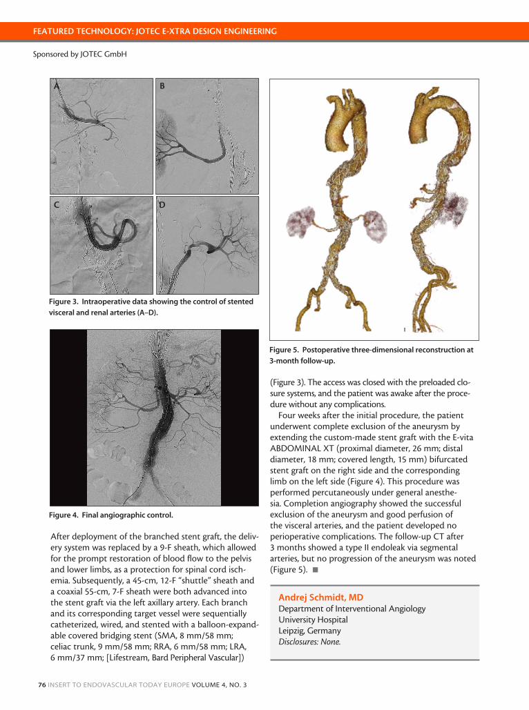
After deployment of the branched stent graft, the deliv-ery system was replaced by a 9-F sheath, which allowed for the prompt restoration of blood flow to the pelvis and lower limbs, as a protection for spinal cord isch-emia. Subsequently, a 45-cm, 12-F “shuttle” sheath and a coaxial 55-cm, 7-F sheath were both advanced into the stent graft via the left axillary artery. Each branch and its corresponding target vessel were sequentially catheterized, wired, and stented with a balloon-expand-able covered bridging stent (SMA, 8 mm/58 mm; celiac trunk, 9 mm/58 mm; RRA, 6 mm/58 mm; LRA, 6 mm/37 mm; [Lifestream, Bard Peripheral Vascular])
(Figure 3). The access was closed with the preloaded clo-sure systems, and the patient was awake after the proce-dure without any complications.
Four weeks after the initial procedure, the patient underwent complete exclusion of the aneurysm by extending the custom-made stent graft with the E-vita ABDOMINAL XT (proximal diameter, 26 mm; distal diameter, 18 mm; covered length, 15 mm) bifurcated stent graft on the right side and the corresponding limb on the left side (Figure 4). This procedure was performed percutaneously under general anesthe-sia. Completion angiography showed the successful exclusion of the aneurysm and good perfusion of the visceral arteries, and the patient developed no perioperative complications. The follow-up CT after 3 months showed a type II endoleak via segmental arteries, but no progression of the aneurysm was noted (Figure 5). n
Figure 3. Intraoperative data showing the control of stented
visceral and renal arteries (A–D).
Figure 4. Final angiographic control.
Figure 5. Postoperative three-dimensional reconstruction at
3-month follow-up.
A B
C D
Andrej Schmidt, MD Department of Interventional AngiologyUniversity Hospital Leipzig, GermanyDisclosures: None.