Embed Size (px)
Citation preview
Epilepsy Research (2013) 107, 115—120
jo ur nal ho me p ag e: www.elsev ier .com/ locate /ep i lepsyres
Eating epilepsy: Phenotype, MRI, SPECT andvideo-EEG observations
M. Patela, P. Satishchandraa, J. Sainib, R.D. Bharathb,S. Sinhaa,∗
a Department of Neurology, NIMHANS, Bangalore, Indiab NIIR, NIMHANS, Bangalore, India
Received 18 April 2013 ; received in revised form 30 July 2013; accepted 14 August 2013Available online 2 September 2013
KEYWORDSEating epilepsy;MRI;Perisylvian area;SPECT;Video-EEG
SummaryBackground: Eating epilepsy is one of the rare forms of reflex epilepsy precipitated by eating.Previous studies have demonstrated lesions due to variable aetiology involving the temporolim-bic and suprasylvian regions.Objective: To study anatomical correlates of reflex eating epilepsy using multimodality inves-tigations (MR imaging, video-EEG and SPECT).Methodology: Six patients (M:F = 3:3; mean age: 20.7 ± 4.9 years) with eating epilepsy weresubjected to MRI of brain, video-EEG studies and SPECT scan. These were correlated withphenotypic presentations.Results: Among the five patients with ictal recording of eating epilepsy during video-EEG, semi-ology was characterized by behavioural arrest followed by either flexion or extension of trunkand neck and two patients had speech arrest and four had salivation from angle of mouth.Another patient had EEG changes during ‘‘thought about eating’’. Four patients had perisylvianfrontal lobe lesions and one had high frontal lesion on MRI. Ictal EEG (n = 6) showed ictal rhythmicslowing/fast activity in parieto-temporal (n = 2) or fronto-temporal (n = 4) regions with subse-quent secondary generalization in three. Ictal and interictal SPECT imaging showed changes in
frontal lobe (n = 1), anterior temporal lobe (n = 1), and parieto-insular region (n = 1) suggestingit to be seizure onset zone. Three of four patients with structural lesions in MRI had concordantictal EEG and ictal SPECT changes.Conclusion: Lesions near the perisylvian region might play a major role in eating epilepsy.© 2013 Elsevier B.V. All rights re∗ Corresponding author at: National Institute of Mental Health andNeurosciences (NIMHANS), Hosur Road, Bangalore 560029, India.Tel.: +91 80 26995150; fax: +91 80 26564830.
E-mail address: sanjib [email protected] (S. Sinha).
I
Ete
0920-1211/$ — see front matter © 2013 Elsevier B.V. All rights reserved.http://dx.doi.org/10.1016/j.eplepsyres.2013.08.005
served.
ntroduction
ating epilepsy is characterized by seizures closely relatedo one or several parts of eating. Although described asating epilepsy, seizures triggered by eating often occur in

1
pnsTLiColaiget(
bi(e
P
StcsiowtS(w
(NilwemFSiofSitoi5f
SiDpnw
iwSaaSftifeafa
R
Sir1ymctpophho
eOriTopgcici(miaitc
Ttat
16
atients who also have spontaneous seizures and are notow classified as a separate epileptic syndrome but as aeizure type in the most recent proposal (Engel, 2001).he unusually high figures had been reported from Srianka (Senanayake, 1994). Earlier, thirteen cases of eat-ng epilepsy were reported from south India (Nagaraja andhand, 1984). Seizures with eating are typically of complexr simple partial type, almost always related to symptomaticocalization-related epilepsy (Remillard et al., 1998). Therere reports with different underlying etiologies like hypoxicschaemic encephalopathy, cortical dysplasia, polymicro-yria causing eating epilepsy (Kishi et al., 1999; Loretot al., 2000; Manyam et al., 2010). There are very few func-ional neuroimaging studies in patients with eating epilepsyBlauwblomme et al., 2011; Loreto et al., 2000).
The aim of this study was to study the regions of therain involved in patients with eating epilepsy by MR imag-ng (MRI), Single photon emission computed tomographySPECT), and video-EEG observations in patients with eatingpilepsy.
atients and methods
ix patients with eating epilepsy were evaluated prospec-ively at a university teaching hospital, a major tertiaryare referral centre for neuro-psychiatric disorders inouth India. Approval from Institute Ethics Committee andnformed written consent from patients or relatives wasbtained. All the patients underwent detailed evaluationith clinical history, physical and neurological examina-
ion, and investigations with MRI of brain, video-EEG andPECT. Patients underwent MRI of brain on a 3T MRI machineAchieva, Philips) with standard epilepsy protocol. The MRIas evaluated independently by the investigators (SS, RDB).
Video-EEG was conducted in the epilepsy monitoring unitEMU), clinical neurophysiology laboratory, Department ofeurology at our centre in all the patients. The record-
ngs of the study were carried out in a semi sound proofaboratory in a standard laboratory setting. Scalp EEGsere recorded on 16-channel ‘‘Galileo NT (EBN)’’ machine,mploying international 10—20 system of electrode place-ent using standard parameters and procedures e.g. High
ilter — 70 Hz; Low Filter — 0.1 Hz; Recording time: 30 min;ensitivity: 7 �V/mm; sampling rate: 256 Hz. The electricalmpedance was kept below 5 K�. The reference electrodesf the monopolar montage were linked to earlobes. Sur-ace EMG electrodes (filter band pass — 20 Hz to 2 KHz;weep speed — 10 s/page) were placed on biceps or tib-alis anterior muscles. Video-EEG was visually analysed byhe investigators (SS, PSC) to look for the clinical semiol-gy, ictal and interictal EEG findings. Activation proceduresncluded hyperventilation, photic stimulation (5—50 Hz for
s at a stretch with eyes open and closed) and after givingood in all during recording to precipitate seizures.
SPECT scans were performed on Symbia T6 True PointPECT machine available at NIMHANS. Radiotracer usedn this study was 99mTc-ECD (ethylenecysteinate dimer).
uring ictal and interictal studies, 20 mCi of freshly pre-ared 99mTc-ECD was injected intravenously followed byormal saline flush. Images of ictal and interictal studiesere acquired, between 45 min and 1 h after radioisotopeTsIh
M. Patel et al.
njection, using a dual-head gamma camera equippedith low energy high resolution collimators (Symbia T6;iemens, Erlanagen, Germany). All the patients underwentn identical protocol. Interictal SPECT was carried out inll the patients with seizure free interval of 24 h. IctalPECT was performed after inducing reflex epilepsy withood and radiotracer (ECD) was injected immediately athe onset of the seizures. Tracer was injected with meannterval of 12.7 s after onset of ictus with range of 10—15 srom the onset of clinical seizure. Video recording of ictalvent was done in all patients during procedure. Interictalnd ictal SPECT images were evaluated for evidence ofocal hyperperfusion or hypoperfusion abnormality by visualnalysis by Nuclear medicine specialist.
The data was entered in digital spread sheet.
esults
ix patients (M:F = 3:3; mean age: 20.7 ± 4.9 years) with eat-ng epilepsy were recruited for the study. The age at onsetanged from 8 to 14 years (mean: 11.3 ± 2.16 years; median:1.5 years). The mean duration of epilepsy was 8.1 ± 4.9ears (median: 7.5; range: 3—16 years). Two patients hadild mental retardation. One patient had history of febrile
onvulsions in childhood. Six (86%) patients had uncon-rolled seizures despite polytherapy. Post-evaluation, oneatient underwent lesionectomy and was seizure free post-peratively. Five patients had abnormal MRI of brain. Fouratients underwent ictal SPECT studies while five patientsad ictus during video-EEG recording. One patient (case 6)ad epileptiform activity in EEG provoked due to ‘thinkingf eating’ during video-EEG recording.
All the patients had ictus during the middle of their eatingxcept one patient who had seizures at beginning of meal.ne of the patient’s seizures was precipitated while eating
ice containing food only. All patients had arrest of activ-ty followed by either flexion/extension of neck and trunk.here was speech arrest in three and drooling from the anglef mouth in five. Interictal EEG was abnormal in four (57%)atients, which included localized sharp waves in three andeneralized spike wave discharges in one. Definite ictal EEGhanges (n = 5) showed ictal rhythmic slowing/fast activityn parieto-temporal (n = 2), fronto-temporal (n = 1), fronto-entral (n = 1) or fronto-centro-temporal (n = 1) regionspsilateral to the MRI lesion in 3 of them. In one of the patientcase 5), obvious ictal EEG changes was not noted due touscle/movement artefacts except for probably focal slow-
ng to theta range across right fronto-central region notedfter modifying the filter settings. One patient (case 6) hadncrease in sharp wave discharges over left fronto-centro-emporal region while thinking of eating without obviouslinical attack.
The MRI of brain showed structural lesion in 5/6 (83.3%).he MRI diagnosis included cortical dysplasia (n = 1), infan-ile stroke (n = 1), calcified healed neurocysticercosis (n = 1),nd unexplained gliosis (n = 2). The lesions were located nearhe perisylvian frontal lobe (n = 4) high frontal lesion (n = 1).
wo patients had only interictal SPECT and one of themhowed hypoperfusion in left frontal lobe with normal MRI.ctal and interictal SPECT in the other 3 patients showedyperperfusion in the frontal (n = 1), temporal (n = 1), and
Eating epilepsy
117
Table 1 Clinical data and results of multi-modal investigations of seven patients of eating epilepsy.
Case 1 Case 2 Case 3 Case 4 Case 5 Case 6
Age 26 26 17 23 16 16Gender F M M F F MAge at seizure
onset10 14 13 11 8 12
Seizure semiology Neck extension,speech arrest,salivation
Neck and trunkextension, flexionof both UL,salivation
Forward Neck andtrunk flexion withnose wiping
Speech arrest,salivation, openingof jaw
Forward Neckflexion, salivation
Forward Neckflexion, salivation,speech arrest
Trigger stimuli Eating Eating rice madefood
Eating Eating Eating Eating
Spontaneousseizures
Yes Yes Yes No Yes Yes
Seizure frequency 5—6/month 3—4/week 1—2/day 1—2/day 1—2/day 1—2/weekMedications Polytherapy Polytherapy Polytherapy Polytherapy Polytherapy PolytherapyNeurological
examinationNo deficit Mild mental
retardationNo deficit No deficit Mild mental
retardationNo deficit
MRI brain Leftfronto-parietalperisylviancortical dysplasia
Right > Left sylvianand suprasylviangliosis
Right frontal andperisylvian gliosis
Left frontalcalcifiedgranuloma
Normal Left frontalperisylvian gliosis
Interictal EEG Left central thetawaves
Left fronto-centralsharp waves
Normal Normal Normal Left fronto centralsharp waves
Ictal EEG Leftparieto-temporalictal slowing
Right Fronto-centro-temporalbeta fast activity
Rightfronto-temporocentral ictalslowing
Left fronto centralictal slowing
Focal slowing totheta range acrossrightfronto-centralregion
Increased sharpwaves across leftfronto-centro-temporal regionon thinking ofeating
SPECT localization Leftfronto-parietal
Left hemispheric-posteriorfrontal/parietal
Normal(inter-ictal)
Left frontal Right medialfrontal
Left frontal(interictal)
F, female; M, male; NCC, neurocysticercosis; SPECT, single photon emission computed tomography.

118 M. Patel et al.
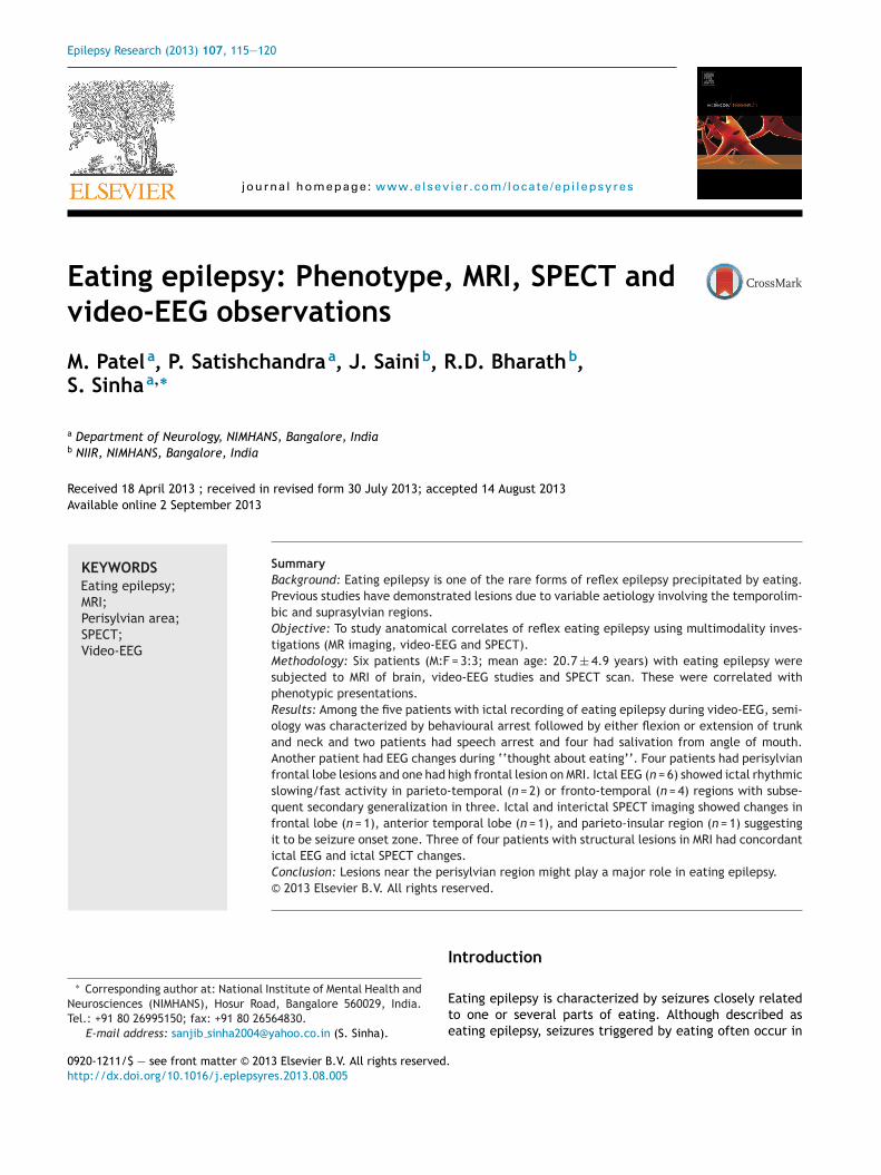
Figure 1 Multi-modality investigation in patient 1: (A and B) MRI FLAIR axial view (A) and MRI T1W sagittal view (B) showed glioticlesion with volume loss in suprasylvian left fronto-parietal lobe; C,c; D,d; E,e: SPECT (axial, sagittal and coronal view) interictals an int eta f
podbdofoF
D
D
Teoo
A
Ir
weiswsdp(mTs(
C
Atsw
can showed hypoperfusion which was well perfused in ictal scheta waves across C3-O1. (G) During ictus, ictal EEG showed b
arieto-insular (n = 1) regions as seizure onset zone. Threef four patients with structural lesions in MRI had concor-ant ictal EEG and ictal SPECT changes. One patient withilateral structural lesion had right hemispheric ictal EEGischarges whereas SPECT showed left hemisphere seizurenset zone suggesting two independent loci operating at dif-erent time. The details are provided in Table 1. The detailsf MRI, EEG and SPECT studies of patient 1 are provided inigs. 1 and 2.
iscussion
emography
he mean age at onset of seizures was in childhood in thearly part of 2nd decade (5/6) in our study. There are reportsf variable age at onset ranging from 4 to 48 years with meanf 21.6 ± 10.3 years (Remillard et al., 1998).
etiology
t is reported that eating epilepsy was almost exclusivelyelated to symptomatic focal epilepsy. There are reports
apis
left fronto-parietal region. (F) Interictal EEG showed runs ofast activity over F3, C3.
ith different underlying etiologies like hypoxic ischaemicncephalopathy, cortical dysplasia, polymicrogyria causingt (Kishi et al., 1999; Loreto et al., 2000). In the presenttudy, 5/6 (83.3%) patients had structural lesion on MRIhich included healed neurocysticercosis, cortical dyspla-
ia, perinatal stroke and gliosis. Previously authors haveocumented aetiological heterogeneity and variable patho-hysiological mechanisms which underlie the eating epilepsyRemillard et al., 1998). In the present cohort, lesions wereostly around perisylvian area: frontal — 4, parietal — 1.he occurrence of opercular and congenital bilateral peri-ylvian syndrome has been documented in eating epilepsyKishi et al., 1999; Manyam et al., 2010; Mateos et al., 1995).
linical attack
ll the patients in this study had seizures at the middle ofheir meal suggesting chewing, swallowing or gastric disten-ion as possible triggers. One patient had history of seizuresithin few minutes of initiating the meal suggesting prob-
bly a non-gustatory stimulus as trigger. Interestingly, oneatient had seizures on eating any ‘rice-containing food’,ndicating role of gustatory, olfactory or absorbed chemicalubstance (if any) as trigger. The same patient had bilateral
Eating epilepsy 119
Figure 2 Multi-modality investigation in patient 2: (A—C) MRI FLAIR axial view (B) and MRI T1W axial (A) and sagittal (C) viewshowed gliosis with volume loss in right > left sylvian and suprasylvian region; D,d; E,e; F,f: SPECT (axial, sagittal and coronal view)interictal scan showed hypoperfusion which was well perfused in ictal scan in left hemisphere — posterior frontal/parietal region.(F) Interictal EEG showed runs of theta waves across F3, C3. (G) During ictus, ictal EEG showed initial artefact followed rhythmic
S
ThtpctIMavw
oafi
beta fast activity right fronto-centro-temporal region.
perisylvian frontal lesion on MRI whereas others had unilat-eral parietal (n = 1) or frontal lesions (n = 2). Some commonpattern in semiology on video was recorded in five patients.All the patients had arrest of behavioural activity followedby either flexion or extension of trunk and neck and threepatients had speech arrest and four had salivation fromangle of mouth suggesting extra-temporal epilepsy. Suchregional involvement might indicate that the final effecterpathways were near face area, pharynx and neck in thefrontal lobe. There were reports of temporal lobe semiologyin eating epilepsy (Remillard et al., 1998).
EEG changes
Interictal abnormality was seen in 4/6 (57%) patients. In pre-vious studies, 32% to 76% of patients have reported interictalEEG (Koul, 1991; Nagaraja and Chand, 1984). In previous
study, ictal EEG had shown involvement of fronto-centraland temporal regions (Remillard et al., 1998) similar tothe finding of parietotemporal and frontocentral ictal onsetzone on EEG.ai(s
PECT observations
here are no studies of SPECT in eating epilepsy. In our study,ypoperfused areas on interictal scan correlated with struc-ural lesion on MRI in 3/4 patients with abnormal MRI. Threeatients with structural lesion on MRI and ictal SPECT hadoncordance. This finding further confirms the role of struc-ural lesion in seizure onset zone in eating epilepsy patients.t was interesting to note that case 3 with bilateral lesion onRI had ictal onset zones in right hemisphere on ictal EEGnd left hemisphere on SPECT scan. It is likely that duringideo-EEG and SPECT scan different seizures were recordedith onset from either hemisphere.
Functional MRI had also demonstrated that therbitofrontal and temporo-insulo-opercular corticesre parts of the central neuronal networks processingood stimuli (Porubska et al., 2006). Recent study haddentified interconnections between the gustatory cortex
nd insulo-hippocampal epileptogenic circuit to be involvedn eating epilepsy by fMRI and intracranial EEG observationsBlauwblomme et al., 2011). In the present study, MRIhowed structural lesion in perisylvian frontal area but
1
iido(
ertiiashfoanlaisoagiaolls
pfms(
C
N
R
B
E
K
K
L
M
M
N
P
R
20
ctal EEG and ictal SPECT showed wider ictal onset zonenvolving temporal and parietal lobe. It is difficult toetermine the exact ictal onset zone based on surface EEGr SPECT observations due to rapid seizure propagationVan Paesschen, 2004).
Remillard et al. (1998) had studied 10 surgically cases ofating epilepsy and found two types of localization, tempo-al and opercular. In the temporal subgroup, it was proposedhat context of the eating and meal is important. Whilen the opercular group, the type of food might play anmportant role. The thalamic sensory input to the dam-ged sensory cortex might have a role. Based on the presenttudy involving 6 patients with eating epilepsy, it might beypothesized that perisylvian region is an important zoneor eating epilepsy. This was documented in this study basedn clinical, MRI abnormalities, EEG changes and SPECT scanlterations. However except in one patient, this cohort didot undergo surgery. Nevertheless, there might be an over-ap between the 2 seizure onset subgroups. The triggers of
seizure are usually stereotyped for each patient, whichnclude gastric distension, chewing, swallowing, the pas-age of food along the oesophagus, specific chemical factorsr the mere sight of food and other complex stimuli, suchs movement and proprioceptive afferents of muscular ori-in, have been considered. Interestingly one of the patientsn this cohort had documented seizure during the thinkingbout eating. Seizures with eating are typically of complexr simple partial type, almost always related to symptomaticocalization-related epilepsy (Remillard et al., 1998). Simi-arly, the patients in this series also had localization relatedymptomatic (n = 5) and cryptogenic (n = 1) epilepsy.
To conclude, there might be an involvement of com-lex neuronal circuits around sylvian fissure (near area of
ace) responsible for eating epilepsy, one of the uncom-only encountered reflex epilepsy. Further studies with PET,imultaneous fMRI—EEG and possible intracranial recordingif clinically indicated) might provide better understanding.
S
V
M. Patel et al.
onflict of interest
one declared.
eferences
lauwblomme, T., Kahane, P., Minotti, L., Grouiller, F., Krainik,A., Vercueil, L., Chabardes, S., Hoffmann, D., David, O.,2011. Multimodal imaging reveals the role of gamma activityin eating-reflex seizures. J. Neurol. Neurosurg. Psychiatry 82,1171—1173.
ngel Jr., J., 2001. A proposed diagnostic scheme for people withepileptic seizures and with epilepsy: report of the ILAE TaskForce on Classification and Terminology. Epilepsia 42, 796—803.
ishi, T., Moriya, M., Kimoto, Y., Nishio, Y., Tanaka, T., 1999. Con-genital bilateral perisylvian syndrome and eating epilepsy. Eur.Neurol. 42, 241—243.
oul, R.L., 1991. Eating epilepsy. J. Assoc. Physicians India 39,393—395.
oreto, V., Nocerino, C., Striano, P.F.D.A., Boccella, P., Striano, S.,2000. Eating epilepsy: heterogeneity of ictal semiology: the roleof video-EEG monitoring. Epileptic Disord. 2, 93—98.
anyam, S.C., Kung, D.H., Rhodes, L.B., Newmark, M.E., Fried-man, D.E., 2010. Unilateral opercular lesion and eating-inducedseizures. Epileptic Disord. 12, 309—313.
ateos, V., Salas-Puig, J., Campos, D.M., Carrero, V., Ander-mann, F., 1995. Acquired bilateral opercular lesions orFoix—Chavany—Marie syndrome and eating epilepsy. J. Neurol.Neurosurg. Psychiatry 59, 559—560.
agaraja, D., Chand, R.P., 1984. Eating epilepsy. Clin. Neurol. Neu-rosurg. 86, 95—99.
orubska, K., Veit, R., Preissl, H., Fritsche, A., Birbaumer, N., 2006.Subjective feeling of appetite modulates brain activity: an fMRIstudy. Neuroimage 32, 1273—1280.
emillard, G.M., Zifkin, B.G., Andermann, F., 1998. Seizures
induced by eating. Adv. Neurol. 75, 227—240.enanayake, N., 1994. Reflex epilepsies: experience in Sri Lanka.Ceylon Med. J. 39, 67—74.
an Paesschen, W., 2004. Ictal SPECT. Epilepsia 45 (Suppl. 4), 35—40.