Embed Size (px)
Citation preview

Early results of ex vivo cultivated autologous limbal
epithelium transplantation for limbal stem cell deficiency
Himanshu Matalia, MD1,2
Arokiaraj Vincent, MSc2
D Kamesh, MSc2
Authors have no financial interest Poster 610
1. Cornea & Refractive Services, Narayana Nethralaya2. Narayana Nethralaya Stem Cell Research Laboratory
Narayana NethralayaNarayana Health City
Super Specialty Eye Hospital & Post Graduate Institute of Ophthalmology
Bangalore - INDIA

Clinical Features of LSCD Loss of palisades of Vogt
Kinoshita S et al JpJ Clin Ophth 1986
Tseng SC et al OCNA 1990
Conjunctivalization
Stippled appearance with
fluorescein
Dua HS Am J Ophthalmol 1990
Huang AJ IOVS 1991
Ch. Inflammation
Recurrent epi. break down
Limbal Stem Cell Defiency (LSCD)

Algorithm for LSCD
Establish Diagnosis of Stem cells deficiency(Clinical features & Impression Cytology)
Unilateral Disease Bilateral Disease
Focal or Partial
Total Deficiency
AMT CLAG Debridment
Cadaveric kerato-limbal allograft (KLAL) Live-related conjunctivo limbal allograft (Lr-CLAL)
Cultured limbal auto graft (best option )
KLAL Lr-CLAL Cultured live-related
limbal allograft
Outcome not very encouraging
Cultivated Autologous Limbal Epithelium Transplantation (CALET)
Most recent technique Best for unilateral LSCD Autologous cells, no immune suppression required No risk at donor site RepeatableLimitations Availability of the tissue culture facility Long-term outcome awaited
Pellegrini et al Lancet 1997; 349: 900-993 Tsai RJ, Li LM, Chen JK. N Engl J Med 2000;343(2):86-93Schwab et al Cornea 2000;19(4):421-6Koizumi et al IOVS 2002;43(7):2114-21

Purpose
• To describe the early results of our technique of ex vivo cultivated autologous limbal epithelial transplantation (CALET) for limbal stem cell deficiency (LSCD).
Methods
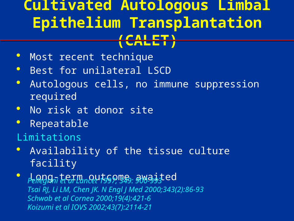
• Prospective, non-randomized interventional case series
• 14 eyes of 14 consecutive patient with LSCD underwent CALET from October
2008-October 2009.
• Primary outcome measures:
– Successful ocular surface reconstruction: stable ocular surface
– Best corrected visual acuity (BCVA)
• Secondary outcome measures:
– Complications
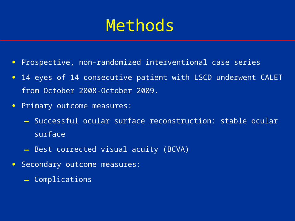
Culture technique Limbal biopsy
Normal limbal epithelium from contra lateral normal eye or normal area of same eye
2 x 2 mm (One clock hour) of normal limbal with cornea epithelium
Amniotic membrane De-epithelialized, processed &
preserved amniotic membrane used as substrate / carrier
Unique from others*
Monolayer only
No air lifting, no fibroblast feeder
Culture duration: 10-14 days
Human Corneal Epithelium medium
*Sangwan VS, Matalia HP et al Arch Ophthalmol 2005

Surgical technique• One drop 1:1000 epinephrine on pannus to reduce bleeding
(chemical cauterization)
• Pannus resection done
• Conjunctival peri-ectomy (2 mm) was done
• Cauterization of bleeders done
• Ex vivo cultivated autologous limbal epithelium grown over human
amnotic membrane (HAM) was spread over the ocular surface
• HAM was secured to the eye with fibrin glue (Tisseal) and 8-0 Vicryl
• Bandage contact lens applied at the end

Results
From July 2008
14 eyes of 14 patients underwent CALET
Female: Male -
Etiology of LSCD:
− Chemical injury 10 eyes (72%)
− 8 of these 10 were lime injury
7 eyes (50%): Previous history of AMT
Follow up: Mean 9 months
Mean duration of culture: 174.40 days
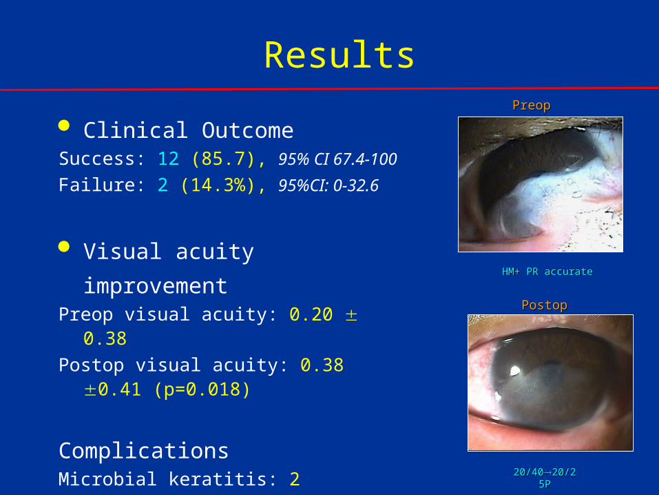
20/4020/4020/25P20/25P
PreopPreop
PostopPostop
Results
HM+ PR accurateHM+ PR accurate
Clinical OutcomeSuccess: 12 (85.7), 95% CI 67.4-100
Failure: 2 (14.3%), 95%CI: 0-32.6
Visual acuity improvementPreop visual acuity: 0.20 0.38
Postop visual acuity: 0.38 0.41 (p=0.018)
ComplicationsMicrobial keratitis: 2
Repeat transplants: 2
Repeat limbal biopsy: 2
Results
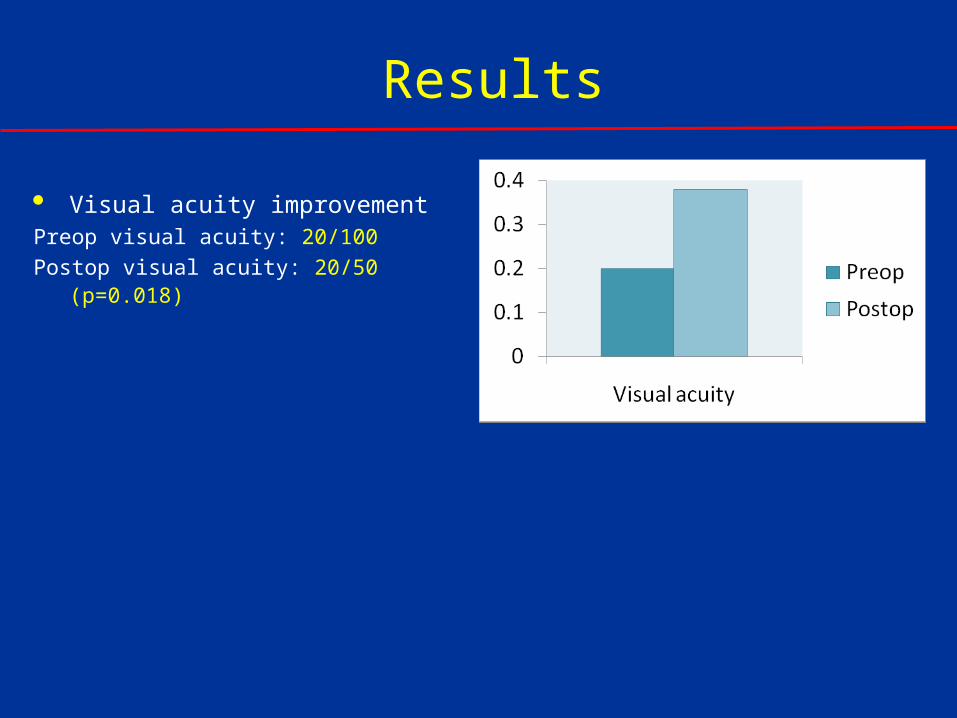
Visual acuity improvementPreop visual acuity: 20/100
Postop visual acuity: 20/50 (p=0.018)
Conclusions
• Cultivated autologous limbal epithelial
transplantation (CALET) could successfully
restore the ocular surface in cases with LSCD.