Embed Size (px)
Citation preview

e-News for Somatosensory Rehabilitation of Pain 2014, Tome 11 (Volume 1)
1
Powered by:
www.neuropain.ch
Serge MARCHAND GuGuesteditor
The official e-Journal of the Somatosensory Rehabilitation of Pain Network A peer-reviewed open-access journal
3 Marchand, S. Guesteditorial : Basic neurophysiology of the development, persistency and
treatment of pain [English]
21 Spicher & Létourneau. Somatosensory Rehabilitation Centre’s Statistics [English]
22 Bernardon, L. Article inédit: Fait clinique suite à une lésion cutanée profonde [F, Abstract in English]
29 Atlas des territoires cutanés (2ème édition) : Planche 4.13 - Préface (S.W. Carmichael) [Français]
31 Létourneau, E.. No Comment Nb 48 [English]
32 Spicher & Quintal. Article : La méthode de rééducation sensitive de la douleur (2003-2013) [F]
39 E. R. Témoignage No 48 d’une patiente : «Le mode zombie-cassé » [Français]
40 Hügli, A. Article : La confiance [Français]
41 Bhagavad gita. Aphorisme saisonnier [Hindi, Français, English, Español]
42 Fisher, J. Read for you : The Graded Motor Imagery Handbook [English]
44 Duchesne, D. Rééducation sensitive de la douleur : Module 3- Bruxelles 2013 [Français]
45 Spicher et al. Continuous Education – Weiterbildung - Formation continue [Français, English]
48 Spicher, C.J. Phénomène de la douleur No 1 [Français]
49 Certificat en rééducation sensitive de la douleur : 12 lauréat(e)s 2013 [Français]

e-News for Somatosensory Rehabilitation of Pain 2014, Tome 11 (Volume 1)
2

e-News for Somatosensory Rehabilitation of Pain 2014, Tome 11 (Volume 1)
3
Serge MARCHAND (PhD)1
Introduction
Pain is more then the mere representation of the nociceptive signal. From the periphery to the brain, the nociceptive signal will be modulated at all the levels of the central nervous system. The perception of pain is the finality of a complex series of endogenous mechanisms that will either emphasize the signal (excitatory mechanisms) or reduce the signal (inhibitory mechanisms). A chronic pain condition can produce changes that will affect these mechanisms in different ways. It is then not surprising that two patients presenting apparently similar pain problems may respond quite differently to the same treatment, since the physiopathology behind the pain is totally different.
Changes in the activity of the nociceptive system from the periphery to the cortex can be responsible for the development and maintenance of a chronic pain condition. However, they will implicate several different mechanisms that will respond differently to a treatment. Recent scientific data using brain imaging are supporting a cortical reorganization of white and gray matter in patients suffering from chronic pain [2; 26; 28; 32]. These observations are extremely important since these changes are reversible following an adequate treatment, including rehabilitation [15; 27; 47]. Based on our new understanding of the neurophysiology of pain, we are in front of a change of paradigm in the treatment of chronic pain. We can no longer look at chronic pain as a persistent acute pain condition, since the mechanisms and response to treatment will be totally different.
From nociception to pain perception
As we can see in figure 1, a nociceptive stimulation will recruit peripheral nociceptors that conduct the nociceptive signal in the primary somatosensory neuron to the dorsal horn of the spinal cord. In the dorsal horn, the primary afferent neuron will make a synaptic contact with secondary or projection neurons. Secondary neurons from the spinothalamic (lateral) and spinoreticular (medial) tracts will immediately cross in the spinal cord and send afferent projections to higher centers. A large proportion of afferents will make a second synapse in the lateral and medial nuclei of the thalamus, which subsequently make synaptic contact with tertiary neurons. It is important to emphasize that the secondary neurons may also synapse with neurons in different nuclei of the brainstem including the periaqueductal gray (PAG) and the nucleus raphe magnus (NRM), areas involved in descending endogenous pain modulation. Tertiary neurons from the thalamus send afferents to the primary and secondary somatosensory cortices (S1, S2). The S1 and S2 are involved in the sensory quality of pain,
1 Clinical Research Centre Etienne-le-Bel; 3001, 12th Avenue Nord; University of Sherbrooke, J1H 5N4 Sherbrooke (Qc), Canada.
GUESTEDITORIAL
Basic neurophysiology of the development, persistency and treatment of pain
To MD To neuroscientist To patient To therapist

e-News for Somatosensory Rehabilitation of Pain 2014, Tome 11 (Volume 1)
4
which includes location, duration and intensity. Tertiary neurons also project to limbic structures, including the anterior cingulate cortex (ACC) and the insula, which are involved in the affective or emotional component of pain.
Figure 1: Pain pathways from the periphery to the cortex. From [38]
Whenever one of the three levels of nociceptive neurons is making a synaptic contact, there is an integration of information that undergoes excitatory and inhibitory influences. These areas of integration are the targets of most analgesics. It is interesting to note that non-pharmacological treatments, such as somatosensory rehabilitation, are having therapeutic

e-News for Somatosensory Rehabilitation of Pain 2014, Tome 11 (Volume 1)
5
effects at all theses levels. For example, based on the gate control theory of Melzack and Wall [39], the activation of non-nociceptive signals by massage, vibration, high frequency TENS or any innocuous stimulation in a painful territory, will recruit inhibitory mechanisms in the spinal cord that will block the nociceptive signal. On the other hand, a nociceptive stimulation can recruit descending inhibitory mechanisms that will reduce the nociceptive signal all over the body [34; 56]. The same is also true for the higher centers. Cognitive information, such as expectations related to a treatment, can enhance or reduce the efficacy of endogenous pain modulations such as DNIC [22].
The initial nociceptive stimulus is therefore not the only factor contributing to pain perception. Between these two events are four steps marked by a series of chemical and electrical reactions: transduction, transmission, modulation, and perception. First, sensory transduction is the conversion of mechanical, thermal or chemical stimuli in chemoelectrical signal in specialized sensory nerve endings. Then, the neural signal will be transmitted from the periphery to the spinal cord, spinal cord to the brainstem and thalamus, and finally the thalamus to the cortex.
As previously mentioned, the nociceptive information that reaches the higher centers have undergone many excitatory and inhibitory influences at all levels of the central nervous system. The fourth step, the perception of pain, is the translation from a noxious stimulus to pain perception. However, pain perception can be present without peripheral nociceptive inputs and will be colored by emotions and the sum of the subject's past experiences.
To explain the physiological mechanisms of pain, we will briefly see the steps by which nociceptive information must pass before reaching consciousness. This neurophysiological knowledge is essential to understanding the phenomenon of pain and its modulation.
From the periphery to the spinal cord
Afferent fibers originating in the periphery fall into three groups, namely Aβ, C and Aδ fibers. The Aβ fibers are large myelinated fibers that conduct at high speed and usually transmit non-nociceptive signals. They do however also participate in pain modulation as will be later explained. The other two classes of fibers, the larger myelinated Aδ fibers and the thin unmyelinated C fibers mainly transmit nociceptive signals. It is important to underline that Aβ fibers can also play a role in pain, be responsible for static mechanical allodynia and even central sensitization [14].
We frequently refer to the nociceptors by the characteristics of their fibers. Myelination and increasing size of a nerve fiber facilitate the speed of conduction of the stimulus. The Aδ fibers conduct the signal relatively rapidly from the periphery to the spinal cord. Because of this rapid conduction velocity they are responsible for the sharp localization of pain and for the rapid spinal response, which can be measured in the laboratory as the nociceptive reflex. In contrast, the C fibers, which have a slow conducting velocity, will mediate a second or dull aching pain. They are also responsible for the first and second pain that we will see in details.
First and second pain
The differences in conduction velocity between the Aδ and C fibers can be appreciated when isolating the sensation of first and second pain. Following a brief nociceptive stimulation, the Aδ fibers will rapidly transmit a brief and acute pinprick-like sensation perceived to be precisely located at the point of stimulation. It is this precision and fast conduction that will result in the nociceptive withdrawal reflex. Following this activity, C fibers will transmit their
e-News for Somatosensory Rehabilitation of Pain 2014, Tome 11 (Volume 1)
6
information, with a relatively long delay (hundred of milliseconds to a second depending on the location of the stimulus). This second sensory input results in a more diffuse deep pain sensation.
It is possible to isolate first and second pain in the laboratory. Using a blood pressure cuff, we can temporarily block trophic factors present in the blood from reaching to the nerves. The first fibers that will show reduced activity are those with largest diameter, including the Aδ fibers. This allows the activity of C fibers to be isolated and independently studied. Following this procedure, a nociceptive stimulation, independent of the nature of the stimulation, hot, cold or mechanical, will be perceived with a certain delay as a deeper pain sensation.
The application of capsaicin, the hot pepper extract, will produce a burning sensation due to the activation of the vanilloid receptors on the C fibers. However, at higher doses, the C fibers will be blocked as a result of a specific action on ionic calcium channels, with resulting isolation of the Aδ fibers at the skin surface. This time, the same nociceptive sensation will be perceived as a sharp pinprick-like sensation without the second burning pain sensation.
The dorsal horns of the spinal cord contain a large network of synaptic convergence involving collateral fibers and interneurons. The passage in the sensory spinal cord is an important step in the modulation of the nociceptive signal. Its complex neural network, which comprises the primary nociceptive neuron terminals, interneurons, secondary projection neurons and neurons from the descending modulatory pathways, contains a variety of neurotransmitters and a large mosaic of receptors that will modulate nociceptive afferents before they are transmitted to the higher centers. Nociceptive activity can lead to excitatory activities and hyperalgesia.
Primary and Secondary Hyperalgesia
Hyperalgesia is defined as an exaggerated response to a normally painful stimulation. In the 1950s, Hardy proposed that two kinds of hyperalgesia could affect the skin: primary hyperalgesia, occurring directly at the site of injury, and secondary hyperalgesia, with its origins in the central nervous system (CNS) [23]. Primary hyperalgesia can be explained by the release of different inflammatory factors in the periphery, which leads to the recruitment of nociceptors near the site of the injury. After an injury, several pronociceptive substances are released in the periphery (potassium, prostaglandins, bradykinin, histamine, substance P, and serotonin), which has the effect of recruiting nociceptors and producing sensitization. The injury site as well as the neighboring tissues will thus have lower pain thresholds.
Secondary hyperalgesia, on the other hand, can be explained by a central phenomenon that is known by the general term ‘central sensitization’ [51]. Repeated recruitment of C fibers after an injury can cause a series of events at the spinal level, which could have the effect of sensitizing the projection neurons in the dorsal horns of the spinal cord. High-frequency recruitment of C fibers (small nociceptive fibers) is known as ‘wind-up’ [16]. Wind-up is a relatively short-lived transient phenomenon, but the repeated recruitment of C fibers can also lead to spinal sensitization, which may extend over several hours or even several days [55].
Thus, an intense, long-lasting stimulation will result in the recruitment of nociceptive fibers, including C fibers, which release excitatory amino acids (EAAs), glutamate, and peptides, such as substance P and CGRP (calcitonin gene-related peptide). These neurotransmitters recruit postsynaptic glutamatergic receptors such as AMPA (α-amino-3-hydroxyl-5-methyl-4-isoxazole propionate) and NMDA (N-methyl-D-aspartate) in the case of EAAs, and

e-News for Somatosensory Rehabilitation of Pain 2014, Tome 11 (Volume 1)
7
neurokinin-1 receptors in the case of substance P. Prolonged stimulation of the NMDA receptors will produce long-lasting cellular sensitization through the activation of gene transcription factors (c-fos and c-jun). These transcription factors induce the expression of some rapidly responding nuclear genes, in turn leading to nociceptor sensitization. This structural plasticity will have the effect of reducing the recruitment threshold of the nociceptors and thus producing hyperalgesia or allodynia, which could persist after the healing of injury. On the clinical side, the phenomenon of central sensitization allows us to better understand the importance of relieving pain as early as possible in order to avoid chronification.
We can measure this phenomenon of central sensitization using either nociceptive stimulation at high frequency or continuous stimulation for minutes [52].
The identification of the source of hyperalgesia is mandatory since a patient suffering from primary hyperalgesia (nociception and inflammation) may have a good response to anti-inflammatory while if the patient is suffering from secondary hyperalgesia (central sensitization), s/he will need a treatment that will have a central effect on the neuronal hyperactivity such as anticonvulsants.
From the spinal cord to the higher centers
Before projecting their axons toward the higher centers, secondary neurons of the spinothalamic and spinoreticular tracts project toward the thalamic nuclei. The thalamic nuclei that receive afferents projections from the nociceptive tracts can be divided into two groups: nuclei of the ventrobasal complex (VPL, VPM) and those of the centromedian (CM) or intralaminar complex. The nuclei of the ventrobasal complex mainly receive their afferents from the spinothalamic tract and project in turn toward the primary (S1) and secondary (S2) somatosensory cortices. The sensory-discriminative component of pain, i.e., information about the location and identification of the painful stimulus (its nature and intensity), is attributed to these somatosensory projections. The CM nuclei mainly receive their afferents from the spinoreticular tract and project in turn toward the various structures of the limbic system. In the area of the medial thalamus, more than half of the neurons are nociceptive, and their receptive field often extends across the entire organism. In this area, impulses originate from the deeper laminae, through the spinothalamic tract and the paramedial tract (spinoreticulothalamic tracts and collaterals of the deep laminae of the spinothalamic tract). Fibers of this region emit signals in several areas of the ipsilateral cortex, particularly in the frontal lobe and the limbic system [25]. These last two targets are also responsible for the motivational-affective component of pain, a component associated with an unpleasant sensation and the desire to escape from the suffering.
This simplified division allows us to understand how, relatively early in the CNS, the various pain pathways project into regions that are specialized, serving either the sensory-discriminative component (spinothalamic tract), or the motivational-affective component (spinoreticular tract).
The cortex and pain perception
We have known for a long time that pain is a complex sensory and emotional experience demanding the participation of the higher centers of the CNS.
It is only once the nociceptive information is sent to the cortex that we can really speak of pain, since pain is a perception. Because an animal cannot tell us its perception of pain, we

e-News for Somatosensory Rehabilitation of Pain 2014, Tome 11 (Volume 1)
8
must refer to its nociceptive behaviors, suspecting that these behaviors are generally responses to pain. The last few decades have been crucial in identifying the role of the different cortical regions in pain. Dividing the cortical regions responsible for the sensory-discriminative and motivational-affective components of pain can simplify the presentation of the cerebral structures implicated in pain perception.
Since the first studies of cerebral imagery of the regions that play a role in pain using positron emission tomography (PET) [50], several subsequent studies have confirmed the participation of the four principal cerebral centers (Fig. 2): the primary somatosensory cortex (S1), in the postcentral gyrus of the parietal lobe; the secondary somatosensory cortex (S2), in the parietal operculum; the anterior cingulate cortex (ACC), in the cingulate gyrus; and the insula, in the lobe of the insular cortex (IC), which is found under the temporal and frontal lobes, in the Sylvian fissure [10]. Methods that involve making a lesion specific to structures or recording nerve cells in these same localized regions have only allowed us to have a fragmented view of the role of the cortex in pain. We have sufficient data to conclude that cortical structures such as S1 contribute to the sensory-discriminative component of pain, whereas the frontal, cingulate, and insular cortices are involved in the motivational-affective component [10; 30; 50].
Figure 2 : Pain matrix from [38]
Endogenous pain modulation mechanisms
The pain signal needs to be clear and emotionally salient for an individual to react rapidly and adequately to the nociceptive stimulus and care about the injury. However, in some conditions, the nociceptive signals have to be temporarily silenced to focus on actions required to reduce further harm and thus, increasing chances of survival. The pain perceived following a nociceptive stimulus would then be completely different depending on the context and situation. It is most likely for these reasons that the CNS has developed several complex

e-News for Somatosensory Rehabilitation of Pain 2014, Tome 11 (Volume 1)
9
endogenous facilitatory and inhibitory mechanisms that can either emphasize or reduce the perception of pain following a nociceptive stimulus depending on the circumstances.
It is then not surprising that endogenous pain modulation mechanisms is one of the domains in which there has been a major breakthrough in our understanding of pain and its treatment. Since the publication of the gate control theory by Melzack and Wall in 1965 [39], which states that pain information does not circulate in a linear manner, but is rather modulated upon its arrival at the spinal cord, several studies have confirmed that nociceptive information is modulated at all levels of the CNS. This modulation can be excitatory, increasing the nociceptive response, or inhibitory, producing analgesia [41]. Therefore, it is possible that disruption of these endogenous pain modulation mechanisms may be the source of certain types of chronic pain. Thus, persistent pain does not originate solely from an increase in nociceptive afferents; it can also result from a decrease in inhibition or an increase in central excitation. As Millan [41] described in his review of the literature, these endogenous mechanisms are extremely numerous and employ a large quantity of neurotransmitters, some of which, such as serotonin and noradrenaline (which are normally associated with the inhibitory endogenous pain mechanisms), can also play an excitatory role. The excitatory or inhibitory role of certain cells of the rostroventral medulla has been understood for some time [4; 18; 34].
Recent studies have highlighted the complexity of certain nerve impulse transmission mechanisms in the presence of a chronic condition, as is the case with the GABAergic response, which normally inhibits nociceptive responses, but under certain conditions can become excitatory [12].
Endogenous Facilitatory Mechanisms
The work completed by Fields describing the activation of “ON” cells and inhibition of “OFF” cells in the brainstem during nociceptive activity has demonstrated the importance of facilitatory mechanisms in amplifying the nociceptive response [19]. Central sensitization and descending facilitatory mechanisms are involved in the development and persistence of some chronic pain conditions [43-45]. Part of the neurophysiological effects of the nocebo comes from the activation of these facilitatory mechanisms [6; 17].
Considering the importance of these facilitatory mechanisms in pain, it will not be surprising that they may play a role in some pain conditions related to mental health such as anxiety disorders or depression. For instance, cholecystokinin (CCK) is an antagonist of placebo analgesia or a pro-nocebo [5]. In healthy subjects, it will block the placebo effect by antagonizing the analgesic effect of opioids induced by a placebo manipulation [5]. It is suggested that the effect of CCK on placebo and nocebo is related to an anxiety factor. CCK antagonists have been found to prevent this anxiety-induced hyperalgesia [11]. Considering the dual role of CCK in both pain modulation and in the persistence of anxiety or major depression [24; 37], it may play a role in the co-occurrence of pain in relation to anxiety or depression.
Endogenous Inhibitory Mechanisms
In order to clarify their role in the manifestation and treatment of pain, these endogenous mechanisms will be presented according to three levels of inhibition of CNS nociceptive afferents (see Fig. 3). These are: i) spinal mechanisms that produce localized effects; ii)

e-News for Somatosensory Rehabilitation of Pain 2014, Tome 11 (Volume 1)
10
descending inhibitory controls that produce diffuse effects; and iii) mechanisms of the higher centers that, depending on the circumstances, may be diffuse or local in nature.
Figure 3: Endogenous pain inhibition mechanisms. From [40]
Spinal Mechanisms: The Gate Control Theory
Since the famous gate control theory of Melzack and Wall [39], the modulation of nociceptive information when it enters the spinal cord has been well documented. Melzack and Wall proposed that the selective stimulation of large-caliber afferents—Aβ fibers—recruits inhibitory interneurons into the substantia gelatinosa of the dorsal horns of the spinal cord.

e-News for Somatosensory Rehabilitation of Pain 2014, Tome 11 (Volume 1)
11
According to their theory, represented in a simplified way in Fig. 3, level 1, the selective stimulation of large afferent Aβ fibers blocks the small nociceptive Aδ and C fibers in the substantia gelatinosa (laminae II) of the dorsal horn of the spinal cord.
According to the gate theory, the selective stimulation of non-nociceptive afferent fibers relieves pain by reducing the transmission of nociceptive information directly upon its entry into the spinal cord. This type of inhibition is segmentary, have an inhibitory effect in the territory of the dermatome stimulated.
It is interesting to analyze the gate control theory in order to better understand its implications in light of current knowledge. In the first place, it shows that nociceptive afferents (Aδ and C fibers) have excitatory contact with afferent fibers of the spinal cord, the secondary nociceptive neurons. The theory also argues that nociceptive afferents block the inhibitory interneurons of the substantia gelatinosa of the spinal cord, which results in the easier passage of the nociceptive impulse.
In addition, in the presence of certain neuropathic types of pain, non-nociceptive neurons can recruit secondary nociceptive neurons from the spinal cord and cause pain, which explains the phenomenon of allodynia or painful sensations following a normally painless stimulation. It is interesting to emphasize that certain types of pain involving allodynic and hyperalgesic responses may result in the loss of tonic inhibitors of the spinal cord [40; 53].
Descending Mechanisms: Diffuse Noxious Inhibitory Control (DNIC)—Conditioned Pain Modulation (CPM)
It was at the end of the 1970s that the concept of diffuse noxious inhibitory control (DNIC) was proposed [34; 35]. This model reveals how a localized nociceptive stimulation can produce a generalized hypoalgesia of nociceptive afferents. In the DNIC model, Le Bars and his colleagues argue that following a painful stimulation, in addition to transporting nociceptive information to the higher centers via the spinothalamic tract, afferent messages are sent to various centers of the brainstem, including the PAG and NRM, which will send inhibitory efferent messages to the various spinal segments and thus cause diffuse inhibition (see Fig. 3, level 2).
According to the DNIC theory, nociceptive stimulation activates the pool of nociceptive neurons corresponding to the spinal segment that they innervate while inhibiting the other nociceptive neurons of the spinal cord serving the rest of the body. By reducing the activity of multireceptive neurons, DNIC lessens the background noise and highlights the activity of specific neurons recruited by nociceptive stimulation [20]. According to this hypothesis, pain is not solely triggered by excitatory processes, but by the perception of a contrast between the activities of the excitatory and inhibitory neurons. This model gives a good explanation for the generalized inhibition of the pain produced by intense stimulation.
Several neurotransmitters are involved in the modulation exerted by the descending inhibitory system, including biogenic amine transmitters and endogenous opioids (endorphins) [41]. Biogenic amine transmitters, including serotonin and noradrenaline, are found in the brainstem, in the NRM and PAG, among other sites. Their spinal action is made possible because of noradrenergic receptors concentrated in the higher laminae of the spinal cord. Serotonin, coming from the serotonergic neurons, acts directly on the neurons of the dorsal horn to inhibit them.
In animals, injury to the dorsolateral funiculus, the principal efferent tract of the descending inhibitory system, causes hyperalgesia [1; 13], suggesting a tonic inhibition of the nociceptive

e-News for Somatosensory Rehabilitation of Pain 2014, Tome 11 (Volume 1)
12
message. The low concentration of serotonin or noradrenaline in the cerebrospinal fluid of certain patients who have chronic pain, such as fibromyalgia [46], raises the possibility of a deficiency in these inhibitory mechanisms. Some recent studies support this idea [29; 31; 33].
A better understanding of the descending inhibitory mechanisms allows us to take advantage of the potential role of certain serotonergic and noradrenergic drugs in pain, as in the case of the analgesic role of certain antidepressants. Since a disruption of the descending inhibitory systems has a good chance of producing diffuse pain [29; 36], this knowledge enables more rapid targeting of the populations that respond better to antidepressant drugs acting on these serotonergic or adrenergic inhibitory mechanisms [57].
Modulation of the Higher Centers of the Central Nervous System
Finally, the higher centers of the CNS play a dominant role in pain modulation (see Fig. 3, level 3). The past few years have provided abundant progress in knowledge of the contribution of the cortical regions responsible for the sensory and emotional components of pain. Improvements in cerebral imaging techniques have shed light on the action of the different higher centers in pain perception [9].
As we have seen previously, several regions of the higher nervous system participate in pain perception, including the S1 cortex, which acts on the sensory-discriminative component, and the limbic structures (cingulate cortex, insula), which have a role in the motivational-affective component. These regions are important with regard to pain modulation. A good example is the fact that frontal lobes lobotomy has been used to relieve pain in patients suffering from terminal phase cancer [3]. Severing the link between the frontal lobe, responsible for rational thought, and the limbic system, linked to the emotions, caused a dissociation between the intensity and the unpleasant aspect of pain in certain patients, who stated that their pain was just as intense as before, but less unpleasant. These surgical interventions illustrate the contribution of the higher centers, but reveal little about the natural functions of these centers in pain modulation. Today surgical techniques are mostly giving place to central nervous stimulation approaches.
Another interesting example is the demonstration that manipulating the expectation related to an analgesic procedure can completely reverse the analgesic effect of endogenous pain modulation and the related pain experience. By suggesting that a procedure that is normally analgesic would produce more pain, subjects indeed reported more pain. Experimental pain was evoked through intermittent electrical stimulations of the left ankle over the retromalleolar path of the sural nerve. When sufficiently intense, this type of stimulation triggers a nociceptive spinal withdrawal reflex (measured by electromyographic recordings of the knee flexor muscle—RIII) and somatosensory evoked potentials (SEP) (by scalp electroencephalographic electrodes), whose amplitude correlates with stimulation intensity. During immersion, there was a significant reduction in perceived sural nerve pain, reflex amplitude and SEP in patients who correctly expected that the immersion would have analgesic properties. On the other hand, participants who expected that the immersion would have pain-enhancing properties showed an increase in perceived sural nerve pain and a complete abolition of the normal reduction in reflex amplitude and significantly reduced SEP. Therefore, suggestion was able to totally block the endogenous analgesia normally recorded with DNIC [22]. Another group of investigators obtained similar results with a strong exogenous analgesic, morphine. Morphine analgesia was potentiated or inhibited depending on the instruction that was given to the subject [7].
e-News for Somatosensory Rehabilitation of Pain 2014, Tome 11 (Volume 1)
13
These results support the idea that cognitive information can modulate the efficacy of endogenous and exogenous analgesia and emphasizes the importance of the patient’s expectations regarding analgesia.
It is increasingly clear that brain plasticity contributes to the perception of pain, which may explain the role of higher centers in certain chronic pain conditions. Changes in brain gray matter have been reported in patients suffering from chronic pain [8]. Studies in patients suffering from fibromyalgia, low back pain or headache have reported loss of cortical gray or white matter (or connectivity) [2; 26; 28; 32]. As disturbing as brain changes with chronic pain can sound, it is important to underline that studies are also reporting that this loss of brain matter can be reversed after an adequate treatment [47]. Interestingly, these cortical changes can happen quite rapidly and are related to personal characteristics. In a study where nociceptive stimulations were repeated daily over a period of eleven days, healthy subjects who sensitized (more temporal summation over time) had a significant reduction of cortical gray matter density in the anterior cingulate cortex, the insular cortex and the frontal cortex than the non sensitized subjects [49]. These results raise the possibility that some subjects (the sensitizers) may be more prone at developing chronic pain than others.
Conclusion
Based on the understanding of pain neurophysiology, we may devise treatment plans for pain management in the clinical setting. Treatments could be aimed towards either reducing excitatory mechanisms or enhancing inhibitory activity. The first goal is to identify as best as possible the mechanisms implicated. For a nociceptive acute pain, depending on the nature of the injury, topical or systemic anti-inflammatory (NSAIDs) or analgesic treatments would be primarily indicated. However, even if the nociceptive activity is clearly identified to be peripheral, central sensitization may also have occurred.
Chronic pain is even more difficult to manage because of the complexity of pain mechanisms and the evolution of the pathology over time. Considering the central sensitization that is frequently implicated in chronic pain, the use of opioid, anticonvulsant or antidepressant drugs could be included in pharmacological treatment. However, the prediction of the better outcomes seems to depend on the mechanisms implicated. Therefore, strategies for the treatment of functional neuropathic pain will be focused either on reduction of nociceptive hyperactivity or on activation of endogenous inhibition. Anticonvulsants or gabapentinoids [25] will reduce sensory input by their effect on ion channels in hyperactive nociceptive neurons, whereas antidepressant medications will augment inhibition by their effect on serotonergic and noradrenergic systems [21]. There is good example of correlations between specific sensory profiles in chronic pain patients with specific treatments [54]. The same is also true for non-pharmacological treatments. For instance, the presence of allodynia, hyperalgesia, localized hypoalgesia, distortion of the body schema in the painful area (such as complex regional pain syndrome–CPRS) or a deficit of CPM may all ends up with a painful territory but will respond differently to different sensory approaches [21; 42; 48]. It is then important to try to better characterize the mechanisms implicated to choose the treatment with highest chance of success (Fig. 4).

e-News for Somatosensory Rehabilitation of Pain 2014, Tome 11 (Volume 1)
14
MECHANISTIC APPROACHES TO PAIN TREATMENT
PERIPHERAL
SPINAL
DESCENDING INHIBITION
SUPERIOR CENTERS
MECHANISMS
MECHANISMS
MECHANISMS
MECHANISMS
- Peripheral hyperalgesia
- Inflammation
- Spinal excitatory – inhibitory mechanisms
- NMDA, GABA, endorphins,
- Serotoninergic – noradrenergic descending inhibition
- …
- Cortical reorganisation
- Psychological factors
- …
TREATMENTS
TREATMENTS
TREATMENTS
TREATMENTS
Pharmacological
Pharmacological
Pharmacological
Pharmacological
- Antihistaminic
- Opioidergic
- Endocannabinoidergic
- …
- Anticonvulsants
- NMDA Antagonists
- Opioids
- …
- Antidepressants
- SNRIs (venlafaxine, duloxetine)
- …
- Tricyclic antidepressants
- (expl: amitriptyline)
- SNRIs (venlafaxine, duloxetine)
- …
Non pharmacological
Non pharmacological
Non pharmacological
Non pharmacological
- Hot
- Cold
- Massages
- …
- TENS, massages
- …
- Acu-TENS
- Deep massage,
- Exercices
- …
- Relaxation
- Somatosensory rehabilitation
- Cognitive-behavioral therapy
Figure 4: Pain treatments based on identified mechanisms Schematic representation of mechanistic based pain treatment. The proposed pharmacological and non-pharmacological treatments are only examples and should be adapted according to available treatments and interaction with the targeted pain mechanisms in each patient.
e-News for Somatosensory Rehabilitation of Pain 2014, Tome 11 (Volume 1)
15
REFERENCES
[1] Abbott FV, Hong Y, Franklin KB. The effect of lesions of the dorsolateral funiculus on formalin pain and morphine analgesia: a dose-response analysis. Pain 1996;65(1):17-23.
[2] Absinta M, Rocca MA, Colombo B, Falini A, Comi G, Filippi M. Selective decreased grey matter volume of the pain-matrix network in cluster headache. Cephalalgia 2012;32(2):109-115.
[3] Barber TX. Toward a new theory of pain: Relief of chronic pain by prefrontal leucotomy, opiates, placebos and hypnosis. Psychological Bulletin 1959;56:430-460.
[4] Basbaum AI, Fields HL. Endogenous pain control mechanisms: Review and hypothesis. Annals of Neurology 1978;4(5):451-462.
[5] Benedetti F, Armanzio M, Casadio C, Oliaro A, Maggi G. Blockade of nocebo hyperalgesia by the cholecystokinin antagonist proglumide. Pain 1997;71:135-140.
[6] Benedetti F, Lanotte M, Lopiano L, Colloca L. When words are painful: unraveling the mechanisms of the nocebo effect. Neuroscience 2007;147(2):260-271.
[7] Bingel U, Wanigasekera V, Wiech K, Ni Mhuircheartaigh R, Lee MC, Ploner M, Tracey I. The effect of treatment expectation on drug efficacy: imaging the analgesic benefit of the opioid remifentanil. Sci Transl Med 2011;3(70):70ra14.
[8] Borsook D, Erpelding N, Becerra L. Losses and gains: chronic pain and altered brain morphology. Expert Rev Neurother 2013;13(11):1221-1234.
[9] Casey KL. The imaging of pain: Background and rational. In: KL Casey, MC Bushnell, editors. Pain Imaging. Seattle: IASP Press, 2000. pp. 1-29.
[10] Coghill RC, Talbot JD, Evans AC, Meyer E, Gjedde A, Bushnell MC, Duncan GH. Distributed processing of pain and vibration by the human brain. Journal of Neurosciences 1994;14(7):4095-4108.
[11] Colloca L, Benedetti F. Nocebo hyperalgesia: how anxiety is turned into pain. CurrOpinAnaesthesiol 2007;20(5):435-439.
[12] Coull JA, Boudreau D, Bachand K, Prescott SA, Nault F, Sik A, De Koninck P, De Koninck Y. Trans-synaptic shift in anion gradient in spinal lamina I neurons as a mechanism of neuropathic pain. Nature 2003;424(6951):938-942.
[13] Davies JE, Marsden CA, Roberts MH. Hyperalgesia and the reduction of monoamines resulting from lesions of the dorsolateral funiculus. Brain Research 1983;261(1):59-68.
[14] Devor M. Ectopic discharge in Abeta afferents as a source of neuropathic pain. Exp Brain Res 2009;196(1):115-128.
[15] Diers M, Yilmaz P, Rance M, Thieme K, Gracely RH, Rolko C, Schley MT, Kiessling U, Wang H, Flor H. Treatment-related changes in brain activation in patients with fibromyalgia syndrome. Experimental brain research Experimentelle Hirnforschung Experimentation cerebrale 2012;218(4):619-628.
[16] Eide PK. Wind-up and the NMDA receptor complex from a clinical perspective. EurJPain 2000;4(1):5-15.
[17] Enck P, Benedetti F, Schedlowski M. New insights into the placebo and nocebo responses. Neuron 2008;59(2):195-206.
[18] Fields HL, Heinricher M. Anatomy and physiology of a nociceptive modulatory system. PhilTransRSocLond 1985;308(1136):361-374.
[19] Fields HL, Malick A, Burstein R. Dorsal horn projection targets of ON and OFF cells in the rostral ventromedial medulla. Journal of Neurophysiology 1995;74(4):1742-1759.
[20] Gall O, Bouhassira D, Chitour D, Le Bars D. Involvement of the caudal medulla in negative feedback mechanisms triggered by spatial summation of nociceptive inputs. Journal of Neurophysiology 1998;79(1):304-311.

e-News for Somatosensory Rehabilitation of Pain 2014, Tome 11 (Volume 1)
16
[21] Geletka BJ, O'Hearn MA, Courtney CA. Quantitative sensory testing changes in the successful management of chronic low back pain. The Journal of manual & manipulative therapy 2012;20(1):16-22.
[22] Goffaux P, Redmond WJ, Rainville P, Marchand S. Descending analgesia--when the spine echoes what the brain expects. Pain 2007;130(1-2):137-143.
[23] Hardy JD, Wolff GH, Goodell H. Pain sensation and reactions. Baltimore: Williams & Wilkins, 1952.
[24] Hebb AL, Poulin JF, Roach SP, Zacharko RM, Drolet G. Cholecystokinin and endogenous opioid peptides: interactive influence on pain, cognition, and emotion. Prog Neuropsychopharmacol Biol Psychiatry 2005;29(8):1225-1238.
[25] Hodge CJ, Jr., Apkarian AV. The spinothalamic tract. Crit RevNeurobiol 1990;5(4):363-397.
[26] Ivo R, Nicklas A, Dargel J, Sobottke R, Delank KS, Eysel P, Weber B. Brain structural and psychometric alterations in chronic low back pain. Eur Spine J 2013;22(9):1958-1964.
[27] Jensen KB, Kosek E, Wicksell R, Kemani M, Olsson G, Merle JV, Kadetoff D, Ingvar M. Cognitive Behavioral Therapy increases pain-evoked activation of the prefrontal cortex in patients with fibromyalgia. Pain 2012;153(7):1495-1503.
[28] Jensen KB, Loitoile R, Kosek E, Petzke F, Carville S, Fransson P, Marcus H, Williams SC, Choy E, Mainguy Y, Vitton O, Gracely RH, Gollub R, Ingvar M, Kong J. Patients With Fibromyalgia Display Less Functional Connectivity In The Brain's Pain Inhibitory Network. Molecular pain 2012;8(1):32.
[29] Julien N, Goffaux P, Arsenault P, Marchand S. Widespread pain in fibromyalgia is related to a deficit of endogenous pain inhibition. Pain 2005;114(1-2):295-302.
[30] Kenshalo DR, Jr., Douglass DK, Bromm B, Desmedt JE. The role of the cerebral cortex in the experience of pain. In: B Bromm, JE Desmedt, editors. Pain and the brain: From nociception to cognition. New York: Raven Press, 1995. pp. 21-34.
[31] Kosek E, Hansson P. Modulatory influence on somatosensory perception from vibration and heterotopic noxious conditioning stimulation (HNCS) in fibromyalgia patients and healthy subjects. Pain 1997;70(1):41-51.
[32] Kuchinad A, Schweinhardt P, Seminowicz DA, Wood PB, Chizh BA, Bushnell MC. Accelerated brain gray matter loss in fibromyalgia patients: premature aging of the brain? Journal Neuroscience 2007;27(15):4004-4007.
[33] Lautenbacher S, Rollman GB. Possible deficiencies of pain modulation in fibromyalgia. ClinJPain 1997;13(3):189-196.
[34] Le Bars D, Dickenson AH, Besson JM. Diffuse noxious inhibitory controls (DNIC). 1. Effects on dorsal horn convergent neurones in the rat. Pain 1979;6(3):283-304.
[35] Le Bars D, Dickenson AH, Besson JM. Diffuse noxious inhibitory controls (DNIC). II.Lack of effect on non-convergent neurones,supraspinal involvement and theoretical implications. Pain 1979;6(3):305-327.
[36] Leonard G, Goffaux P, Mathieu D, Blanchard J, Kenny B, Marchand S. Evidence of descending inhibition deficits in atypical but not classical trigeminal neuralgia. Pain 2009;147(1-3):217-223.
[37] Lovick TA. Pro-nociceptive action of cholecystokinin in the periaqueductal grey: a role in neuropathic and anxiety-induced hyperalgesic states. Neurosci Biobehav Rev 2008;32(4):852-862.
[38] Marchand S, International Association for the Study of Pain. The phenomenon of pain. Seattle: IASP Press, 2012.
[39] Melzack R, Wall PD. Pain mechanisms: A new theory. Science 1965;150:971-979.
e-News for Somatosensory Rehabilitation of Pain 2014, Tome 11 (Volume 1)
17
[40] Millan MJ. The induction of pain: an integrative review. ProgNeurobiol 1999;57(1):1-164.
[41] Millan MJ. Descending control of pain. ProgNeurobiol 2002;66(6):355-474. [42] Moseley GL, Flor H. Targeting cortical representations in the treatment of chronic pain:
a review. Neurorehabil Neural Repair 2012;26(6):646-652. [43] Nijs J, Meeus M, Van Oosterwijck J, Roussel N, De Kooning M, Ickmans K, Matic M.
Treatment of central sensitization in patients with 'unexplained' chronic pain: what options do we have? Expert opinion on pharmacotherapy 2011;12(7):1087-1098.
[44] Ossipov MH, Dussor GO, Porreca F. Central modulation of pain. J Clin Invest 2010;120(11):3779-3787.
[45] Porreca F, Ossipov MH, Gebhart GF. Chronic pain and medullary descending facilitation. Trends Neurosci 2002;25(6):319-325.
[46] Russell IJ. Neurochemical pathogenesis of fibromyalgia. Z Rheumatol 1998;57 Suppl 2:63-66.
[47] Seminowicz DA, Wideman TH, Naso L, Hatami-Khoroushahi Z, Fallatah S, Ware MA, Jarzem P, Bushnell MC, Shir Y, Ouellet JA, Stone LS. Effective treatment of chronic low back pain in humans reverses abnormal brain anatomy and function. J Neurosci 2011;31(20):7540-7550.
[48] Spicher CJ, Mathis F, Degrange B, Freund P, Rouiller EM. Static mechanical allodynia (SMA) is a paradoxical painful hypo-aesthesia: observations derived from neuropathic pain patients treated with somatosensory rehabilitation. Somatosens Mot Res 2008;25(1):77-92.
[49] Stankewitz A, Valet M, Schulz E, Woller A, Sprenger T, Vogel D, Zimmer C, Muhlau M, Tolle TR. Pain sensitisers exhibit grey matter changes after repetitive pain exposure: a longitudinal voxel-based morphometry study. Pain 2013;154(9):1732-1737.
[50] Talbot JD, Marrett S, Evans AC, Meyer E, Bushnell MC, Duncan GH. Multiple Representations of Pain in Human Cerebral Cortex. Science 1991;251(1999):1355-1358.
[51] Terman GW, Bonica JJ, Loeser JD. Spinal Mechanisms and their modulation. In: JD Loeser, SH Butler, CR Chapman, DC Turk, editors. Management of pain, Vol. 3. Philadelphia: Lippincott Williams & Wilkins, 2001. pp. 73-152.
[52] Tousignant-Laflamme Y, Page S, Goffaux P, Marchand S. An experimental model to measure excitatory and inhibitory pain mechanisms in humans. Brain Research 2008;1230:73-79.
[53] Traub RJ. Spinal modulation of the induction of central sensitization. Brain Research 1997;778(1):34-42.
[54] von Hehn CA, Baron R, Woolf CJ. Deconstructing the neuropathic pain phenotype to reveal neural mechanisms. Neuron 2012;73(4):638-652.
[55] Woolf CJ. Windup and central sensitization are not equivalent. Pain 1996;66(2-3):105-108.
[56] Yarnitsky D, Arendt-Nielsen L, Bouhassira D, Edwards RR, Fillingim RB, Granot M, Hansson P, Lautenbacher S, Marchand S, Wilder-Smith O. Recommendations on terminology and practice of psychophysical DNIC testing. European Journal of Pain 2010;14(4):339.
[57] Yarnitsky D, Granot M, Nahman-Averbuch H, Khamaisi M, Granovsky Y. Conditioned pain modulation predicts duloxetine efficacy in painful diabetic neuropathy. Pain 2012;153(6):1193-1198.
e-News for Somatosensory Rehabilitation of Pain 2014, Tome 11 (Volume 1)
18
SOMATOSENSORY REHABILITATION of
PAIN NETWORK www.neuropain.ch
6, Hans-Geiler Street
Departement of CH - 1700 FRIBOURG Continuous education [email protected]
Sin
ce 2
00
6,
2
01
4’s
SO
MA
TOS
ENS
OR
Y R
EHA
BIL
ITA
TIO
N o
f P
AIN
What can we offer our patients suffering from neuropathic pain?
www.neuropain.ch/education/calendar
The 6th course for somatosensory rehabilitation of pain is a four day comprehensive theoretical and hands-on course for therapists, physicians and others, about a method to treat neuropathic pain patients (NPP).
Somatosensory Rehabilitation of Pain (Spicher, 2006) includes: Assessment of cutaneous sense disorders and their painful complications (CRPS, mechanical allodynia, neuralgia i.e post carpal tunnel syndrome release) and also rehabilitation.
Problem
Cutaneous sense disorders, including hypoaesthesia and/or mechanical allodynia are often significant contributors to chronic pain.
The normalisation of the cutaneous sense has a positive impact on neuropathic pain. The shooting pain, the burning sensations decrease, offering NPP a better quality of life.
Concepts
The concept of Aβ pain was proposed by Marshall Devor [Exp Brain Res 2009] many years after Tinel (1917) suggested that neuropathic pain is conducted partly through the Aβ fibers. The etiology of neuropathic pain hinges on this idea. It means that chronic neuropathic pain can arise from the alteration of the somaesthetic system and not only from the alteration of the C fibers. Therefore, the painful area must be carefully assessed in order to determine the presence of Aβ fibers lesions (hypoaesthesia and/or mechanical allodynia). Consequently, the normalisation of the cutaneous sense has a positive impact on neuropathic pain.
e-News for Somatosensory Rehabilitation of Pain 2014, Tome 11 (Volume 1)
19
Overall Learning Aims
To rehabilitate the disorders of the cutaneous sense on the basis of the neuroplasticity of the somaesthetic system;
To avert the outbreak of painful complications by rehabilitating the cutaneous sense;
To build bridges between rehabilitation, medicine and the neurosciences.
Instructors of the Somatosensory Rehab of Pain Network
Claude Spicher, Scientific collaborator (University of Fribourg – Neurophysiology Unit), Swiss certified HT; www.unifr.ch/neuro/rouiller/collaborators/spicher.php
Eva Létourneau, BSc OT (University of Montreal), Certified Somatosensory Rehabilitation of Pain, therapist in the Somatosensory Rehabilitation Ctr (Fribourg – Switzerland). linkedin.com/profile/letourneau
Course Informations
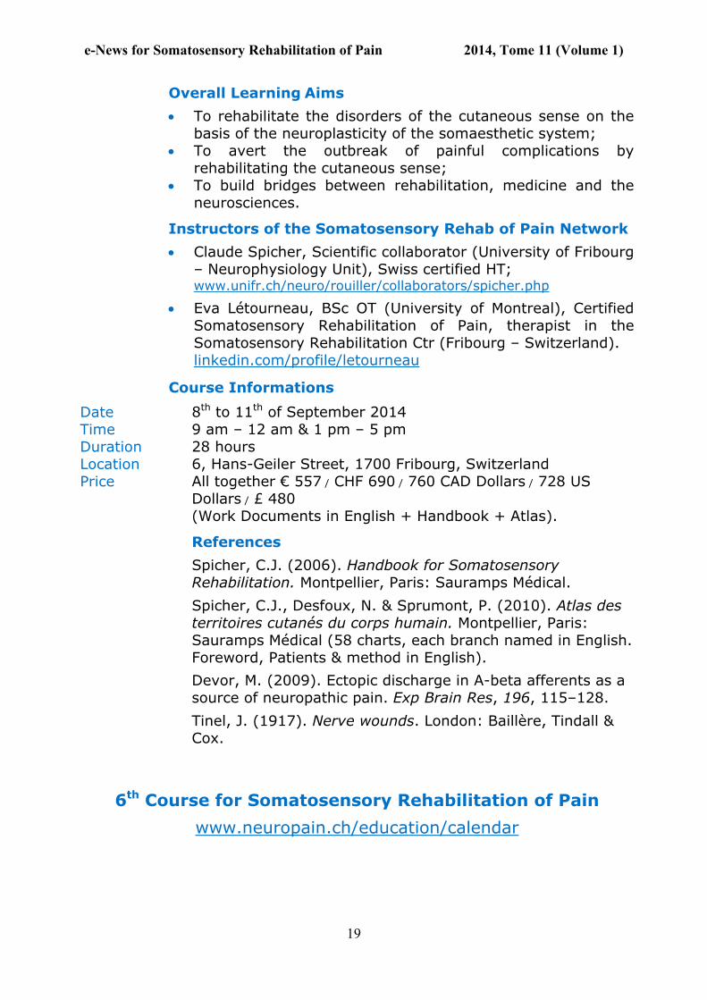
Date Time Duration Location Price
8th to 11th of September 2014 9 am – 12 am & 1 pm – 5 pm 28 hours 6, Hans-Geiler Street, 1700 Fribourg, Switzerland All together € 557 / CHF 690 / 760 CAD Dollars / 728 US Dollars / £ 480 (Work Documents in English + Handbook + Atlas).
References
Spicher, C.J. (2006). Handbook for Somatosensory Rehabilitation. Montpellier, Paris: Sauramps Médical.
Spicher, C.J., Desfoux, N. & Sprumont, P. (2010). Atlas des territoires cutanés du corps humain. Montpellier, Paris: Sauramps Médical (58 charts, each branch named in English. Foreword, Patients & method in English).
Devor, M. (2009). Ectopic discharge in A-beta afferents as a source of neuropathic pain. Exp Brain Res, 196, 115–128.
Tinel, J. (1917). Nerve wounds. London: Baillère, Tindall & Cox.
6th Course for Somatosensory Rehabilitation of Pain
www.neuropain.ch/education/calendar

e-News for Somatosensory Rehabilitation of Pain 2014, Tome 11 (Volume 1)
20
6th Course for Somatosensory Rehabilitation of Pain
(Since May 2006)
8th to 11th of September 2014
REGISTRATION FORM
Deadline: Monday, 30th June 2014
Name: First (given) name: Professional occupation: Address: e-mail address: Please fill and return to: Somatosensory Rehabilitation of Pain Network Department of Continuous Education 6, Hans-Geiler Street CH-1700 Fribourg Switzerland e-mail : [email protected] or Fax: +41 26 350 06 35

e-News for Somatosensory Rehabilitation of Pain 2014, Tome 11 (Volume 1)
21
Spicher, C.J., Quintal, I. & Létourneau, E. From the 1st of July 2004 until the 7th of November 2013, 2000 patients, with 3595 positive diagnostic testing of axonal lesions, have been assessed or treated.
Stage n Percentage
I 482 13 %
II 134 4 %
III 1510 2735
42 % 76 %
IV 1225 34 %
V 244 7 %
TOTAL 3595 100 %
Table I : 76 % of our clinical work among the five somaesthesic and / or neuropathic status of the 3595 axonal lesions (n= 2000 patients).
Neuralgia n
Trigeminal neuralgia i.e. : Right maxillary nerve (after domestic violence)
85
Occipital neuralgia i.e. : greater occipital nerve (after a whiplash injury)
86
Cervical neuralgia i.e. : posterior branch of C8
75
Brachial neuralgia i.e. : palmar branch of ulnar nerve
583
Dorso-intercostal neuralgia i.e. : anterior perforating branch of the 7th thoracic nerve
212
Lombo-abdominal neuralgia i.e. : abdominal branch of the ilioinguinal nerve (after a c-section)
109
Lombo-femoral neuralgia i.e. : lateral cutaneous nerve of thigh (meralgia paresthetica)
104
Crural neuralgia i.e. : medial cutaneous nerve of thigh
364
Sciatic neuralgia i.e. : lateral calcaneal branches of sural nerve
959
Sacral neuralgia i.e. : posterior labial nerves (pudendal neuralgia)
158
TOTAL 2735
Table II : Repartition of 2735 neuralgias – intermittent or incessant – of the 10 cutaneous departments of the human body (Spicher et al., 2013) assessed or treated from the 1st of July 2004 until the 7th of November 2013 at Somatosensory Rehabilitation Centre.
Somatosensory Rehabilitation Centre’s Statistics
To MD. To neuroscientist To patient To therapist

e-News for Somatosensory Rehabilitation of Pain 2014, Tome 11 (Volume 1)
22
BERNARDON, L.2 (erg DE, RSDC®)
ABSTRACT Background : Neuropathic pain, as described by patients with deep cutaneous lesion covered with split thickness skin graft, can limit therapeutics interventions that are necessary for a good scar evolution. Usual treatments, such as massages, water therapy and orthotics are difficult to tolerate or are used with great pain for patients during the course of therapy. The somatosensory rehabilitation of pain method is used to reduce neuropathic pain and decrease hypoaesthetic skin. Purpose : The aim of this clinical case report is to demonstrate the effectiveness of the somatosensory rehabilitation of pain method in facilitating the classical interventions of scar maturation with a patient with split thickness skin graft and neuropathic pain. Case presentation : Somatosensory rehabilitation of pain method, with its evaluations and its treatments, was proposed to Ms. M, a patient with split thickness skin graft on the dorsal part of the right foot. Ms M. described her neuropathic pain as stabbing, shooting and pricking. After the axonal lesions testing, the results suggest that intermittent femoropopliteal neuralgia of the superficial peroneal nerve (Spicher et al., 2010a) was present, with mechanical allodynia (stage III of axonal lesions). Mechanical allodynia was first treated by distant vibrotactile counter-stimulation approach. Rehabilitation of the underlying hyposensitivity was then performed using the somatosensory rehabilitation method. Results : After two months of treatment, the mechanical allodynia was removed, and after one month the patient had stability in progression with the hypoesthesia, but with better vibro-tactile perception than before the treatment begins. The results show a great evolution of the neuropathic pain and also a better tolerance with the usual treatment proposed to treat the scars. Conclusion : The somatosensory rehabilitation of pain method is a treatment of choice for patients with neuropathic pain that limits the treatment of the scar maturation after a split thickness skin graft. This method has even a positive effect on the skin graft, by increasing the vibrotactile perception. Keywords : Neuropathic pain, deep cutaneous lesion, skin graft, mechanical allodynia, hypoesthesia
2 Ergothérapeute DE, RSDC® Rééducatrice Sensitive de la Douleur Certifiée, Centre Médical de Rééducation Pédiatrique Romans Ferrari ; Rue de La Chanal ; BP 10118 ; 01701 Miribel cedex (France) [email protected]
ARTICLE INEDIT
La rééducation sensitive : Traitement proposé pour douleurs neuropathiques suite à une lésion cutanée profonde
Aux médecins Aux scientifiques en neurosciences Aux patients Aux thérapeutes
e-News for Somatosensory Rehabilitation of Pain 2014, Tome 11 (Volume 1)
23
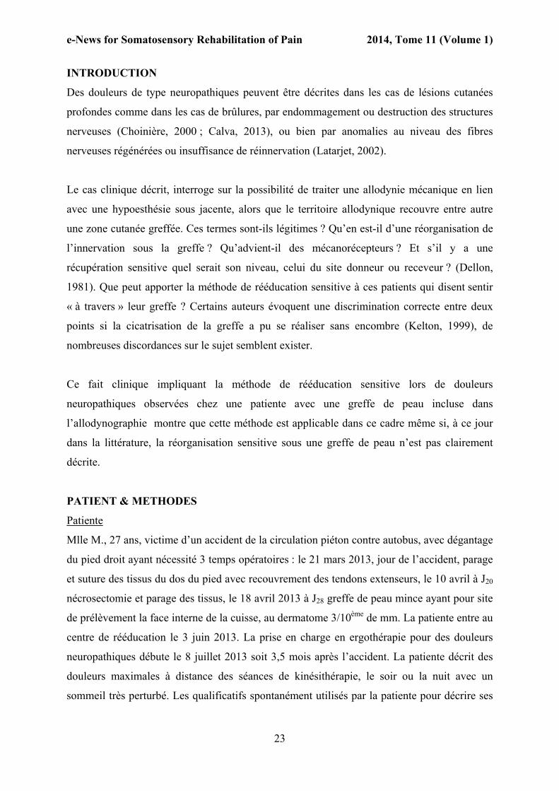
INTRODUCTION
Des douleurs de type neuropathiques peuvent être décrites dans les cas de lésions cutanées
profondes comme dans les cas de brûlures, par endommagement ou destruction des structures
nerveuses (Choinière, 2000 ; Calva, 2013), ou bien par anomalies au niveau des fibres
nerveuses régénérées ou insuffisance de réinnervation (Latarjet, 2002).
Le cas clinique décrit, interroge sur la possibilité de traiter une allodynie mécanique en lien
avec une hypoesthésie sous jacente, alors que le territoire allodynique recouvre entre autre
une zone cutanée greffée. Ces termes sont-ils légitimes ? Qu’en est-il d’une réorganisation de
l’innervation sous la greffe ? Qu’advient-il des mécanorécepteurs ? Et s’il y a une
récupération sensitive quel serait son niveau, celui du site donneur ou receveur ? (Dellon,
1981). Que peut apporter la méthode de rééducation sensitive à ces patients qui disent sentir
« à travers » leur greffe ? Certains auteurs évoquent une discrimination correcte entre deux
points si la cicatrisation de la greffe a pu se réaliser sans encombre (Kelton, 1999), de
nombreuses discordances sur le sujet semblent exister.
Ce fait clinique impliquant la méthode de rééducation sensitive lors de douleurs
neuropathiques observées chez une patiente avec une greffe de peau incluse dans
l’allodynographie montre que cette méthode est applicable dans ce cadre même si, à ce jour
dans la littérature, la réorganisation sensitive sous une greffe de peau n’est pas clairement
décrite.
PATIENT & METHODES
Patiente
Mlle M., 27 ans, victime d’un accident de la circulation piéton contre autobus, avec dégantage
du pied droit ayant nécessité 3 temps opératoires : le 21 mars 2013, jour de l’accident, parage
et suture des tissus du dos du pied avec recouvrement des tendons extenseurs, le 10 avril à J20
nécrosectomie et parage des tissus, le 18 avril 2013 à J28 greffe de peau mince ayant pour site
de prélèvement la face interne de la cuisse, au dermatome 3/10ème de mm. La patiente entre au
centre de rééducation le 3 juin 2013. La prise en charge en ergothérapie pour des douleurs
neuropathiques débute le 8 juillet 2013 soit 3,5 mois après l’accident. La patiente décrit des
douleurs maximales à distance des séances de kinésithérapie, le soir ou la nuit avec un
sommeil très perturbé. Les qualificatifs spontanément utilisés par la patiente pour décrire ses

e-News for Somatosensory Rehabilitation of Pain 2014, Tome 11 (Volume 1)
24
douleurs sont des coups de couteaux, des décharges électriques ou des aiguilles qui traversent
le pied.
Méthodes
L’application du questionnaire de la douleur St-Antoine (QDSA) et la réalisation d’une
allodynographie positive à 15 grammes (monofilament Semmes-Weinstein 5.18) de la
branche terminale intermédiaire du nerf péronier superficiel (face dorsale du pied),
provoquant une douleur à 3 (sur une EVA de 10 cm) à J0 me permettent d’évoquer une
névralgie fémoro-poplitée intermittente de la branche terminale intermédiaire du nerf péronier
superficiel (Spicher et al., 2010a) avec allodynie mécanique (stade III de lésions axonales). Le
traitement d’une allodynie mécanique (douleur résultant d’un stimulus normalement non
douloureux) est donc mis en place. Le contact étant contre indiqué pour que l’allodynie
mécanique puisse se résorber et pour éviter la flambée des douleurs a posteriori (Spicher et
al., 2010b), les stimulations mécaniques de la zone allodynique sont levées autant que
possible. Après présentation du bilan au médecin et au kinésithérapeute, suspension des
séances de kinésithérapie et des dispositifs de maturation et de croissance dermique (DMDG)
(Guillot, 2010) mis en place pour le traitement de la greffe cutanée. En revanche, maintien de
la chaussette compressive et poursuite de la déambulation avec béquilles.
Détermination de la zone de contre stimulation confortable au niveau du territoire du nerf
cutané sural latéral, 8 fois 1 minute par jour ou moins longtemps avec un morceau de cuir
doux, car nous ne possédons pas de peau de lapin.
Le jour où l’allodynographie (J56) est devenue négative, et où une esthésiographie secondaire
est cartographiée (Spicher et al., 2008), nous mettons en place le protocole de rééducation de
l’hyposensibilité secondaire durant 4 semaines, à savoir des stimulations de durée et
fréquence variant chaque semaine : de 12 fois 15 secondes par jour ou moins longtemps la 1ère
semaine, puis 8 fois 30 secondes, puis 6 fois 1 minute, puis 4 fois 3 minutes puis 4 fois 5
minutes à la 5ème semaine qui signe l’entrée dans la rééducation de l’hyposensibilité comme
s’il n’y avait jamais eu d’allodynie mécanique. Toutefois, l’esthésiographie secondaire (J56)
n’a pas pu être réalisée à 0,4g (face dorsale du pied Spicher et al., 2010a) car la peau greffée
ne permettait pas la perception d’une telle pression et la cartographie n’était donc pas
réalisable. Pour permettre une détection au travers de la greffe, l’esthésiographie secondaire a
donc été testée à 0,7g.

e-News for Somatosensory Rehabilitation of Pain 2014, Tome 11 (Volume 1)
25
RÉSULTATS
Les qualificatifs sensoriels notés au QDSA étaient les suivants :
A J0 = décharges électriques, coups de poignard, en étau sont cotées à 4, chaleur à 3,
irradiation, déchirure et fourmillements à 2.
A J28= pulsations et chaleur cotées à 2, piqure, compression et picotements à 1.
Temps en jours QDSA Allodynographie Arc-en-ciel des douleurs
J0 66 pts Positive
J7 Indigo 8,7 g
J28 13 pts Indigo 8,7 g
J35 Positive
J42 Violet 15,0 g
J56 0 pt Négative
Tableau I : Evolution de l’allodynie mécanique jusqu’au jour où l’allodynographie devient
négative après 2 mois de traitement.
L’allodynie mécanique a été levée après deux mois de contre stimulation vibrotactile à
distance (Tableau I). Nous retrouvons une zone d’hypoesthésie sous jacente à
l’allodynographie (Spicher et al., 2008), sur le territoire de la branche terminale intermédiaire
du nerf péronier superficiel, territoire correspondant à la greffe cutanée. Egalement, deux
autres territoires sont inclus dans la petite esthésiographie (correspondant aux branches
terminales médiale et latérale du nerf péronier superficiel) mais recouverts en partie par la
greffe de peau, ce qui peut expliquer la sensibilité diminuée et donc leur inclusion dans la
petite esthésiographie. La suspension des séances de kinésithérapie aura duré 3 mois
comprenant 2 mois de traitement par contre stimulation à distance pour l’allodynie mécanique
et 1 mois de rééducation d’hyposensibilité sous-jacente évoluant ensuite vers le traitement de
l’hyposensibilité (Tableau II, voir page suivante).
e-News for Somatosensory Rehabilitation of Pain 2014, Tome 11 (Volume 1)
26
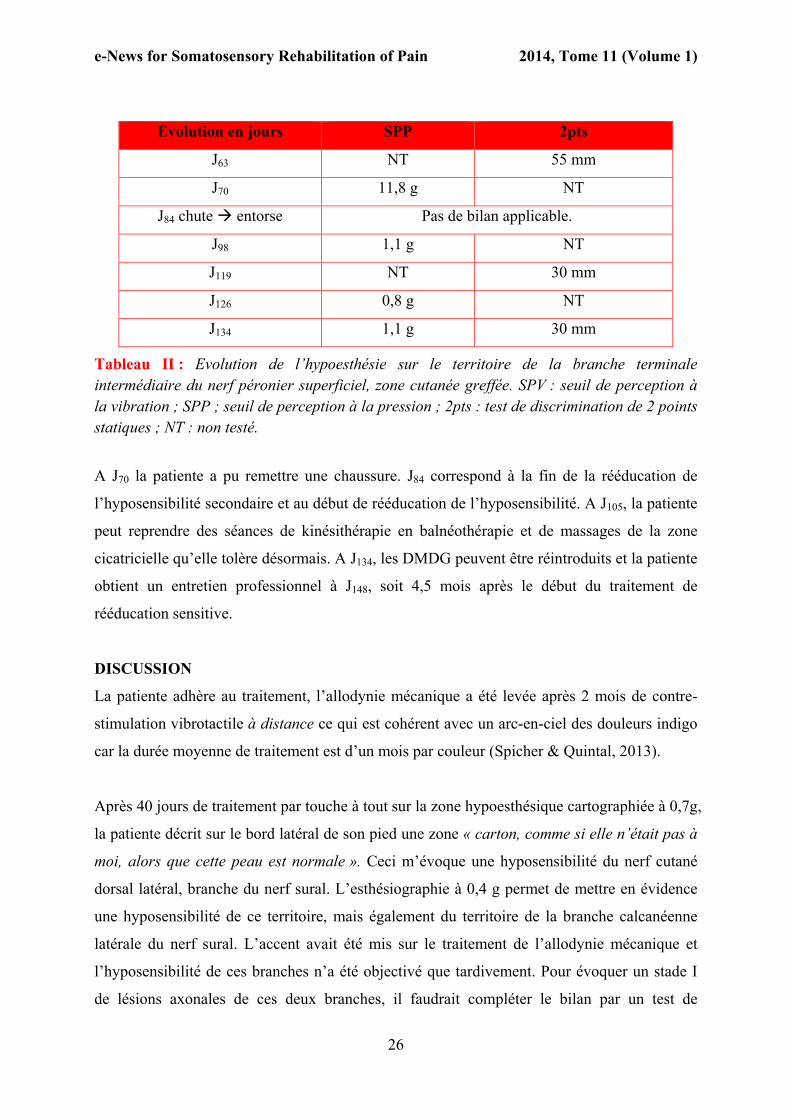
Evolution en jours SPP 2pts
J63 NT 55 mm
J70 11,8 g NT
J84 chute entorse Pas de bilan applicable.
J98 1,1 g NT
J119 NT 30 mm
J126 0,8 g NT
J134 1,1 g 30 mm
Tableau II : Evolution de l’hypoesthésie sur le territoire de la branche terminale intermédiaire du nerf péronier superficiel, zone cutanée greffée. SPV : seuil de perception à la vibration ; SPP ; seuil de perception à la pression ; 2pts : test de discrimination de 2 points statiques ; NT : non testé.
A J70 la patiente a pu remettre une chaussure. J84 correspond à la fin de la rééducation de
l’hyposensibilité secondaire et au début de rééducation de l’hyposensibilité. A J105, la patiente
peut reprendre des séances de kinésithérapie en balnéothérapie et de massages de la zone
cicatricielle qu’elle tolère désormais. A J134, les DMDG peuvent être réintroduits et la patiente
obtient un entretien professionnel à J148, soit 4,5 mois après le début du traitement de
rééducation sensitive.
DISCUSSION
La patiente adhère au traitement, l’allodynie mécanique a été levée après 2 mois de contre-
stimulation vibrotactile à distance ce qui est cohérent avec un arc-en-ciel des douleurs indigo
car la durée moyenne de traitement est d’un mois par couleur (Spicher & Quintal, 2013).
Après 40 jours de traitement par touche à tout sur la zone hypoesthésique cartographiée à 0,7g,
la patiente décrit sur le bord latéral de son pied une zone « carton, comme si elle n’était pas à
moi, alors que cette peau est normale ». Ceci m’évoque une hyposensibilité du nerf cutané
dorsal latéral, branche du nerf sural. L’esthésiographie à 0,4 g permet de mettre en évidence
une hyposensibilité de ce territoire, mais également du territoire de la branche calcanéenne
latérale du nerf sural. L’accent avait été mis sur le traitement de l’allodynie mécanique et
l’hyposensibilité de ces branches n’a été objectivé que tardivement. Pour évoquer un stade I
de lésions axonales de ces deux branches, il faudrait compléter le bilan par un test de

e-News for Somatosensory Rehabilitation of Pain 2014, Tome 11 (Volume 1)
27
discrimination de deux points statiques et la recherche des signes de fourmillements. Les
qualificatifs sont négatifs. Le territoire hyposensible recouvre donc les territoires de
distribution cutanée de 5 branches nerveuses dont 3 sont inclus dans la greffe de peau.
Aussi, il semble que les derniers bilans effectués [Seuil de Perception à la Pression (SPP) et
test de discrimination de 2 pts statiques (2 pts)] sur le territoire cutané greffé de la branche
terminale intermédiaire du nerf péronier superficiel, montrent une stagnation des résultats
depuis 4 semaines avec un SPP oscillant entre 0,8 et 1,1 g et un 2 pts à 30 mm, alors que le
SPP normal pour cette zone devrait être de 0,4 g et le 2 pts de 15 mm (Spicher & Quintal,
2013). Les semaines qui vont suivre me diront peut être si nous sommes bien au maximum du
potentiel de récupération sensitive de cette zone cutanée greffée. Ceci m’interroge sur la
présence ou non des mécanorécepteurs dans le tissu greffé et leur densité (Dellon 1981) et sur
la récupération sensitive évoquée par Kelton (1999).
CONCLUSION
L’organisation de l’innervation sous la greffe de peau n’est à ce jour pas clairement décrite
dans la littérature, mais la méthode de rééducation sensitive a été efficace dans ce cas de
douleurs neuropathiques. Elle a permis de reprendre les traitements habituellement mis en
place, d’éviter les préjudices esthétiques et surtout fonctionnels de la cicatrice, notamment au
niveau des rétractions et adhérences mais aussi des reprises d’appui et de la rééducation à la
marche. L’allodynie a pu être levée et l’hyposensibilité diminuée. La sensibilité retrouvée
semble être certes limitée mais nettement améliorée par rapport à la sensibilité présente avant
l’application de la méthode.
RÉFÉRENCES BIBLIOGRAPHIQUES
Calva, V. (2013). Névralgie fémoro-poplitée avec allodynie mécanique, suite à une lésion cutanée profonde greffée, traitée par la rééducation sensitive de la douleur. e-News Somatosens Rehab, 10(2), 64-69. http://www.neuropain.ch/sites/default/files/e-news/le-news_10_2_.pdf#page=11 (5.1.2013)
Choinière, M. (2000). Le point sur le traitement de la douleur chez les patients brûlés. Brûlures, 1(3), 128-135.
Dellon, A.L. (1981). Sensory corpuscules after nerve repair in Evaluation of sensibility and re-education of sensation in the hand. (pp 65-91). Baltimore : Williams and Wilkins.
Guillot, M., (2010). Principes généraux de rééducation fonctionnelle du brûlé. In Echinard, C., Latarjet, J., Les brûlures. (pp. 233-262). Paris : Elsevier Masson.

e-News for Somatosensory Rehabilitation of Pain 2014, Tome 11 (Volume 1)
28
Kelton, P.L., (1999) http://crafd.com.sg/files/lams_ext/Module6/9-01%20SKIN%20GRAFTS.pdf (15.12.2013)
Latarjet, J. (2002). La douleur du brûlé. Pathol Biol, 50, 127-133.
Spicher, C.J. & Quintal, I. (2013). La méthode de rééducation sensitive de la douleur. Montpellier, Paris : Sauramps Médical.
Spicher, C.J., Ribordy, F., Mathis, F., Desfoux, N., Schönenweid, F. & Rouiller, E.M. (2008). L’allodynie mécanique masque une hypoesthésie : Observations topographiques de 23 patients douloureux neuropathiques chroniques. Doul & Analg, 21, 239-251. http://doc.rero.ch/lm.php?url=1000,43,2,20090123161239-ME/rouiller_amm.pdf
(24.1.2014)
Spicher, C.J., Desfoux, N. & Sprumont, P. (2010a) Atlas des territoires cutanés du corps humain : Esthésiologie de 240 branches. Montpellier, Paris : Sauramps Médical.
Spicher, C.J., Anitiglio, D., Delaquaize, F., Crohas, A. & Vianin, M. (2010b). L’allodynie mécanique : une contre-indication temporaire pour certains traitements physiques. Mains libres, 5, 199-205.

e-News for Somatosensory Rehabilitation of Pain 2014, Tome 11 (Volume 1)
29
Branche antérieure du nerf cutané médial de l’avant-bras Anterior branch of medial antebrachial cutaneous nerve
Ramus anterior, nervus cutaneous antebrachii medialis
Branche antérieure du nerf brachial cutané interne
Le plus proximal
Face médiale
Le plus postérieur Face médiale
Territoire autonome Face médiale
Le plus antérieur Face médiale
Le plus distal Face palmaire
Département brachial
Planche anatomique 4.13 : les 5 éléments topographiques de la sensibilité vibrotactile
Le territoire autonome et les quatre bornes limitrophes du territoire maximal de distribution cutanée
Spicher, C., Buchet (- Desfoux), N. & Sprumont, P. (novembre 2013) Atlas des territoires cutanés du corps humain : Esthésiologie de 240 branches
2ème édition – Préface S.W. Carmichael (Mayo clinique) Montpellier, Paris : Sauramps médical
Aux médecins Aux scientifiques en neurosciences Aux patients Aux thérapeutes

e-News for Somatosensory Rehabilitation of Pain 2014, Tome 11 (Volume 1)
30
Préface de la 2ème édition : S.W. Carmichael
Les auteurs l'expliquent d'emblée: le point de vue de cet atlas est radicalement différent de celui des autres atlas décrivant la distribution des nerfs cutanés. Ils ont spécifiquement cartographié la sensibilité cutanée en partant de la périphérie vers le centre. Ils ont commencé par cartographier l'extension la plus distale du territoire cutané de chaque branche nerveuse. Puis ils ont progressé en direction proximale pour en déterminer les limites latérale et médiale jusqu'à atteindre finalement la limite la plus proximale. Cette approche est très utile pour les cliniciens parce que les neuropathies, typiquement, progressent du distal vers le proximal.
L’emphase mise sur la clinique est évidente puisque les cartes publiées proviennent d'observations faites chez près de 2000 patients souffrant de douleurs neuropathiques, soit presque le double du nombre de patients cartographiés dans la 1ère édition de cet atlas. Cet échantillonnage élevé a été validé grâce aux contributions de plusieurs experts internationaux. On trouve aussi une centaine de références d'anatomie clinique. L'observateur attentif remarquera des variations entre les cartes publiées dans ces ouvrages de référence et dans cet atlas. Ces fluctuations proviennent du fait que la distribution cutanée de chaque nerf présente des variations individuelles. Afin de rendre cet atlas valide malgré les variations de distribution cutanée, les auteurs ont fait le choix d'illustrer "le territoire maximal de distribution cutanée". On peut donc s'attendre à ce que, chez un patient donné, la distribution d'un nerf cutané s'établisse toujours à l'intérieur des frontières décrites dans l’atlas. Ces dernières ne peuvent pas être parfaitement précises mais les limites qu'elles indiquent seront précieuses pour le praticien confronté à des plaintes de type neuropathique.
En plus des territoires d’innervation, les grandes modalités de perception sensible ont également été présentées. Par exemple, la douleur est perçue pour des seuils inférieurs à ceux du toucher, donc le territoire des sensations douloureuses est typiquement plus étendu que celui du toucher pour un nerf donné. Ceci est dû au fait que des récepteurs répondant à diverses modalités de stimulus sont impliqués et que ce sont des fibres nerveuses différentes qui conduisent ces influx nerveux. En raison de ces différences, le tact (défini par la détection de 0,7 gramme appliqué sur la peau) est diminué alors que la douleur est perçue dans le même territoire.
Parmi l’ensemble des cartographies représentées, les planches anatomiques résumant les territoires des nerfs thoraciques sont l’une des parties les plus utiles de cet atlas. Ils fournissent au clinicien une image qui est, à la fois, facile à saisir et qui s'applique à beaucoup de situations cliniques. De plus, de nombreux schémas de territoires cutanés s'accompagnent de photographies explicitant l'extension des ramifications nerveuses. De même des tableaux judicieusement choisis fournissent aux cliniciens forts occupés des références synoptiques. Enfin, on trouvera à la fin de l'atlas les index des planches anatomiques et des tableaux, ce qui permet de mener le lecteur directement au nerf cutané qui l'intéresse.
Stephen W. Carmichael, Ph.D., D.Sc. Professeur émérite d’anatomie et de chirurgie orthopédique Mayo Clinique, Rochester, Minnesota, Etats-Unis d’Amérique

e-News for Somatosensory Rehabilitation of Pain 2014, Tome 11 (Volume 1)
31
Létourneau, E. (BSc OT, CSTP®)3
At first a static mechanical allodynia was found on the palmar branch of ulnar nerve on the left hand. With the evolution of the treatment, a second axonal lesion site was found with static mechanical allodynia for the anterior branch of medial cutaneous antebrachial nerve on the left forearm. With the treatment of the two lesions, the two statics mechanical allodynia disappear at the same time after 4 years of intermittent neuropathic pain.
Static Mechanical Allodynia Static Mechanical Allodynia
t0 t38
t19 t43
Allodynography : negative t58 t58 Allodynography : negative
15.0 gram - successive allodynographies for the palmar branch of ulnar nerve on the left hand tested from the 16th of July 2013 (t0) to the 28th of August 2013 (t43) on the palmar side of the left hand
15.0 gram - successive allodynographies for the anterior branch of medial antebrachial cutaneous nerve on the left forearm tested from the 9th of August 2013 (t0) to the 28th of August 2013 (t19) on the medial side of the left elbow
The allodynographies outlines the hypersensitive territories. The points are the ones at which, the application of the 15.0 gram - aesthesiometer (Semmes-Weinstein monofilament mark 5.18) is perceived as painful (3 / VAS 10 cm). The arrows indicate the axes along which the pain perception of 15.0 gram has been tested.
3 BSc OT, CSTP® Certified Somatosensory Therapist of Pain, Somatosensory Rehabilitation Centre; 6, Hans-Geiler St.; CH - 1700 Fribourg [email protected]
No Comment Nb 48 To MD To neuroscientist To patient To therapist

e-News for Somatosensory Rehabilitation of Pain 2014, Tome 11 (Volume 1)
32
Cet article est à référer de la manière suivante :
Spicher, C.J & Quintal, I. (2013). La méthode de rééducation sensitive de la douleur (2003 – 2013). In M.H. Izard (Ed.), Expériences en ergothérapie, 26ème série (pp. 71-77). Montpellier,
Paris : Sauramps médical.
La méthode de rééducation sensitive de la douleur (2003 – 2013)
C. SPICHER4 & I. QUINTAL5,
4 Ergothérapeute, rééducateur de la main re-certifié SSRM, Centre de rééducation sensitive du corps humain – 6, rue Hans-Geiler, Clinique Générale, CH-1700 Fribourg & collaborateur scientifique Université de Fribourg, Faculté des sciences, Département de médecine, Unité de physiologie – 5, ch. du Musée 5, CH-1700 Fribourg [email protected] 5 Ergothérapeute graduée de l’université de Montréal, RSDC® Rééducatrice Sensitive de la Douleur Certifiée, Membre de l’ordre des ergothérapeutes du Québec, Centre Professionnel d’Ergothérapie - 6960, rue Sherbrooke est, H1N 1E4 Montréal, Qc, Canada.
ARTICLE L’article suivant a été publié dans Expériences en ergothérapie 2013.
Nous le rééditons avec la gracieuse permission : de M.-H. IZARD et de Sauramps Médical
Aux médecins Aux scientifiques en neurosciences Aux patients Aux thérapeutes

e-News for Somatosensory Rehabilitation of Pain 2014, Tome 11 (Volume 1)
33
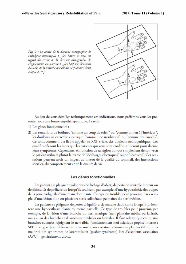
e-News for Somatosensory Rehabilitation of Pain 2014, Tome 11 (Volume 1)
34

e-News for Somatosensory Rehabilitation of Pain 2014, Tome 11 (Volume 1)
35
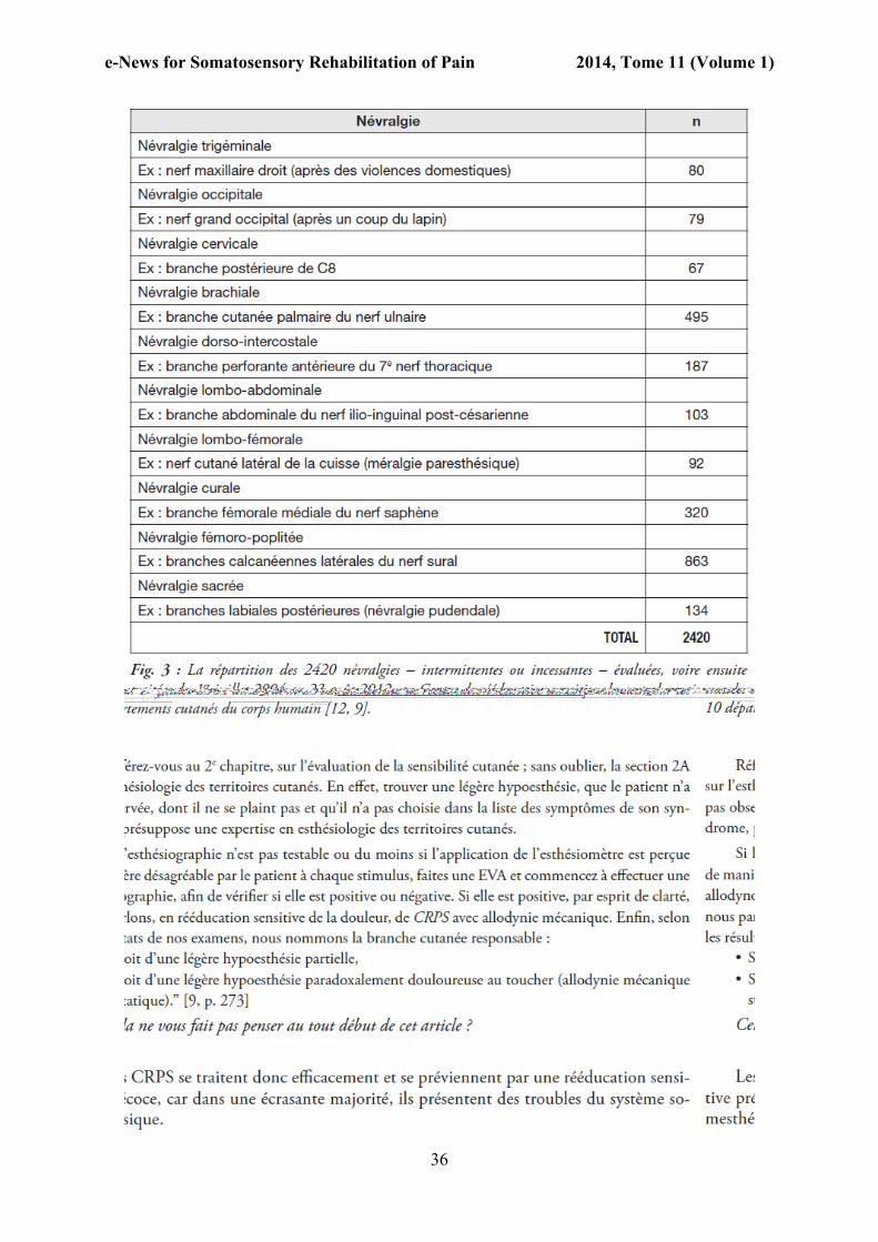
e-News for Somatosensory Rehabilitation of Pain 2014, Tome 11 (Volume 1)
36

e-News for Somatosensory Rehabilitation of Pain 2014, Tome 11 (Volume 1)
37
e-News for Somatosensory Rehabilitation of Pain 2014, Tome 11 (Volume 1)
38
e-News for Somatosensory Rehabilitation of Pain 2014, Tome 11 (Volume 1)
39
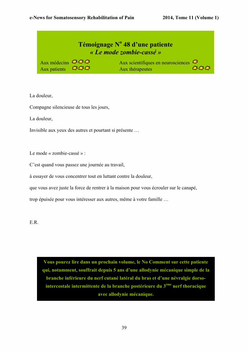
La douleur,
Compagne silencieuse de tous les jours,
La douleur,
Invisible aux yeux des autres et pourtant si présente …
Le mode « zombie-cassé » :
C’est quand vous passez une journée au travail,
à essayer de vous concentrer tout en luttant contre la douleur,
que vous avez juste la force de rentrer à la maison pour vous écrouler sur le canapé,
trop épuisée pour vous intéresser aux autres, même à votre famille …
E.R.
Vous pourez lire dans un prochain volume, le No Comment sur cette patiente
qui, notamment, souffrait depuis 5 ans d’une allodynie mécanique simple de la
branche inférieure du nerf cutané latéral du bras et d’une névralgie dorso-
intercostale intermittente de la branche postérieure du 3ème nerf thoracique
avec allodynie mécanique.
Aux médecins Aux scientifiques en neurosciences Aux patients Aux thérapeutes
Témoignage No 48 d’une patiente « Le mode zombie-cassé »

e-News for Somatosensory Rehabilitation of Pain 2014, Tome 11 (Volume 1)
40
HÜGLI, A. (MD)6
Bien souvent, je suis frappée par la confiance presque inconditionnelle que nos patients nous accordent. Ce lien très privilégié nous honore, justifie le choix de cette profession tout en chargeant parfois nos épaules.
Ce lien se tisse d’une manière très subtile dans les premiers échanges; un nouveau patient se présente, avec dans ses bagages la complexité de sa personnalité, son passé, son contexte, et pour l’oncologue un problème qui menace a priori son existence.
Nous connaissons tous les ingrédients qui vont permettre d’établir une relation : c’est notre quotidien. Si le langage, la précision des mots, les explications modulées en fonction de chacun sont les bases de l’échange, le non verbal me semble capital pour établir la confiance. Certains de nos patients viennent munis d’informations puisées sur internet et nous les soumettent. Leur propos n’est jamais une confrontation scientifique, mais plutôt un test quant à notre ouverture d’esprit, quant à la maîtrise des techniques et des ressources de notre profession. Si nous entrons en matière dans cette discussion technologique qu’ils proposent, très vite, rassurés par nos compétences, ils abandonnent ce chapitre.
Mais je crois que c’est en nous transmettant les émotions survenues au cours des évènements récents que la confiance s’établit. Lorsque, à travers le récit de nos patients, nous revivons les étapes à l’origine de cette visite médicale, lorsque nous adoptons la logique des évènements, un climat d’échange s’installe. Le regard, le sourire, l’humour sont alors de précieux alliés. On ressent parfois à quel moment le « fluide » passe, à quel instant la relation se scelle. Quelle alchimie permet, la plupart du temps cette cristallisation ? Elle varie avec chacun, elle confirme que nous sommes dans une relation unique.
Parfois un mur s’installe. Le courant ne passe pas. On cherche vainement la clé, le commutateur qui permettra la communication. Lorsque ces difficultés premières se résolvent, on s’aperçoit que l’absence de dialogue résulte souvent d’un décalage, d’un déphasage entre le patient et nous. Pressé par l’inquiétude ou la nature de la maladie nous brûlons les étapes.
6 Rue de Candolle 18; CH – 1205 Genève [email protected]
ARTICLE
La confiance
L’article suivant a été publié dans : Revue Médicale Suisse 2013 ; 9 : 2130 (une page).
Nous le rééditons avec la gracieuse permission de l’auteure et de l’éditeur Médecine & Hygiène
Aux médecins Aux scientifiques en neurosciences Aux patients Aux thérapeutes
e-News for Somatosensory Rehabilitation of Pain 2014, Tome 11 (Volume 1)
41
Avant notre patient, nous avons accepté la maladie et sommes prêts à la soigner et ne réalisons pas que celui-ci n’adhère pas encore au statut de malade. Un décalage s’installe, parfois temporaire, parfois irrémédiable si nous n’arrivons pas à faire machine arrière et à cheminer avec lui.
Mais n’oublions pas que cette relation thérapeutique exige beaucoup de nos patients. Ils doivent lâcher prise, s’abandonner tout en restant partenaire. L’acceptation de cette requête, en apparence contradictoire, s’enracine dans l’échange. La qualité de nos réponses, que ce soit à travers les mots, notre écoute ou notre regard, va permettre au patient de libérer des tensions intérieures, de partager un poids et d’accepter une nouvelle dynamique, celle des soins.
Cette alliance thérapeutique sous-tend toute notre démarche, elle est la clé de beaucoup de succès et celle de notre plaisir au quotidien. Cultivons là.
amanitvam, adambhitvam, ahimsa, ksantir, arjavam, […] atma-vinigrahah, […] anahankara, […] – nityatvam tattva-jñanartha-darsanametaj jñanam iti proktamajñanam yad ato ‘nyata Bhagavad gita / 13.8-12
"L’humilité, la modestie, la non-violence, la tolérance, la simplicité, […] la maîtrise de soi, […] le non-ego, […] telle est, je l’affirme, la connaissance. Le contraire de cela est ignorance."
"Humbleness, modesty, non-violence, tolerance, simplicity, […] self-control, […] absence of false ego, […] - all these I declare to be knowledge, and besides this whatever there may be is ignorance."
"La humildad, la ausencia de orgullo, la no violencia, la tolerancia, la sencillez, […] el autocontrol, […] la ausencia de ego falso, […] - todo eso Yo declaro que es conocimiento, y cualquier otra cosa que haya aparte de eso es ignorancia."
Aphorisme saisonnier Aux médecins Aux scientifiques en neurosciences Aux patients Aux thérapeutes
Seasonal aphorism To MD To neuroscientistt
To patient To therapist
Temporada de aforismos
Para Médicos Para científicos en neurociencias Para pacientes Para terapeutas

e-News for Somatosensory Rehabilitation of Pain 2014, Tome 11 (Volume 1)
42
A review by Jan Fisher, wife of 38 years and a mother of 2 fantastic adults, pain sufferer (edited for brevety)
G. Lorimer Moseley, David S. Butler, Timothy B. Beame sand Thomas J. Giles. The Graded Motor Imagery Handbook. Noigroup Publications, 2012 - 143 pages. ISBN : 0987246755, 9780987246752.
"This new Noigroup publication is born of a firm conviction that, as Lorimer Moseley says, "people in pain do better if they are given the resources to master their situation". To this end The Graded Motor Imagery Handbook builds on and extends much further the knowledge that Explain Pain began. A persistent theme is that treatment requires patience and persistence, courage and commitment.... The book is divided into five sections - knowledge, background, treatment, stories and tool boxes. David Butler in the first section tells us about this exciting new era in rehabilitation powered by the neuroscience revolution of which Graded Motor Imagery Program (GMIP) is an important part. David explodes some myths and builds on the plain common sense of Explain Pain. He offers suggestions and encouragement, including encouragement to share your experience and thoughts. This open mindedness is refreshing. David explains that GMIP, unlike a program is not preset. It's a series of novel treatment strategies which remind us that representation of body in the brain should be considered in all patients. Importantly he says that patients cannot be just passive recipients of treatment. They need to self manage and understand that gaining knowledge is part of treatment - knowledge is power - knowledge can be therapy."If you have knowledge of GMI you have skills to know why it hurts so much today and how to deal with it." In the second section of The Graded Motor Imagery Handbook Lorimer Moseley gives us the science underpinning the concept of Graded Motor Imagery. He also explains such things as delayed reaction time in left/right hand judgements and why sometimes even imagined movements make some feel pain. He gives a great explanation of possible mechanism of
To MD To neuroscientist To patient To therapist
READ FOR YOU
The Graded Motor Imagery Handbook

e-News for Somatosensory Rehabilitation of Pain 2014, Tome 11 (Volume 1)
43
spreading pain which can manifest as imprecise movements or dystonia. Lorimer explains the pattern of primary sensory cortex (outermost layer of brain) activation caused by stimulating different points on the skin....Interestingly Dr Moseley (page 50) describes comparison between patients who used imaginary and mirror therapy with those who used the three step GMIP (that is left/right judgements followed by imagined movements and then mirror therapy). In his results with ungraded motor imagery "patients tended to get worse in those phases but improved when left/right judgements were used and then further improved when the next steps were followed. " I'm putting this here because what I like about this book is that the authors don't pretend to know it all. I did not have the benefit of left/right judgement before I began mirror work, nor did I know anything about imagined movements as therapy. I read about mirror therapy, worked out what might work and experimented. With persistence, a very little and often over a long period I improved. I suspect that keeping the training very short prevented worsening of symptoms as described. The important message here is that beyond doubt, pain can be reduced by brain retraining methods. Tim Beames authors the third and no less important section on treatment. He goes much further into the practical aspects of treatment, the stages, adaptive techniques, how to use Recognise, implicit and explicit motor imagery and on to mirror therapy This is a valuable section for clinicians as many examples are given. Telling photos of mirror therapy in practice will be an asset for both therapists and patients. There's a great "how to mirror therapy" table on page 89. Each section is clearly summarized and on page 92, Timothy brings it all together and encourages us to be creative and "not be satisfied with just managing pain." I like his attitude. David takes the fourth section to a new level. I like the way he cuts to the chase, a no nonsense approach explaining how stories and metaphors can "get us through". He has a great knack of keeping it simple and really getting his message across. I like too the analogy on page 114 of our brain being "our own drug cabinet". I agree with what he says at the end,"Basically, knowledge is the greatest pain liberator of all. The key is in your hands." Now to the final section written by Thomas Giles.... He's the one who shows us how to access and use the tools to make it work. It's refreshing to see this depth of information and practical support. From system requirements and connecting to Recognize to making your own resources, Tom explains it all simply and logically. He encourages us to share our ideas. Just contact NOI Group. Many people with CRPS contact me asking where they can find out more, who can help them, where there's someone in their area. Now I can suggest they go to their local clinician. If the clinician/physical therapist is unfamiliar with Graded Motor Imagery Program this link and this wonderful book will provide the answers. Ignorance of something new isn't a problem. It's an opportunity.... I believe The Graded Motor Imagery Handbook is a great resource for patients but I consider it is a must for those who treat people in pain. I add my encouragement for clinicians who use it to document what you do and add to the growing body of evidence by sharing your results and experience. This book gets a big thumbs up from me."
e-News for Somatosensory Rehabilitation of Pain 2014, Tome 11 (Volume 1)
44
z
Diane DUCHESNE7
Merci à toi Claude ! Merci à Frédéric ! Et merci à ce groupe riche et généreux !
Je suis revenue littéralement changée, comme empreinte d'un nouveau souffle ; comme
l'impression qu'une immensité vient de s'ouvrir à moi ; comme la découverte d’une vérité
dont j’avais jusqu’à ce jour été privée. Et que ce dernier module de 3 jours à Bruxelles n'est
finalement qu'un début... Le début d'une aventure, d'un parcours. Le début de nombreuses
expériences à venir ; d’obstacles certainement, de difficultés aussi. Mais n’est-ce pas la ce
qui fait la richesse de notre profession ?
J’aime beaucoup la phrase d’Eva que tu nous a retranscrit à plusieurs reprises et sur quoi je
la rejoins à 200% aujourd’hui : on n’a pas fait tout ça pour être « tech »[nicien(ne)s].
Grâce à toi, à vous tous qui nous avez ouvert la voie, nous disposons aujourd’hui d’outils,
d’une méthode, de ressources. J’espère réussir à en faire bon usage et apporter, à ces patients
qui souffrent trop et depuis trop longtemps, un peu de répit, une petite libération pour une
meilleure qualité de vie, un retour à leur vie. Loin de moi l’idée de faire des miracles8, j’ai
bien compris la leçon.
7 Ergothérapeute DE, RSDC® Rééducatrice Sensitive de la Douleur Certifiée, Hôpital de La Tour ; 3, Av. J.-D. Maillard ; CH-1217 Meyrin e-mail : [email protected]
8 Ndlr : en lien avec la toute puissance, un des thèmes du cours sur la gestion du lien thérapeutique avec Frédéric Soum, psychothérapeute, lors du premier jour de ce module 3.
Aux médecins Aux scientifiques en neurosciences Aux patients Aux thérapeutes
Formation en rééducation sensitive de la douleur Module 3 – Bruxelles 2013

e-News for Somatosensory Rehabilitation of Pain 2014, Tome 11 (Volume 1)
45
Continuous Education – Weiterbildung - Formation continue
Xème COURS Depuis 2005
Le traitement des syndromes douloureux neuropathiques
par la rééducation sensitive de la douleur
Date: 17 - 20 mars 2014
Rééducation sensitive de la douleur Troubles de base I & II, Complications douloureuses I & II
Nadège Buchet (-Desfoux), ergothérapeute DE, RSDC® Rééducatrice Sensitive de la
Douleur Certifiée
Claude Spicher, ergothérapeute, rééducateur de la main re-certifié SSRM,
collaborateur scientifique universitaire
Lieu : Enseignement Permanent de l’Ergothérapie, Montpellier, France
Info : http://www.ergotherapiemontpellier.com/formation.html
Ces formations peuvent être comptabilisées pour l’obtention du titre : RSDC® Rééducatrice Sensitive de la Douleur Certifiée
e-News for Somatosensory Rehabilitation of Pain 2014, Tome 11 (Volume 1)
46
10-12 février 2014 Module 4 en rééducation sensitive de la douleur : Mettre à jour vos connaissances, lire un article en 15 min., écrire un rapport de thérapie, intégrer la méthode dans votre institution
Lieu Centre de rééducation sentive du corps humain (Fribourg) Info [email protected]
www.neuropain.ch/fr/enseignement/calendrier Formatrices Isabelle Quintal, graduée de l’Université de Montréal, RSDC® Mélanie Kaeser, PhD, unité de physiologie, université de
Fribourg ; Pascal Latière, Fanny Horisberger & Séverine Glanowski RSDC® ; Claude Spicher, fondateur de la méthode
Cette formation est exclusivement réservée aux 51 licencié(e)s en Rééducation sensitive de la douleur RSDC®
10-11 avril 2014 Module 1 en rééducation sensitive de la douleur: Troubles de base I & II – Comment traiter les syndromes du canal carpien, algodystrophies et hémiplégies 8ème promotion
Lieu Université de Montréal, Faculté de médecine, Ecole de réadaptation
Info www.readap.umontreal.ca Formatrice Isabelle Quintal, graduée de l’Université de Montréal, RSDC®
est ergothérapeute graduée de l’Université de Montréal. Après plus de 5 ans d'expérience en clinique privée au Québec, elle a été engagée au Centre de rééducation sensitive du corps humain (Suisse). Elle travaille actuellement au Centre Professionnel d’Ergothérapie (Montréal). Son activité d’enseignante pour le RRSD l’a déjà menée à Bruxelles, Fribourg, Montpellier et Montréal. Elle a publié des articles dans différentes revues, dont l’Encyclopédie Médico-Chirugicale (EMC). Elle est responsable du Département de la méthode du RRSD depuis sa fondation.
Ces formations peuvent être comptabilisées pour l’obtention du titre : RSDC® Rééducatrice Sensitive de la Douleur Certifiée
14–16 avril 2014 Module 3 en rééducation sensitive de la douleur: Gestion du lien thérapeutique, Anatomie clinique II & Complications douloureuses II 7ème promotion
Lieu Université de Montréal, Faculté de médecine, Ecole de réadaptation
Info www.readap.umontreal.ca
Formateurs Claude Spicher et Dr Marc Zaffran Ces formations peuvent être comptabilisées pour l’obtention du titre :
RSDC® Rééducatrice Sensitive de la Douleur Certifiée

e-News for Somatosensory Rehabilitation of Pain 2014, Tome 11 (Volume 1)
47
14-16 mai 2014 Module 2 en rééducation sensitive de la douleur: Analyse de pratiques, Anatomie clinique I & Complications douloureuses I 8ème promotion
Lieu CREA-HELB, Campus ERASME, Bruxelles Info www.crea-helb.be / [email protected] www.anfe.fr / [email protected]
Ces formations peuvent être comptabilisées pour l’obtention du titre : RSDC® Rééducatrice Sensitive de la Douleur Certifiée
18-21 June 2014 19th FESSH & 11th EFSHT Congresses Place Paris (France) Palais des Congrès – Porte Maillot Info www.fessh2014.com/en/welcome
8-10 octobre 2014 Module 3 en rééducation sensitive de la douleur: Gestion du lien thérapeutique, Anatomie clinique II & Complications douloureuses II 8ème promotion
Lieu CREA-HELB, Campus ERASME, Bruxelles Formateurs Claude Spicher et Frédéric Soum, psychothérapeute FSP Info www.crea-helb.be / [email protected] www.anfe.fr / [email protected]
Ces formations peuvent être comptabilisées pour l’obtention du titre : RSDC® Rééducatrice Sensitive de la Douleur Certifiée
13-14 novembre 2014 48ème Congrès annuel SSCM & 16ème Congrès suisse SSRM Lieu Kultur- und Kongresszentrum Thun (KK Thun) Thoune, Suisse Info www.sgh-sghr-kongresse.ch
6-11 October 2015 9th Congress of the European Federation of IASP® Chapter EFIC®
Place Vienna (Austria) Info www.efic.org
24-28 October 2016 13th IFSSH & 10th IFSHT Congresses Place Buenos Aires (Argentina) Info www.ifssh-ifsht2016.com/home/index.php

e-News for Somatosensory Rehabilitation of Pain 2014, Tome 11 (Volume 1)
48
Claude J. SPICHER9
La douleur qui aliène. La douleur bien réelle, mais surréaliste aussi
comme un monde fantastique. La douleur insurmontable, qui travestit la réalité. Douleur d’une incommensurable lourdeur,
comme des briques que l’on ne peut plus porter. Douleur intolérable. Silence, pesant, glauque et poisseux. Silence insoutenable. Silence d’une solitude absolue. et pourtant : Silence habité ?
9 Rédacteur en chef du e-News Somatosens Rehab depuis sa création en 2004.
PHENOMENE DE LA DOULEUR
No 1
Aux médecinss Aux scientifiques en neurosciences Aux patients Aux thérapeutes

e-News for Somatosensory Rehabilitation of Pain 2014, Tome 11 (Volume 1)
49
Le titre de RSDC® Rééducateur/trice Sensitif/ve de la Douleur Certifié(e) se porte à merveille : une 5ème promo de lauréats est sortie de l’Université de Montréal le printemps passé et une 6ème promo à Bruxelles cette semaine. Une 7ème promo commencera en février à Bruxelles sous l’égide d’Isabelle Quintal de Montréal et une 8ème promo commencera ce printemps à l’Université de Montréal avec la sus-nommée et Claude Spicher. A ce jour, les patients douloureux neuropathiques peuvent compter sur 51 RSDC®. Ils sont coordonnés par la responsable du Département de rééducation du Réseau de Rééducation Sensitive de la Douleur : Marion Vittaz, RSDC®. Les 12 lauréates et lauréat des 5ème promo et 6ème promo 2013 (par ordre alphabétique) sont :
- Laurence Bernardon, Miribel, France,
- Julie Chénier, St-Jérôme, Canada (Qc),
- Laure-Anne Clerc, Mendrisio Svizzera,
- Lucie Crego-Adil, Libourne, France,
- Diane Duchesne, Meyrin, Suisse,
- Gaëlle Faure-Perathoner, Paris, France,
- Andrea Grass, Biel, Schweiz,
- Marie Kubezyk, Paris, France,
- Céline Langlois, Saint-Gobin, France,
- Eva Létourneau, Lévis, Canada (Qc),
- Tara Packham, Hamilton, Canada (Ont.),
- Vincent Soriot, Amiens, France.
Les travaux de validation ont été expertisés par Mme Julie Masse, professeure adjointe de clinique de l’Université de Montréal sous la direction de Claude Spicher. Cette formation compte 56 heures de cours ou équivalents et nécessite environ 64 heures de travail personnel, sans compter la trentaine d’heures pour le travail final de validation. Lisez, en page 22 un travail final de cette 6ème promo.
Aux médecins Aux scientifiques en neurosciences Aux patients Aux thérapeutes
Certificat en rééducation sensitive de la douleur

e-News for Somatosensory Rehabilitation of Pain 2014, Tome 11 (Volume 1)
50
You are 38’432 neuroscientists, medical doctors, therapists & patients all over the world on the 5 continents, in 138 countries who are receiving the e-News for Somatosensory Rehabilitation of Pain.
Who are you?
IMPRESSUM
International Standard Serial Number (ISSN): 1664-445X Editor-in-chief
Claude J SPICHER, University scientific collaborator, Swiss Certified HT, OT. Co-editor
Fanny HORISBERGER, OT, Certified Somatosensory Therapist of Pain CSTP® Assistant editors
Marion VITTAZ, OT, Certified Somatosensory Therapist of Pain CSTP® International assistant editors
Tara L PACKHAM, MSc, OT Reg. CSTP® (Ontario, Canada) Elinor BEHAR, OT, CSTP® (Belgium, Israel)
Julie MASSE, MSc OT (Québec, Canada) Renée HAMILTON, BSc OT (Québec, Canada) Séverine GLANOWSKI, OT, CSTP® (France)
Catherine COUVREUR, OT, CSTP® (Belgium) Guillaume LEONARD, PhD, MSc, pht (Québec, Canada)
Sibele de ANDRADE MELO, PhD, pht (Brazil) Ivano DONES, MD (Italy)
Rebekah DELLA CASA, OT, CSTP® (Switzerland) Sandra B FRIGERI, OT (Argentina)
Honorary members Prof EM ROUILLER, PhD (Switzerland)
Prof AL DELLON, MD, PhD (USA) Prof R MELZACK, OC, OQ, FRSC, PhD (Québec, Canada)
Peer-reviewed (since 2012)
Published: 4 times per year since 2004 Deadline: 10Pth P January, 10 Pth P April, 10 Pth P July, 10Pth P October
Price: Free Sponsor: Somatosensory Rehabilitation of Pain Network, Switzerland, Europe.
20 languages: Français, English, Deutsch, Español, Portugues, Рускиӣ, Italiano, Lingala,
Shqipe, Srpski i Hrvatski, Corse, Česky, Svenska, Türkçe, Suomea, Ελληνικά, Nederlands,
hindi, עברית, e-News’s Library: H www.neuropain.ch
e-mail : [email protected]