Embed Size (px)
Citation preview

J. Zool., Lond. (1972) 168, 107--117
Dynamics of uptake and binding of 35S-~~Iphate by the cartilage of Rainbow trout (SaZmo gaivdneri) and the influence of thyroxine
SANDRA BARBER A N D E. J. W. BARRINGTON Department of Zoology, The University, Nottingham
(Accepted 14 March 1972)
(With 2 plates and 4 figures in the text)
35S-sulphate has been injected into the body cavity of yearling Rainbow trout (Sabno gairdneri) and the time sequence of its excretion, and of its uptake and binding by branchial cartilage, determined by scintillation counting. The data are shown to conform well with the time sequence of movement of 35S-sulphate into the cells and matrix of the cartilage, as visualized by autoradiography. Peak content of both unbound and bound sulphate is reached within the cartilage at about 30 hours after injection, in both control and thryoxine- treated fish, but the uptake in the latter is significantly higher. In conformity with this, the rate of excretion of 35S-sulphate is lower in the hormone-treated animals. The results confirm and extend earlier work in showing that thyroxine, by enhancing sulphate uptake, could exert a growth-promoting influence upon cartilage, but it is pointed out that this can be only one component of a complex structural and hormonal situation.
Contents
Introduction and previous work , . . . . . . . . . . . Material and methods . . . . . . . . . . . . . . Results . . . . . . . . . . . . . . . . . .
Excretion . . . . . . . . . . . . . . . . Branchial cartilages . . . . . . .. . . . . .. Autoradiography . . . . . . Assay of extracts of cartilage . . . . . . . .
Discussion . . . . . . . . . . . . . . . . References . . . . . . . . . . . . . . . . . .
. . . . . . .. . . . .
. .
..
..
. .
..
. .
..
..
. .
Page . . 107 .. 108 .. 109 .. 109 .. 111 .. 113 .. 114 .. 114 .. 117
Infxoduction and previous work It is well established for mammals that 35S-sulphate, injected intraperitoneally, is rapidly
taken up by cartilage and incorporated into chondroitin sulphate (Dziewiatkowski, 1958). The radioisotope appears first within the chondrocytes, where it is stored, and where synthesis of the mucopolysaccharide is believed to take place (Pelc & Gliicksmann, 1955; Dziewiatkowski, 1962). Subsequently the labelled product is slowly passed into the matrix. This cellular uptake provides a reliable index of the activity of the cells, variation in their radioactive content being related to their contribution to the growth of the cartilage. Uptake is less, for example, in the more peripheral and younger chondrocytes (Pelc & Gliickmann, 1 955).
Little comparable evidence is available for the sulphate metabolism of the cartilage of fish, apart from a report from this laboratory by Barrington & Rawdon (1967). They
107

108 S. BARBER AND E. J. W. BARRINGTON
showed,in a further analysis of the growth-promoting action of thyroxine in Rainbow trout (established by Barrington, Barron & Piggins, 1961), that the uptake of 35S-sulphate into the bone and cartilage of the branchial skeleton of yearling fish, and its binding into chondroitin sulphate, is enhanced by prior immersion of the animals in 1/500,000 thyroxine solution. Their evidence, however, was derived only from a study of uptake values at one period. This was at 24 hours after administration of the isotope, an interval selected after consideration of the published data for mammals.
The purpose of the present work has been to extend this finding by investigating the dynamics of radiosulphate uptake into the cartilage of the branchial skeleton. It was hoped in this way to determine the time relationships of the movement of the isotope into the cartilage from the site of injection, and thus to establish that the enhancing effect of thyroxine treatment is expressed during peak uptake, at a time when it could have a maxi- mum effect upon cartilage growth. The work has been planned to provide a basis for a study (to be reported later) of the influence of hormones upon the radiosulphate uptake of individual arch elements in vitro. The consequent introduction into the extraction and counting procedure of various refinements has also facilitated the determination of time relationships in viva
Material and methods This account is based upon the results of five experiments, of which the first three, using only
small numbers of fish, were exploratory. Rainbow trout, 6 to 18 months old, were obtained from a local hatchery (Trent Fish Company)
and were acclimatized to laboratory conditions in stock aquaria. During experiments they were maintained individually in well-aerated 8 1 tanks, and were fed on alternate days with proprietary trout pellets (River Pride). The tank water was changed every 48 h, at 24 h after feeding.
Carrier-free 35S-sulphate (Amersham), in distilled water at a concentration of 1 mCi ~ m - ~ , was injected intraperitoneally through the rectal wall or the ventral body wall. The volume injected was adjusted to give an average level in the fish of 5 pCi g-l. To faciliate accurate injec- tion, the fish were anaesthetized with 65 parts/million MS222 (Sandoz).
For thyroxine treatment, the fish were immersed for 6 to 14 days (prior to injection of radio- sulphate) in 2 parts/million thyroxine (sodium salt, BDH), dissolved in a minimal volume of 0.1 M NaOH. This was added to the tank water at the time of changing. A similar volume of the solvent was added to the control tanks. Fish were not fed after injection, in order to avoid the data for excretion being complicated by adsorption of radioisotope on residual food. Only occasion- ally did this mean that the animals were without food for a greater length of time than that of the usual routine maintenance.
For determination of radioisotope excretion, 10cm3 samples of tank water were taken at specified times after the injection, and measurements made of the radioactivity of aliquots. The maintenance of constant tank-water volume and of vigorous agitation contributed to ensure con- sistent accuracy of sampling.
For measurements of the radioactivity of the blood, 20 pl. samples were taken from the hearts of anaesthetized fish when they were killed. These samples were transferred to a solution of heparin, which was dissolved in 1 % Teepol in order to facilitate an even spreading on the disks used for scintillation counting (see below).
For measurements of the radioactivity of the peritoneal fluid, 2 cm3 of heparin in 1 % Teepol was injected into the peritoneal cavity and then withdrawn into the syringe, which was not re- moved between the two processes. Any sample containing blood was rejected.
Cartilage units (for structural details, see later) were briefly dried on filter paper to remove
UPTAKE A N D BINDING OF 35S-SULPHATE BY RAINBOW TROUT 109
surface moisture and were weighed on a torsion balance. A carefully standardized procedure ensured uniformity of treatment. Each unit was homogenized, using a hand-operated tissue grinder, in a saturated solution (about 0.5 % w/v) of papain in phosphate buffer at pH 5.5. This treatment, which was not included by Barrington & Rawdon (1967), was intended to break down the mucopolysaccharide/protein complex and thus facilitate solution of the acid mucopoly- saccharide moiety. Extraction was then carried out by adding a standard volume of 25% KCI, with continued shaking for a further 24 h (KCI was used in preference to the 2 % NaOH of the previous study (Barrington & Rawdon, 1967) in order to avoid risk of possible degradation of the chondroitin sulphate molecule). The extract was made up to standard volume (2-0 to 3.5 cm3) with papain solution, and centrifuged at 2000 revs/min for 30 sec. The supernatant was decanted, and aliquots used to determine total 35S content. For estimation of bound 35S, the extract was dialysed (using Visking tubing) against running tap-water for 24 h, and then against distilled water for a further 24 h.
Barrington & Rawdon (1967) assayed the radioactivity of skeletal extracts with a Geiger- Muller end-window counter. Scintillation counting has been used in the present work, and has improved sensitivity so much that it has been possible to determine the radioactivity of individual cartilage units instead of pooled specimens. 0.1 cm3 aliquots of the various samples and extracts were pipetted onto glass fibre disks (Whatman GF/A, 2-1 cm) which were dried and introduced into 0.3 cm3 of liquid scintillator (NE 215) in a counting vial. They were assayed for 5x 100 seconds on a Panax (SC/LP) scintillation counter in association with an IDL (1700) scaler, or else on a Packard Tricarb Spectrometer until a 1 % standard deviation was recorded. The usual corrections were made for background activity, which was kept to a minimum.
Results Excretion
In an exploratory experiment involving four pairs of fish, one member of each pair was exposed to thyroxine for 12 days, the other member serving as a control to it. All of the fish then received an injection of radiosulphate. All four pairs were killed at one day after injection. Determination of excreted radiosulphate showed that in each of the pairs the control specimen had excreted more radiosulphate than had the thyroxine-treated one.
This result was only suggestive, but the reality of the hormone influence was confirmed in a definitive experiment using 36pairs of fish, the two members of each pair being carefully matched for weight. One member of each pair was pretreated with thyroxine for six days, after which all fish were injected with radiosulphate. Six pairs, with an approximate mean weight of 35 g, received 180 pCi of radiosulphate, six pairs of mean weight 44 g received 220 pCi, and six pairs of mean weight 50 g received 250 pCi. Water samples were taken from the tanks at the times shown in Table I, the number of samples at each point of time being the same for the control group and for the thyroxine-treated group. The number was restricted by considerations of time, and also by the killing of fish at intervals for the determination of blood 35S (see later).
The results (Table I) show an initially high rate of excretion, which falls off to a low rate after about 18 hours, so that in control fish about 76 % of the injected dose has been excreted by 18 hours. Thereafter, radiosulphate continues to be lost at a very slow rate. This pattern (of an early and brief phase of rapid loss), agrees with that reported by Rosenthal(1961) for Lebistes, and by Barrington & Rawdon (1967) for Rainbow trout.
I t is further shown in Table I that the excretion of radiosulphate is consistently a t a lower rate in the thyroxine-treated fish, this effect being apparent from the first (one hour)

110 S . BARBER A N D E. J. W . BARRINGTON
TABLE I
Excretion of and the infiuence of thyroxine. Standard deviations and significance are indicated
Student’s t test Time after Number of % injected dose excreted (control versus
injection (hours) water samples Control Thyroxine-treated thyroxine-treated) P
1 13 11 &4 8 k 2 0.02 2 5 16k3 1 5 5 2 n.s. 3 12 26k6 19k6 0.02 6 6 42f9 31&15 n s .
18 8 76k10 69&9 n s . 22 9 82&8 71f14 n.s. 25 9 80&7 71 5 9 0.05 28 3 82f6 74+20 n.s. 48 6 83f8 83 5 10 n s . 96 3 94513 9Okl9 n.s.
n.s., Not significant.
sample. The numbers of water samples were not always large enough to be statistically analysable, but the differences at one and three hours were significant at the 2 % level, and at 25 hours at the 5 % level (Table I).
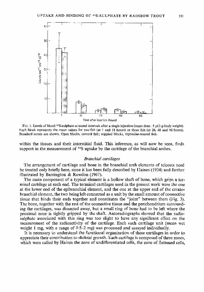
These same fish were also used for measurement of blood 36S. It was intended that three pairs of fish (one pair from each of the three weight groups) should be killed at 1,5, 18,24, 48 and 96 hours after injection. Two pairs, however, were missing from the final analysis owing to technical difficulties. The results (Fig. 1) show that a high level of blood 35S is attained within one hour of the injection. Thereafter, there is a rapid fall to a very low level by 24 hours, at which time, as we have seen, the excretory rate has also become low. There is no clear influence of thyroxine treatment upon these blood levels.
In a further experiment, involving 12 days of thyroxine pre-treatment, these observations were amplified by studying six pairs of weight-matched fish over a shorter period (from 1 to 18 hours after injection of radiosulphate). Results (Fig. 2) were similar in principle, showing an initial high rate of excretion (rising in this instance to 50 % of the injected dose at 18 hours). The difference between this value and that of the previous experiment is typical of the variation in detail found in these experiments. There was a comparable fall of blood 35S, from an initial level somewhat lower than in the previous experiment.
In this further experiment some measurements were made of the radioactivity of the peritoneal fluid (Fig. 2). They conform well with the other data, showing that already at one hour after injection the level of activity is low, and that it is negligible by six hours. Evidently the injected radiosulphate passes very rapidly from the peritoneal fluid into the blood stream.
As in the preceding experiment, there is no clear evidence of an influence of thyroxine upon the distribution of the radioisotope within the body fluids, although much more data would be needed to establish this negative finding. However, the results from both of the experiments reported here suggest that the increased amount of 35S retained in the thy- roxine-treated fish (as indicated by their reduced excretory rate) must be located mainly
UPTAKE A N D B I N D I N G OF 35S-SULPHATE BY RAINBOW TROUT 111
Time after injection (hours)
FIG. 1. Levels of blood 35S-sulphate at stated intervals after a single injection (mean dose: 5 pCi g-body weight). Each block represents the mean values for two fish (at 1 and 18 hours) or three fish (at 24, 48 and 96 hours). Standard errors are shown. Open blocks, control fish; stippled blocks, thyroxine-treated fish.
within the tissues and their interstitial fluid. This inference, as will now be seen, finds support in the measurement of 35S uptake by the cartilage of the branchial arches.
Branchial cartilages The arrangement of cartilage and bone in the branchial arch elements of teleosts need
be treated only briefly here, since it has been fully described by Haines (1934) and further illustrated by Barrington & Rawdon (1967).
The main component of a typical element is a hollow shaft of bone, which grips a ter- minal cartilage at each end. The terminal cartilages used in the present work were the one at the lower end of the epibranchial element, and the one at the upper end of the cerato- branchial element, the two being left connected as a unit by the small amount of connective tissue that binds their ends together and constitutes the “joint” between them (Fig. 3). The bone, together with the rest of the connective tissue and the perichondrium surround- ing the cartilages, was dissected away, but a small ring of bone had to be left where the proximal zone is tightly gripped by the shaft. Autoradiographs showed that the radio- sulphate associated with this ring was too slight to have any significant effect on the measurement of the radioactivity of the cartilage. Each such cartilage unit (mean wet weight 1 mg, with a range of 0-5-2 mg) was processed and assayed individually.
It is necessary to understand the functional organization of these cartilages in order to appreciate their contribution to skeletal growth. Each cartilage is composed of three zones, which were called by Haines the zone of undifferentiated cells, the zone of flattened cells,

112
6(
5(
m g 4! U m
a, +
.- ._ % 3( al Ln
c m +
S. BARBER A N D E. J . W. BARRINGTON
1 1 I I I I
Time after injection (hours)
FIG. 2. Distribution and excretion of 85S-sulphate at stated intervals after injection. Mean weight of fish 40 g. Each fish received 200 pCi. Each point represents the mean for four fish (1 and 6 hours) or for two fish (18 hours). Standard errors are indicated where possible; if not shown, they lie within the area of the point; A, Peritoneal s5S; w, blood 36S; 0, excreted s5S.
and the zone of hypertrophic cells. We prefer, because of the autoradiographic evidence to be described below, to term them respectively the articular (i.e. outermost), medial and basal (i.e. innermost) zones.
As Haines (1934) pointed out, cells are recruited into the articular zone from the peri- chondrium. The cells in this zone are frequently associated in pairs, which suggests that the zone grows by division of the recruited cells, and by the laying down of new matrix. Thus the articular zone is continuously enlarged and moulded, which is one reason why it seems unsuitable to term it the zone of undifferentiated cells.
Growth in length of the whole skeletal element, however, must be mainly secured by the activity of the medial zone, the cells of which are also recruited from the perichondrium, and perhaps also from the articular zone as well. Mitoses are readily seen in transverse sections of the flattened cells of the median zone (Barrington & Rawdon, 1967). These lead to the cells moving towards the basal zone, new matrix being laid down with consequent increase in intercell distance.
The situation in the basal zone is quite different, for here the striking feature is the enlargement of the cells. This increase is accompanied by increasing nuclear pycnosis and cell disintegration, the cells, and their surrounding matrix, being finally destroyed by the erosive action of the bone marrow (Haines, 1934). Haines, who emphasized this hyper- trophy in his terminology, believed that the transition from the flattened cells of the medial zone to the enlarged cells of the basal zone was accompanied by a great increase in the

UPTAKE AND BINDING OF 35S-SULPHATE BY RAINBOW TROUT 113
FIG. 3. Simplified diagram to show the main features of the cartilage unit, formed by the lower terminal cartilage
az, Articular zone; b, bone; bm, bone marrow; bz, basal zone; ct, connective tissue at “joint”; mz, medial zone; of the epibranchial element and the upper terminal cartilage of the ceratobranchial cartilage.
pz, perichondrium. The units are finally trimmed at the junction of marrow and basal zone.
amount of intercellular matrix. The change in cell shape makes it difficult to assess the extent of this increase, but the evidence of autoradiographs will be seen to indicate that there can be virtually no new formation of chondroitin sulphate by the hypertrophic cells. They are inactive in sulphate metabolism, and must be steadily degenerating. It is not wholly clear what does happen in this zone, but it could be that there is uptake of fluid prior to its erosion by the marrow. The overall growth of the branchial element clearly depends primarily on vigorous sdphate metabolism in the cartilage of the distal and medial zones, as just outlined, in conjunction with the addition of new periosteal bone to the shaft. This latter aspect is outside the scope of the present study.
Autoradiography Autoradiography shows that within one hour of injection there is abundant radio-
sulphate in the cartilage of the articular (Plate I (a)) and medial (Plate I (b)) zones. This is another reason for not regarding the cells of the articular zone as “undifferentiated”. The radioactivity is located mainly, but not exclusively, in the chondrocytes, sometimes so intensely as to give a dense black image over individual cells. This distribution indicates vig- orous uptake and binding of radiosulphate by these cells, including those in the outer region of the matrix, close to the perichondrium from which they have been derived. As in

114 S. BARBER A N D E. J. W. BARRINGTON
mammals, however, these peripheral cells are less active, and do not show the dense images seen over the more centrally situated ones.
The basal zone presents a sharp contrast to the other two, for only traces of radio- activity are seen in it (Plate I (c)), located mainly near to the edge of the medial zone. Sulphate metabolism must be negligible in this region; indeed, it could be that any radio- sulphate present results only from ionic exchange with that of the blood.
At 5 hours after injection there is heavy concentration of radioactivity in the cells of the articular and medial zones (Plate I1 (a), cf. Plate I (b)), and it is spreading beyond the borders of the cell bodies. By 28 hours (Plate I1 (b)) the radioactivity is becoming less concentrated over many of the chondrocytes, and there is considerable dispersion into the neighbouring matrix. This is apparent also at 96 hours (Plate I1 (c), cf. Plate I1 (a)) at which time there is still much bound radiosulphate in the tissue.
Evidence for any influence of thyroxine upon radiosulphate metabolism in the cartilage is best obtained by direct assay of cartilage extracts. Differences between autoradiographs of control and thyroxine-treated specimens are not sharply defined, owing to the general density of the images, and the considerable individual variability of the fish. Nevertheless, the autoradiographs do show some enhancement of binding in thyroxine-treated fish. This may be judged from Plate I (d), which shows the medial zone from a thyroxine-treated fish at one hour. This may be compared with Plate I (b), which is from the paired control fish at one hour. Another comparison, in this instance of the two members of a pair at 28 hours, shows a much greater dispersion of bound radiosulphate in the matrix of the thy- roxine-treated fish (Plate I1 (d)) as compared with the control member of the pair (Plate I1 (b)).
Assay of extracts of cartilage Data for the uptake of total 35S and organically-bound 35S, as determined by direct assay
of cartilage extracts, are shown in Fig. 4. The pattern of uptake is basically similar in the control and in the hormone-treated fish, with peaks at about 30 hours. Thyroxine, however, markedly increases the total uptake, the difference between control and thyroxine-treated fish at the peak being significant at the 1 % level by Student’s t test. Even with the inclusion at 30 hours of one of the anomalous fish mentioned in the legend, with an exceptionally tow value, there is still an 80% increase at this peak. There is a suggestion in the data (Fig. 4) that uptake in the thyroxine-treated fish not only rises more rapidly to its peak than it does in the control fish, but also that it declines more rapidly. This could mean that the hormone may be stimulating both the anabolism and the catabolism of sulphate. However, given the influence of experimental conditions upon uptake and excretion, to which we have already referred, a good deal more study would be needed to establish this point, and we mention it only as a possibility.
Discussion The results presented here provide an integrated account of some of the events interven-
ing between the injection of radiosulphate into the Rainbow trout and its binding into chondroitin sulphate. Radiosulphate moves so rapidly from the peritoneal fluid into the blood stream that within two hours the level of 35S in the blood is falling from its peak value. This fall is accompanied by a comparable increase in 35S excretion, so that as much as 80 % of the injected dose may have been excreted within about 24 hours, by which time

UPTAKE A N D BINDING OF "S-SULPHATE BY RAINBOW TROUT 115
1 I
-
300
200 -
- 'V I
2 400- (b) -
m
3 0 0
- 300-
I I I , _ _ 1 . 0 10 20 30 40 50 60 90 100
Time after injection (hours) FIG.
0, Total 35S; 0, organic
content of cartilage units removed from control fish (a) and thyroxine-treatedfish (I, at stated intervals after injection of 36Ssulphate. The fish were those illustrated also in Fig. 1.
the blood level has fallen to a very low value. Thereafter, excretion continues, but at a slow rate. A significant percentage of the injected dose is thus retained within the body for a considerable time, certainly for longer than the four days over which our present study has extended.
These results are in partial agreement with Rosenthal's (1961) data for the loss oftotal radiosulphate from the body of Lebistes after equilibration with the radiosulphate content of the surrounding water. He, too, found an early rapid loss, which lasted for the first 1.4 days. From then until about 15 days the rate of loss was lower. Thereafter, it passed into a third phase of very slow loss. He found that much of the earlier and more rapid loss took place from the spine and head, and concluded that there was little sequestration of 3% into mineral components of these regions during the first 20 days. In our experiments, however, such sequestration certainly did take place, even during the period of very rapid loss, although the proportion of the injected dose retained within the body was small.
Study of young and adult mammals has shown that 35S-sulphate accumulates preferenti- ally in those tissues where it is used in the synthesis of sulphated mucopolysaccharides

116 S. BARBER AND E. J . W . BARRINGTON
(Dziewiatkowski, 1958). Uptake is rapid, 35S-sulphate appearing in the cartilage cells of young rats (BClanger, 1954) within an hour or two of administration. It is believed (Stoolmiller & Dorfman, 1969) that the sulphate ion is activated within the chondrocytes and incorporated into protein-polysaccharide chains, which are later secreted into the matrix. Because of this, the radioactivity of the cartilage declines only very slowly after maximal sulphate concentration has been attained within it.
Our autoradiographic results show that uptake in fish follows a similar pattern. As in the rat, 35S-sulphate is abundantly present in the chondrocytes within one hour of administra- tion, a speed that is impressive, considering the lower temperature (not exceeding 16°C.) of our experimental fish in comparison with the body temperature of mammals. The fact that in fish, as in mammals, the radiosulphate is first concentrated within the chondrocytes, shows that we are here observing the results of sulphate metabolism, and not merely ionic exchange between the cartilage and the body fluids. Further evidence for this is the pattern of distribution of the radiosulphate within the several zones of the branchial cartilage. This indicates vigorous synthesis of chondroitin sulphate in the apical and medial zones, but virtually none in the proximal zone. The autoradiographic evidence corresponds well with our data relating to the movement of radiosulphate in the body at successive time levels. Rapid uptake into the chondrocytes within 1 hour parallels the rapid passage of the isotope by that time into the blood stream. The subsequent reduction in intracellular concentration agrees with the decline in blood level, while the prolonged retention in the matrix is associated with a regular rate of excretion.
Scintillation counting of extracts of the cartilages of control fish confirms the autoradio- graphic evidence, and gives more precise definition to the dynamics of uptake. Peak content of 35S-sulphate (both total and bound) is reached at about 30 hours, and thereafter there is only a very slow fall in level, just as in mammals. This conforms well with our evidence that by 30 hours the blood level is already very low, while the rate of excretion has markedly slowed down.
An influence of thyroxine upon the dynamics of 35S distribution is seen in the data for ex- cretion, which is consistently at a lower rate in the hormone-treated fish than in the control ones. Further evidence, correlated with this, is that the uptake of radiosulphateinto thebranc- hial cartilage is significantly higher in thyroxine-treated fish than in controls. This confirms the earlier report of Barrington & Rawdon (1967), but adds much weight to it by defining the time relationships. The peak uptake in hormone-treated fish is at about 30 hours, just as in control ones, but the level in the former is at least 80 % higher. Barrington & Rawdon studied only the activity at 24 hours after injection, having selected this interval by analogy with published data for mammals. Present results show that this period is sufficiently near to the peak to give a significant result. Times substantially earlier or later, however, might well have failed to do this.
The pattern of distribution of bound sulphate in the branchial cartilages shows that thyroxine, by enhancing sulphate uptake, could exert a growth-promoting influence, but it is impossible to judge from our present data how this influence might be exerted. In any case, the hormone must certainly be acting in conjunction with others, and in particular with pituitary growth hormone. No growth-promoting action of thyroxine is detectable in Poecilia in the absence of growth hormone (Ball, Olivereau, Slicher & Kallman, 1965), while both of these hormones are required for the maintenance of normal concentrations of chondroitin sulphate in young rats (Schiller, 1963). Moreover, hypophysectomy of juvenile

UPTAKE A N D B I N D I N G OF 35S-SULPHATE BY RAINBOW T R O U T 117
rats markedly diminishes the sulphate uptake by the costal cartilage, injection of growth hormone partially rectifying the condition (Ellis, HublC & Simpson, 1953). There are other considerations also that complicate the analysis ; in particular, the involvement of protein metabolism. Much of the chondroitin sulphate in cartilage is in combination with a non- collagenous protein, the two forming a protein-polysaccharide complex (Stoolmiller & Dorfman, 1969). In addition, the formation of the matrix requires the coordinated synthesis of collagen. Finally, growth of the matrix depends not only on the synthesis of chondroitin sulphate, but also upon chondrocyte proliferation; this, too, is influenced by growth hor- mone (Bois, BClanger & Le Buis, 1963). Not surprisingly, it is impossible as yet to define the consequent hormone-enzyme relationships in terms of any unitary mechanism (Schiller, 1966). Our present study is very much an abstraction from a complex field of hormonal interaction. It is to be hoped, however, that current in vitro studies of the branchial cartilages, based upon the methods developed in the present work, may help to clarify the problems involved.
R E F E R E N C E S Ball, J . N., Olivereau, M., Slicher, A. M. & Kallman, K. D. (1965). Functional capacity of ectopic pituitary trans-
plants in the teleost Poecilia formosa, with a comparative discussion on the transplanted pituitary. Phil. Trans. R . Soc. (B) 249: 69-99.
Barrington, E. J. W., Barron, N. & Piggins, D. J. (1961). The inlluence of thyroid powder and thyroxine upon the growth of rainbow trout (Salmo gairdneri). Gen. comp. Endocr. 1: 170-178.
Barrington, E. J. W. & Rawdon, B. B. (1967). Influence of thyroxine upon the uptake of 35S-labelled sulphate into the branchial arch skeleton of the rainbow trout (Salmo gairdnerii). Gen. comp. Endocr. 9: 116128.
Btlanger, L. F. (1954). Autoradiographic visualization of the entry and transit of S35 in cartilage, bone and dentine of young rats and the effect of hyaluronidase in vitro. Can. J. Biochem. Physiol. 32: 161-169.
Bois, P. L., Bklanger, L. F. & Le Buis, J. (1963). Effect of growth hormone and aminoacetonitrile on the mitotic rate of epiphysial cartilage in hypophysectomized rats. Endocrinology 73: 507-509.
Dziewiatkowski, D. D. (1958). Autoradiographic studies with S35-sulfate. Znt. Rev. Cytol. 7: 159-193. Dziewiatkowski, D. D. (1962). Intracellular synthesis of chondroitin sulfate. J. Cell Biol. 13: 359-364. Ellis, S., Hublk, J. & Simpson, M. E. (1953). Influence of hypophysectomy and growth hormone on cartilage
Haines, R. W. (1934). Epiphysial growth in the branchial skeleton of fishes. Q. JI microsc. Sci. 77: 77-97. Pelc, S. R. & Gliicksmann, A. (1955). Sulphate metabolism in the cartilage of the trachea, pinna and xiphoid
Rosenthal, H. L. (1961). The uptake and turnover of S35 sulfate by Lebistes. Biol. Bull. mar. biol. Lab., Woods Hole
Schiller, S. (1963). Mucopolysaccharides in relation to growth and thyroid hormones. J. chron. Dis. 16: 291-304. Schiller, S. (1966). Connective and supporting tissues: mucopolysaccharides of connective tissues. A . Rev. Physiol.
Stoolmiller, A. C. & Dorfman, A. (1969). The metabolism of glycosaminoglycans. In Comprehensive Biochemistry
sulfate metabolism. Proc. SOC. exp. Biol. Med. 84: 603-605.
process of the adult mouse as indicated by autoradiographs. Expl Cell Res. 8: 336-344.
120: 183-191.
28: 137-158.
17: 241-275. Florkin, M. and Stotz, E. H. (Eds). Amsterdam: Elsevier.