Embed Size (px)
Citation preview

ANRV373-PC60-12 ARI 25 February 2009 16:35
Dynamics of Light Harvestingin PhotosynthesisYuan-Chung Cheng and Graham R. FlemingDepartment of Chemistry and QB3 Institute, University of California, Berkeley and PhysicalBioscience Division, Lawrence Berkeley National Laboratory, Berkeley, California 94720;email: [email protected]
Annu. Rev. Phys. Chem. 2009. 60:241–62
First published online as a Review in Advance onNovember 14, 2008
The Annual Review of Physical Chemistry is online atphyschem.annualreviews.org
This article’s doi:10.1146/annurev.physchem.040808.090259
Copyright c© 2009 by Annual Reviews.All rights reserved
0066-426X/09/0505-0241$20.00
Key Words
excitation energy transfer, quantum coherence, FMO complex,two-dimensional electronic spectroscopy, pigment-protein complex
AbstractWe review recent theoretical and experimental advances in the elucidation ofthe dynamics of light harvesting in photosynthesis, focusing on recent the-oretical developments in structure-based modeling of electronic excitationsin photosynthetic complexes and critically examining theoretical models forexcitation energy transfer. We then briefly describe two-dimensional elec-tronic spectroscopy and its application to the study of photosynthetic com-plexes, in particular the Fenna-Matthews-Olson complex from green sulfurbacteria. This review emphasizes recent experimental observations of long-lasting quantum coherence in photosynthetic systems and the implicationsof quantum coherence in natural photosynthesis.
241
Ann
u. R
ev. P
hys.
Che
m. 2
009.
60:2
41-2
62. D
ownl
oade
d fr
om w
ww
.ann
ualr
evie
ws.
org
by U
nive
rsid
ade
de S
ao P
aulo
(U
SP)
on 1
0/29
/10.
For
per
sona
l use
onl
y.

ANRV373-PC60-12 ARI 25 February 2009 16:35
RC: reaction center
PPC: pigment-protein complex
EET: excitationenergy transfer
FMO: Fenna-Matthews-Olsonprotein of green sulfurbacteria
LHCII: the majorlight-harvestingcomplex of plants
1. INTRODUCTION
Photosynthesis provides chemical energy for almost all life on Earth. The primary event in pho-tosynthesis involves the absorption of sunlight to create electronic excitations in the peripheralantenna of photosynthetic systems and the subsequent transfer of the excitations to a reactioncenter (RC) (1, 2). An efficient light-harvesting step is critical for the success of photosynthesis,and photosynthetic organisms have evolved sophisticated pigment-protein complexes (PPCs) forthis function, with very high yield for light-to-charge conversion (>95%) (2). Rapid excitationenergy transfer (EET) from the outer antenna to the RC is required to compete with normalexcited-state quenching (3). However, the precise molecular principles that enable such high ef-ficiency have remained elusive because of the lack of both experimental and theoretical tools thatcan unambiguously reveal couplings and dynamics in complex multicomponent PPCs.
Two fundamental questions regarding the principles of light harvesting can be formulated (1).What are the characteristics of the spatial organization and electronic and electron-nuclear inter-actions in photosynthetic PPCs that enable effective energy transport (2)? What is the mechanismof EET in light harvesting, and to what extent are quantum mechanical effects involved in thisprocess? Recent advances in structural-based theoretical modeling and emerging spectroscopictools have provided deeper insight into the interactions and dynamics of photosynthetic systems.In particular, two-dimensional (2D) electronic spectroscopy appears to be well suited for systemssuch as PPCs because it records couplings and dynamics of energy flow on a 2D map and allowsthe direct observation of coherence between electronic excitations. We review these recent ad-vances, with a focus on experiments on the Fenna-Matthews-Olson (FMO) protein of green sulfurbacteria.
A full review of the current state of knowledge of the dynamics of light harvesting is impossible.We therefore refer readers to recent reviews covering the peripheral light-harvesting antenna andRC of purple bacteria (4–10), and photosystem I (7, 11) and photosystem II (12) of cyanobacteriaand green plants. In particular, van Grondelle & Novoderezhkin (8) present a current review onultrafast spectroscopy and energy relaxation dynamics in ring-like bacterial antenna complexesand the major light-harvesting complex II (LHCII) of higher plants. Scholes & Fleming (7) alsoreviewed energy transfer and light harvesting in purple bacterial antenna complexes with a detailedpresentation of theoretical descriptions of electronic couplings and EET dynamics. More recently,Sundstrom (10) reviewed the applications of ultrafast spectroscopy to light-induced biological pro-cesses, including the role of carotenoids in light harvesting and photoprotection in photosynthesis.
2. STRUCTURE AND ELECTRONIC EXCITATIONIN PHOTOSYNTHETIC COMPLEXES
The availability of high-resolution crystal structures of major photosynthetic complexes hasrevolutionized our understanding of photosynthetic light harvesting (13). The structural arrange-ment of pigments and the interactions between the pigments and their protein environmentdetermine the spectral properties and energy transfer characteristics of PPCs. Thus this structuralinformation provides an essential starting point for understanding the design principles of naturallight harvesting.
2.1. Structure of Photosynthetic Complexes
The architecture of antenna light-harvesting complexes varies widely among photosynthetic or-ganisms. For example, the antenna complexes of purple bacteria have highly symmetric ring struc-tures, whereas the major light-harvesting complex (LHCII) of higher plants, the most important
242 Cheng · Fleming
Ann
u. R
ev. P
hys.
Che
m. 2
009.
60:2
41-2
62. D
ownl
oade
d fr
om w
ww
.ann
ualr
evie
ws.
org
by U
nive
rsid
ade
de S
ao P
aulo
(U
SP)
on 1
0/29
/10.
For
per
sona
l use
onl
y.

ANRV373-PC60-12 ARI 25 February 2009 16:35
a
1
2
3
4
5
6
7
cb
Chlorosome antenna
Cytoplasm
Periplasm
Reactioncenter
Baseplate
FMO
Figure 1(a) Top-view of the Fenna-Matthews-Olson (FMO) protein trimer from green sulfur bacterium Prosthecochloris aestuarii. The protein isdepicted in yellow, and the bacteriochlorophyll (BChl) molecules are in green. (b) The FMO protein is located between the light-harvesting antenna (chlorosome) and the reaction center, with the C3 symmetry axis of the trimer perpendicular to the membraneplane of the baseplate. (c) Side view of the BChl arrangement in the FMO trimer. Seven BChl a molecules belonging to one of themonomeric subunits are highlighted in black. The BChl numbers correspond to the original labeling used in Reference 14.
BChl:bacteriochlorophyll
light-harvesting complex on Earth, has a structure with no apparent symmetry. Nevertheless,the structures of photosynthetic complexes share some characteristics that are critical for theirfunctions. A well-studied example is the water-soluble FMO protein of green sulfur bacteria(Figure 1a). Green sulfur bacteria utilize large antenna structures called chlorosomes to harvestsunlight, and the FMO proteins, situated between the chlorosomes and the RC, are responsiblefor transferring excitation energy from the antenna to the RC (Figure 1b). The FMO complexwas the first chlorophyll-protein structure solved by X-ray crystallography with atomic resolution(14). Currently, high-resolution crystal structures of the FMO protein from Chlorobium tepidum(15, 16) and Prosthecochloris aestuarii (17, 18) are available.
The FMO complex comprises three identical subunits that each contains seven bacteriochloro-phyll (BChl) a pigments enclosed in beta sheets. Figure 1c shows the arrangement of BChls in theFMO trimer. The nearest center-to-center distance between neighboring intrasubunit BChls is∼11 A, whereas that between intersubunit BChls is ∼24 A. The highly compact packing of BChlsfacilitated by the protein scaffolding enables strong couplings between BChls and energy tuningvia pigment-protein interactions, both of which are important for efficient EET.
2.2. Structure-Based Modeling of Electronic Excitations
The nanoscale dimensions of photosynthetic PPCs produce strong pigment-pigment interactions,resulting in delocalized electronically excited states. Consequently, optical excitations in PPCs aredescribed by the Frenkel exciton model, in which a system made of N chromophores is representedby the Hamiltonian (1, 19)
He =N∑
n=1
εn|n〉〈n| +∑n < m
Jnm(|n〉〈m| + |m〉〈n|), (1)
where |n〉 denotes a molecular excited state at site n, εn is the site energy of |n〉, and Jnm is theexcitonic coupling between the n-th and m-th chromophores. Diagonalization of He gives rise toeigenstates |ψα〉 (exciton states) such that He |ψα〉 = Eα|ψα〉. Exciton states in a photosynthetic
www.annualreviews.org • Light Harvesting in Photosynthesis 243
Ann
u. R
ev. P
hys.
Che
m. 2
009.
60:2
41-2
62. D
ownl
oade
d fr
om w
ww
.ann
ualr
evie
ws.
org
by U
nive
rsid
ade
de S
ao P
aulo
(U
SP)
on 1
0/29
/10.
For
per
sona
l use
onl
y.

ANRV373-PC60-12 ARI 25 February 2009 16:35
PPC are usually delocalized and described as the linear combination of molecular excited states:
|ψα〉 =N∑
n=1
φαn |n〉. (2)
Exciton states form the basis for the description of optical properties and EET dynamics of PPCs.For example, the optical transition dipole moments of exciton states are the linear combinationof molecular transition dipoles, �μα = ∑
n φαn �μn. As a result, relating the spatial structure of a
protein complex to its function (i.e., optical and electronic transfer properties) is a nontrivial task.It requires the determination of the site energies εn and electronic couplings Jnm to define theexciton states. A model electronic Hamiltonian, however, provides critical insights into the designof PPCs and is indispensable for the interpretation of experimental observations.
2.2.1. Excitonic coupling. The excitonic coupling that gives rise to the EET results from theinteractions between the transition dipoles of the chromophores. Conventionally, the excitoniccoupling is calculated using a point-dipole approximation as suggested by Forster (20). Convincingevidence has emerged in the past decade that point-dipole approximation is inadequate for thistask because of the compact packing of pigments (7, 21). Nevertheless, the excitonic couplingscan be obtained straightforwardly based on structural information. For an interpigment distanceof >10 A, the excitonic coupling is determined by the Coulomb interaction between transitions(22–26), which can be calculated exactly using the transition density cube method (24, 25, 27, 28).Based on the same transition density idea, Renger and coworkers (29, 30) recently developed anumerically efficient transition charge from the electrostatic potential method, which has beenshown to provide similar accuracy to the transition density cube method.
A critical but less well-explored issue is the influence of the protein environment (21). Theprotein screens the Coulomb interactions and modifies excited states to alter their transitiondipole densities (reaction-field effects). Typically, one uses a screening factor to calculate the ef-fective Coulomb interactions. An effective dipole strength smaller than the solution value forthe Qy transition of BChl is usually required to reproduce experimental observations in PPCs(31), which was explained by the lower electronic polarizability of the protein (29, 30). However,Hsu et al. (32) showed that the polarization of the protein can either decrease or increase thecoupling, depending on the orientation and position of the donor and acceptor, indicating thatthe scaling factor should be used with caution. Recently, Mennucci and coworkers (33, 34) de-veloped a quantum mechanical model that combines a polarizable solvation continuum modeland a linear response approach to calculate excitonic couplings in protein environments. Thissophisticated treatment reveals a nontrivial distance dependence of excitonic couplings in morethan 100 pairs of chromophores taken from photosynthetic complexes (35, 36): The effectivenessof solvent screening decays exponentially as distance decreases in the <20-A range because ofthe exclusion of solvent molecules (protein residues) at short interchromophore distances. Thisis precisely the range of interchromophore distances that is relevant in photosynthesis; there-fore, a solvation model with molecular detail seems to be required for the description of EET inphotosynthesis.
2.2.2. Energetics of light harvesting. The site energy of an electronic transition is defined asthe transition energy of a chromophore from its ground state to an excited state in the absence ofelectronic coupling with other chromophores. This energy is controlled by the chemical compo-sition of the chromophore and by interactions with the environment. It is difficult to accuratelycalculate this energy using only structural information because the protein environment as well
244 Cheng · Fleming
Ann
u. R
ev. P
hys.
Che
m. 2
009.
60:2
41-2
62. D
ownl
oade
d fr
om w
ww
.ann
ualr
evie
ws.
org
by U
nive
rsid
ade
de S
ao P
aulo
(U
SP)
on 1
0/29
/10.
For
per
sona
l use
onl
y.

ANRV373-PC60-12 ARI 25 February 2009 16:35
as long-range electrostatic interactions play important roles in defining this energy. Furthermore,specific interactions between protein residues and the chromophores can be important (37, 38).
Conventionally, the site energies are obtained by fitting to optical experiments, which becomesunreliable when the number of fitting parameters is large, as in large PPCs with highly congestedspectra. In addition, the empirical values do not relate the energy shifts to the molecular detailsof the PPC, obscuring the mechanisms of energy tuning. Recently, Renger and coworkers (30,39, 40) calculated the site energies of the seven BChls in a subunit of the FMO complex usingonly structural information. They treated the BChl Qy excited state using time-dependent densityfunctional theory and considered a simple electrostatic model that includes the whole protein inatomic detail (39). They showed that the collective electric fields of two α-helices determine theenergy shift of the lowest energy site (BChl 3) in the FMO complex and concluded that the maincontribution to the site energy tuning arises from electrostatic interactions between the pigmentsand their surroundings (39, 40). We do not know if these energy-tuning mechanisms are universal,and such detailed modeling of other complexes will be highly informative.
3. THEORETICAL MODELS FOR ENERGY TRANSFER DYNAMICS
In addition to the electronic Hamiltonian, the environment that modulates the electronic excita-tions and gives rise to relaxation has to be included in a model for EET. The electronic system iscoupled to a harmonic bath so that the total system-plus-bath Hamiltonian reads (41)
HTOT = He + HB + HSB .
A linear system-bath coupling term is usually employed (19, 42):
HSB =∑
n
|n〉〈n| · qn, (3)
where qn is a collective bath coordinate coupled to the n-th chromophore. It is generally assumedthat the bath modes modulating different chromophores are uncorrelated, so that 〈qn(t)qm(0)〉 = 0when n �= m. This form of HSB represents independent phonon-induced fluctuations of the siteenergies on each chromophore. Within this model, the details of the bath are not important, andthe dynamics is related to the spectral density, Jn(ω), which represents the coupling strength anddensity of states of the phonon bath. In principle, the spectral density can be obtained from ultrafastnonlinear spectroscopic techniques (43). The strength of system-bath coupling is measured by thebath reorganization energy λn = ∫ ∞
0 dωJn(ω)/ω.This microscopic model provides a consistent means to calculate various linear and nonlinear
spectra of the system, as well as the EET dynamics through a perturbative treatment (19, 44).Two limits of EET dynamics can be identified: When the electronic coupling (Jnm) is small, alocalized description of the donor and acceptor excited states is an appropriate representation,and a perturbation theory treatment of excitonic couplings gives rise to the Forster picture ofEET dynamics. Conversely, when the system-bath coupling is weak (i.e., λ � J), a delocalizedexcitonic representation is needed, and a perturbation theory treatment of HSB yields the Redfieldequations. We discuss these limits of EET dynamics in the following section.
3.1. Weak Electronic Coupling Limit: Forster Theoryand Multichromophoric Effects
The classic Forster theory of EET assumes incoherent hopping between chromophores inducedby point dipole–point dipole interaction between chromophore transition dipoles (21). The simple
www.annualreviews.org • Light Harvesting in Photosynthesis 245
Ann
u. R
ev. P
hys.
Che
m. 2
009.
60:2
41-2
62. D
ownl
oade
d fr
om w
ww
.ann
ualr
evie
ws.
org
by U
nive
rsid
ade
de S
ao P
aulo
(U
SP)
on 1
0/29
/10.
For
per
sona
l use
onl
y.

ANRV373-PC60-12 ARI 25 February 2009 16:35
Forster equation is inadequate in photosynthetic systems because the nanoscale packing of thepigments results in the breakdown of the point-dipole approximation and, more importantly,because the coherence within donor or acceptor subunits can modify the spectral properties ofchromophores and electronic interactions between them (21). A key insight recognized by Scholesand coworkers (45, 46) and independently by Sumi (47, 48) is that EET between blocks of exci-tonically coupled molecules proceeds via delocalized exciton states, not the molecular units. Silbeyand coworkers (49, 50) later formulated a multichromophoric generalization of the Forster theorythat gives the idea a firm theoretical footing. Their multichromophoric Forster resonance energytransfer theory treats nonequilibrium effects (50) and has been extended to treat correlated fluc-tuations between donor and acceptor modes (51). This model considers excitations delocalizedover a small group of molecules and a picture of hopping between these groups, which seemsto provide a satisfactory description for the motion of excitations in energetically well-separatedcomponents, such as the antenna of purple bacteria (5, 52, 53). However, multichromophoricForster resonance energy transfer theory alone cannot provide a complete description of EET ina PPC because EET within strongly coupled donor (or acceptor) groups still requires a coherentdescription of dynamics.
3.2. Weak System-Bath Coupling: Redfield Dynamics
When the electronic coupling is strong and the system-bath coupling is weak, it is necessaryto consider relaxation between delocalized exciton states. In this limit, the EET dynamics aredescribed by the coupled Redfield equations in the exciton basis (41, 54):
ddt
ραβ (t) = −iωαβραβ (t) −∑γ δ
Rαβ,γ δ(t)ργδ(t), (4)
where ωαβ = Eβ − Eα is the energy gap between excitons. The first term in Equation 4 describescoherent evolution governed by He (Equation 1). The second term corresponds to bath-induceddissipative dynamics. The Redfield tensor element Rαβ,γ δ(t) describes the transfer rate from ργδ toραβ at time t. A second-order perturbation treatment based on HSB (Equation 3) yields the tensorelements in terms of correlation functions:
Rαβ,γ δ(t) = −∫ t
0dτ
[〈Vδβ (0)Vαγ (τ )〉e−iωδβ τ + 〈Vδβ (τ )Vαγ (0)〉e−iωαγ τ
− δδβ
∑s
〈Vαs (τ )Vs γ (0)〉e−iωs γ τ−δγα
∑s
〈Vδs (0)Vsβ (τ )〉e−iωδs τ
], (5)
where, according to Equations 2 and 3,
〈Vδβ (0)Vαγ (τ )〉 =∑
n
φαn φβ
n φγn φδ
n · 〈qn(0)qn(τ )〉. (6)
In general, Rαβ,γ δ(t) is time dependent; however, when the timescale of interest is long com-pared with the bath relaxation time τ b (55, 56), the Markovian approximation can be applied usingRαβ,γ δ(t = ∞). The Redfield equations given in Equations 4 and 5 describe the full dynamicsof the system, including (a) Rαα,ββ , population transfer from |β〉 to |α〉; (b) Rαβ,αβ (α �= β), de-phasing of the |α〉〈β| coherence; and (c) Rαβ,γ δ (ωαβ − ωγδ �= 0), coherence transfer from |γ 〉〈δ|to |α〉〈β|. We note that the correlation functions in Equation 6 are determined by the spatialoverlap of the exciton wave functions and bath correlation functions (57). Thus, Redfield the-ory predicts rapid energy transfer between exciton states that have strong overlap of their wavefunctions.
246 Cheng · Fleming
Ann
u. R
ev. P
hys.
Che
m. 2
009.
60:2
41-2
62. D
ownl
oade
d fr
om w
ww
.ann
ualr
evie
ws.
org
by U
nive
rsid
ade
de S
ao P
aulo
(U
SP)
on 1
0/29
/10.
For
per
sona
l use
onl
y.

ANRV373-PC60-12 ARI 25 February 2009 16:35
LH2: the LH2antenna complex ofpurple bacteria
The contribution from the Redfield tensor element Rαβ,γ δ is calculated by integrating over an in-tegrand that oscillates at a frequency ω = |ωαβ −ωγδ|, and thus the contribution averages out on atimescale of ω−1. Consequently, the population transfer (Rαα,ββ ) and dephasing (Rαβ,αβ ) terms aremore effective in the relaxation process because ω = 0. This is the basis of the popular secular ap-proximation (54), in which the coherence transfer terms with ω �= 0 are assumed to be negligible.However, a nonsecular term can have significant contribution when (a) the oscillating frequenciesof the two coherences (ωαβ and ωγδ) are close to each other, so that ω is small and ω−1 is longerthan the timescale of interest (58), and (b) when the exciton states are delocalized, so that the crosscorrelation of bath-induced energy fluctuations is significant (Equation 6) (59). A photosyntheticcomplex with strong excitonic coupling and dense, almost equally spaced exciton levels satisfiesthese criteria. Ultrafast fluorescence anisotropy studies on the LH2 antenna complex indicatethat the full Redfield treatment is necessary to reproduce the long-lived coherences and higheranisotropy values observed in the experiments at <2 ps (60, 61). A recent 2D experiment on theFMO complex also indicates that coherence transfer can play a role in light-harvesting EET (62). Itis well established in vibrational relaxation that these nonsecular coherence transfer terms can actu-ally contribute to the nuclear wave-packet dynamics (63, 64). However, in electronic EET dynam-ics, the effect of these coherence transfer terms has not been systematically studied in the literature.
Redfield theory assumes that the electron-phonon coupling is weak and that the spectrum ofthe phonon bath is broad (compared to the energy gap between exciton states) so that only single-phonon processes are required. Yang & Fleming (42) examined the limitations of the Redfieldformalism and concluded that the later point is more stringent and limits the applicability. Inother words, Redfield theory breaks down when multiphonon processes are important, which islikely to be the case for large energy gaps or low temperatures.
3.3. Intermediate Coupling Regime
Modeling EET dynamics in the intermediate coupling regime, in which the electronic coupling iscomparable to the bath organization energy, is a challenging problem. This is evident in treatmentsof exciton migration in molecular crystals (65–67). It is becoming clear that in many photo-synthetic systems, a small parameter does not exist, and a theory applicable to the intermediatecoupling regime is required (8, 68–71). First suggested by Zhang et al. (72) and further exploredby Yang & Fleming (42), the modified-Redfield approach treats diagonal bath reorganization inthe exciton basis exactly and the off-diagonal fluctuations using second-order perturbation theory.Because multiphonon reorganization effects are treated exactly, the modified-Redfield approachgives reasonable results when conventional Redfield equations break down (42). Consequently,the modified-Redfield approach has gained popularity. For example, van Grondelle and coworkersshowed that the modified-Redfield approach provided a significantly better description for EETdynamics in LH2 (69) and LHCII (68). Renger et al. (71) found that modified-Redfield theoryyields a better prediction for the downhill population transfer rate in a Chl a/Chl b heterodimer inthe water-soluble chlorophyll binding protein and concluded that modified-Redfield theory needsto be used if the energy gap between the exciton states is larger than approximately 200 cm−1.However, the modified-Redfield approach is based on a diagonal projection operator approach,and therefore, unlike the full Redfield approach, it does not describe coherence dynamics. Thislimits its applications to population dynamics in the exciton basis. The pure dephasing referencesystem approach developed by Golosov & Reichman (73, 74) adopts a similar framework and treatsthe full coherence dynamics. However, applying their approach to treat EET dynamics in PPCsappears to be a formidable task because of the highly complicated inhomogeneous terms in itsequations.
www.annualreviews.org • Light Harvesting in Photosynthesis 247
Ann
u. R
ev. P
hys.
Che
m. 2
009.
60:2
41-2
62. D
ownl
oade
d fr
om w
ww
.ann
ualr
evie
ws.
org
by U
nive
rsid
ade
de S
ao P
aulo
(U
SP)
on 1
0/29
/10.
For
per
sona
l use
onl
y.

ANRV373-PC60-12 ARI 25 February 2009 16:35
Issues such as dynamical localization effects, coherence transfer dynamics, and more generalforms of system-bath couplings are largely overlooked in present models of light-harvesting EETdynamics. For example, almost all existing theories assume independent fluctuations of energies atdifferent sites; however, recent experiments (62, 75) suggest that correlated energetic fluctuationsand bath modulation of the electronic couplings might be important in photosynthetic PPCs (52).In summary, a complete and practical theoretical description of full coherent EET dynamics inthe intermediate coupling regime is not yet available.
4. TWO-DIMENSIONAL ELECTRONIC SPECTROSCOPY
Excitation energy transfer in photosynthetic systems often occurs on a subpicosecond timescale,making ultrafast spectroscopic methods indispensable for probing EET dynamics (8, 10). The dif-ficulty of directly probing electronic couplings and of resolving dynamics in optically congestedmultichromophoric systems makes it extremely challenging to obtain detailed dynamical informa-tion. However, the advent of 2D electronic spectroscopy, a new type of photon echo measurement,has changed this situation. In this section, we briefly review the principles of 2D electronic spec-troscopy and the applications of the 2D technique to the study of photosynthetic systems and referthe reader to several recent surveys of the theoretical underpinning (76–78), experimental setup(77), and other applications (78, 79) of 2D electronic spectroscopy.
A 2D experiment utilizes three laser pulses to interrogate the sample and uses heterodyne detec-tion for the signal field in the phase-matching direction ks = −k1 +k2 +k3. Fourier transformationwith respect to the time delay between the first and second pulses (coherence time, τ ) and thetime delay between the third pulse and signal (rephasing time, t) yields the frequency domain 2Delectronic spectrum at a fixed time delay between the second and third pulses (population time,T ). Figure 2a shows a model 2D spectrum for a three-component system (80). In contrast to
a b
Re
ph
asi
ng
fre
qu
en
cy (
ωt)
Excited-state absorption
Total line width
Homogeneouslybroadened peak
Inhomogeneouslybroadened peak
Cross peaks
e1
e1
e2
e2
e3
e3
T = 500 fs
Homogeneous
line w
idth
Excitonic quantum beating
Beating pluspopulation decay
0
200
400
Population time (fs)
12,20012,400
12,600
Frequency (cm
–1 )
CCoheoherrenencce e ffrrequenequenccy (y (ωωττ))Coherence frequency (ωτ)
Figure 2Information content in 2D spectra. (a) Model 2D spectrum for a three-component system (e1, e2, and e3). The 2D spectrum can beviewed as a map that correlates the absorption (ωτ ) and emission (ωt) frequencies of the system. (b) Time evolution of the diagonal sliceof the 2D spectrum of the Chlorobium tepidum Fenna-Matthews-Olson complex (62). The 2D technique is most informative whenspectra measured at different population times are connected to visualize the dynamics of spectral features. For example, the black linesemphasize the excitonic coherence dynamics in the amplitude beating of the exciton 1 peak and the additional population relaxationdynamics in the decay of the exciton 3 peak.
248 Cheng · Fleming
Ann
u. R
ev. P
hys.
Che
m. 2
009.
60:2
41-2
62. D
ownl
oade
d fr
om w
ww
.ann
ualr
evie
ws.
org
by U
nive
rsid
ade
de S
ao P
aulo
(U
SP)
on 1
0/29
/10.
For
per
sona
l use
onl
y.

ANRV373-PC60-12 ARI 25 February 2009 16:35
vibrational 2D spectra, electronic 2D spectra of excitonic systems show no symmetry around thediagonal. The geometry of the complex determines how the oscillator strength is redistributedbetween the various ground to one-exciton and one- to two-exciton transitions (79).
The 2D spectrum (Figure 2a) provides a map that correlates frequencies of the system betweenthe initial (τ ) and final (t) time intervals. Excitation and the subsequent emission from the sametransition give rise to a diagonal peak (ωτ = ωt), showing the energy level of the exciton. Energyfluctuations induced by system-bath coupling result in a symmetric, homogeneously broadenedpeak (at large T ) because the dynamical processes rapidly scramble the energy of the exciton. Incontrast, when a system has a wide distribution of transition energies due to different environments,each exciton keeps the same transition frequency during the duration of the experiment. Thiscorrelation leads to an elongation of the peak along the diagonal, showing an inhomogeneouslybroadened peak (Figure 2a). Thus, the transient behavior of a 2D lineshape reveals solvationdynamics and solute-solvent interactions (81–83). Cross peaks at T = 0 arise because the samemolecule, or molecules, contributes to, for example, two different transitions in the absorptionspectrum. In other words, they reveal delocalized excitations and electronic couplings in thesystem (84, 85). At longer population times, cross peaks appear through energy transfer. Weplot the spectrum such that these cross peaks appear below the diagonal. For example, the twocross peaks in Figure 2a show that energy transfer from e3 → e1 and e2 → e1 occurs on the500-fs timescale. Such correlations allow an unambiguous assignment of parallel pathways, whichmakes the 2D technique a particularly powerful tool for monitoring EET dynamics in networksof coupled chromophores (57, 80, 84).
Moreover, because 2D electronic spectroscopy records the signal at the level of the field ratherthan the intensity, it is sensitive to the quantum phase evolution of the system. A broadband pulseinteracts with all excitons and can produce superpositions (coherences) of exciton states. Figure 3
Rephasing
Off-diagonal peak (ωα, ω
β)
Nonrephasing
Diagonal peak (ωβ, ω
β)
Time
τ
T
t
R2
(αβ) R1
(βα) R1
*(αβf) R2
*(βαf)
βα βα βαβα
fα fαβg βg
βg βggα gα
gg gg gg gg
ωβ
ωβ
ωβ
ωβ
ωα ω
α
ωα
ωβ
ωβk
2ω
β
k2
ωβ
k2
k2
k3
ωα
k3
k3
ωα
k3
k1
ωα
k1
ωα
k1
ωα
k1
Figure 3Impulsive limit double-sided Feynman diagrams representing contributions to the 2D signals from thecoherence pathways. These diagrams describe the evolution of density matrix elements, where g denotes theground state, the Greek letters denote one-exciton states, and f represents a two-exciton state. The time axisgoes from bottom to top, and the arrows represent system-field interactions, in which the three laser pulsesin a 2D experiment are labeled by k1, k2, and k3. For these pathways, the system is prepared in a coherencestate |β〉〈α| during the population time delay; therefore, an oscillating phase factor with a frequency of ωαβ isassociated with all these terms. These diagrams can be divided into the rephasing and the nonrephasingcontributions. In rephasing contributions (left), the system evolves in conjugate frequencies during τ and t,which gives rise to a photon echo signal. In nonrephasing contributions (right), the phase factors associatedwith the evolution of coherences during τ and t have the same sign, resulting in free induction decay duringt. Here we show that coherence pathways contribute to cross peaks in rephasing spectra; however, innonrephasing spectra, the coherence pathways contribute to diagonal peaks.
www.annualreviews.org • Light Harvesting in Photosynthesis 249
Ann
u. R
ev. P
hys.
Che
m. 2
009.
60:2
41-2
62. D
ownl
oade
d fr
om w
ww
.ann
ualr
evie
ws.
org
by U
nive
rsid
ade
de S
ao P
aulo
(U
SP)
on 1
0/29
/10.
For
per
sona
l use
onl
y.

ANRV373-PC60-12 ARI 25 February 2009 16:35
shows double-sided Feynman diagrams representing contributions from such coherence pathways.These diagrams describe the evolution of density matrix elements: The first two pulses interactwith two different exciton states (denoted |α〉 and |β〉) to prepare a coherence |β〉〈α| during thepopulation time T, and the third pulse then interacts again with |α〉 to induce a signal. The timeevolution of the |β〉〈α| coherence during T has an oscillating phase factor with a frequency ωαβ
(Equation 4), resulting in quantum beats in the 2D spectra that can be related quantitatively tothe coherence dynamics of the system (80, 84–86). Such quantum beats were observed in 2Delectronic spectra of the C. tepidum FMO complex (Figure 2b) (62). In addition, the rephasing(τ > 0) and nonrephasing (τ < 0) coherence contributions give rise to beating on the cross peaksand on the diagonal peaks, respectively (Figure 3) (64, 86–88).
Linear absorption and conventional pump-probe spectroscopy are insensitive to the natureof electronic couplings, whereas 2D spectroscopy provides a direct probe for interactions anddynamics even in overlapping optical bands. For example, in the purple bacterial light harvestingcomplex LH3 (89), the 2D spectra clearly show that EET to dark states dominates B800 toB820 energy transfer (89), as had been predicted earlier by theory (24, 45). Furthermore, theB800 diagonal peak shows pronounced asymmetry around the diagonal even for weak electroniccouplings (∼30 cm−1) and substantial static energetic disorder (∼80 cm−1) (89). If the B800molecules were completely uncoupled, the diagonal peak corresponding to the B800 band wouldbe symmetric about the diagonal. This experiment demonstrated the exquisite sensitivity of the2D lineshape to weak couplings. Through the redistribution of oscillator strength in the one- totwo-exciton manifold and the subsequent interference between the diagonal peak (positive) andoff-diagonal excited-state absorption contributions (negative), real 2D lineshape reports electroniccoupling even when no cross peak is visible.
The 2D spectra of light-harvesting PPCs generally contain many overlapping bands that com-plicate the interpretation. Thus developing variants of 2D electronic spectroscopy for enhancedspecificity and resolution is currently an active field of research. First suggested by Hochstrasser(90) and Zanni et al. (91) and later explored by Dreyer et al. (92), the polarization of laser pulsescan be rotated to manipulate the relative contributions of Liouville pathways to enhance or sup-press specific spectral features. For example, by using a cross-peak specific polarization sequence(π/3, −π/3, 0, 0), Read et al. (93) showed that cross peaks can be isolated from the stronger di-agonal peaks in the 2D spectrum of the FMO complex from Pelodictyon phaeum. Mukamel andcoworkers (94) have taken a further step and proposed using polarization pulse shaping to suppressdiagonal peaks and amplify cross peaks based on the complex chirality and fundamental symme-tries of multidimensional optical signals (95–97). These extensions to 2D electronic spectroscopytake advantage of the relative orientations of excitonic transitions and can provide powerful toolsto pick apart spectral signatures in a congested 2D spectrum to probe specific dynamics of interest,much like the programming of pulse sequences in multidimensional nuclear magnetic resonance.Read et al. (98) showed that nonrephasing spectra yield important information not apparent inthe rephasing or total correlation spectra and are especially powerful when combined with varyingpolarization conditions.
5. DYNAMICS OF ENERGY TRANSFER IN THE FMO COMPLEX
The FMO complex represents an important model in photosynthesis because of its relativelysmall size and lack of symmetry. In addition to structural data, theoretical studies (30, 31, 57,99–101) and ultrafast spectroscopic experiments (62, 102–104) have paved the way to a detailedunderstanding of this system. Here we review recent measurements on the FMO complex ob-tained via 2D electronic spectroscopy. These results generally corroborate previous studies and
250 Cheng · Fleming
Ann
u. R
ev. P
hys.
Che
m. 2
009.
60:2
41-2
62. D
ownl
oade
d fr
om w
ww
.ann
ualr
evie
ws.
org
by U
nive
rsid
ade
de S
ao P
aulo
(U
SP)
on 1
0/29
/10.
For
per
sona
l use
onl
y.

ANRV373-PC60-12 ARI 25 February 2009 16:35
provide unprecedented details about the electronic structure and dynamics of energy transferin FMO.
5.1. Excitonic Structure of the FMO Complex
The development of a general method to measure excitonic coupling in photosynthetic com-plexes is a challenging task. Methods based on two-color three-pulse photon echo peak shiftmeasurements have been successful for spectrally well-resolved systems (105–108); however, formost PPCs, the spectrum is highly congested, and the two-color selectivity is unattainable. Readet al. (98) took polarization-dependent 2D electronic spectra of the FMO complex from P. aes-tuarii using three-pulse polarization conditions: 〈0◦, 0◦, 0◦, 0◦〉, 〈45◦, −45◦, 0◦, 0◦〉, and 〈75◦,−75◦, 0◦, 0◦〉 (Figure 4). The polarization-dependent 2D nonrephasing spectra are sensitiveto the relative orientations between transition dipoles of excitons, which are determined by thepigment arrangement and the redistribution of transition dipoles owing to electronic couplings.Spectra taken at different polarization conditions together provide a stringent test of a model elec-tronic Hamiltonian. The model Hamiltonian proposed by Renger and coworkers (30, 39) basedsolely on structural information provides a semiquantitative description of the measurements(98).
Furthermore, Read et al. (98) focused on nonrephasing spectra and the cross peak produced byenergy transfer from exciton 4 to exciton 2 (Figure 4c). The amplitude of this peak depends onthe polarization conditions. In the 〈45◦, −45◦, 0◦, 0◦〉 spectrum, the peak is suppressed, whereasit is enhanced in the 〈75◦, −75◦, 0◦, 0◦〉 spectrum. Model calculations showed that the ampli-tude variation is only reproduced when the angle between the two transition dipoles is ∼40◦.This observation cannot be explained by the orientations of the transition dipole moments of thelocalized molecular excitations and requires redistribution of transition dipole moments due todelocalization of the exciton states (Figure 4b). In other words, this technique enables visualiza-tion of the relative orientations of the excitons. If the structure is known, the excitonic transitiondipole moments are determined by wave functions of the excitons, which is directly related to theelectronic Hamiltonian (Equations 1 and 2). The result indicates that the combination of 2D non-rephasing and polarization techniques enables a direct probe for the coarse-grained informationabout the relative orientations of the excitonic transition dipoles and a direct link to the electroniclandscape.
5.2. Energy Transfer Pathways
The excitonic couplings and site energies of excited states in the FMO complex have been usedto describe the dynamics of excitation transfer (30, 31, 57, 99–101). Brixner et al. (104) studiedEET in the FMO complex from C. tepidum using the 2D technique. They took 2D spectraat several population time points, providing snapshots of excitation motion within the FMOcomplex. Figure 5a shows the 2D spectrum at T = 0 fs. The cross peaks below the diagonal revealelectronic couplings between excitonic states. The data were combined with theoretical modelingto demonstrate that the energy flow within the FMO complex occurs primarily through twoenergy transfer pathways (Figure 5b) (57, 104), which connect spatially proximate and excitonicallycoupled dimers. Cho et al. (57) presented a detailed theoretical analysis of the experimental dataand suggested that population dynamics in the FMO complex can be described by a combinationof Forster theory and modified-Redfield equations.
Brixner et al. (104) showed that although the two pathways affect an overall funneling of theenergy from higher-energy sites to the lowest-energy trap, the energy transport is not a simple
www.annualreviews.org • Light Harvesting in Photosynthesis 251
Ann
u. R
ev. P
hys.
Che
m. 2
009.
60:2
41-2
62. D
ownl
oade
d fr
om w
ww
.ann
ualr
evie
ws.
org
by U
nive
rsid
ade
de S
ao P
aulo
(U
SP)
on 1
0/29
/10.
For
per
sona
l use
onl
y.

ANRV373-PC60-12 ARI 25 February 2009 16:35
Polarizationsequence
a Site basis b Exciton basis
c d
e
3
12
Sample
LO
θ –θ
Absorption/laser spectrum
1
0.5
0830 810 790
Ab
s/I
(a.u
.)
λ (nm)
λ t (n
m)
λ t (nm
)
λ–τ
(nm) λ–τ
(nm) λ–τ
(nm)
λ–τ
(nm) λ–τ
(nm) λ–τ
(nm)
<0°, 0°, 0°, 0°> <45°, –45°, 0°, 0°> <75°, –75°, 0°, 0°>
S(λ–τ
, T, λt) |S(λ
–τ, T, λ
t)| |S(λ
–τ, T, λ
t)|
790
810
830
790
810
830
790
810
830
790
810
830
790
810
830
790
810
830
790810830 790810830 790810830
790810830 790810830 790810830
Site 5Site 6Site 2Site 4Site 1Site 7Site 3
En
erg
y
En
erg
y
Exciton 7Exciton 6Exciton 5Exciton 4Exciton 3Exciton 2Exciton 1
38o
Th
eo
ryE
xp
eri
me
nt
Figure 4Orientations of Qy transitions of the seven bacteriochlorophyll (BChl) molecules in the Fenna-Matthews-Olson (FMO) protein fromProsthecochloris aestuarii (98). The molecular transition dipole moments shown in the site basis (a) are redistributed among excitonsowing to electronic couplings between BChls, resulting in different positions and orientations of the excitonic transition dipoles in theexciton basis (b). The dominant features in the short-time 2D spectra arise from excitons 2 and 4, whose transition dipoles are shown inred and cyan, respectively. (c) Theoretical and experimental nonrephasing spectra of the FMO complex measured at T = 400 fs and77 K using polarization conditions 〈0◦, 0◦, 0◦, 0◦〉, 〈45◦, −45◦, 0◦, 0◦〉, and 〈75◦, −75◦, 0◦, 0◦〉. The black arrows indicate the exciton4 to exciton 2 energy transfer cross peak that is suppressed in the 〈45◦, −45◦, 0◦, 0◦〉 spectrum and enhanced in the 〈75◦, −75◦, 0◦, 0◦〉spectrum. (d ) The polarization-dependent 2D pulse sequence. (e) The experimental and theoretical linear absorption spectra are shownin cyan and red, respectively, and the dotted line is the laser pulse profile. Figure reprinted with permission by Biophysical Journal; figureoriginally appeared in E.L. Read, G.S. Schlau-Cohen, G.S. Engel, J. Wen, R.E. Blankenship & G.R. Fleming, Biophys. J. 95:847–56( July 2008).
252 Cheng · Fleming
Ann
u. R
ev. P
hys.
Che
m. 2
009.
60:2
41-2
62. D
ownl
oade
d fr
om w
ww
.ann
ualr
evie
ws.
org
by U
nive
rsid
ade
de S
ao P
aulo
(U
SP)
on 1
0/29
/10.
For
per
sona
l use
onl
y.

ANRV373-PC60-12 ARI 25 February 2009 16:35
5, 6
3, 7
2, 4 1
ba
12,600
12,300
12,000
–12,000 –12,300 –12,600
ωτ (cm–1)
ωt (
cm–
1)
1 2 3 4 5 6 7
1
2
345
67
Figure 5Exciton delocalization and energy flow in the Fenna-Matthews-Olson (FMO) complex (104). (a) The linearabsorption and T = 0 fs 2D spectrum of the Chlorobium tepidum FMO complex. Individual exciton bands areshown in green, and the red curve indicates the laser spectrum in the experiment. (b) The FMO structuralarrangement of the seven bacteriochlorophyll molecules (italic numbers) overlaid qualitatively with thedelocalization patterns of the excitons (colored shading, bold numbers). Two main excitation energy transferpathways are indicated by the red and green arrows. Figure reprinted by permission from MacmillanPublishers Ltd: Nature; figure originally appeared in T. Brixner, J. Stenger, H.M. Vaswani, M. Cho, R.E.Blankenship & G.R. Fleming, Nature, 434:625–28 (2005); copyright 2005.
process of stepwise energy decrease from one exciton level to the next lowest level. Instead, itdepends sensitively on detailed spatial properties of the excited-state wave functions, as predictedby Redfield theory. In addition, BChl 3 was also identified as the energy trap within the FMO,which is consistent with recent theoretical calculations (30, 31, 57, 99, 101). The result suggeststhat the FMO complex is organized so that BChls 3 and 4 are in contact with the RC, which hasbeen recently confirmed by Blankenship and coworkers (109). The combined spatial and energeticlandscape of the complex enables the excitation to move to the lowest energy state in two or at mostthree steps, no matter where the excitation starts. This overall picture of EET in light harvestingis notably different from a random diffusion model that has been considered previously (110, 111).
5.3. Mechanism of Energy Transfer
That chromophores in photosynthetic PPCs produce delocalized states has been evident forsome time (112–115), but techniques to reveal whether quantum coherence effects were relevantto their function were lacking. Hints of quantum coherence in photosynthetic complexes havebeen observed using pump-probe anisotropy techniques (60, 61, 116); however, it was difficult tounambiguously assign these observations to an electronic origin. Indeed, it is generally assumedthat electronic coherence decays so rapidly that it does not affect the EET.
Two-dimensional electronic spectroscopy provides a unique probe that is sensitive to the co-herent phase evolution of quantum systems. Recently, Engel et al. (62) investigated the FMOcomplex of C. tepidum and took multiple 2D spectra from T = 0 to T = 660 fs at 77 K. Theevolution of the diagonal slice of the 2D spectrum reveals a complex quantum beating pattern
www.annualreviews.org • Light Harvesting in Photosynthesis 253
Ann
u. R
ev. P
hys.
Che
m. 2
009.
60:2
41-2
62. D
ownl
oade
d fr
om w
ww
.ann
ualr
evie
ws.
org
by U
nive
rsid
ade
de S
ao P
aulo
(U
SP)
on 1
0/29
/10.
For
per
sona
l use
onl
y.
ANRV373-PC60-12 ARI 25 February 2009 16:35
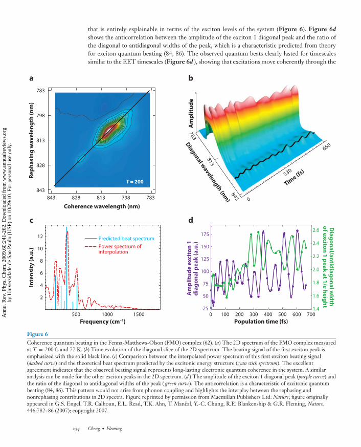
that is entirely explainable in terms of the exciton levels of the system (Figure 6). Figure 6d
shows the anticorrelation between the amplitude of the exciton 1 diagonal peak and the ratio ofthe diagonal to antidiagonal widths of the peak, which is a characteristic predicted from theoryfor exciton quantum beating (84, 86). The observed quantum beats clearly lasted for timescalessimilar to the EET timescales (Figure 6d ), showing that excitations move coherently through the
c
a b
d
348
318
387
0
066
033
Time (f
s)D
iagon
al wavelen
gth
(nm
)
Am
pli
tud
e
Population time (fs)
0 100 200 300 400 500 600 70025
50
75
125
100
150
175
1.4
1.6
1.8
2.0
2.2
2.4
2.6
Am
pli
tud
e e
xci
ton
1
dia
go
na
l p
ea
k (
a.u
.)
Dia
go
na
l/an
tidia
go
na
l wid
th
of e
xcito
n 1
pe
ak
at 1
/e h
eig
ht
843
843
813
813
783
783
798
798
828
828
Re
ph
asi
ng
wa
ve
len
gth
(n
m)
Coherence wavelength (nm)
T = 200
Predicted beat spectrum
Power spectrum of interpolation
500 1000 1500
2
4
6
8
10
12
Frequency (cm–1)
Inte
nsi
ty (
a.u
.)
Figure 6Coherence quantum beating in the Fenna-Matthews-Olson (FMO) complex (62). (a) The 2D spectrum of the FMO complex measuredat T = 200 fs and 77 K. (b) Time evolution of the diagonal slice of the 2D spectrum. The beating signal of the first exciton peak isemphasized with the solid black line. (c) Comparison between the interpolated power spectrum of this first exciton beating signal(dashed curve) and the theoretical beat spectrum predicted by the excitonic energy structure (cyan stick spectrum). The excellentagreement indicates that the observed beating signal represents long-lasting electronic quantum coherence in the system. A similaranalysis can be made for the other exciton peaks in the 2D spectrum. (d ) The amplitude of the exciton 1 diagonal peak (purple curve) andthe ratio of the diagonal to antidiagonal widths of the peak ( green curve). The anticorrelation is a characteristic of excitonic quantumbeating (84, 86). This pattern would not arise from phonon coupling and highlights the interplay between the rephasing andnonrephasing contributions in 2D spectra. Figure reprinted by permission from Macmillan Publishers Ltd: Nature; figure originallyappeared in G.S. Engel, T.R. Calhoun, E.L. Read, T.K. Ahn, T. Mancal, Y.-C. Chung, R.E. Blankenship & G.R. Fleming, Nature,446:782–86 (2007); copyright 2007.
254 Cheng · Fleming
Ann
u. R
ev. P
hys.
Che
m. 2
009.
60:2
41-2
62. D
ownl
oade
d fr
om w
ww
.ann
ualr
evie
ws.
org
by U
nive
rsid
ade
de S
ao P
aulo
(U
SP)
on 1
0/29
/10.
For
per
sona
l use
onl
y.
ANRV373-PC60-12 ARI 25 February 2009 16:35
FMO complex, rather than by incoherent hopping as had usually been assumed (110). In addition,the beating amplitude of the cross peak between exciton 1 and 3, which traces the amplitude ofthe coherence between the two excitons (80), is minimum at T = 0 and increases as a function ofT. This indicates that coherence is transferred into the 1–3 coherence. According to the discussionin Section 3.2, such a coherence transfer effect requires an energy-matching condition and cor-related bath fluctuations (58, 59, 63, 64, 117); therefore, a more general theoretical model thatincludes a correlated nonlocal bath, coherence transfer dynamics, and non-Markovian dynamicswill be required to describe the observations. We note that the beating on the diagonal in Figure 6arises solely from the nonrephasing contribution and therefore provides another example of theutility of recording the separate rephasing and nonphasing spectra. Nonrephasing spectra providea precise way to determine beat frequencies and dephasing times, as recently demonstrated byCalhoun et al. (118).
The observation of long-lasting quantum coherence also led to the suggestion that quantumeffects might be utilized in the FMO complex to promote light harvesting. Engel et al. (62)proposed that the FMO complex performs a quantum search algorithm that is more efficient thana classical random walk suggested by the hopping mechanism. Quantum coherence enables theexcitation to rapidly and reversibly sample multiple pathways to search for the site (BChl 3) thatconnects to the RC (119). A second advantage of the reversible motion is that it is more difficultto become trapped in a subsidiary energy minimum. Further studies are required before it canbe stated that quantum coherence is a general feature of photosynthetic PPCs, but it seems clearthat any accurate description of the dynamics (and design principles) of these systems will requireproper consideration of this true quantum effect.
The long-lasting quantum coherence revealed in the FMO complex is not unique. In an inde-pendent study using a different method that is specifically sensitive to coherences, the two-colorelectronic coherence photon echo technique, Fleming and coworkers (75) directly observed coher-ence dynamics in a photosynthetic bacterial RC and revealed a long-lasting electronic coherencebetween two electronic states that are formed by mixing of the bacteriopheophythin and accessoryBChl excited states. This experiment yielded Gaussian dephasing times of 440 and 310 fs at 77and 180 K, respectively. Furthermore, the long-lasting coherence can be explained only by strongcorrelations between protein motions that modulate electronic transition energies of neighboringchromophores. The combined results of References 62 and 75 suggest that correlated proteinenvironments preserve electronic coherence in photosynthetic complexes and contribute to theoptimization of photosynthetic EET.
6. WHAT DOES QUANTUM COHERENCE HAVE TO DO WITHNATURAL LIGHT HARVESTING?
Do these observations made using coherent laser excitation represent the reality of natural lightharvesting? A frequent concern is that the coherence is created only by the ultrashort laser pulsesused in the experiments, and the sun, being an incoherent light source, would not produce thesame effect. It is certainly true that by synchronizing the entire ensemble, the femtosecond laserpulses enable us to observe the quantum aspects of the system, but the function of light harvestingdoes not depend on such ensemble coherence. Phase coherence within an individual complexwould allow quantum coherence to affect the dynamics of EET. The sun is a source of broadbandblack-body radiation that can be viewed as a series of random ultrashort spikes with a durationas short as the bandwidth allows (120–122). At the single system level, excitation by sunlight alsogenerates a coherent superposition of excited states. Preparation by energy transfer from anotherlight-harvesting complex can also produce delocalized superposition states, and phonon-induced
www.annualreviews.org • Light Harvesting in Photosynthesis 255
Ann
u. R
ev. P
hys.
Che
m. 2
009.
60:2
41-2
62. D
ownl
oade
d fr
om w
ww
.ann
ualr
evie
ws.
org
by U
nive
rsid
ade
de S
ao P
aulo
(U
SP)
on 1
0/29
/10.
For
per
sona
l use
onl
y.

ANRV373-PC60-12 ARI 25 February 2009 16:35
coherence transfer dynamics can induce excitonic coherence after the transfer (63, 117). Generallyspeaking, in natural light harvesting, coherence at an individual system level can be generated,and the quantum effect can affect the outcome of energy trapping, as long as the coherencewithin a single system is maintained. If a dozen quantum computers each independently executeda quantum algorithm, for example, no one would imagine that 12 computers should be started atthe same instant for each to arrive independently at the correct result.
The mechanisms responsible for long-lasting coherence observed by coherent laser experi-ments are also relevant in natural light harvesting. These experiments are carried out with weaklight pulses whose electronic fields do not alter the Hamiltonian of the complex. The terms inthe Hamiltonian that give rise to the long-lasting coherence observed are intrinsic. The key char-acteristic of the photosynthetic complexes is the small reorganization energy, λ (7). With thespin-boson model, one can estimate a lower bound for the Gaussian dephasing time as a functionof temperature and λ in the high-temperature limit using (43, 123)
1τg
=√
2λkb T.
For λ = 80 cm−1, which is a typical value measured for chlorophyll Qy excitation in photosyntheticcomplexes, τ g = ∼60 fs at 77 K and τ g ∼ 30 fs at 298 K. Additionally, if we consider that not allthe reorganization energy may be expressed on the timescale of very rapid energy transfer and thatcorrelated protein environments and phonon-induced coherence transfer processes can furtherextend dephasing times (75), it is plausible that the long-lasting coherence observed in complexphotosynthetic PPCs could play a role in light harvesting.
7. CONCLUDING COMMENTS
The efficiency of light harvesting and hence photosynthesis is controlled by the competition be-tween loss through radiative and nonradiative decay and trapping at the reaction center; thus therapid EET on a subpicosecond timescale is crucially important to the success of photosynthesis.Theoretical studies based on high-resolution structures have shown that the nanoscale dimensionsin photosynthetic PPCs are responsible for their effective function. This leads to strong couplingsbetween molecular excitations and provides a means for the protein matrix to modify the energeticsof light harvesting. Such intricate pigment-protein interactions result in organized energy gradi-ents and energy transfer pathways that are utilized to transfer excitation energy toward the RC,even in apparently irregular structures such as the FMO complex. In short, the protein scaffold-ing constructs a network of highly interconnected chlorophylls whose zeroth-order Hamiltonian,describing partially delocalized exciton states, already favors energy transfer toward the RC.
Emerging experimental data indicate that the dynamics of light harvesting is not fully de-scribed by a classical random-walk picture. Strong electronic couplings between spatially proxi-mate chromophores give rise to delocalized exciton states and spatially connected EET pathwaysthat are highly efficient. An intriguing addition to this picture is the quantum coherence effect,which can enable the excitation to rapidly and coherently sample multiple pathways in space. Wesuggest that such long-lasting excitonic coherence can be explained by the intrinsically small re-organization energy and potential for correlated bath motions in the protein environment insidePPCs. The fundamental issue of quantum coherence effects in natural biological systems de-mands more thorough theoretical and experimental investigations, perhaps using tools developedin quantum information science. The research can potentially open a revolutionary avenue forthe effective use of biological systems as quantum devices or resources for quantum informationprocessing.
256 Cheng · Fleming
Ann
u. R
ev. P
hys.
Che
m. 2
009.
60:2
41-2
62. D
ownl
oade
d fr
om w
ww
.ann
ualr
evie
ws.
org
by U
nive
rsid
ade
de S
ao P
aulo
(U
SP)
on 1
0/29
/10.
For
per
sona
l use
onl
y.

ANRV373-PC60-12 ARI 25 February 2009 16:35
SUMMARY POINTS
1. The nanoscale dimensions of light-harvesting PPCs result in strong couplings betweenpigments and energy tuning via pigment-protein interactions, which create energy trans-fer pathways that can channel energy flow efficiently toward the RC.
2. Based on structural information, site energies and excitonic couplings of excitations in aPPC can be determined using modern quantum chemical methods. However, includingdielectric screening of the protein matrix and specific interactions between chromophoresand protein residues is crucial for the accurate description of electronic excitations inphotosynthetic PPCs.
3. Two-dimensional electronic spectroscopy is a particularly powerful tool for monitoringEET dynamics in photosynthetic PPCs because it is sensitive to electronic couplingsand coherent dynamical processes in the system. Moreover, variants of 2D electronicspectroscopy can be developed to enhance specificity and resolution of 2D spectra.
4. Two energy transfer pathways control the energy flow in the FMO complex, and theenergy transfer occurs via a wavelike, coherent mechanism, which can promote the effi-ciency of energy trapping in photosynthesis.
5. In natural light harvesting, coherence at an individual system level may be generatedthrough the absorption of sunlight or EET from another light-harvesting complex, andthe quantum effect can affect the outcome of energy trapping as long as the coherencewithin the system is maintained.
DISCLOSURE STATEMENT
The authors are not aware of any biases that might be perceived as affecting the objectivity of thisreview.
ACKNOWLEDGMENTS
G.R.F. thanks Prof. D.M. Jonas for valuable discussions. This work was supported by the Director,Office of Science, Office of Basic Energy Sciences, of the U.S. Department of Energy undercontract DE-AC02-05CH11231 and by the Chemical Sciences, Geosciences and BiosciencesDivision, Office of Basic Energy Sciences, U.S. Department of Energy under contract DE-AC03-76SF000098.
LITERATURE CITED
1. van Amerongen H, Valkunas L, van Grondelle R. 2000. Photosynthetic Excitons. Singapore: World Sci.2. Blankenship RE. 2002. Molecular Mechanisms of Photosynthesis. London: Blackwell Sci.3. Duysens L. 1964. Photosynthesis. Prog. Biophys. Mol. Biol. 14:1–1044. Sundstrom V, Pullerits T, van Grondelle R. 1999. Photosynthetic light-harvesting: Reconciling dynamics
and structure of purple bacterial LH2 reveals function of photosynthetic unit. J. Phys. Chem. B 103:2327–46
5. Yang M, Agarwal R, Fleming GR. 2001. The mechanism of energy transfer in the antenna of photosyn-thetic purple bacteria. J. Photochem. Photobiol. A 142:107–19
6. Comprehensivereview of structure-based theoreticalstudies of EETdynamics inphotosyntheticapparatus of purplebacteria.
6. Hu X, Ritz T, Damjanovic A, Autenrieth F, Schulten K. 2002. Photosynthetic apparatus of purplebacteria. Q. Rev. Biophys. 35:1–62
www.annualreviews.org • Light Harvesting in Photosynthesis 257
Ann
u. R
ev. P
hys.
Che
m. 2
009.
60:2
41-2
62. D
ownl
oade
d fr
om w
ww
.ann
ualr
evie
ws.
org
by U
nive
rsid
ade
de S
ao P
aulo
(U
SP)
on 1
0/29
/10.
For
per
sona
l use
onl
y.

ANRV373-PC60-12 ARI 25 February 2009 16:35
7. Scholes GD, Fleming GR. 2005. Energy transfer and photosynthetic light harvesting. Adv. Chem. Phys.132:57–130
8. Clear and currentreview of experimentaland theoretical studiesof energy transfer in thephotosynthetic light-harvesting complexesLH1, LH2, and LHCII.
8. van Grondelle R, Novoderezhkin VI. 2006. Energy transfer in photosynthesis: experimentalinsights and quantitative models. Phys. Chem. Chem. Phys. 8:793–807
9. Cogdell RJ, Gall A, Kohler J. 2006. The architecture and function of the light-harvesting apparatus ofpurple bacteria: from single molecules to in vivo membranes. Q. Rev. Biophys. 39:227–324
10. Sundstrom V. 2008. Femtobiology. Annu. Rev. Phys. Chem. 59:53–7711. Sener MK, Jolley C, Ben-Shem A, Fromme P, Nelson N, et al. 2005. Comparison of the light-harvesting
networks of plant and cyanobacterial photosystem I. Biophys. J. 89:1630–4212. Vassiliev S, Bruce D. 2008. Toward understanding molecular mechanisms of light harvesting and charge
separation in photosystem II. Photosyn. Res. 97:75–8913. Cogdell RJ, Lindsay JG. 2000. The structure of photosynthetic complexes in bacteria and plants: an
illustration of the importance of protein structure to the future development of plant science. NewPhytol. 145:167–96
14. Fenna RE, Matthews BW, Olson JM, Shaw EK. 1974. Structure of a bacteriochlorophyll protein fromthe green photosynthetic bacterium Chlorobium limicola: crystallographic evidence for a trimer. J. Mol.Biol. 84:231–40
15. Li YF, Zhou W, Blankenship RE, Allen JP. 1997. Crystal structure of the bacteriochlorophyll a proteinfrom Chlorobium tepidum. J. Mol. Biol. 271:456–71
16. Camara-Artigas A, Blankenship RE, Allen JP. 2003. The structure of the FMO protein from Chlorobiumtepidum at 2.2 A resolution. Photosyn. Res. 75:49–55
17. Matthews BW, Fenna RE, Bolognesi MC, Schmid MF, Olson JM. 1979. Structure of a bacteriochloro-phyll a protein from the green photosynthetic bacterium Prosthecochloris aestuarii. J. Mol. Biol. 131:259–85
18. Tronrud DE, Schmid MF, Matthews BW. 1986. Structure and X-ray amino acid sequence of a bacteri-ochlorophyll a protein from Prosthecochloris aestuarii refined at 1.9 A resolution. J. Mol. Biol. 188:443–54
19. In-depth review ofthe theoreticalmodeling of opticalproperties and EETdynamics inphotosynthetic PPCs.
19. Renger T, May V, Kuhn O. 2001. Ultrafast excitation energy transfer dynamics in photosyntheticpigment-protein complexes. Phys. Rep. 343:137–254
20. Forster T. 1948. Zwischenmolekulare Energiewanderung und Fluoreszenz. Ann. Phys. (Berlin) 437:55–75
21. Summarizes thevarious approximationsin Forster theory andthe development of amore generaltheoretical descriptionof resonance energytransfer in donor-acceptor pairs.
21. Scholes GD. 2003. Long-range resonance energy transfer in molecular systems. Annu. Rev. Phys.
Chem. 54:57–8722. Alden RG, Johnson E, Nagarajan V, Parson W, Law C, et al. 1997. Calculations of spectroscopic
properties of the LH2 bacteriochlorophyll-protein antenna complex from Rhodopseudomonas acidophila.J. Phys. Chem. B 101:4667–80
23. Cory M, Zerner M, Hu X, Schulten K. 1998. Electronic excitations in aggregates of bacteriochlorophylls.J. Phys. Chem. B 102:7640–50
24. Krueger BP, Scholes GD, Fleming GR. 1998. Calculation of couplings and energy-transfer pathwaysbetween the pigments of LH2 by the ab initio transition density cube method. J. Phys. Chem. B 102:5378–86
25. Scholes GD, Gould IR, Cogdell RJ, Fleming GR. 1999. Ab initio molecular orbital calculations ofelectronic couplings in the LH2 bacterial light-harvesting complex of Rps. acidophila. J. Phys. Chem. B103:2543–53
26. Hsu CP, You ZQ, Chen HCH. 2008. Characterization of the short-range couplings in excitation energytransfer. J. Phys. Chem. C 112:1204–12
27. Jordanides X, Scholes GD, Fleming GR. 2001. The mechanism of energy transfer in the bacterialphotosynthetic reaction center. J. Phys. Chem. B 105:1652–69
28. Jordanides X, Scholes GD, Shapley W, Reimers JR, Fleming GR. 2004. Electronic couplings and energytransfer dynamics in the oxidized primary electron donor of the bacterial reaction center. J. Phys. Chem.B 108:1753–65
29. Madjet MEA, Abdurahman A, Renger T. 2006. Intermolecular Coulomb couplings from ab initio elec-trostatic potentials: application to optical transitions of strongly coupled pigments in photosyntheticantennae and reaction centers. J. Phys. Chem. B 110:17268–81
258 Cheng · Fleming
Ann
u. R
ev. P
hys.
Che
m. 2
009.
60:2
41-2
62. D
ownl
oade
d fr
om w
ww
.ann
ualr
evie
ws.
org
by U
nive
rsid
ade
de S
ao P
aulo
(U
SP)
on 1
0/29
/10.
For
per
sona
l use
onl
y.

ANRV373-PC60-12 ARI 25 February 2009 16:35
30. Adolphs J, Renger T. 2006. How proteins trigger excitation energy transfer in the FMO complex ofgreen sulfur bacteria. Biophys. J. 91:2778–97
31. Louwe R, Vrieze J, Hoff A, Aartsma TJ. 1997. Toward an integral interpretation of the optical steady-state spectra of the FMO complex of Prosthecochloris aestuarii. 2. Exciton simulations. J. Phys. Chem. B101:11280–87
32. Hsu C, Fleming GR, Head-Gordon M, Head-Gordon T. 2001. Excitation energy transfer in condensedmedia. J. Chem. Phys. 114:3065–72
33. Iozzi MF, Mennucci B, Tomasi J, Cammi R. 2004. Excitation energy transfer (EET) between moleculesin condensed matter: a novel application of the polarizable continuum model (PCM). J. Chem. Phys.120:7029–40
34. Curutchet C, Mennucci B. 2005. Toward a molecular scale interpretation of excitation energy transferin solvated bichromophoric systems. J. Am. Chem. Soc. 127:16733–44
35. Scholes GD, Curutchet C, Mennucci B, Cammi R, Tomasi J. 2007. How solvent controls electronicenergy transfer and light harvesting. J. Phys. Chem. B 111:6978–82
36. Curutchet C, Scholes GD, Mennucci B, Cammi R. 2007. How solvent controls electronic energy transferand light harvesting: toward a quantum-mechanical description of reaction field and screening effects.J. Phys. Chem. B 111:13253–65
37. Fowler GJ, Visschers RW, Grief GG, van Grondelle R, Hunter CN. 1992. Genetically modified pho-tosynthetic antenna complexes with blueshifted absorbance bands. Nature 355:848–50
38. Fowler GJ, Sockalingum GD, Robert B, Hunter CN. 1994. Blue shifts in bacteriochlorophyll absorbancecorrelate with changed hydrogen bonding patterns in light-harvesting 2 mutants of Rhodobacter sphaeroideswith alterations at α-Tyr-44 and α-Tyr-45. Biochem. J. 299:695–700
39. Calculates the siteenergies of the sevenBChls in the FMOcomplex includingmolecular details of theprotein environment,based only on thestructural information.
39. Muh F, Madjet MEA, Adolphs J, Abdurahman A, Rabenstein B, et al. 2007. α-Helices direct exci-tation energy flow in the Fenna Matthews Olson protein. Proc. Natl. Acad. Sci. USA 104:16862–67
40. Adolphs J, Muh F, Madjet MEA, Renger T. 2008. Calculation of pigment transition energies in theFMO protein: from simplicity to complexity and back. Photosyn. Res. 95:197–209
41. Breuer HP, Petruccione F. 2002. The Theory of Open Quantum Systems. Oxford: Oxford Univ. Press
42. Explains thelimitations and domainsof applicability of theRedfield, Forster, andmodified-Redfieldequations.
42. Yang M, Fleming GR. 2002. Influence of phonons on exciton transfer dynamics: comparison ofthe Redfield, Forster, and modified Redfield equations. Chem. Phys. 282:163–80
43. Fleming GR, Cho M. 1996. Chromophore-solvent dynamics. Annu. Rev. Phys. Chem. 47:109–3444. Mukamel S. 1995. Principles of Nonlinear Optical Spectroscopy. Oxford: Oxford Univ. Press45. Scholes GD, Fleming GR. 2000. On the mechanism of light harvesting in photosynthetic purple bacteria:
B800 to B850 energy transfer. J. Phys. Chem. B 104:1854–6846. Scholes GD, Jordanides X, Fleming GR. 2001. Adapting the Forster theory of energy transfer for
modeling dynamics in aggregated molecular assemblies. J. Phys. Chem. B 105:1640–5147. Sumi H. 1999. Theory on rates of excitation energy transfer between molecular aggregates through
distributed transition dipoles with application to the antenna system in bacterial photosynthesis. J. Phys.Chem. B 103:252–60
48. Along withReference 49,generalizes the Forsterapproach to treatmultichromophoriceffects arising from thecoherence within donoror acceptor groups.
48. Sumi H. 2002. Bacterial photosynthesis begins with quantum-mechanical coherence. Chem. Rec.
1:480–9349. Jang S, Newton MD, Silbey RJ. 2004. Multichromophoric Forster resonance energy transfer. Phys. Rev.
Lett. 92:21830150. Jang S, Jung Y, Silbey RJ. 2002. Nonequilibrium generalization of Forster-Dexter theory for excitation
energy transfer. Chem. Phys. 275:319–3251. Jang S. 2007. Generalization of the Forster resonance energy transfer theory for quantum mechanical
modulation of the donor-acceptor coupling. J. Chem. Phys. 127:17471052. Cheng YC, Silbey RJ. 2006. Coherence in the B800 ring of purple bacteria LH2. Phys. Rev. Lett. 96:02810353. Jang S, Newton MD, Silbey RJ. 2007. Multichromophoric Forster resonance energy transfer from b800
to b850 in the light harvesting complex 2: evidence for subtle energetic optimization by purple bacteria.J. Phys. Chem. B 111:6807–14
54. Redfield AG. 1957. On the theory of relaxation processes. IBM J. Res. Dev. 1:19–3155. Suarez A, Silbey RJ, Oppenheim I. 1992. Memory effects in the relaxation of quantum open systems.
J. Chem. Phys. 97:5101–7
www.annualreviews.org • Light Harvesting in Photosynthesis 259
Ann
u. R
ev. P
hys.
Che
m. 2
009.
60:2
41-2
62. D
ownl
oade
d fr
om w
ww
.ann
ualr
evie
ws.
org
by U
nive
rsid
ade
de S
ao P
aulo
(U
SP)
on 1
0/29
/10.
For
per
sona
l use
onl
y.

ANRV373-PC60-12 ARI 25 February 2009 16:35
56. Cheng YC, Silbey RJ. 2005. Markovian approximation in the relaxation of open quantum systems.J. Phys. Chem. B 109:21399–405
57. Cho M, Vaswani HM, Brixner T, Stenger J, Fleming GR. 2005. Exciton analysis in 2D electronicspectroscopy. J. Phys. Chem. B 109:10542–56
58. Ohtsuki Y, Fujimura Y. 1996. Theoretical study of bath-induced coherence transfer effects on a time-and frequency-resolved resonant light scattering spectrum. 2. Energy mismatch effects. J. Chem. Phys.104:8321–31
59. Ohtsuki Y, Fujimura Y. 1989. Bath-induced vibronic coherence transfer effects on femtosecond time-resolved resonant light scattering spectra from molecules. J. Chem. Phys. 91:3903–15
60. Kuhn O, Sundstrom V, Pullerits T. 2002. Fluorescence depolarization dynamics in the B850 complexof purple bacteria. Chem. Phys. 275:15–30
61. Novoderezhkin VI, Wendling M, van Grondelle R. 2003. Intra- and interband transfers in the B800–B850 antenna of Rhodospirillum molischianum: Redfield theory modeling of polarized pump-probe kinetics.J. Phys. Chem. B 107:11534–48
62. Observes long-lasting excitoniccoherence and coherentenergy transfer in theFMO complex.
62. Engel GS, Calhoun TR, Read EL, Ahn TK, Mancal T, et al. 2007. Evidence for wavelike energytransfer through quantum coherence in photosynthetic systems. Nature 446:782–86
63. Jean J, Fleming GR. 1995. Competition between energy and phase relaxation in electronic curve crossingprocesses. J. Chem. Phys. 103:2092–101
64. Khalil M, Demirdoven N, Tokmakoff A. 2004. Vibrational coherence transfer characterized with Fourier-transform 2D IR spectroscopy. J. Chem. Phys. 121:362–73
65. Kenkre VM, Knox RS. 1974. Generalized-master-equation theory of excitation transfer. Phys. Rev. B9:5279–90
66. Silbey R. 1976. Electronic energy transfer in molecular crystals. Annu. Rev. Phys. Chem. 27:203–2367. Munn R, Silbey RJ. 1980. Remarks on exciton-phonon coupling and exciton transport. Mol. Cryst. Liq.
Cryst. 57:131–4468. Novoderezhkin VI, Palacios MA, van Amerongen H, van Grondelle R. 2004. Energy-transfer dynamics
in the LHCII complex of higher plants: modified Redfield approach. J. Phys. Chem. B 108:10363–7569. Novoderezhkin VI, Rutkauskas D, van Grondelle R. 2006. Dynamics of the emission spectrum of a single
LH2 complex: interplay of slow and fast nuclear motions. Biophys. J. 90:2890–90270. Schroder M, Kleinekathofer U, Schreiber M. 2006. Calculation of absorption spectra for light-harvesting
systems using non-Markovian approaches as well as modified Redfield theory. J. Chem. Phys. 124:08490371. Renger T, Trostmann I, Theiss C, Madjet MEA, Richter M, et al. 2007. Refinement of a structural model
of a pigment-protein complex by accurate optical line shape theory and experiments. J. Phys. Chem. B111:10487–501
72. Zhang W, Meier T, Chernyak V, Mukamel S. 1998. Exciton-migration and three-pulse femtosecondoptical spectroscopies of photosynthetic antenna complexes. J. Chem. Phys. 108:7763–74
73. Golosov AA, Reichman DR. 2001. Reference system master equation approaches to condensed phasecharge transfer processes. I. General formulation. J. Chem. Phys. 115:9848–61
74. Golosov AA, Reichman DR. 2001. Reference system master equation approaches to condensed phasecharge transfer processes. II. Numerical tests and applications to the study of photoinduced chargetransfer reactions. J. Chem. Phys. 115:9862–70
75. Observes long-lasting coherence in abacterial RC and showsthat this phenomenon isexplained by acorrelated proteinenvironment.
75. Lee H, Cheng YC, Fleming GR. 2007. Coherence dynamics in photosynthesis: protein protectionof excitonic coherence. Science 316:1462–65
76. Mukamel S. 2000. Multidimensional femtosecond correlation spectroscopies of electronic and vibrationalexcitations. Annu. Rev. Phys. Chem. 51:691–729
77. Jonas DM. 2003. Two-dimensional femtosecond spectroscopy. Annu. Rev. Phys. Chem. 54:425–6378. Cho M. 2008. Coherent two-dimensional optical spectroscopy. Chem. Rev. 108:1331–418
79. Provides an intuitivedescription of the originand information contentof 2D electronic spectra.
79. Cho M, Brixner T, Stiopkin IV, Vaswani HM, Fleming GR. 2006. Two dimensional electronicspectroscopy of molecular complexes. J. Chin. Chem. Soc. 53:15–24
80. Cheng YC, Engel GS, Fleming GR. 2007. Elucidation of population and coherence dynamics usingcross-peaks in two-dimensional electronic spectroscopy. Chem. Phys. 341:285–95
81. Hybl J, Yu A, Farrow D, Jonas DM. 2002. Polar solvation dynamics in the femtosecond evolution oftwo-dimensional Fourier transform spectra. J. Phys. Chem. A 106:7651–54
260 Cheng · Fleming
Ann
u. R
ev. P
hys.
Che
m. 2
009.
60:2
41-2
62. D
ownl
oade
d fr
om w
ww
.ann
ualr
evie
ws.
org
by U
nive
rsid
ade
de S
ao P
aulo
(U
SP)
on 1
0/29
/10.
For
per
sona
l use
onl
y.

ANRV373-PC60-12 ARI 25 February 2009 16:35
82. Stiopkin IV, Brixner T, Yang M, Fleming GR. 2006. Heterogeneous exciton dynamics revealed bytwo-dimensional optical spectroscopy. J. Phys. Chem. B 110:20032–37
83. Roberts ST, Loparo JJ, Tokmakoff A. 2006. Characterization of spectral diffusion from two-dimensionalline shapes. J. Chem. Phys. 125:084502
84. Pisliakov AV, Mancal T, Fleming GR. 2006. Two-dimensional optical three-pulse photon echo spec-troscopy. II. Signatures of coherent electronic motion and exciton population transfer in dimer two-dimensional spectra. J. Chem. Phys. 124:234505
85. Cho M, Fleming GR. 2005. The integrated photon echo and solvation dynamics. II. Peak shifts andtwo-dimensional photon echo of a coupled chromophore system. J. Chem. Phys. 123:114506
86. Cheng YC, Fleming GR. 2008. Coherence quantum beats in two-dimensional electronic spectroscopy.J. Phys. Chem. A 112:4254–60
87. Hybl J, Christophe Y, Jonas DM. 2001. Peak shapes in femtosecond 2D correlation spectroscopy. Chem.Phys. 266:295–309
88. Khalil M, Demirdoven N, Tokmakoff A. 2003. Coherent 2D IR spectroscopy: molecular structure anddynamics in solution. J. Phys. Chem. A 107:5258–79
89. Zigmantas D, Read EL, Mancal T, Brixner T, Gardiner AT, et al. 2006. Two-dimensional electronicspectroscopy of the B800–B820 light-harvesting complex. Proc. Natl. Acad. Sci. USA 103:12672–77
90. Hochstrasser RM. 2001. Two-dimensional IR spectroscopy: polarization anisotropy effects. Chem. Phys.266:273–84
91. Zanni MT, Ge NH, Kim YS, Hochstrasser RM. 2001. Two-dimensional IR spectroscopy can be designedto eliminate the diagonal peaks and expose only the crosspeaks needed for structure determination. Proc.Natl. Acad. Sci. USA 98:11265–70
92. Dreyer J, Moran A, Mukamel S. 2003. Tensor components in three pulse vibrational echoes of a rigiddipeptide. Bull. Kor. Chem. Soc. 24:1091–96
93. Read EL, Engel GS, Calhoun TR, Mancal T, Ahn TK, et al. 2007. Cross-peak-specific two-dimensionalelectronic spectroscopy. Proc. Natl. Acad. Sci. USA 104:14203–8
94. Voronine DV, Abramavicius D, Mukamel S. 2007. Manipulating multidimensional electronic spectra ofexcitons by polarization pulse shaping. J. Chem. Phys. 126:044508
95. Abramavicius D, Mukamel S. 2006. Chirality-induced signals in coherent multidimensional spectroscopyof excitons. J. Chem. Phys. 124:034113
96. Voronine DV, Abramavicius D, Mukamel S. 2006. Coherent control of cross-peaks in chirality-inducedtwo-dimensional optical signals of excitons. J. Chem. Phys. 125:224504
97. Abramavicius D, Voronine DV, Mukamel S. 2008. Unravelling coherent dynamics and energy dissipationin photosynthetic complexes by 2D spectroscopy. Biophys. J. 94:3613–19
98. Read E, Schlau-Cohen G, Engel GS, Wen J, Blankenship RE, et al. 2008. Visualization of excitonic struc-ture in the Fenna-Matthews-Olson photosynthetic complex by polarization-dependent two-dimensionalelectronic spectroscopy. Biophys. J. 95:847–56
99. Vulto SIE, Neerken S, Louwe R, de Baat M, Amesz J, et al. 1998. Excited-state structure and dynamicsin FMO antenna complexes from photosynthetic green sulfur bacteria. J. Phys. Chem. B 102:10630–35
100. Vulto SIE, de Baat M, Louwe R, Permentier HP, Neef T, et al. 1998. Exciton simulations of opticalspectra of the FMO complex from the green sulfur bacterium Chlorobium tepidum at 6 K. J. Phys. Chem.B 102:9577–82
101. Wendling M, Przyjalgowski MA, Gulen D, Vulto SIE, Aartsma TJ, et al. 2005. The quantitative rela-tionship between structure and polarized spectroscopy in the FMO complex of Prosthecochloris aestuarii:refining experiments and simulations. Photosyn. Res. 71:99–123
102. Freiberg A, Lin S, Timpmann K, Blankenship RE. 1997. Exciton dynamics in FMO bacteriochlorophyllprotein at low temperatures. J. Phys. Chem. B 101:7211–20
103. Savikhin S, Buck DR, Struve WS. 1998. Toward level-to-level energy transfers in photosynthesis: theFenna-Matthews-Olson protein. J. Phys. Chem. B 102:5556–65
104. First 2D electronicspectroscopicexperiment on aphotosyntheticcomplex, showing theEET pathways in theFMO complex.
104. Brixner T, Stenger J, Vaswani HM, Cho M, Blankenship RE, et al. 2005. Two-dimensionalspectroscopy of electronic couplings in photosynthesis. Nature 434:625–28
105. Yang M, Fleming GR. 1999. Two-color three-pulse photon echoes as a probe of electronic coupling inmolecular complexes. J. Chem. Phys. 110:2983–90
www.annualreviews.org • Light Harvesting in Photosynthesis 261
Ann
u. R
ev. P
hys.
Che
m. 2
009.
60:2
41-2
62. D
ownl
oade
d fr
om w
ww
.ann
ualr
evie
ws.
org
by U
nive
rsid
ade
de S
ao P
aulo
(U
SP)
on 1
0/29
/10.
For
per
sona
l use
onl
y.

ANRV373-PC60-12 ARI 25 February 2009 16:35
106. Mancal T, Fleming GR. 2004. Probing electronic coupling in excitonically coupled heterodimer com-plexes by two-color three-pulse photon echoes. J. Chem. Phys. 121:10556–65
107. Parkinson DY, Lee H, Fleming GR. 2007. Measuring electronic coupling in the reaction center of purplephotosynthetic bacteria by two-color, three-pulse photon echo peak shift spectroscopy. J. Phys. Chem. B111:7449–56
108. Cheng YC, Lee H, Fleming GR. 2007. Efficient simulation of three-pulse photon-echo signals withapplication to the determination of electronic coupling in a bacterial photosynthetic reaction center.J. Phys. Chem. A 111:9499–508
109. Wen J, Zhang H, Gross ML, Blankenship RE. 2008. Membrane orientation of the FMO antenna pro-tein from Chlorobium tepidum as determined by mass spectrometry-based footprinting. Unpublishedmanuscript
110. Robinson GW. 1966. Excitation transfer and trapping in photosynthesis. In Brookhaven Symposium inBiology, No. 19, pp. 16–48. Upton, NY: Brookhaven Natl. Lab.
111. Pearlstein RM. 1982. Exciton migration and trapping in photosynthesis. Photochem. Photobiol. 35:835–44112. Leegwater J. 1996. Coherent versus incoherent energy transfer and trapping in photosynthetic antenna
complexes. J. Phys. Chem. 100:14403–9113. Monshouwer R, Abrahamsson M, van Mourik F, van Grondelle R. 1997. Superradiance and exciton
delocalization in bacterial photosynthetic light-harvesting systems. J. Phys. Chem. B 101:7241–48114. Chachisvilis M, Kuhn O, Pullerits T, Sundstrom V. 1997. Excitons in photosynthetic purple bacteria:
wavelike motion or incoherent hopping? J. Phys. Chem. B 101:7275–83115. Meier T, Chernyak V, Mukamel S. 1997. Multiple exciton coherence sizes in photosynthetic antenna
complexes viewed by pump-probe spectroscopy. J. Phys. Chem. B 101:7332–42116. Savikhin S, Buck DR, Struve WS. 1997. Oscillating anisotropies in a bacteriochlorophyll protein: evi-
dence for quantum beating between exciton levels. Chem. Phys. 223:303–12117. Jean J. 1996. Vibrational coherence effects on electronic curve crossing. J. Chem. Phys. 104:5638–46118. Calhoun T, Schlau-Cohen G, Ginsberg N, Fleming GR. 2008. Unpublished manuscript119. Gaab KM, Bardeen CJ. 2004. The effects of connectivity, coherence, and trapping on energy transfer in
simple light-harvesting systems studied using the Haken-Strobl model with diagonal disorder. J. Chem.Phys. 121:7813–20
120. Gouy M. 1886. Sur le mouvement lumineux. J. Phys. Theor. Appl. 5:354–62121. Rayleigh J. 1889. On the character of the complete radiation at a given temperature. Philos. Mag. 27:460–
69122. Bradley D, New G. 1974. Ultrashort pulse measurements. Proc. IEEE 62:313–45123. Hwang H, Rossky PJ. 2004. An analysis of electronic dephasing in the spin-boson model. J. Chem. Phys.
120:11380–85
262 Cheng · Fleming
Ann
u. R
ev. P
hys.
Che
m. 2
009.
60:2
41-2
62. D
ownl
oade
d fr
om w
ww
.ann
ualr
evie
ws.
org
by U
nive
rsid
ade
de S
ao P
aulo
(U
SP)
on 1
0/29
/10.
For
per
sona
l use
onl
y.

AR373-FM ARI 25 February 2009 17:55
Annual Review ofPhysical Chemistry
Volume 60, 2009 Contents
Frontispiece � � � � � � � � � � � � � � � � � � � � � � � � � � � � � � � � � � � � � � � � � � � � � � � � � � � � � � � � � � � � � � � � � � � � � � � � � � � � � � � � � � � � � xiv
Sixty Years of Nuclear MomentsJohn S. Waugh � � � � � � � � � � � � � � � � � � � � � � � � � � � � � � � � � � � � � � � � � � � � � � � � � � � � � � � � � � � � � � � � � � � � � � � � � � � � � � � � � 1
Dynamics of Liquids, Molecules, and Proteins Measured with Ultrafast2D IR Vibrational Echo Chemical Exchange SpectroscopyM.D. Fayer � � � � � � � � � � � � � � � � � � � � � � � � � � � � � � � � � � � � � � � � � � � � � � � � � � � � � � � � � � � � � � � � � � � � � � � � � � � � � � � � � � � �21
Photofragment Spectroscopy and Predissociation Dynamics of WeaklyBound MoleculesHanna Reisler � � � � � � � � � � � � � � � � � � � � � � � � � � � � � � � � � � � � � � � � � � � � � � � � � � � � � � � � � � � � � � � � � � � � � � � � � � � � � � � � � �39
Second Harmonic Generation, Sum Frequency Generation, and χ (3):Dissecting Environmental Interfaces with a Nonlinear Optical SwissArmy KnifeFranz M. Geiger � � � � � � � � � � � � � � � � � � � � � � � � � � � � � � � � � � � � � � � � � � � � � � � � � � � � � � � � � � � � � � � � � � � � � � � � � � � � � �61
Dewetting and Hydrophobic Interaction in Physical and BiologicalSystemsBruce J. Berne, John D. Weeks, and Ruhong Zhou � � � � � � � � � � � � � � � � � � � � � � � � � � � � � � � � � � � � � � � � �85
Photoelectron Spectroscopy of Multiply Charged AnionsXue-Bin Wang and Lai-Sheng Wang � � � � � � � � � � � � � � � � � � � � � � � � � � � � � � � � � � � � � � � � � � � � � � � � � � � � � � 105
Intrinsic Particle Properties from Vibrational Spectra of AerosolsOmar F. Sigurbjörnsson, George Firanescu, and Ruth Signorell � � � � � � � � � � � � � � � � � � � � � � � � � 127
Nanofabrication of Plasmonic StructuresJoel Henzie, Jeunghoon Lee, Min Hyung Lee, Warefta Hasan, and Teri W. Odom � � � � 147
Chemical Synthesis of Novel Plasmonic NanoparticlesXianmao Lu, Matthew Rycenga, Sara E. Skrabalak, Benjamin Wiley,and Younan Xia � � � � � � � � � � � � � � � � � � � � � � � � � � � � � � � � � � � � � � � � � � � � � � � � � � � � � � � � � � � � � � � � � � � � � � � � � � � � � � 167
Atomic-Scale Templates Patterned by Ultrahigh Vacuum ScanningTunneling Microscopy on SiliconMichael A. Walsh and Mark C. Hersam � � � � � � � � � � � � � � � � � � � � � � � � � � � � � � � � � � � � � � � � � � � � � � � � � � 193
DNA Excited-State Dynamics: From Single Bases to the Double HelixChris T. Middleton, Kimberly de La Harpe, Charlene Su, Yu Kay Law,Carlos E. Crespo-Hernández, and Bern Kohler � � � � � � � � � � � � � � � � � � � � � � � � � � � � � � � � � � � � � � � � � � � � 217
viii
Ann
u. R
ev. P
hys.
Che
m. 2
009.
60:2
41-2
62. D
ownl
oade
d fr
om w
ww
.ann
ualr
evie
ws.
org
by U
nive
rsid
ade
de S
ao P
aulo
(U
SP)
on 1
0/29
/10.
For
per
sona
l use
onl
y.

AR373-FM ARI 25 February 2009 17:55
Dynamics of Light Harvesting in PhotosynthesisYuan-Chung Cheng and Graham R. Fleming � � � � � � � � � � � � � � � � � � � � � � � � � � � � � � � � � � � � � � � � � � � � � 241
High-Resolution Infrared Spectroscopy of the Formic Acid DimerOzgür Birer and Martina Havenith � � � � � � � � � � � � � � � � � � � � � � � � � � � � � � � � � � � � � � � � � � � � � � � � � � � � � � � 263
Quantum Coherent Control for Nonlinear Spectroscopyand MicroscopyYaron Silberberg � � � � � � � � � � � � � � � � � � � � � � � � � � � � � � � � � � � � � � � � � � � � � � � � � � � � � � � � � � � � � � � � � � � � � � � � � � � � � 277
Coherent Control of Quantum Dynamics with Sequences of UnitaryPhase-Kick PulsesLuis G.C. Rego, Lea F. Santos, and Victor S. Batista � � � � � � � � � � � � � � � � � � � � � � � � � � � � � � � � � � � � � 293
Equation-Free Multiscale Computation: Algorithms and ApplicationsIoannis G. Kevrekidis and Giovanni Samaey � � � � � � � � � � � � � � � � � � � � � � � � � � � � � � � � � � � � � � � � � � � � � � 321
Chirality in Nonlinear OpticsLevi M. Haupert and Garth J. Simpson � � � � � � � � � � � � � � � � � � � � � � � � � � � � � � � � � � � � � � � � � � � � � � � � � � � 345
Physical Chemistry of DNA VirusesCharles M. Knobler and William M. Gelbart � � � � � � � � � � � � � � � � � � � � � � � � � � � � � � � � � � � � � � � � � � � � � 367
Ultrafast Dynamics in Reverse MicellesNancy E. Levinger and Laura A. Swafford � � � � � � � � � � � � � � � � � � � � � � � � � � � � � � � � � � � � � � � � � � � � � � � � 385
Light Switching of Molecules on SurfacesWesley R. Browne and Ben L. Feringa � � � � � � � � � � � � � � � � � � � � � � � � � � � � � � � � � � � � � � � � � � � � � � � � � � � � � 407
Principles and Progress in Ultrafast Multidimensional NuclearMagnetic ResonanceMor Mishkovsky and Lucio Frydman � � � � � � � � � � � � � � � � � � � � � � � � � � � � � � � � � � � � � � � � � � � � � � � � � � � � � � 429
Controlling Chemistry by Geometry in Nanoscale SystemsL. Lizana, Z. Konkoli, B. Bauer, A. Jesorka, and O. Orwar � � � � � � � � � � � � � � � � � � � � � � � � � � � � � 449
Active Biological MaterialsDaniel A. Fletcher and Phillip L. Geissler � � � � � � � � � � � � � � � � � � � � � � � � � � � � � � � � � � � � � � � � � � � � � � � � � 469
Wave-Packet and Coherent Control DynamicsKenji Ohmori � � � � � � � � � � � � � � � � � � � � � � � � � � � � � � � � � � � � � � � � � � � � � � � � � � � � � � � � � � � � � � � � � � � � � � � � � � � � � � � � 487
Indexes
Cumulative Index of Contributing Authors, Volumes 56–60 � � � � � � � � � � � � � � � � � � � � � � � � � � � 513
Cumulative Index of Chapter Titles, Volumes 56–60 � � � � � � � � � � � � � � � � � � � � � � � � � � � � � � � � � � � � 516
Errata
An online log of corrections to Annual Review of Physical Chemistry articles may befound at http://physchem.annualreviews.org/errata.shtml
Contents ix
Ann
u. R
ev. P
hys.
Che
m. 2
009.
60:2
41-2
62. D
ownl
oade
d fr
om w
ww
.ann
ualr
evie
ws.
org
by U
nive
rsid
ade
de S
ao P
aulo
(U
SP)
on 1
0/29
/10.
For
per
sona
l use
onl
y.