Embed Size (px)
Citation preview

Technical Note
Dynamic MR Venography: An Intrinsic Benefit ofTime-Resolved MR Angiography
Jiang Du, PhD,1,3* Frank J. Thornton, MD,2 Charles A. Mistretta, PhD,1,2 andThomas M. Grist, MD1,2
Purpose: To investigate the possibility of obtaining dy-namic contrast-enhanced magnetic resonance venography(DCE-MRV) images of the lower extremities.
Materials and Methods: Peripheral contrast-enhancedmagnetic resonance angiography (CE-MRA) was performedon 20 patients using a time-resolved sequence that com-bined undersampled projection reconstruction (PR) in-plane and Cartesian slice encoding through-plane. Thecontrast dynamics of distal vessels were depicted. An au-tomated segmentation algorithm based on a contrast ar-rival time (CAT) threshold was used to generate contrastdynamics in the venous system. The signal difference be-tween the vein and artery was measured to evaluate theeffectiveness of this technique in isolating the venous con-trast dynamics.
Results: The automatically generated image series de-picted the contrast dynamics of both the arterial and ve-nous systems, including asymmetric venous enhancementand background tissue enhancement. Quantitative mea-surement showed a mean venous/arterial signal ratio in-crease from 1.58 to 4.82 for the peak venous frame afterarterial signal suppression.
Conclusion: DCE-MRV is a minimally invasive techniquefor evaluating the venous side of the systemic vascularanatomy. Time-resolved MRA has the potential clinical ben-efit of enabling both arterial and venous disease to be de-tected in patients undergoing CE-MRA.
Key Words: contrast enhancement; magnetic resonance(MR); dynamic imaging; peripheral vasculature; venogra-phyJ. Magn. Reson. Imaging 2006;24:922–927.© 2006 Wiley-Liss, Inc.
DEEP VENOUS THROMBOSIS (DVT) has been reportedto be a major cause of morbidity and mortality, with5,000,000 episodes occurring annually in the UnitedStates alone (1). Conventional contrast venographycontinues to serve as a gold standard for diagnosis, butthis technique is invasive and not without risks, includ-ing exposure to ionizing radiation, complications asso-ciated with the use of iodinated contrast material, andprocedure-induced DVT (2). Duplex sonography hasbeen shown to have both high sensitivity and specificityfor femoral and popliteal DVT, but it is operator-depen-dent and has reduced accuracy in the distal lower ex-tremities due to the low volume and flow velocity ofblood within the calf veins (3).
Magnetic resonance imaging (MRI) has been used todetect DVT of the lower and upper extremities and pel-vis for at least a decade (4–10). Techniques such astime-of-flight (TOF) and phase contrast (PC) angiogra-phy have been extensively investigated and proven to bereliable for the assessment of the deep venous system(4). However, these methods have limited value for de-picting paired veins, perforating veins, and venousvalves below the knee, mostly because of high sensitiv-ity to artifacts caused by complex flow and long scantimes (7). Flow-enhanced multishot TOF echo-planarimaging (EPI) can result in better visualization of calfveins by exploiting transient flow-augmenting followingthe use of venous occlusion, but this technique pro-vides little separation of the arterial venous circula-tions, and suffers from edge blurring due to T2* decay(8). Fast-spin-echo (FSE) techniques have a high sensi-tivity for detecting small veins and arteries with slowflow rates, and a low sensitivity for large veins with highflow rates (9). High signal from edema may lead tosuboptimal depiction of the venous anatomy in flow-independent MR venography (MRV) based on inversion-recovery rapid acquisition with relaxation enhance-ment (RARE) sequences (10).
Contrast-enhanced (CE) MRV was recently proposedas a means of evaluating the venous anatomy of theextremities (11–15). This technique requires the injec-tion of an extracellular contrast material that shortensthe T1 of blood. The arterial system is enhanced duringthe first pass of the contrast bolus, with subsequentenhancement of venous structures. Single-phase ac-quisition combined with pedal injections of diluted con-
1Department of Medical Physics, University of Wisconsin–Madison,Wisconsin, USA.2Department of Radiology, University of Wisconsin–Madison, Wiscon-sin, USA.3Department of Radiology, University of California–San Diego, San Di-ego, California, USA.*Address reprint requests to: J.D., Department of Radiology, Universityof California–San Diego, 200 West Arbor Drive, San Diego, CA 92103-8756. E-mail: [email protected] July 11, 2005; Accepted June 30, 2006.DOI 10.1002/jmri.20716Published online 6 September 2006 in Wiley InterScience (www.interscience.wiley.com).
JOURNAL OF MAGNETIC RESONANCE IMAGING 24:922–927 (2006)
© 2006 Wiley-Liss, Inc. 922
trast material can provide a comprehensive display ofthe venous system in the pelvis and lower extremities(11). Multiphase acquisitions are typically used to de-pict the differences in arterial and venous enhance-ment, followed by a subtraction algorithm wherebysubtraction of an early arterial phase acquisition from alate arterial-venous equilibrium phase image producesa selective venogram (12). However, most current dy-namic imaging techniques produce time-resolved im-ages with poor temporal resolution. For example, thevenous enhanced subtracted peak arterial (VESPA)MRV technique generates eight successive images witha temporal resolution of 30 seconds (13). Higher tem-poral resolution is desirable to better depict the con-trast dynamics in both the arterial and venous systems.This may also help with image processing, such aseigenimage filtering and correlation analysis (16,17).
In this study a time-resolved undersampled hybridprojection reconstruction (PR) sequence that combinesundersampled PR in-plane and Cartesian slice encod-ing through-plane was applied for data acquisition inthe distal extremities (18). Both high-temporal-resolu-tion and high-spatial-resolution angiograms andvenograms were obtained, and contrast dynamics inthe venous system were depicted with the use of auto-mated contrast enhancement (CE) and contrast arrivaltime (CAT) analysis.
MATERIALS AND METHODS
A previously reported time-resolved hybrid PR se-quence was used to image the distal extremities (18). Inthis sequence the whole k-space was divided into fourregions (A, B, C, and D) from low-spatial-frequency sliceencoding to high-spatial-frequency slice encoding. Re-gion D accounted for the higher half of the slice-encod-ing values. Regions A, B, and C were of equal size, andwere acquired in the order used for the time resolvedimaging of contrast kinetics (TRICKS) sequence (ABA-CABAC. . .) (18). Region D was acquired repeatedly dur-ing the steady state of contrast material to increase theslice resolution.
The CAT (defined as the time to reach 60% of the peaksignal) threshold is a simple but robust criterion forseparating signals with different contrast dynamics. Ithas been used to suppress background tissue signalfrom CE vascular signal in extended dynamic carotidimaging in swine (17). In dynamic imaging with ante-cubital venous injection, the arterial system is en-hanced earlier than the venous system in the distalextremities. If the time-resolved images have a highenough temporal resolution, a simple CAT thresholdcan remove the arterial signals and maintain all of thevenous signals in the dynamic time frames, thus pro-ducing isolated contrast dynamics in the venous sys-tem alone. It is desirable to generate the CAT maps, andto find the arterial and venous CATs automatically.Furthermore, it is important to recognize that the CATsmay vary based on the location in the imaging field ofview (FOV), especially given the inherent spatial vari-ability of contrast arrival in the large coronal imagingFOV. For example, caudally located arterial voxels have
a later CAT than those located cephalad, since the con-trast bolus flows from the heart to the distal extremity.
An automatic estimation of the contrast bolus arrivaltimes (BATs) for arteries and veins can be performedbased on the dynamic-contrast information. The algo-rithm we adapted was based on the iterative CE (de-fined as the signal difference between the CAT frameand the first time frame) and CAT threshold, as shownin Fig. 1. A CAT map and a CE map were generated inthe first step. A histogram of the CAT was then gener-ated for voxels above the CE threshold. We automatedthe latter step by reducing the CE threshold iterativelyuntil there were only two peaks in the CAT histogram,corresponding to the arterial and venous CATs, respec-tively. To deal with any possible variation in CATs, thewhole FOV was divided into multiple regions (typically16), and the same iterative threshold algorithm wasapplied to generate region-specific arterial/venousCATs and their corresponding contrast dynamics basedon region-specific CAT thresholds. We evaluated theeffectiveness of this venous contrast-dynamics genera-tion algorithm by measuring the venous/arterial sig-nal-to-noise ratio (SNR) before and after arterial signalsuppression for the peak venous frame.
Twenty patient studies were performed on a standard1.5 T MR scanner (Signa LX, GE Medical System,Waukesha, WI, USA). A peripheral phased-array coil(MRI Devices, Milwaukee, WI, USA) was used to imagethe contrast dynamics in the calf vascular system. Theacquisition parameters were as follows: FOV � 40 � 40cm2, TR/TE � 7.5/2.7 msec, flip angle � 30°, BW �62.5 KHz, oblique acquisition readout � 384, numberof projections � 80, number of slices � 72, slice thick-ness � 1.5 mm. An injection of 14 mL of gadolinium(Gd)-based contrast agent (Omniscan; Nycomed Amer-sham, Princeton, NJ, USA) was followed by a flush of 14mL of saline. A computer-controlled power injector(Spectris, Medrad, Indianola, PA, USA) was used toensure a precise injection rate of 2.0 mL/second for thefirst 7 mL of contrast agent, and 0.5 mL/second for thesecond 7 mL. Written informed consent was obtainedfrom each subject before the imaging procedures wereperformed, in accordance with regulations set forth bythe institutional review board.
RESULTS
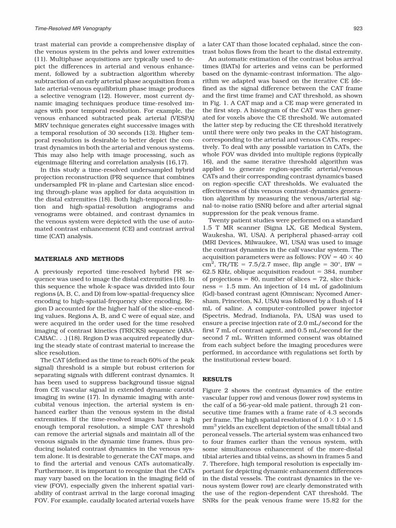
Figure 2 shows the contrast dynamics of the entirevascular (upper row) and venous (lower row) systems inthe calf of a 56-year-old male patient, through 21 con-secutive time frames with a frame rate of 4.3 secondsper frame. The high spatial resolution of 1.0 � 1.0 � 1.5mm3 yields an excellent depiction of the small tibial andperoneal vessels. The arterial system was enhanced twoto four frames earlier than the venous system, withsome simultaneous enhancement of the more-distaltibial arteries and tibial veins, as shown in frames 5 and7. Therefore, high temporal resolution is especially im-portant for depicting dynamic enhancement differencesin the distal vessels. The contrast dynamics in the ve-nous system (lower row) are clearly demonstrated withthe use of the region-dependent CAT threshold. TheSNRs for the peak venous frame were 15.82 for the
Time-Resolved MR Venography 923

artery and 21.56 for the vein, resulting in a vein/arterySNR of 1.36, which was increased to 5.23 after the CATthreshold. There was some persistent arterial edge sig-nal from the arterial arrival time threshold, which wasdue to the distorted contrast time course resulting froma low SNR. Despite this, the venous contrast dynamics
are well depicted with high spatial resolution and hightemporal resolution.
Figure 3 shows the contrast dynamics (upper row) ina 48-year-old female patient. The asymmetric venousenhancement and background tissue enhancementwere well depicted through the automatically generated
Figure 1. Flow chart for automatic ar-terial/venous CAT generation. CAT andCE maps were generated from the dy-namic image series and then a CAT his-togram was generated for voxels abovethe CE threshold. The CE thresholdvalue was reduced iteratively until therewere two peaks in the histogram corre-sponding to the arterial and venousCATs, respectively. The venous contrastdynamics were generated for voxels witha CAT longer than the arterial CAT.
Figure 2. Time series of coro-nal MIP images before (upperrow) and after (lower row) theautomated region-specific CATthreshold was applied in a 56-year-old male patient. Theseimages are five of 21 timeframes and correspond toframes 5, 7, 9, 11, and 13, re-spectively, with a temporal res-olution of 4.3 seconds/frame.The venous valves in the tibialveins of both legs are well de-picted (short arrows) due to thehigh spatial resolution of 1.0 �1.0 � 1.5 mm3. This was zero-padded to 0.78 � 0.78 � 0.78mm3 after image reconstruc-tion.
924 Du et al.

venous contrast dynamics (lower row). The SNRs for thepeak venous frame were 12.64 for the artery and 19.98for the vein, resulting in a vein/artery signal ratio of1.58, which was increased to 4.82 after CAT segmenta-tion. The tibial venous branches were depicted signifi-cantly better after arterial signal suppression, as indi-cated by the short arrow in Fig. 3. The left venoussystem enhanced quickly after the peak arterial frame,
while the right venous system enhanced moderately.Furthermore, the left great saphenous vein could beseen better in frames 10–12, but was degraded by thebackground tissue enhancement, as shown in frame16.
High spatial resolution is demonstrated in Fig. 4,where the peak venous frame is shown through bothcoronal and sagittal reprojections of the three-dimen-
Figure 3. Time series of coro-nal MIP images before (upperrow) and after (lower row) CATsegmentation for a 48-year-oldfemale patient. The contrastdynamics for the tibial veins(short arrows) and great saphe-nous veins (long arrows) aredepicted with much bettervein/artery signal ratio. Thiswas increased from 1.58 to4.82 for the peak arterialframe. The left great saphe-nous vein is best seen inframes 10–12, and is obscuredby the background tissue en-hancement after frame 16.
Figure 4. High spatial resolu-tion and broad slice coverageare demonstrated in the peakvenous frame (arterial signalsuppressed) via (a) sagittal MIPof the right leg, (b) coronal MIPof both legs, and (c) sagittalMIP of the left leg. The acquiredvoxel size of 1.0 � 1.0 � 1.5mm3 yields an excellent depic-tion of the small veins in thecalf.
Time-Resolved MR Venography 925

sional data sets. The high in-plane spatial resolution of1.0 � 1.0 mm2 is demonstrated through the coronalmaximum intensity projection (MIP) image, while thehigh slice resolution of 1.5 mm is demonstratedthrough the sagittal MIP image of both the left and rightlegs.
The contrast-to-noise ratio (CNR) before and after theregion-dependent CAT threshold was measured in thepeak venous frame for all 20 patients. Analysis of datashowed that the mean venous/arterial CNR ratio wasaugmented from 1.38 to 4.95 after the arterial signalsuppression. This increase in venous/arterial signaldifference is obvious from the venous dynamic imageseries shown in Figs. 2 and 3.
DISCUSSION
Contrast dynamics in the distal vascular system can bewell depicted with a time-resolved hybrid under-sampled PR acquisition. This sequence provides time-resolved angiograms with both high spatial resolutionand high temporal resolution. The contrast dynamics inthe venous system can be separated from those in thearterial system with the use of a simple automatedregion-dependent CAT threshold. The automatic BATestimation based on an iterative CE and CAT thresholdalgorithm offers totally operator-independent postpro-cessing. The high-spatial-resolution MRV with an ac-quired voxel size of 1.5 mm3 shows very small subcu-taneous or muscular branches (about 1 mm indiameter) that might be missing by other low-spatial-resolution imaging techniques.
MR venograms can be generated by subtracting theselective arterial phase images from later arterial-ve-nous images (12). In most cases this technique cangenerate venograms with good image quality. However,sometimes the venograms can be contaminated by re-sidual arterial signals due to prolonged arterial signaldecay from the peak arterial phase to the later arterial-venous equilibrium phase. Proper signal scaling canpartly resolve the residual arterial signals but cannotcompletely resolve them because of the variable arterialsignal decay rates in large imaging FOVs. Double sub-traction improves the CNR of venograms, but still can-not completely resolve this problem. Other techniques,such as eigenimage filtering, factor analysis, and correla-tion analysis, can generate higher-quality venograms(16,17); however, these techniques require a longer post-processing time. In addition, they generate only a singlevenous image without contrast dynamics in the venoussystem. As a result, the venous structures, such as theleft great saphenous vein shown in Fig. 3, may beobscured due to soft-tissue enhancement. The vein iswell depicted in frame 12, but is totally obscured inframe 16.
The simple BAT threshold works well for depictingvenous contrast dynamics in the distal extremities.This is primarily because of the high temporal resolu-tion of 4.3 seconds per frame provided by the time-resolved hybrid undersampled PR sequence. A single-subtraction (12), double-subtraction, or VESPA study(13,14) with a temporal resolution of 30 seconds perframe cannot provide details about the contrast dynam-
ics in the vascular system, or enough information toseparate venous from arterial structures based on theBAT threshold. It is important to implement the region-specific CAT threshold algorithm to robustly isolate thevenous contrast dynamics from the arterial contrastdynamics. It is not uncommon to find a variation of upto two or three time frames for the arterial CATs in thelarge coronal imaging FOV. Using a global CAT thresh-old may leave strong arterial residual signals in thevenous contrast dynamics.
The use of venous contrast dynamics permits accu-rate visualization of the venous structure, especially insites where soft-tissue enhancement quickly obscuresthe vasculature. Furthermore, the time-resolved datacan be used to generate contrast dynamics in soft tis-sues, which may help to identify sites of subclinicalsoft-tissue injury (19). High temporal resolution is crit-ical for the simultaneous generation of arterial and ve-nous contrast dynamics. Future studies will focus onimproving the temporal resolution by combining paral-lel imaging reconstruction (20) with undersampled hy-brid PR TRICKS acquisition, and systematically inves-tigating patients with diseases of the venous system.
In conclusion, we have demonstrated the feasibility ofperforming three-dimensional DCE-MRV in the distalextremities. Time-resolved hybrid undersampled PR ac-quisition provides both high spatial resolution and hightemporal resolution dynamic images. Contrast BATthresholds separate veins from arteries simply and ef-fectively. Region-specific arterial/venous CATs can beautomatically generated with an iterative CE and CATthreshold algorithm. The high spatial and temporal res-olution dynamic venous image series provide accuratedepiction of the venous valves, and asymmetric CE inthe venous system, as well as good separation of venousand background-tissue enhancement.
ACKNOWLEDGMENT
The authors thank Dr. Graeme Bydder for useful dis-cussions and editorial help.
REFERENCES1. Anderson FA, Wheeler HB, Goldberg RJ, et al. A population-based
perspective of the hospital incidence and case-fatality rates of deepvein thrombosis and pulmonary embolism. The Worcester DVTstudy. Arch Intern Med 1991;151:933–938.
2. Redman HC. Deep venous thrombosis: is contrast venography stillthe diagnostic “gold standard”? Radiology 1988;168:277–278.
3. Yucel EK, Fisher JS, Egglin TK, Geller SC, Waltman AC. Isolatedcalf venous thrombosis: diagnosis with compression US. Radiology1991;179:443–446.
4. Evans AJ, Sostman HD, Witty LA, et al. Detection of deep venousthrombosis: prospective comparison of MR imaging and sonogra-phy. J Magn Reson Imaging 1996;1:44–51.
5. Erdman WA, Jayson HT, Redman HC, Miller GL, Parkey RW, Pe-shock RW. Deep venous thrombosis of extremities: role of MR im-aging in the diagnosis. Radiology 1990;174:425–431.
6. Spritzer CE, Sostman HD, Wilkes DC, Coleman E. Deep venousthrombosis: experience with gradient-echo MR imaging in 66 pa-tients. Radiology 1990;177:235–241.
7. Lanzer P, Gross G, Keller F, Pohost G. Sequential 2D inflow venog-raphy: initial clinical observations. Magn Reson Med 1991;19:470–476.
8. Holtz DJ, Debatin JF, McKinnon GC, et al. MR venography of thecalf: value of flow-enhanced time-of-flight echoplanar imaging. AJRAm J Roentgenol 1996;166:663–668.
926 Du et al.

9. Bluemke DA, Wolf RL, Tani I, Tachiki S, McVeigh ER, Zerhouni EA.Extremity veins: evaluation with fast-spin-echo MR venography.Radiology 1997;204:562–565.
10. Gallix BP, Achard-Lichere C, Dauzat M, Bruel JM, Lopez FM. Flow-independent magnetic resonance venography of the calf. J MagnReson Imaging 2003;17:421–426.
11. Ruehm SG, Wiesner W, Debatin JF. Pelvic and lower extremity veins:contrast-enhanced three-dimensional MR venography with a dedi-cated vascular coil—initial experience. Radiology 2000;215:421–427.
12. Lebowitz JA, Rofsky NM, Krinsky GA, Weinreb JC. Gadolinium-enhanced body MR venography with subtraction technique. AJRAm J Roentgenol 1997;169:755–758.
13. Fraser DGW, Moody AR, Davidson IR, Martel AL, Morgan PS. Deepvenous thrombosis: diagnosis by using venous enhanced sub-tracted peak arterial MR venography versus conventional venogra-phy. Radiology 2003;226:812–820.
14. Martel AL, Fraser DGW, Delay GS, Morgan PS, Moody AR. Sepa-rating arterial and venous components from 3D dynamic contrast-enhanced MRI studies using factor analysis. Magn Reson Med2003;49:928–933.
15. Sandstede JJW, Krause U, Pabst T, et al. Deep venous thrombosisand consecutive pulmonary embolism as the first sign of an ovariancancer: MR angiography using an intravascular contrast agent(CLARISCAN). J Magn Reson Imaging 2000;12:497–500.
16. Kaandorp DW, Kopinga K, Wijn PF. Venous signal suppression in3D dynamic Gd-enhanced carotid artery imaging using the eigen-image filter. Magn Reson Med 1999;42:307–313.
17. Mazaheri Y, Carroll TJ, Du J, et al. Combined time-resolved andhigh spatial resolution 3D MRA using an extended adaptive acqui-sition. J Magn Reson Imaging 2002;15:291–301.
18. Du J, Carroll TJ, Wagner HJ, et al. Time-resolved, undersampledprojection reconstruction imaging for high resolution CE-MRA ofthe distal runoff vessels. Magn Reson Med 2002;48:516–522.
19. Zhang HL, Kent KC, Bush HL, et al. Soft tissue enhancement ontime-resolved peripheral magnetic resonance angiography. J MagnReson Imaging 2004;19:590–597.
20. Pruessmann KP, Weiger M, Bornert P, Boesiger P. Advances insensitivity encoding with arbitrary k-space trajectories. Magn Re-son Med 2001;46:638–651.
Time-Resolved MR Venography 927