Embed Size (px)
Citation preview

RESEARCH Open Access
Dynamic expression of cytokine andtranscription factor genes duringexperimental Fasciola gigantica infection inbuffaloesWei Shi1,2, Zhi-Yong Wei1,2, Hany M. Elsheikha3, Fu-Kai Zhang2, Zhao-An Sheng1,2, Ke-Jing Lu1, Dong-Ying Wang1,2,Wei-Yi Huang1,2* and Xing-Quan Zhu2,4*
Abstract
Background: Determining the mechanisms involved in the immune-pathogenesis of the tropical liver fluke,Fasciola gigantica, is crucial to the development of any effective therapeutic intervention. Here, we examined thedifferential gene expression of cytokines and transcription factors in the liver of F. gigantica-infected buffaloes, overthe course of infection.
Methods: Water buffaloes (swamp type) were infected orally with 500 F. gigantica encysted metacercariae. Livertissue samples were collected 3, 10, 28, 42, 70 and 98 days post-infection (dpi). Levels of gene expression of ninecytokines (IFN-γ, TGF-β, IL-1β, IL-4, IL-6, IL-10, IL-12B, IL-13 and IL-17A) and four transcription factors (T-bet, GATA-3,Foxp3 and ROR-γτ) were determined using quantitative real-time PCR (qRT-PCR). We evaluated any correlation betweengene expression of these immune-regulatory factors and the severity of liver pathology.
Results: Histopathological examination revealed that cellular infiltration, hemorrhage and fibrosis without calcification inthe liver parenchyma of infected buffaloes, increased over the course of infection. This progressive pathology wasattributed to dysregulated and excessive inflammatory responses induced by infection. The early infection phase(3–10 dpi) was marked by a generalized immunosuppression and elevated TGF-β expression in order to facilitateparasite colonization. A mixed Th1/Th2 immune response was dominant from 28 to 70 dpi, to promote parasitesurvival while minimizing host tissue damage. During late infection (98 dpi), the response was biased towards Th1/Tregin order to inhibit the host’s Th2 protective response and promote chronic infection. Both IL-10 and IL-17A and theTh17/Treg balance, played key roles in mediating the inflammatory and immunoregulatory mechanisms in the liverduring chronic fasciolosis.
Conclusions: Our data showed distinct CD4+ T helper (Th) polarization and cytokine dysregulation in response toF. gigantica infection in water buffaloes over the course of infection. Characterizing the temporal expression profiles forhost immune genes during infection should provide important information for defining how F. gigantica adapts andsurvives in the liver of buffaloes and how host immune responses influence F. gigantica pathogenicity.
Keywords: Fasciola gigantica, Buffaloes, Liver, Cytokines, Transcription factors, Gene expression
* Correspondence: [email protected]; [email protected] of Animal Science and Technology, Guangxi University, Nanning530005, Guangxi Zhuang Autonomous Region, People’s Republic of China2State Key Laboratory of Veterinary Etiological Biology, Key Laboratory ofVeterinary Parasitology of Gansu Province, Lanzhou Veterinary ResearchInstitute, Chinese Academy of Agricultural Sciences, Lanzhou 730046, GansuProvince, People’s Republic of ChinaFull list of author information is available at the end of the article
© The Author(s). 2017 Open Access This article is distributed under the terms of the Creative Commons Attribution 4.0International License (http://creativecommons.org/licenses/by/4.0/), which permits unrestricted use, distribution, andreproduction in any medium, provided you give appropriate credit to the original author(s) and the source, provide a link tothe Creative Commons license, and indicate if changes were made. The Creative Commons Public Domain Dedication waiver(http://creativecommons.org/publicdomain/zero/1.0/) applies to the data made available in this article, unless otherwise stated.
Shi et al. Parasites & Vectors (2018) 10:602DOI 10.1186/s13071-017-2538-1
BackgroundFasciolosis is a zoonotic parasitic disease caused by in-fection with the digenetic trematode flukes of the genusFasciola. While Fasciola hepatica is prevalent in temper-ate regions, F. gigantica is more widespread in Africaand Asia [1, 2]. Migration of these flukes inside the bodyof the host causes severe damage to the liver parenchymaand gall-bladder [3–5]. Buffaloes are economically import-ant animals for the farming communities in developingcountries. Infection of buffaloes with F. gigantica is com-mon in southern China and other geographic regions ofthe world [6]. Infection can cause poor animal health andsignificant loss of meat and milk production, with consid-erable financial implications [3, 7]. Fasciola giganticaflukes specifically target the liver of their definitive host.Effective and balanced local immunity is therefore essen-tial for detecting and controlling these hepatotropic para-sites, and for limiting hepatic damage.Liver flukes are, however, efficient immune-modulators
and produce many effectors in order to exploit the hostimmune response to ensure their survival. A recent studyin experimentally infected buffaloes reported a modest in-crease in the level of Th2-type immune cytokines duringearly F. gigantica colonization and immunosuppressionduring chronic F. gigantica infection [8]. Other studies re-ported a pro-inflammatory or a mixed Th1/Th2 immuneresponse during early infection, and heightened Th2 andTreg responses during chronic infection. This heightenedresponse was assumed to play roles in restoring the hosttissue integrity by damping excessive inflammatory re-sponse [9, 10]. How inflammation contributes to thepathogenesis of F. gigantica is a complex and multi-faceted story that is still unfolding.Hepatic immune-inflammatory mechanisms are essen-
tial to maintain liver homeostasis and, if dysregulated (e.g.due to parasite infection), can lead to liver pathology anddysfunction. The abnormal production of cytokines and/or transcription factors can lead to inadequate control ofFasciola infection [11, 12]. CD4+ T-cells are subdividedinto Th1, Th2, Th17 and regulatory T-cells (Treg) subsets,based on their pattern of cytokine production [13]. Tran-scription factors T-bet, GATA-3, Foxp3 and ROR-γτ playimportant roles in the differentiation of Th1, Th2, Tregand Th17 cells respectively, and mediate the productionof cytokines in these cells [14–16]. Although immuno-logical impairment and polarization of the Th1/Th2 bal-ance are major consequences of F. gigantica-induced liverpathology, the expression profile and dynamic changes ofTh1/Th2 cytokines during F. gigantica infection has notbeen completely elucidated. Also, the role of Th17 andTreg cells in the pathogenesis of F. gigantica infection isstill not well-defined.In the present study, we hypothesized that F. gigantica
infection impairs the balance of Th subsets (Th1/Th2/
Th17) and Treg, thus contributing to the immune-pathogenesis of fasciolosis. A temporal study of gene ex-pression of nine cytokines (IFN-γ, IL-1β, IL-12B, IL-4,IL-6, IL-10, IL-13, IL-17A and TGF-β) and four tran-scription factors (T-bet, GATA-3, Foxp3 and ROR-γτ) inthe livers from buffaloes infected with F. gigantica wasconducted using quantitative real-time PCR (qRT-PCR).Data analyses revealed a large number of differentiallyregulated genes, which exhibited temporal profiles of ex-pression across the time course study. Our resultsshowed evidence of a step-change in gene expressionfrom an ‘early’ TGF-β-associated immune-suppersiveresponse (3–10 dpi), to a mixed Th1/Th2 immune re-sponse (28–70 dpi) and a ‘late’ predominantly Th1/Treg-driven response (98 dpi). These results provide new in-sights into the dynamic immune response of buffaloes toF. gigantica over the course of experimental infection.
MethodsParasite strainEggs of F. gigantica were obtained from the gall bladderand faeces of naturally infected buffaloes slaughtered forhuman consumption at local abattoirs (Nanning,Guangxi, P.R. China). Protocols used for the preparationof F. gigantica eggs, snail infection with miracidia andharvesting of encysted metacercariae (EM), were per-formed as previously described [16]. EM were stored insterile phosphate buffered solution (PBS) at 4 °C. Weemployed PCR amplification and sequencing of the sec-ond internal transcribed spacer (ITS-2) of ribosomalDNA (rDNA) to genotype EM, as described previously[17]. Species identity was confirmed as F. giganticabased on absolute homology to the known ITS-2sequence of F. gigantica from Guangxi province (Gen-Bank: AJ557569). The viability of EM was examinedmicroscopically and only those with viability greater than90% were used.
AnimalsEight to ten month-old (80–100 kg body weight) waterbuffaloes (n = 35) were obtained from a local breederand were identified as swamp type by karyotypic ana-lysis. Buffaloes were kept in separate concrete floor pens.Commercial feed and clean water were provided ad libi-tum for all animals during the entire study period. Noneof the buffaloes had been used previously for any experi-mental procedure. Animals were confirmed as negativein terms of prior infection with liver flukes, by negativefecal examination and negative serum F. gigantica-spe-cific IgG-antibody-based ELISA prior to the start of thestudy. All animals were treated with a single dose of tri-clabendazole (5% w/v) in order to eliminate any poten-tial existing fluke infection that may have been missedon laboratory examination. Following triclabendazole
Shi et al. Parasites & Vectors 2017, 10:602 Page 2 of 12

treatment, buffaloes were allowed to acclimatize for30 days to avoid any residual efficacy of the treatment onthe establishment of experimental F. gigantica infection.
Animal inoculation and tissue collectionThirty-five buffaloes were assigned randomly to sevendifferent groups (5 buffaloes/group). Group I was com-posed of 5 buffaloes that were mock-incoulated withPBS only. Buffaloes in Groups II-VII were each infectedwith 500 viable metacercariae by oral gavage. Controlbuffaloes were euthanized at the start of the experimentto obtain baseline values for hepatic tissue pathologyand gene expression. Animals from each of the six in-fected groups were sacrificed and their livers were har-vested at 3, 10, 28, 42, 70 and 98 days post infection(dpi), for histopathological and molecular studies. GroupIII-VII buffaloes were examined clinically on a weeklybasis for the development of clinical signs of fasciolosis.The control group served as a baseline point of refer-ence for monitoring the progressive changes in gene ex-pression over the course of infection. However, theinclusion of matched control groups receiving placebo(treated with vehicle only) and euthanized at the sametime points as infected groups would have strengthenedthe power of the study. Alternatively, control buffaloescould have been kept alive (rather than killing them atthe start) and liver punch biopsy samples obtained fromthem at each of the above time points. Despite the earlysacrifice of the control animals for reasons of economyand resources, we believe the effects we have observedto be due to the experimental manipulation.
Gross examination and histopathological evaluationAt six time points after infection (indicated above), ani-mals in each infected group were sacrificed, their liverswere harvested and examined for pathological lesionsand the presence of the flukes. Parasite eggs were recov-ered by filtering bile fluid through a 0.15 mm pore sizemesh. Fasciola gigantica infection was confirmed by ob-serving gross pathological lesions, associated with flukesin the livers and/or by the presence of flukes and eggs inthe bile ducts. Samples of liver tissue (~8 g) showingpathological lesions were collected from each animal.Tissue samples were resuspended in 10% PBS-bufferedformalin solution overnight, then dehydrated in alcohol,rinsed in xylene, and embedded in paraffin. 3 μm sec-tions of paraffin-embedded tissue were mounted ontoglass slides, and stained with hematoxylin and eosin(H&E). Stained tissue sections were examined micro-scopically at 400× magnification and imaged using aZeiss Axio Imager manual upright research microscope.Additional liver tissue samples for RNA extraction werecollected and kept in RNA store buffer (Tiangen
Biotech, Beijing, China), snap frozen in liquid nitrogenand stored at -80 °C.
RNA isolationTotal RNA was extracted from frozen liver tissue samplesby RNAprep Pure Tissue Kit (Tiangen Biotech, Beijing,China) following the manufacturer’s instructions. RNA in-tegrity was examined by 2% agarose gel electrophoresisand quantified by NanoDrop 2000/2000c Spectrophotom-eter analysis (Thermo Scientific, Waltham, US).
Quantification of cytokine and transcription factor geneexpressionQuantitative gene expression analysis was performed onliver samples obtained from uninfected control animals,and from infected animals on 3, 10, 28, 42, 70 and 98dpi. Levels of mRNA expression of nine cytokines (IFN-γ, IL-1β, IL-12B, IL-4, IL-6, IL-10, IL-13, IL-17A andTGF-β) and four transcription factors (T-bet, GATA-3,Foxp3 and ROR-γτ) were determined using quantitativereal-time PCR (qRT-PCR). All qRT-PCR primers used inthe study are described in Table 1. ComplementaryDNA (cDNA) was synthesized from 500 ng RNA sam-ples using a PrimerScript™ RT reagent kit (TaKaRa Bio,Dalian, China). qRT-PCR was performed using SYBR®-Premix Ex Taq™ II (Tli RNaseH Plus, TaKaRa Bio) and aCFX96 real-time PCR instrument (Bio-Rad, Hercules,US). To determine the specificity of amplification, melt-ing curve analysis was applied to all final PCR products.The efficiency of qRT-PCR, and relative quantification(RQ) of gene expression, were analyzed using the com-parative 2–ΔΔCq method [18]. The level of expression ofeach gene was normalized using glyceraldehyde-3-phosphate dehydrogenase (GAPDH) as the referencehousekeeping gene.
Data analysisStatistical analysis and graph production were performedusing GraphPad Prism (GraphPad Software Inc., La Jolla,CA, USA, version 6.02.). Levels of cytokine and tran-scription factor mRNA expression between uninfectedand infected groups were compared at different timepoints after infection using one-way analysis of variance(ANOVA) with post-hoc LSD multiple comparison tests.Results were presented as F(DFn, DFd) and P-value. Pear-son’s correlation coefficients (r-value) were used to de-tect any correlation between the measured level of Th1,Th2, Treg and Th17 immune cytokines and transcrip-tion factors (T-bet, GATA-3, Foxp3 and ROR-γτ) geneexpression between infected groups, followed by a two-tailed post-hoc test and presented as a P-value. Datashown represent the mean ± SEM of results from fivebuffaloes. The level of significance for all analyses wasevaluated with a confidence interval > 95% (P < 0.05).
Shi et al. Parasites & Vectors 2017, 10:602 Page 3 of 12

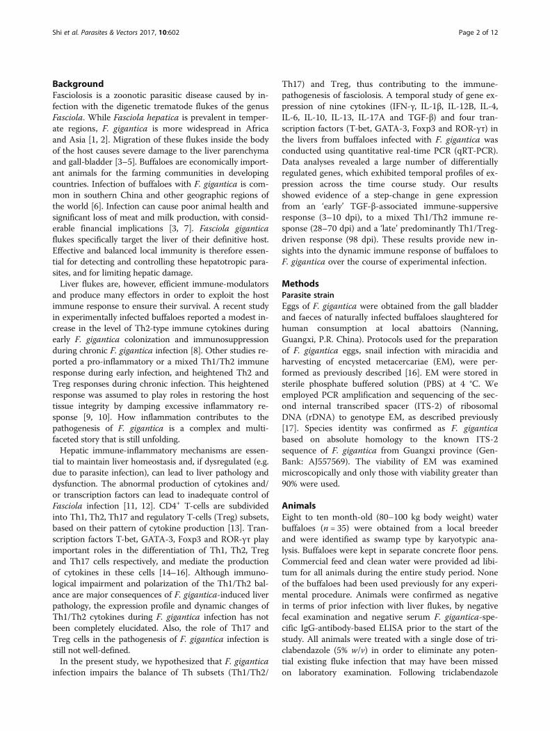
ResultsGross and histopathological attributesEven though buffaloes did not exhibit clear clinical signs,F. gigantica induced a wide range of pathological lesionsover the course of infection (Table 2). During the earlystage of infection (3–70 dpi), immature flukes migratedthrough the intestinal wall, abdominal cavity, liver cap-sule and liver parenchyma, to reach the bile ducts wherethey attain sexual maturity. Migration of the juvenileflukes through the host’s tissues was characterized byclassical inflammatory signs and accumulation of fibro-blasts towards the end of this stage, but without obviousfibrosis. The total number (and average length) of flukesrecovered from the livers of infected groups at 28, 42, 70and 98 dpi were: 3 flukes (1.5 mm), 22 flukes (2.5 mm),65 flukes (8 mm), and 36 flukes (14 mm), respectively.No fluke was observed in the liver or bile duct before 28
dpi, probably because they were too small to be visibleto the naked-eye. Hepatic hemorrhage, swelling, necrosisand viscous bile were first detected at 10 dpi. Tissue fi-brosis was observed at 70 dpi, along with disappearanceof hemorrhage. Seven parameters were used to describethe histopathological features in buffalo livers during in-fection (Table 3). Histopathological characteristics of theinfected liver tissues (Fig. 1) included an infiltration ofinflammatory cells, such as neutrophils and lymphocytesat 3 dpi. As infection progressed, an accumulation ofeosinophils, monocytes and red blood cells (RBCs) wasdetected (10–28 dpi), while fibroblast formation and bileduct hyperplasia were observed later (42 dpi). Duringthe chronic stage (70–98 dpi), significant fibrosis andnecrosis, without calcification, were observed. Flukeeggs were detected in the bile and feces of infectedanimals at 98 dpi. Granulomas with necrotic centers,
Table 1 List of primers used in the SYBR green-based qRT-PCR analysis
Gene target Primer sequence (5′–3′) Product length (bp) Reference
GAPDH F CCTGCACCACCAACTGCTTG 222 [62]
R TTGAGCTCAGGGATGACCTTG
IFN-γ F GTCTCCTTCTACTTCAAACT 253 [63]
R ATTCTGACTTCTCTTCCGCT
TGF-β F CGTGCTAATGGTGGAATAC 208 Present study
R GCCAGGAATTGTTGCTATA
IL-1β F CTAGCCCATGTGTGCTGAAG 59 [62]
R CCTTTACTTGGCTCTTCACC
IL-4 F CAGCATGGAGCTGCCT 177 [64]
R ACAGAACAGGTCTTGCTTGC
IL-6 F CTGCAATGAGAAAGGAGATA 191 [63]
R GGTAGTCCAGGTATATCTGA
IL-10 F CTGTGCCTCTCCCCTAGAGT 236 [62]
R GCAGCTAGCTCCACAAGGAA
IL-12B F CAGGGACATCATCAAACCAG 213 [63]
R CTTGTGGCATGT GACTTTGG
IL-13 F AGAACCAGAAGGTGCCGCT 50 [65]
R GGTTGAGGCTCCACACCATG
IL-17A F CTACAGTGAACTGGAAGGAAC 554 Present study
R AAAAGGGGCTGGGTCT
T-bet F CCTGGACCCAACTGTCAACT 171 [66]
R GAAACTCGGCCTCATAGCTG
GATA-3 F GATCAAGCCCAAGCGAAGG 124 Present study
R CCGCAGGCATTGCAGACA
Foxp3 F GACAGCACCCTTTCGACTGT 191 [66]
R CTCCAGAGATTGCACCACCT
ROR-γτ F CTACAGTGAACTGGAAGGAAC 554 Present study
R AAAAGGGGCTGGGTCT
Abbreviations: F forward primer, R reverse primer
Shi et al. Parasites & Vectors 2017, 10:602 Page 4 of 12

heavy infiltrates of lymphocytes, RBCs and eosinophilswere present at 98 dpi.
Gene expression of cytokinesTo determine how immune response changed over thecourse of infection, gene expression of nine cytokineswas assessed in the liver tissue of 29 infected buffaloesand five uninfected buffaloes using qRT-PCR. RNA fromone animal from group VII did not pass the quality con-trol check and was therefore excluded from the analysis.Results showed that infection had a significant impacton gene expression of Th1, Th2, Th17, and Treg cyto-kines (Fig. 2a and Table 4). The transcriptional profile ofcytokine genes showed an immunosuppressive stateduring early infection, a mixed Th1/Th2 response as theinfection progressed, and a shifting to Th1/Treg re-sponse, associated with greater histopathological changesand fibrosis during late stage infection.The gene expression of Th1 cytokines (IFN-γ and IL-
12B) and IL-10 showed no significant change duringearly infection. IFN-γ showed significantly higher ex-pression at 70 dpi (P = 0.0079) and 98 dpi (P = 0.0003),when compared with the control. Relative gene expres-sion of both of IL-12B (P < 0.0001) and IL-10 (P =0.0036) peaked at 98 dpi. Fluke infection also upregu-lated IL-4 expression at 28 dpi (P = 0.0028), whereas IL-13 significantly increased at 28 dpi (P = 0.0259) and 70dpi (P = 0.0426), peaking at 98 dpi (P < 0.0001). TGF-β
mRNA was rapidly elevated at 3 dpi (P = 0.0003),followed by a decrease to the basal level and remainedlow thereafter. Upregulation of IL-17A was noted at 28dpi (P = 0.0114), however expression was relatively lowbased on a high Cq value, suggesting it to be of littlediagnostic value as a marker for Th17 cytokine in thisstudy. The kinetics of the cytokines IL-1β and IL-6mRNA were similar, both peaking at 28 dpi (P = 0.0007for IL-1β; P = 0.0002 for IL-6) and 98 dpi (P < 0.0001 forIL-1β; P = 0.0008 for IL-6).Spearman’s correlation analysis between each pair of
cytokines was performed based on the RQ values frominfected groups (Fig. 3a). This analysis revealed a signifi-cant positive correlation between the expression ofIL-12B mRNA and Th2 cytokines (IL-4: r(29) = 0.44, P =0.0336; IL-13: r(29) = 0.41, P = 0.0542), and between IFN-γ and IL-12B (r(29) = 0.62, P = 0.0017) or IL-13 (r(29) =0.51, P = 0.0124). The gene expression of IL-10 wassignificantly correlated with both Th1 and Th2 cytokines(IFN-γ: r(29) = 0.49, P = 0.0171; IL-12B: r(29) = 0.50, P =0.014; IL-4: r(29) = 0.80, P < 0.0001; IL-13: r(29) = 0.51, P =0.014) and with IL-17A (r(29) = 0.73, P < 0.0001). Positivecorrelation was also found between IL-4 and IL-13 (r(29)= 0.44, P = 0.0336) gene expression. There was only astatistically significant relationship between IL-17A ex-pression and IL-4 r(29) = 0.87, P < 0.0001) and IL-10(r(29) = 0.73, P < 0.0001), but not with any of the othercytokines. Although not significant, the relative mRNA
Table 3 The presence (+) and absence (−) of the histopathological changes observed in the liver of buffaloes experimentallyinfected with F. gigantica
Group Dpi No. of animals Inflammatory infiltration RBCs Eosinophils Neutrophils Fibroblasts Lymphocytes Monocytes
I 0 5 – – – – – – –
II 3 5 + – – + – + –
III 10 5 + + + + – + –
IV 28 5 + + + + – + +
V 42 5 + + + + + + +
VI 70 5 + + + + + + +
VII 98 4 + + + + + + +
Table 2 The presence (+) and absence (−) of gross pathological lesions, flukes and fluke eggs in the liver of buffaloesexperimentally infected with F. gigantica
Group Dpi No. of animals Hemorrhage Swelling Parasite tunnel Fibrosis Necrosis Bile duct hyperplasia Viscous bile Visible flukes Eggs
I 0 5 – – – – – – – –
II 3 5 – – – – – – – – –
III 10 5 + + – – + – + – –
IV 28 5 + + – – + – + + –
V 42 5 – + + – + + + + –
VI 70 5 – + + + + + + + –
VII 98 4a – + + + + + + + +aRNA from one animal in this group VII did not pass the quality control check and was therefore excluded from the analysis
Shi et al. Parasites & Vectors 2017, 10:602 Page 5 of 12
expression of TGF-β showed an inverse relationship toany other cytokine.
Expression profiles of transcription factorsFasciola gigantica infection altered the expression of T-bet, GATA-3 and ROR-γτ, with the expression of Foxp3being least affected (Fig. 2b and Table 4). Compared with
the control, mRNA of T-bet was highly expressed at 98dpi (P = 0.0113), while the expression of the GATA-3,Foxp3 and ROR-γτ genes was upregulated at 28 dpi (P =0.0035, P = 0.0424 and P = 0.0002, respectively). Spear-man’s correlation analysis indicated that the expressionof T-bet and Foxp3 was correlated (r(29) = 0.63, P =0.0011). Positive correlation was also found between theexpression of ROR-γτ and GATA-3 (r(29) = 0.43, P =0.039) or Foxp3 (r(29) = 0.42, P = 0.0439) (Fig. 3b). Asshown in Fig. 3c, the mRNA expression of GATA-3 cor-related significantly with that of IL-4 (r(29) = 0.54, P =0.0072), but was less correlated with another Th2 cyto-kine IL-13 (r(29) = 0.017, P = 0.9387), whereas, the ex-pression of ROR-γτ was correlated with that of IL-17A(r(29) = 0.54, P = 0.0072). There was a tendency of correl-ation between the expression of T-bet and Th1 cytokineIFN-γ (r(29) = 0.22, P = 0.305) or IL-12B (r(29) = 0.33, P =0.1265) and between the expression of Foxp3 and IL-10(r(29) = 0.38, P = 0.0718).
Th1/Th2/Th17/Treg balanceTo better understand the temporal trends in gene expres-sion of Th1, Th2, Th17 and Treg in the liver post-infection, the ratios between the expression of transcrip-tion factors were evaluated using the pairwise comparisonmethod (Fig. 2c). The RQ ratio of T-bet/GATA-3 wasemployed as an indicator of Th1/Th2 cytokine gene ex-pression pattern. This ratio was higher in the liver at 98dpi when compared to the uninfected group (P = 0.0049).This analysis also revealed that Th1/Th17 (P = 0.0246)and Treg/Th17 (P = 0.0126) were both upregulated at 98dpi, when compared with the samples tested at other timepoints (3–70 dpi). However, no significant change wasfound in either Th1/Treg, Th2/Treg, or Th2/Th17 expres-sion ratios at any of the examined time points. These datasuggest a predominance of the Th1 and Treg cellularimmune response at 98 dpi.
DiscussionOur findings provide an increased understanding of theimmuno-inflammatory response of buffaloes to F. giganticainfection through examining the correlation between geneexpression of nine cytokines and four transcription factorsin Th1, Th2, Th17 and Treg cells, together with the liverpathology during early, mid and late stages of infection.We initially characterized gross and histopathologicalchanges in the liver at six time points following infectioncompared to uninfected control. Although infected buffa-loes did not exhibit clinical signs, they developed hepaticgross pathologies similar to what have been observed inother studies [15, 19]. Histopathological features alsoagreed with previous studies on F. gigantica [9, 20, 21] andF. hepatica [22]. An unexpected finding was that none ofthe examined liver from infected animals showed calcium
Fig. 1 Histopathological characteristics of the livers of buffaloesinfected with Fasciola gigantica. a At 3 dpi, there was local hyperemiaassociated with mild filtration of lymphocytes and neutrophils. b At 10dpi, there was scattered vacuolation of hepatocytes, consistent withfat, along with mild to moderate focal necrosis. c At 28 dpi, diffuseintravascular coagulation, severe infiltration of inflammatory cellsmainly neutrophils and lymphocytes, and granular degeneration of thecytoplasm were observed. d At 42 dpi, moderate to severe multifocalhemorrhages and necrosis, infiltration of eosinophils, RBCs andmonocytes, and accumulation of fibroblasts were detected. e At70 dpi, there were mild to moderate multifocal bile duct hyperplasiaand focal coagulative necrosis associated with collagen deposition. f At98 dpi, severe periportal fibrosis associated with multifocal inflammatoryinfiltrate, cellular debris, moderate multifocal hemorrhages, andgranulomas with necrotic centres were detected. g Liver tissue fromuninfected animal showed normal histological architecture of thehepatic tissue. h Adult flukes in the intrahepatic bile duct, along withepithelial hyperplasia of the duct. In all figures, tissue sections werestained with H&E and arrows point at the corresponding morphologicalfeatures described above. Scale-bars: a-g, 50 μm; h, 100 μm
Shi et al. Parasites & Vectors 2017, 10:602 Page 6 of 12

deposits. The difference in liver calcification between ourstudy and previous reports may relate to variations in theexperimental conditions, such as using different infectiousdoses, different parasite strains, the permissibility of the
host species, the duration of the experimental infection,and using single infection vs repeated infections [21–23].Next, we have shown that buffaloes showed a substantial
liver immune response subsequent to oral challenge with
Fig. 2 Temporal changes of the mRNA expression of cytokines and transcription factors in the liver of buffaloes experimentally infected withFasciola gigantica. The X axis represents days post infection (dpi), control animals are represented by empty bars and Y axis represents the mRNArelative expression of target genes relative to GAPDH based on 2–ΔΔCq calculation. Columns show the means and error bars show SEMs. The lognumber of mRNA relative expression of a IFN-γ, IL-1β, IL-4, IL-6, IL-10, IL-12B, IL-13, IL-17A, TGF-β and b T-bet, GATA-3, Foxp3, and ROR-γτ, areshown. c Trends in the ratios between each pair of transcription factors during the course of infection, which was used as indicators of the balancebetween Th1/Th2 and Treg/Th17. Significant differences of each time-point compared with control non-infected (NC) group: *P < 0.05; **P < 0.01;***P < 0.001 or ****P < 0.0001 (analyzed by one-way ANOVA, post-hoc LSD test)
Shi et al. Parasites & Vectors 2017, 10:602 Page 7 of 12

500 F. gigantica encysted metacercariae. Certain genes ex-hibited shifting temporal expression patterns as the infec-tion proceeded. Expression of cytokine and transcriptionfactor genes were decreased at 3 and 10 dpi, suggesting ageneral immunosuppression state to allow parasitecolonization. Cytokine reduction during the early stage ofF. gigantica infection has been reported previously [24, 25].A previous study has documented the downregulation ofMHC-II related genes, as well as the suppression of thehost pro-inflammatory (Th1) immune response duringearly F. gigantica infection [16]. This immunosuppressionmight be attributed to an increased expression of the im-munosuppressive cytokine TGF-β. While TGF-β was up-regulated at 3 dpi, Foxp3 (Treg transcription factor) wasupregulated at a later stage of infection (28 dpi), indicatingthat Tregs (also known as Foxp3-expressing cells) play amore diverse role than causing early immunosuppressionin regulating immune responses. Although increasedTGF-β at 3 dpi may have promoted the generation ofFoxp3-expressing Tregs, increased IL-6 at 28 dpi may haveabrogated the suppressive function of Tregs [26] by antag-onizing TGF-β-induced Treg generation and stimulatingTreg differentiation to Th17, as reported previously [27].The inflammatory cascades triggered by F. gigantica
must be tightly coordinated in order to avoid severe liverpathology. Th2 cells mediate humoral responses via in-duction of IL-4, IL-5, IL-9, IL-10 and IL-13 cytokines,which are important for controlling extracellular para-sites [28]. In agreement with this, our results at 28 dpishowed a significant increase in the expression of theanti-inflammatory Th2-associated cytokines (IL-4, IL-13)and transcription factor (GATA-3), indicating the
induction of Th2 response in liver tissue, probably tolimit the fluke development and to balance the increasedlevels of inflammatory cytokines IL-1β, IL-6 and IL-17A.This response was expected because IL-4 is known tohave the greatest effect in inducing Th2 differentiation[29] and the GATA-3 transcription factor is a key regu-lator of Th2 differentiation [30]. IL-4 stimulation mayhave activated STAT6, which is the major signal trans-ducer in IL-4-mediated Th2 differentiation [31]. STAT6is known to promote Th2 differentiation by stimulatingGATA-3 [32]. Therefore, this coordinated response maybe mediated by the parasite and/or the host in order topromote parasite survival while minimizing host hepaticdamage. Th2-predominant response together with thesuppression of Th1/Th17 response has been reported pre-viously [33–36]. Interestingly, some molecules secreted byFasciola species can suppress the differentiation of Th17cells independently of Th2 cells differentiation, by alteringthe function of dendritic cells [37].Although CD4+ Th17 cells play a role in the control of
a variety of parasitic infections, the role of IL-17 pro-duced by CD4+ Th17 cells in immunity to F. gignaticahas not been clearly defined [38, 39]. Our current andprevious studies [8], demostrated that IL-17 may play arole in the inflammatory process during early F. gigan-tica infection. Th17 cells are a newly-identified class ofeffector T cell, which produces IL-17A and IL-17F. Th17cells can contribute to resistance to many intracellular[40] and extracellular parasites [8]. In addition to con-trolling infection, IL-17 expression has been associatedwith inflammatory and allergic responses [41]. TGF-βand IL-6 act cooperatively and non-redundantly to
Table 4 Relative gene expression in the liver of buffaloes experimentally infected with F. gigantica compared to the control group.The relative quantities were obtained according to the comparative Cq method (2–ΔΔCq). P values indicate statistical significance (P < 0.05)
GeneTarget
ANOVA Multiple comparisons
3 dpi 10 dpi 28 dpi 42 dpi 70 dpi 98 dpi
F(DFn, DFd) P RQ P RQ P RQ P RQ P RQ P RQ P
IFN-γ 4.693(6, 21) 0.0035 2.08 0.4326 5.51 0.1282 2.06 0.7358 3.21 0.4590 9.27 0.0079 13.91 0.0003
TGF-β 4.738(6, 19) 0.0041 13.93 0.0003 3.07 0.4884 1.67 0.8036 3.85 0.3408 1.63 0.8371 2.66 0.6062
IL-1β 11.05(6, 26) < 0.0001 1.31 0.9767 1.27 0.9811 14.25 0.0007 0.26 0.8137 2.31 0.8025 28.45 < 0.0001
IL-4 2.877(6, 24) 0.0291 7.01 0.5847 19.4 0.4892 97.21 0.0028 27.9 0.3121 9.43 0.7749 67.14 0.0630
IL-6 7.636(6, 24) 0.0001 5.50 0.8529 2.81 0.9366 78.1 0.0002 1.94 0.9728 23.14 0.2805 94.56 0.0008
IL-10 2.514(6, 26) 0.0471 5.99 0.7003 9.92 0.3184 21.18 0.0694 2.3 0.9579 6.24 0.4902 32.67 0.0036
IL-12B 5.595(6, 26) 0.0008 5.92 0.4786 3.99 0.4746 13.68 0.0759 0.86 0.9825 5.37 0.5312 24.84 < 0.0001
IL-13 6.56(6, 20) 0.0006 2.09 0.8459 7.80 0.1522 11.87 0.0259 5.45 0.3499 10.8 0.0426 27.33 < 0.0001
IL-17A 3.532(6, 21) 0.0142 0.33 0.4612 0.07 0.3694 5.95 0.0114 0.26 0.4327 0.28 0.4400 2.99 0.6354
T-bet 2.173(6, 28) 0.076 2.16 0.7923 0.25 0.7830 4.73 0.1018 3.93 0.4158 3.99 0.4606 11.38 0.0113
GATA-3 3.319(6, 28) 0.0135 0.84 0.8101 1.42 0.9579 5.18 0.0035 4.80 0.0889 1.34 0.9397 1.8 0.6952
Foxp3 1.455(6, 28) 0.2296 0.70 0.9054 1.07 0.9749 3.03 0.0424 3.97 0.3705 3.40 0.4458 3.05 0.1372
ROR-γτ 4.387(6, 28) 0.003 2.25 0.7567 4.57 0.3780 10.28 0.0002 3.24 0.5637 2.68 0.7076 2.55 0.7148
Abbreviations: RQ, relative quantity (mean); F(DFn, DFd), degrees of freedom with numerator and degrees of freedom denominator inside the subscript parentheses
Shi et al. Parasites & Vectors 2017, 10:602 Page 8 of 12

promote Th17 activity [27]. In agreement with this,TGF-β was upregulated at 3 dpi, and both IL-6 and IL-17A were upregulated at 28 dpi. IL-6 was found to beimportant in suppressing Treg generation in order topromote Th17 differentiation as discussed above. Theexpression of retinoic acid-related orphan receptorsROR-γt, a key transcription factor in Th17 differenti-ation [42, 43], was positively correlated with the RQ levelof IL-17A at 28 dpi.At 70 and 98 dpi a mixed Th1/Th2 type profile, sup-
ported by co-dominance of IFN-γ, IL-1β, IL-12B, IL-6,IL-4 and IL-13 cytokines, was observed as consistent
with the result obtained by Kumar [44]. The high ex-pression of Th1 cytokines (IL-1β and IL-6), suggestingacute inflammatory activity, correlated with hemorrhage,necrosis and severe infiltration of inflammatory cells inthe liver. Th2 cytokines, on the other hand, often lead tochronic inflammatory response by promoting fibrosisand tissue remodeling [45, 46]. The accumulation offibroblasts during chronic infection suggests that Th2cytokine-mediated tissue repair was taking place. Thisstate of immune homeostasis is probably required forparasite persistence and was correlated with an in-creased number of flukes. The T-bet/GATA-3 ratio, a
Fig. 3 Pearson’s correlation analysis between mRNA expressions of Th1/Th2/Th17/Treg type a cytokines, b transcription factors, and c cytokinesvs transcription factors in the liver of buffaloes infected with Fasciola gigantica (tested samples, n = 29) by pairwise comparison. Connecting linesillustrate positive (r > 0) and negative (r < 0) correlation indices
Shi et al. Parasites & Vectors 2017, 10:602 Page 9 of 12
marker of Th1/Th2 [47], was not altered during acuteinfection, but was significantly increased when the infec-tion became chronic, suggesting more bias towards aTh1-mediated inflammatory response during late infec-tion. This observation contradicts previous studies inmice, cattle and buffaloes infected with F. gigantica thatreported a mixed Th1/Th2 response with a predomin-ance of a Th2-biased pattern [48, 49]. Other studies havereported a Th2 response in early infection and an in-creased Th0-type response during chronic F. giganticainfection in cattle [35] and immunosuppression duringchronic F. gigantica infection in buffaloes [8]. These dif-fering findings between studies may result from differentexperimental conditions.A synergism between IL-1β and IL-17 has previously
been reported [50]. In our study, a strong correlationwas observed between IL-17A and IL-4 and IL-10, aswell as between ROR-γτ and GATA-3 and Foxp3, imply-ing that induction of Th17 was paralleled by inductionof both Th2 and Treg. This refutes the antagonistic rela-tionship previously reported between a polarized Th2/Treg immune response and suppression of Th1/Th17cytokines [11, 51–53]. A gradual upregulation of IL-10mRNA expression was observed during early infection,in agreement with our previous finding in buffalo’sserum [8]. IL-10 levels significantly increased at 98 dpi,which was also reported in a study on F. hepatica-in-fected sheep [54]. IL-10 is considered to be a regulatorycytokine rather than a Th2 type cytokine [55, 56]. Highlevels of IL-10 can suppress excessive inflammation in-duced by the parasite [57, 58]. In our study, elevatedlevels of IL-10 and the concurrent upregulation of Th1and Th2 cytokine genes in the liver suggest that IL-10was required for the Th2 response to F. gigantica infec-tion, instead of serving an anti-inflammatory function.Our data also showed that the RQ level of Foxp3 in-creased at 28 dpi. This level remained relatively highover the course of infection, which may have contributedto the Th1/Th2 balance in order to enhance parasite sur-vival in the liver and to protect the host from over-inflammation [59]. Foxp3-expressing Treg cells are crit-ical to maintaining immune homeostasis by minimizingtissue pathology, while modulating host immune re-sponse against helminth infections [60, 61]. The moni-toring of Th17 and Treg cell offers a promising newperspective on the pathogenesis of F. gigantica infectionand deserves further exploration.
ConclusionsThis is the first study to characterize the correlation be-tween the expression of Th1, Th2, Th17, and Treg cyto-kines and transcription factors with liver pathology inbuffaloes, during the course of experimental F. giganticainfection. The expression patterns for the examined
genes indicated that there were periods of differentialregulation during F. gigantica infection, which may sug-gest either a mechanism of immune evasion based onmodulation of transcription or a mechanism used byhost tissue to limit the infection and tissue damage.Gene expression profiles showed a significant T-celladaptive immune suppression between 3 and 10 dpi tofacilitate parasite colonization and a more substantialtranscript differential expression change (mixed Th1/Th2 immune response) from 28 to 70 dpi, which maycontribute to the parasite survival while minimizing hosttissue damage. During late infection (98 dpi), the re-sponse was biased towards Th1/Treg in order to limitthe host’s Th2 protective response and promote chronicinfection, which conincided with an increase in thenumber of recovered flukes and greater histopathologicalchanges and fibrosis. Clearly, regulation of immunecytokine and transcription factor gene expression duringF. gigantica infection is a complex mechanism involvinga variety of parasite-specific and host-specific factors,perhaps depending on the parasite’s needs for survivalduring various phases of infection. Research into themechanisms governing differential expression of theseimmune-related genes, may shed light on the actual roleof these immune regulatory factors in the pathogenesisof F. gigantica infection in buffaloes. Much remains tobe elucidated about the role of various types of inflam-matory responses, and the cross-talk between types ofT-helper responses, in fasciolosis.
AbbreviationsANOVA: Analysis of variance; cDNA: Complementary deoxyribonucleic acid;Cq: Quantification cycle; DFd: Degrees of freedom denominator;DFn: Degrees of freedom numerator; DNA: Deoxyribonucleic acid; DPI: Dayspost-infection; ELISA: Enzyme linked immunosorbent assay; EM: Encystedmetacercariae; Foxp3: Forkhead box P3; GAPDH: Glyceraldethde-3-phosphatedehydrogenase; GATA: GATA binding protein; H&E: Hematoxylin and eosin;IFN: Interferon; IL: Interleukin; ITS: Internal transcribed spacer; LSD: Leastsignificant difference; mRNA: Messenger ribonucleic acid; qRT-PCR: Quantitative reverse transcription-polymerase chain reaction; RBC: Redblood cells; rDNA: Ribosomal DNA; RNA: Ribonucleic acid; ROR: Retinoid-related orphan nuclear receptor; RQ: Relative quantitation; SEM: Standarderror of the mean; T-bet: T-box expressed in T cells; TGF: Transforminggrowth factor; Th: T helper; Treg: Regulatory T cell; WPI: Weeks post-infection
AcknowledgementsWe are grateful to Xiao-Xuan Zhang, Wen-Bin Zheng and Jian-Gang Ma fromLanzhou Veterinary Research Institute, Chinese Academy of Agricultural Sciences,and Yang-Qun Kang, Xue-Fang Mei, Chang-Hong Hu, Yu Zhang, Yao-Yao Zhang,and Yi-Ying Liang from Guangxi University for their technical assistance.
FundingProject financial support was provided by National Key Basic ResearchProgram (973 Program) of China (Grant No. 2015CB150300) and by NationalNatural Science Foundation of China (Grant No. 31260605).
Availability of data and materialsThe data supporting the findings of this article are included within the article.
Authors’ contributionsWS, WYH and XQZ conceived and designed the study, and critically revisedthe paper. WS, ZYW, FKZ and KJL prepared the experimental samples. WS,
Shi et al. Parasites & Vectors 2017, 10:602 Page 10 of 12

ZYW and ZAS performed the experiments. WS, ZYW, FKZ and DYW analyzedthe data. WS drafted the manuscript. HME helped in the data analysis andcritical revision of the manuscript. All authors read and approved the finalmanuscript.
Ethics approvalThe study design was reviewed and approved by the Animal EthicsCommittee of Guangxi University. Animals used in the study were handledin accordance with good animal practices as required by the Animal EthicsProcedures and Guidelines of the People’s Republic of China.
Consent for publicationNot applicable.
Competing interestsThe authors declare that they have no competing interests.
Publisher’s NoteSpringer Nature remains neutral with regard to jurisdictional claims inpublished maps and institutional affiliations.
Author details1College of Animal Science and Technology, Guangxi University, Nanning530005, Guangxi Zhuang Autonomous Region, People’s Republic of China.2State Key Laboratory of Veterinary Etiological Biology, Key Laboratory ofVeterinary Parasitology of Gansu Province, Lanzhou Veterinary ResearchInstitute, Chinese Academy of Agricultural Sciences, Lanzhou 730046, GansuProvince, People’s Republic of China. 3Faculty of Medicine and HealthSciences, School of Veterinary Medicine and Science, University ofNottingham, Sutton Bonington Campus, Loughborough LE12 5RD, UK.4Jiangsu Co-innovation Center for the Prevention and Control of ImportantAnimal Infectious Diseases and Zoonoses, Yangzhou University College ofVeterinary Medicine, Yangzhou, Jiangsu Province 225009, People’s Republicof China.
Received: 22 August 2017 Accepted: 14 November 2017
References1. Mas-Coma S, Funatsu I, Bargues M. Fasciola hepatica and lymnaeid snails
occurring at very high altitude in South America. Parasitology. 2001;123:115–27.
2. Cywinska A. Epidemiology of fascioliasis in human endemic areas. J Helminthol.2005;79:207–16.
3. Yadav S, Sharma R, Kalicharan A, Mehra U, Dass R, Verma A. Primaryexperimental infection of riverine buffaloes with Fasciola gigantica. VetParasitol. 1999;82:285–96.
4. Ahmedullah F, Akbor M, Haider M, Hossain M, Khan M, Hossain M, et al.Pathological investigation of liver of the slaughtered buffaloes in Barisaldistrict. Ban J Vet Med. 2007;5:81–5.
5. Raadsma H, Kingsford N, Spithill T, Piedrafita D. Host responses duringexperimental infection with Fasciola gigantica or Fasciola hepatica in merinosheep: I. Comparative immunological and plasma biochemical changesduring early infection. Vet Parasitol. 2007;143:275–86.
6. Yuan W, Liu JM, Lu K, Li H, Duan MM, Feng JT, et al. Molecular identificationand seasonal infections of species of Fasciola in ruminants from twoprovinces in China. J Helminthol. 2016;90:359–63.
7. Chai JY. Intestinal flukes. In: Murrell KD, Fried B, editors. Food-borne parasiticzoonoses. World class parasites, vol. 11. Boston: Springer; 2007. p. 53–115.
8. Zhang FK, Guo AJ, Hou JL, Sun MM, Sheng ZA, Zhang XX, et al. Serumlevels of cytokines in water buffaloes experimentally infected with Fasciolagigantica. Vet Parasitol. 2017;244:97–101.
9. Dalton JP (Editor). Fasciolosis. Wallingford: CABI Publishing; 1999.10. Mas-Coma S, Bargues MD, Valero MA. Fascioliasis and other plant-borne
trematode zoonoses. Int J Parasitol. 2005;35:1255–78.11. O'neill SM, Brady MT, Callanan JJ, Mulcahy G, Joyce P, Mills KH, et al. Fasciola
hepatica infection downregulates Th1 responses in mice. Parasite Immunol.2000;22:147–55.
12. Zhang W, Moreau E, Hope J, Howard C, Huang W, Chauvin A. Fasciolahepatica and Fasciola gigantica: comparison of cellular response toexperimental infection in sheep. Exp Parasitol. 2005;111:154–9.
13. Sallusto F, Lanzavecchia A. Heterogeneity of CD4+ memory T cells:functional modules for tailored immunity. Eur J Immunol. 2009;39:2076–82.
14. Infante-Duarte C, Kamradt T. Th1/Th2 balance in infection. SeminImmunopathol. 1999;21:317–38.
15. Zhang WY, Moreau E, Yang BZ, Li ZQ, Hope JC, Howard CJ, et al. Humoraland cellular immune responses to Fasciola gigantica experimental infectionin buffaloes. Res Vet Sci. 2006;80:299–307.
16. Zhang FK, Zhang XX, Elsheikha HM, He JJ, Sheng ZA, Zheng WB, et al.Transcriptomic responses of water buffalo liver to infection with thedigenetic fluke Fasciola gigantica. Parasit Vectors. 2017;10:56.
17. Huang WY, He B, Wang CR, Zhu XQ. Characterisation of Fasciola species frommainland China by ITS-2 ribosomal DNA sequence. Vet Parasitol. 2004;120:75–83.
18. Schmittgen TD, Livak KJ. Analyzing real-time PCR data by the comparativeCT method. Nat Protoc. 2008;3:1101–8.
19. Molina EC, Skerratt LF. Cellular and humoral responses in liver of cattle andbuffaloes infected with a single dose of Fasciola gigantica. Vet Parasitol.2005;131:157–63.
20. Shaikh AA, Bilqees F, Khan MM. Bile duct hyperplasia and associatedabnormalities in the buffaloes infected with Fasciola gigantica. Pak J Zool.2004;36:231–8.
21. Okaiyeto SO, Salami OS, Dnbirni SA, Allam L, Onoja II. Clinical, gross andhistopathological changes associated with chronic fasciolosis infection in adairy farm. J Vet Adv. 2012;2:444–8.
22. Marcos LA, Yi P, Machicado A, Andrade R, Samalvides F, Sánchez J, et al. Hepaticfibrosis and Fasciola hepatica infection in cattle. J Helminthol. 2007;81:381–6.
23. Grange D, Dhumeaux D, Couzineau P, Bismuth H, Bader J-P. Hepaticcalcification due to Fasciola gigantica. Arch Surg. 1974;108:113–5.
24. Gironènes N, Valero MA, García-Bodelón MA, Chico-Calero I, Punzón C, PresnoM, Mas-Coma S. Immune suppression in advanced chronic fascioliasis: anexperimental study in a rat model. J Infect Dis. 2007;195:1504–12.
25. Rodríguez E, Noya V, Cervi L, Chiribao ML, Brossard N, Chiale C, et al. Glycansfrom Fasciola hepatica modulate the host immune response and TLR-inducedmaturation of dendritic cells. PLoS Negl Trop Dis. 2015;9:e0004234.
26. Pasare C, Medzhitov R. Toll pathway-dependent blockade of CD4+ CD25+ Tcell-mediated suppression by dendritic cells. Science. 2003;299:1033–6.
27. Mangan PR, Harrington LE, O'quinn DB, Helms WS, Bullard DC, Elson CO,et al. Transforming growth factor-β induces development of the TH17lineage. Nature. 2006;441:231–4.
28. Sher A, Coffman RL. Regulation of immunity to parasites by T cells and Tcell-derived cytokines. Annu Rev Immunol. 1992;10:385–409.
29. Swain SL, Weinberg AD, English M, Huston G. IL-4 directs the developmentof Th2-like helper effectors. J Immunol. 1990;145:3796–806.
30. Zhu J, Min B, Hu-Li J, Watson CJ, Grinberg A, Wang Q, et al. Conditionaldeletion of Gata3 shows its essential function in TH1-TH2 responses. NatImmunol. 2004;5:1157.
31. Takeda K, Kamanaka M, Tanaka T, Kishimoto T, Akira S. Impaired IL-13-mediated functions of macrophages in STAT6-deficient mice. J Immunol.1996;157:3220–2.
32. Zhu J, Guo L, Watson CJ, Hu-Li J, Paul WE. Stat6 is necessary and sufficientfor IL-4’s role in Th2 differentiation and cell expansion. J Immunol.2001;166:7276–81.
33. Mulcahy G, Dalton JP, Cathepsin L. Proteinases as vaccines against infectionwith Fasciola hepatica (liver fluke) in ruminants. Res Vet Sci. 2001;70:83–6.
34. Waldvogel AS, Lepage MF, Zakher A, Reichel MP, Eicher R, Heussler VT.Expression of interleukin 4, interleukin 4 splice variants and interferongamma mRNA in calves experimentally infected with Fasciola hepatica.Vet Immunol Immunopathol. 2004;97:53–63.
35. Ingale SL, Singh P, Raina OK, Mehra UR, Verma AK, Gupta SC, et al.Interferon-gamma and interleukin-4 expression during Fasciola giganticaprimary infection in crossbred bovine calves as determined by real-timePCR. Vet Parasitol. 2008;15:158–61.
36. Ingale SL, Singh P, Raina OK, Verma AK, Channappanavar R, Mehra UR.Interleukin-2 and interleukin-10 gene expression in calves experimentallyinfected with Fasciola gigantica. Livest Sci. 2010;131:141–3.
37. Dowling DJ, Hamilton CM, Donnelly S, La Course J, Brophy PM, Dalton J,et al. Major secretory antigens of the helminth Fasciola hepatica activate asuppressive dendritic cell phenotype that attenuates Th17 cells but fails toactivate Th2 immune responses. Infect Immun. 2010;78:793–801.
38. Noya V, Brossard N, Rodríguez E, Dergan-Dylon LS, Carmona C, RabinovichGA, et al. A mucin-like peptide from Fasciola hepatica instructs dendriticcells with parasite specific Th1-polarizing activity. Sci Rep. 2017;7:40615.
Shi et al. Parasites & Vectors 2017, 10:602 Page 11 of 12
39. Valero MA, Perez-Crespo I, Chillón-Marinas C, Khoubbane M, Quesada C,Reguera-Gomez M, et al. Fasciola hepatica reinfection potentiates a mixedTh1/Th2/Th17/Treg response and correlates with the clinical phenotypes ofanemia. PLoS One. 2017;12:e0173456.
40. Lieberman LA, Cardillo F, Owyang AM, Rennick DM, Cua DJ, Kastelein RA,et al. IL-23 provides a limited mechanism of resistance to acutetoxoplasmosis in the absence of IL-12. J Immunol. 2004;173:1887–93.
41. Yen D, Cheung J, Scheerens H, Poulet F, McClanahan T, Mckenzie B, et al.IL-23 is essential for T cell–mediated colitis and promotes inflammation viaIL-17 and IL-6. J Clin Invest. 2006;116:1310–6.
42. Yang XO, Pappu BP, Nurieva R, Akimzhanov A, Kang HS, Chung Y, et al. Thelper 17 lineage differentiation is programmed by orphan nuclear receptorsRORα and RORγ. Immunity. 2008;28:29–39.
43. Wan YY, Regulatory T. Cells: immune suppression and beyond. Cell MolImmunol. 2010;7:204–10.
44. Kumar N, Raina OK, Nagar G, Prakash V, Jacob SS. Th1 and Th2 cytokinegene expression in primary infection and vaccination against Fasciolagigantica in buffaloes by real-time PCR. Parasitol Res. 2013;112:3561–8.
45. Allen JE, Sutherland TE. Host protective roles of type 2 immunity: parasitekilling and tissue repair, flip sides of the same coin. Semin Immunol.2014;26:329–40.
46. Cortés A, Muñoz-Antoli C, Esteban JG, Toledo R. Th2 and Th1 responses:clear and hidden sides of immunity against intestinal helminths. TrendsParasitol. 2017;33:678–93.
47. Chakir H, Wang H, Lefebvre DE, Webb J, Scott FW. T-Bet/GATA-3 ratio as ameasure of the Th1/Th2 cytokine profile in mixed cell populations:predominant role of GATA-3. J Immunol Methods. 2003;278:157–69.
48. Chantree P, Phatsara M, Meemon K, Chaichanasak P, Changklungmoa N,Kueakhai P, et al. Vaccine potential of recombinant cathepsin B againstFasciola gigantica. Exp Parasitol. 2013;135:102–9.
49. Molina EC. Serum interferon-gamma and interleukins-6 and-8 during infectionwith Fasciola gigantica in cattle and buffaloes. J Vet Sci. 2005;6:135–9.
50. Ilarregui JM, van Beelen AJ, Fehres CM, Bruijns SC, García-Vallejo JJ, vanKooyk Y. New roles for CD14 and IL-β linking inflammatory dendritic cells toIL-17 production in memory CD4+ T cells. Immunol Cell Biol. 2016;94:907–16.
51. Donnelly S, O'Neill SM, Sekiya M, Mulcahy G, Dalton JP. Thioredoxinperoxidase secreted by Fasciola hepatica induces the alternative activationof macrophages. Infect Immun. 2005;73:166–73.
52. Flynn RJ, Mannion C, Golden O, Hacariz O, Mulcahy G. Experimental Fasciolahepatica infection alters responses to tests used for diagnosis of bovinetuberculosis. Infect Immun. 2007;75:1373–81.
53. Donnelly S, Stack CM, O'Neill SM, Sayed AA, Williams DL, Dalton JP.Helminth 2-Cys peroxiredoxin drives Th2 responses through a mechanisminvolving alternatively activated macrophages. FASEB J. 2008;22:4022–32.
54. Hacariz O, Sayers G, Flynn RJ, Lejeune A, Mulcahy G. IL-10 and TGF-β1 areassociated with variations in fluke burdens following experimentalfasciolosis in sheep. Parasite Immunol. 2009;31:613–22.
55. Anderson CF, Oukka M, Kuchroo VJ, Sacks D. CD4+ CD25− Foxp3− Th1 cellsare the source of IL-10–mediated immune suppression in chronic cutaneousleishmaniasis. J Exp Med. 2007;204:285–97.
56. Jankovic D, Kullberg MC, Feng CG, Goldszmid RS, Collazo CM, Wilson M,et al. Conventional T-bet+Foxp3− Th1 cells are the major source of host-protective regulatory IL-10 during intracellular protozoan infection. J ExpMed. 2007;204:273–83.
57. Mahanty S, Mollis SN, Ravichandran M, Abrams JS, Kumaraswami V,Jayaraman K, et al. High levels of spontaneous and parasite antigen-driveninterleukin-10 production are associated with antigen-specifichyporesponsiveness in human lymphatic filariasis. J Infect Dis. 1996;173:769–72.
58. Anuradha R, Munisankar S, Dolla C, Kumaran P, Nutman TB, Babu S. Parasiteantigen-specific regulation of Th1, Th2, and Th17 responses in Strongyloidesstercoralis infection. J Immunol. 2015;195:2241.
59. Scalzo K, Plebanski M, Apostolopoulos V. Regulatory T-Cells: immunomodulatorsin health and disease. Curr Top Med Chem. 2006;6:1759–68.
60. Zhou S, Jin X, Chen X, Zhu J, Xu Z, Wang X, et al. Heat shock protein 60 ineggs specifically induces Tregs and reduces liver immunopathology in micewith schistosomiasis japonica. PLoS One. 2015;10:e0139133.
61. Escamilla A, Zafra R, Pérez J, McNeilly TN, Pacheco IL, Buffoni L, et al.Distribution of Foxp3+ T cells in the liver and hepatic lymph nodes of goatsand sheep experimentally infected with Fasciola hepatica. Vet Parasitol.2016;230:14.
62. Castillo-Velázquez U, Aranday-Cortés E, Gutiérrez-Pabello JA. Alternativeactivation modifies macrophage resistance to Mycobacterium bovis. VetMicrobiol. 2011;151:51–9.
63. Mingala CN, Konnai S, Venturina FA, Onuma M, Ohashi K. Quantification ofwater buffalo (Bubalus bubalis) cytokine expression in response to inactivatedfoot-and-mouth disease (FMD) vaccine. Res Vet Sci. 2009;87:213–7.
64. Puech C, Dedieu L, Chantal I, Rodrigues V. Design and evaluation of aunique SYBR green real-time RT-PCR assay for quantification of five majorcytokines in cattle, sheep and goats. BMC Vet Res. 2015;11:65.
65. Pleasance J, Wiedosari E, Raadsma HW, Meeusen E, Piedrafita D. Resistanceto liver fluke infection in the natural sheep host is correlated with a type-1cytokine response. Parasite Immunol. 2011;33:495–505.
66. Maeda Y, Ohtsuka H, Tomioka M. Effect of progesterone on Th1/Th2/Th17and regulatory T cell-related genes in peripheral blood mononuclear cellsduring pregnancy in cows. Vet Res Commun. 2013;37:43–9.
• We accept pre-submission inquiries
• Our selector tool helps you to find the most relevant journal
• We provide round the clock customer support
• Convenient online submission
• Thorough peer review
• Inclusion in PubMed and all major indexing services
• Maximum visibility for your research
Submit your manuscript atwww.biomedcentral.com/submit
Submit your next manuscript to BioMed Central and we will help you at every step:
Shi et al. Parasites & Vectors 2017, 10:602 Page 12 of 12


![CRIP1 expression is correlated with a favorable outcome and ......protein-protein interactions during transcription, immune response, and cytokine expression [6, 7]. Additionally,](https://img.dokumen.tips/doc/110x75/6026c4750c07ba76034bd468/crip1-expression-is-correlated-with-a-favorable-outcome-and-protein-protein.jpg)