Embed Size (px)
Citation preview

Dynamic change in hyoid muscle length associated with trajectory of hyoid boneduring swallowing: analysis using 320-row area detector computed tomography
Takeshi Okada,1,2 Yoichiro Aoyagi,1 Yoko Inamoto,3 Eiichi Saitoh,1 Hitoshi Kagaya,1 Seiko Shibata,1
Kikuo Ota,3 and Koichiro Ueda2
1Department of Rehabilitation Medicine I, School of Medicine, Fujita Health University, Aichi, Japan; 2Department ofDysphagia Rehabilitation, Nihon University School of Dentistry, Tokyo, Japan; and 3Faculty of Rehabilitation, School ofHealth Sciences, Fujita Health University, Aichi, Japan
Submitted 15 April 2013; accepted in final form 18 August 2013
Okada T, Aoyagi Y, Inamoto Y, Saitoh E, Kagaya H, Shibata S,Ota K, Ueda K. Dynamic change in hyoid muscle length associated withtrajectory of hyoid bone during swallowing: analysis using 320-row areadetector computed tomography. J Appl Physiol 115: 1138–1145, 2013. Firstpublished August 22, 2013; doi:10.1152/japplphysiol.00467.2013.—Research on muscle activation patterns during swallowing has beenlimited. Newly developed 320-row area detector computed tomogra-phy (320-ADCT) has excellent spatial and temporal resolution, whichfacilitates identification of laryngopharyngeal structures and quanti-tative kinematic analysis of pharyngeal swallowing. We investigatedmuscle activity patterns by observing the changes in length of hyoidmuscles. 320-ADCT was performed in 26 healthy males while swal-lowing. The following parameters were analyzed three-dimensionally:1) origins and insertions of the stylohyoid, anterior and posteriordigastric, mylohyoid, geniohyoid, and thyrohyoid muscles; and 2)movement of the hyoid bone. The stylohyoid, posterior digastric, andmylohyoid muscles began to shorten simultaneously during the initialstage of swallowing. The shortening of these muscles occurred duringthe upward movement of the hyoid bone. Subsequently, the geniohy-oid, thyrohyoid, and anterior digastric muscles began to shorten,synchronizing with the forward movement of the hyoid bone. Asignificant correlation was observed between the shortened musclelengths of the stylohyoid, posterior digastric, and mylohyoid musclesand the upward movement of the hyoid bone (r � 0.45–0.65). Acorrelation was also observed between the shortened muscle lengthof the geniohyoid muscle and the forward movement of the hyoidbone (r � 0.61). In this study, the sequence of muscle activityduring pharyngeal swallowing remained constant. Serial shorten-ing of the hyoid muscles influenced the trajectory of the hyoidbone. The stylohyoid, posterior digastric, and mylohyoid musclesinitiated the swallowing reflex and contributed to upward move-ment of the hyoid bone. The geniohyoid is a key muscle in theforward movement of the hyoid bone.
muscle; swallowing; movement; hyoid bone
THE HUMAN SWALLOWING REFLEX is a complex process involving thecoordinated activity of swallowing-related muscles. It is impera-tive that we develop an understanding of the muscle activitypattern occurring during normal swallowing. Various techniqueshave been used to study the physiological and biomechanicalaspects of swallowing. Well-known techniques such as videofluo-roscopy (VF) and videoendoscopy provide information on themovement of anatomic structures during swallowing. Manometryand electromyography (EMG) are used to study the pressure and
muscle activation generated during pharyngeal swallowing, re-spectively.
Muscle activation patterns are best determined using EMG.Both animal and human experiments with EMG have contrib-uted to the corpus of knowledge on swallowing. One of theearly and most frequently cited studies on pharyngeal swal-lowing by Doty and Bosma (5) used wire electrodes to recordintramuscular EMG activity in a large complex of oral, pha-ryngeal, and laryngeal muscles in anesthetized animals. Thestudy documented an organized and relatively invariant se-quence of muscle activation during swallowing. AdditionalEMG investigations of oral and pharyngeal swallowing innonhuman species have been reported (15, 16, 21, 33, 34, 37).Data from these studies support the conclusion that the invol-untary portion of the swallowing process is controlled by acentral pattern generator, as stated by Doty and Bosma (5).
Because many muscles of deglutition tend to be located deepbelow the surface, needle or wire electrodes are typically used.This limits the quality and quantity of data obtained fromsimultaneously recorded muscle activity in humans. Therefore,many human studies using intramuscular EMG during pharyn-geal swallowing focused on the activity of a single muscle ormuscle pair (1, 6, 24, 35, 36). Studies that examined EMGactivity on more than two muscles in humans (7, 9, 25, 26, 30,32) have been somewhat limited and did not clearly quantifythe temporal relationships of the activity between muscles.Furthermore, EMG does not provide information about thedynamics of swallowing whereas competent swallowing in-volves a complex sequence of oral and pharyngeal eventsresulting in the passage of a bolus into the esophagus.
Bolus passage occurs as a result of physiological and biome-chanical changes in the anatomic structures involved in swallow-ing. VF has verified and quantified the upward and subsequentforward movement of the hyoid bone during swallowing (4, 31).Because the supra- and infrahyoid muscles attach to the hyoidbone, it has been thought that supra- and infrahyoid muscles playprimary roles in controlling the hyoid movement (18, 19). Simul-taneous VF and EMG data collection offer an opportunity tounderstand how muscle activity is related to the hyoid movement.However, synchronous VF and EMG recordings have been per-formed only in a few animal studies (10, 16). Thus the roles of thesupra- and infrahyoid muscles have not been explicitly or directlyinvestigated (3, 14, 17). Most recently, Pearson et al. proposedthat not only the supra- and infrahyoid muscles but also the longpharyngeal muscles contribute to elevate the hyolaryngeal com-plex by using physiological cross-sectional areas of muscles takenfrom cadavers (28, 29) and muscle functional magnetic resonanceimaging in healthy subjects (27).
Address for reprint requests and other correspondence: Y. Aoyagi, Dept. ofRehabilitation Medicine I, School of Medicine, Fujita Health Univ., 1-98Dengakugakubo, Kutsukake, Toyoake, Aichi 470-1192, Japan (e-mail: [email protected]).
J Appl Physiol 115: 1138–1145, 2013.First published August 22, 2013; doi:10.1152/japplphysiol.00467.2013.
8750-7587 /13 Copyright © 2013 the American Physiological Society http://www.jappl.org1138
by 10.220.33.3 on Novem
ber 4, 2016http://jap.physiology.org/
Dow
nloaded from
The dynamic changes of muscle activity that occur withnormal swallowing can be understood using a newly devel-oped technology: 320-row area detector computed tomog-raphy (320-ADCT). 320-ADCT has excellent spatial andtemporal resolution. It allows quantitative kinematic analysisof pharyngeal swallowing and sequential identification of la-ryngeal structures such as the hyoid bone and vocal cords (8,12). This dynamic three-dimensional (3D) imaging techniquedisplays target structures stereoscopically from multiple direc-tions and enables precise quantitative and dynamic simultane-ous measurement during swallowing (13, 23).
320-ADCT thus allows direct observation of musculatureshortening and the movement of the hyoid bone. It enable us tomeasure the length of the supra- and infrahyoid muscles byidentifying their origins and insertions. In this study, wehypothesized that individual hyoid muscles have specific rolesfor generating the hyoid upward and the following forwardmovement during pharyngeal swallowing. To verify it, we firstevaluated sequential changes in length in six hyoid muscles.We then calculated the trajectory of the hyoid bone separatelyfor the anterior-posterior and the superior-inferior directions.Finally, we analyzed the association between shortened musclelength and the upward vs. forward hyoid bone movements.
METHODS
Subjects. Healthy adult subjects over 20 years old with normalswallowing ability, without neurological diseases, and with no previ-ous surgery in the upper gastrointestinal tract were recruited throughadvertisements at our university hospital. Judgment of having normalswallowing ability was done by a speech language pathologist and aphysiatrist. All subjects gave informed consent for participation in thisstudy that was based on a protocol approved by the institutionalreview board at Fujita Health University. To minimize the effect ofbody height, we analyzed the data for males in this study. Twenty-sixhealthy males with a mean age of 46 � 16 (SD) yr were analyzed.
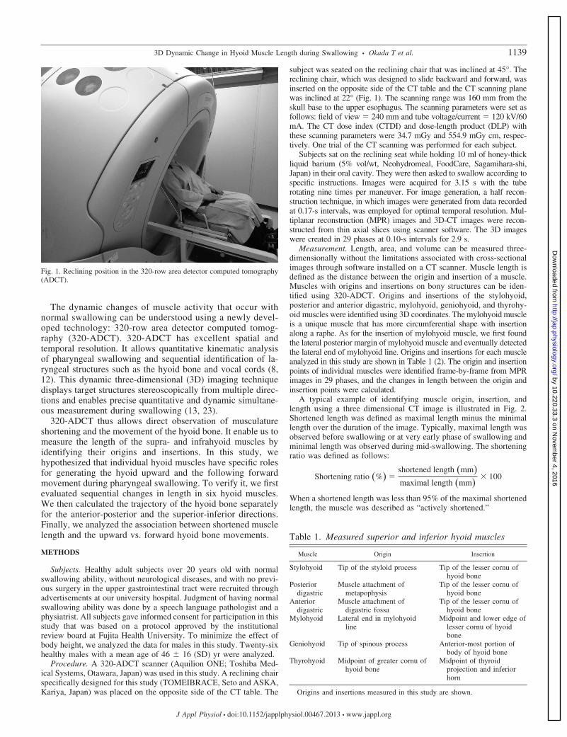
Procedure. A 320-ADCT scanner (Aquilion ONE; Toshiba Med-ical Systems, Otawara, Japan) was used in this study. A reclining chairspecifically designed for this study (TOMEIBRACE, Seto and ASKA,Kariya, Japan) was placed on the opposite side of the CT table. The
subject was seated on the reclining chair that was inclined at 45°. Thereclining chair, which was designed to slide backward and forward, wasinserted on the opposite side of the CT table and the CT scanning planewas inclined at 22° (Fig. 1). The scanning range was 160 mm from theskull base to the upper esophagus. The scanning parameters were set asfollows: field of view � 240 mm and tube voltage/current � 120 kV/60mA. The CT dose index (CTDI) and dose-length product (DLP) withthese scanning parameters were 34.7 mGy and 554.9 mGy cm, respec-tively. One trial of the CT scanning was performed for each subject.
Subjects sat on the reclining seat while holding 10 ml of honey-thickliquid barium (5% vol/wt, Neohydromeal, FoodCare, Sagamihara-shi,Japan) in their oral cavity. They were then asked to swallow according tospecific instructions. Images were acquired for 3.15 s with the tuberotating nine times per maneuver. For image generation, a half recon-struction technique, in which images were generated from data recordedat 0.17-s intervals, was employed for optimal temporal resolution. Mul-tiplanar reconstruction (MPR) images and 3D-CT images were recon-structed from thin axial slices using scanner software. The 3D imageswere created in 29 phases at 0.10-s intervals for 2.9 s.
Measurement. Length, area, and volume can be measured three-dimensionally without the limitations associated with cross-sectionalimages through software installed on a CT scanner. Muscle length isdefined as the distance between the origin and insertion of a muscle.Muscles with origins and insertions on bony structures can be iden-tified using 320-ADCT. Origins and insertions of the stylohyoid,posterior and anterior digastric, mylohyoid, geniohyoid, and thyrohy-oid muscles were identified using 3D coordinates. The mylohyoid muscleis a unique muscle that has more circumferential shape with insertionalong a raphe. As for the insertion of mylohyoid muscle, we first foundthe lateral posterior margin of mylohyoid muscle and eventually detectedthe lateral end of mylohyoid line. Origins and insertions for each muscleanalyzed in this study are shown in Table 1 (2). The origin and insertionpoints of individual muscles were identified frame-by-frame from MPRimages in 29 phases, and the changes in length between the origin andinsertion points were calculated.
A typical example of identifying muscle origin, insertion, andlength using a three dimensional CT image is illustrated in Fig. 2.Shortened length was defined as maximal length minus the minimallength over the duration of the image. Typically, maximal length wasobserved before swallowing or at very early phase of swallowing andminimal length was observed during mid-swallowing. The shorteningratio was defined as follows:
Shortening ratio �%� �shortened length �mm�maximal length �mm�
� 100
When a shortened length was less than 95% of the maximal shortenedlength, the muscle was described as “actively shortened.”
Fig. 1. Reclining position in the 320-row area detector computed tomography(ADCT).
Table 1. Measured superior and inferior hyoid muscles
Muscle Origin Insertion
Stylohyoid Tip of the styloid process Tip of the lesser cornu ofhyoid bone
Posteriordigastric
Muscle attachment ofmetapophysis
Tip of the lesser cornu ofhyoid bone
Anteriordigastric
Muscle attachment ofdigastric fossa
Tip of the lesser cornu ofhyoid bone
Mylohyoid Lateral end in mylohyoidline
Midpoint and lower edge oflesser cornu of hyoidbone
Geniohyoid Tip of spinous process Anterior-most portion ofbody of hyoid bone
Thyrohyoid Midpoint of greater cornu ofhyoid bone
Midpoint of thyroidprojection and inferiorhorn
Origins and insertions measured in this study are shown.
11393D Dynamic Change in Hyoid Muscle Length during Swallowing • Okada T et al.
J Appl Physiol • doi:10.1152/japplphysiol.00467.2013 • www.jappl.org
by 10.220.33.3 on Novem
ber 4, 2016http://jap.physiology.org/
Dow
nloaded from

The timing of the hyoid bone movements (onset of upward andforward movement) was also measured. Onset of the upward movementof the hyoid was defined as the point at which the hyoid bone exceeded5% of total possible vertical displacement just after it began to moveupward. Onset of the forward movement of the hyoid was defined as thepoint at which the hyoid bone began to move forward and it exceeded 5%of total possible horizontal displacement. In this study, onset of theupward movement of the hyoid was labeled as time 0. The line passingthrough the anterior and posterior nasal spine was determined as “hori-zontal line” (Fig. 3). Upward movement of each muscle and the hyoidbone was defined as vertical to the horizontal line and forward movementwas defined as parallel to the horizontal line.
Statistical analysis. Spearman’s rank correlation coefficient wasused to test the relationship between the maximal muscle length,shortened muscle length, and shortening ratio. Spearman’s rank cor-relation coefficient was also used to test the relationship betweenshortened length of each hyoid muscle and the maximal displacementof upward and forward movement of the hyoid bone. SPSS ver. 19.0J for Mac (SPSS, Chicago, IL) was used for the statistical analyses.The level of significance was set at �5%.
RESULTS
Maximal and minimal muscle length. The means and standarddeviations of the maximal and minimal muscle lengths are shownin Table 2. In most cases, maximal muscle length corresponded tothe length at rest. In some thyrohyoid and anterior digastricmuscles, maximal muscle length corresponded to the beginning ormiddle of hyoid bone movement (cf. Fig. 5). The maximal lengthof the longest muscle, the posterior digastric muscle, was 85.2 �
Fig. 2. Anterior, lateral, and superior views ofmandibular bone, hyoid bone, and thyroid car-tilage. The origins, insertions, and lengths ofthe measured hyoid muscles are illustrated.
Anterior nasal spine
Horizontal line Perpendicular line
Posterior nasal spine
Fig. 3. Anterior nasal spine, a part of the maxilla bone, and posterior nasalspine, a part of the palatine bone, are bony projections easily identified using320-ADCT. The line passing through the distal portions of anterior andposterior nasal spines was almost always perpendicular to the cervical spine(cf. the posterior wall at the lower end of C2 and upper end of C4). Cervicalspine does not necessarily represent vertical line in the case of cervicalspondylosis. Therefore, we determined the line including the distal portions ofthe anterior and posterior nasal spines as horizontal line.
1140 3D Dynamic Change in Hyoid Muscle Length during Swallowing • Okada T et al.
J Appl Physiol • doi:10.1152/japplphysiol.00467.2013 • www.jappl.org
by 10.220.33.3 on Novem
ber 4, 2016http://jap.physiology.org/
Dow
nloaded from

8.2 mm. The maximal length of the second longest muscle, thestylohyoid muscle, was 59.3 � 12.3 mm. The geniohyoid andthyrohyoid muscles were the shortest muscles (32.5 � 5.5 mmand 30.6 � 7.5 mm, respectively).
Although the minimal muscle length was relatively constant(8.2–12.8 mm), the shortening ratio differed among hyoidmuscles of the subjects included in this analysis (14%–32%)(Table 2). In Fig. 4A, the maximal and shortened lengths of sixhyoid muscles of all subjects are plotted. No significant changes inminimal muscle lengths were observed regardless of maximalmuscle lengths. Figure 4B plots maximal muscle lengths and theshortening ratio of muscle lengths during swallowing. A signifi-cant correlation was observed between maximal muscle lengthand shortening ratio (P � 0.001, r � �0.380). Higher shorteningratios were associated with shorter muscles.
Changes in muscle length during swallowing. Figure 5 shows anaverage change in muscle length and the trajectory of hyoid bonemovement frame-by-frame when the onset of the upward move-ment of the hyoid bone was set as 0 s. The stylohyoid, posterior
digastric, and mylohyoid muscles simultaneously shortened dur-ing the initial stage of swallowing. The geniohyoid, thyrohyoid,and anterior digastric muscles shortened subsequently and se-quentially. Table 3 shows the average times and standard devia-tions of the onset of shortening for each muscle and the onset ofhyoid bone movement in 26 subjects. The onset of upwardmovement of the hyoid bone was very similar to the onset ofshortening of the stylohyoid, posterior digastric, and mylohyoidmuscles in terms of timing. The onset of forward movement of thehyoid was very similar to the onset of shortening of the geniohy-oid, thyrohyoid, and anterior digastric muscles.
Muscle shortening and hyoid trajectory. The average up-ward movement of the hyoid bone was 16.5 � 9.2 mm and theaverage forward movement was 12.8 � 5.0 mm. Figure 6illustrates the relationships between minimal muscle lengths ofthe six hyoid muscles and upward movement distance of thehyoid bone during swallowing. The correlation coefficients forindividual muscles are shown in Fig. 6. Significant correlationswere observed for the stylohyoid, posterior digastric, andmylohyoid muscles with the distance of upward movement ofthe hyoid bone (r � 0.652, 0.452, and 0.625, respectively).Figure 7 illustrates the relationships between shortened musclelengths of six hyoid muscles and forward movement distanceof the hyoid bone during swallowing. The correlation coeffi-cients are also shown in Fig. 7. A significant correlation wasobserved between the geniohyoid and forward movement dis-tance of the hyoid bone (r � 0.611). Although not significant,moderate correlation coefficients were observed for the ante-rior digastric and thyrohyoid muscles (r � 0.30).
DISCUSSION
Activity of the hyoid muscles. Muscle contraction involved inthe swallowing reflex can be evaluated using EMG. Some EMG
Table 2. Maximal and minimal muscle length, shortenedlength, and the shortening ratio
Muscles
MaximalMuscle
Length, mm
MinimalMuscle
Length, mmShortened
Length, mmShorteningRatio, %
Stylohyoid 59.3 � 12.3 46.6 � 11.6 12.8 � 7.2 21.3 � 10.8Posterior digastric 85.2 � 8.2 72.6 � 9.4 11.7 � 7.2 13.6 � 8.1Mylohyoid 42.3 � 11.3 29.0 � 9.3 13.3 � 5.2 31.6 � 9.6Geniohyoid 32.5 � 5.5 22.8 � 3.8 9.7 � 4.1 29.2 � 9.0Thyrohyoid 30.6 � 7.4 22.1 � 6.6 8.5 � 3.8 28.1 � 11.7Anterior digastric 44.7 � 7.5 36.5 � 6.5 8.2 � 4.7 17.9 � 9.4
Shortened length was defined as maximal length minus minimal length inthe acquired duration. Shortening ratio was calculated by: shortening ratio(%) � {[shortened length (mm)/maximal length (mm)] � 100}.
A B
Fig. 4. A: maximal muscle length and shortened muscle length. There was no significant relationship between them (r � 0.325, P � 0.113). B: maximal musclelength and shortening ratio of muscle length. A significant relationship was seen between them (r � �0.380, P � 0.006).
11413D Dynamic Change in Hyoid Muscle Length during Swallowing • Okada T et al.
J Appl Physiol • doi:10.1152/japplphysiol.00467.2013 • www.jappl.org
by 10.220.33.3 on Novem
ber 4, 2016http://jap.physiology.org/
Dow
nloaded from

experiments have been reported in the past 50 years. Animalstudies using EMG have suggested that the swallowing reflex isinitiated by the contraction of certain suprahyoid muscles (5, 34).The work of Doty and Bosma (5) led to a consensus that the
mylohyoid muscle is a key muscle in the sequential pattern ofmuscle activation. However, they found wide variation in theduration of activity in various muscles; no two EMG measure-ments of deglutition from the same electrodes gave identical oreven similar unit patterns (5). Thexton et al. (34) reinvestigatedEMG activity during reflex pharyngeal swallowing. They sug-gested that the mylohyoid muscle was not activated before othermuscles and that the geniohyoid muscle was not a part of the“leading complex.” However, they also stated that the recordedEMG signals showed both the intramuscular and interanimalvariations, whereas the movements were highly stereotyped (34).Some of this variation could be attributed to slight displacementsof the electrodes during muscle movement (5, 34).
In humans, needle EMG of the swallowing musculature is morechallenging than that of the limb muscles because hyoid musclesinserting into the hyoid bone are located deep to the surface.Although needle EMG appears to be a safe procedure (17, 22),patient discomfort may be a problem; time may be required to
Fig. 5. Changes in muscle length and the trajectoryof hyoid bone movement during swallowing of a10-ml honey-thick liquid. Onset of the upwardmovement of the hyoid bone was set as 0 s. Thegraphs are averaged data from 26 subjects. Anarrow indicates the point when a shortened musclelength became �95% of the maximal shortenedlength at which the muscle is considered as ac-tively shortened. Positive value indicates the up-ward and forward directions in the graph below.
Table 3. Onset of upward and forward movement of thehyoid bone and actively shortened time of individual musclesduring swallowing of a 10-ml honey-thick liquid
Average, s SD, s
Upward movement of hyoid bone 0.00 0.00Forward movement of hyoid bone 0.34 0.10Stylohyoid 0.01 0.05Posterior digastric 0.03 0.06Mylohyoid 0.04 0.09Geniohyoid 0.27 0.12Thyrohyoid 0.29 0.09Anterior digastric 0.39 0.18
The onset of upward movement of hyoid bone was set as 0 s. The average and SDfor each muscle were relative to onset of the upward movement of the hyoid bone.
1142 3D Dynamic Change in Hyoid Muscle Length during Swallowing • Okada T et al.
J Appl Physiol • doi:10.1152/japplphysiol.00467.2013 • www.jappl.org
by 10.220.33.3 on Novem
ber 4, 2016http://jap.physiology.org/
Dow
nloaded from

manipulate the needle to locate the muscle of interest, which maybe deeply embedded in other overlapping muscles not directlyrelated to swallowing. Thus human studies investigating EMGactivity in swallowing-related muscles have been limited. EMGstudies of the stylohyoid or digastric posterior muscles have beenvery rarely conducted in humans (17).
The general consensus is that sequential muscle shorteningrepresents muscle activity during concentric contraction (11, 20).Thus muscle shortening has been measured two-dimensionally forevaluation of hyoid muscle activity during swallowing (20). Inthis study, changes in the distance between the origins andinsertions of supra- and infrahyoid muscles were measured three-dimensionally using a 320-ADCT scanner. We demonstrated thatthe supra- and infrahyoid muscles changed in length duringpharyngeal swallowing and that the overall sequence of muscularactivity remained constant and stereotypical.
Role of hyoid muscles in hyoid bone movement. A crucialevent in the pharyngeal phase of swallowing is the elevationand subsequent anterior movement of the hyoid bone. VF hasverified and quantified the upward and subsequent forwardmovement of the hyoid bone during the passage of a bolus intothe esophagus (4, 31). The 3D dynamic ADCT technique usedin this study allowed calculation of the total upward andforward movement distances of the hyoid bone.
Muscles attached to the hyoid bone presumably affect hyoidbone movement, but their roles have not been explicitly inves-tigated (3, 14, 17). In a recent study by Pearson et al. (28), acadaver model was used to evaluate the architecture of musclespositioned to move the hyoid upward and forward. Based on
structural properties, the geniohyoid muscle may have the mostpotential to displace the hyoid in the forward direction, and themylohyoid may have the most potential to displace the hyoid inthe upward direction. However, as Pearson et al. (28) stated,anatomic data while suggestive of function must be corrobo-rated by functional studies to ensure its clinical usefulness. Inthe current study, we demonstrated not only the role of thegeniohyoid and mylohyoid muscles but that of other hyoidmuscles using the 3D dynamic ADCT technique.
In the analysis of hyoid muscle activity, shortening of thestylohyoid, posterior digastric, and mylohyoid muscles was ob-served before that of other suprahyoid muscles during swallowing(Fig. 5, Table 3). The shortening of these muscles synchronizedand significantly correlated with the upward movement of thehyoid bone (r � 0.45–0.65) (Fig. 6). The correlation of timingand distance between the shortening of these three muscles andthe upward movement of the hyoid bone provides absolute evi-dence that the stylohyoid, posterior digastric, and mylohyoidmuscles cause this upward movement (Fig. 8). Following theshortening of these three muscles, the geniohyoid, thyrohyoid, andanterior digastric muscles began to shorten, synchronizing withthe forward movement of the hyoid bone. The shortening of thegeniohyoid muscle correlated significantly with this forwardmovement (r � 0.61) (Fig. 7). The shortening of the thyrohyoidand anterior digastric muscles was moderately but insignificantlycorrelated with this forward movement (r � 0.3). Although thethyrohyoid shortened associated with onset of the forward move-ment, the primary of this muscle is to pull the larynx closer to thehyoid bone. These results indicate that the geniohyoid is the key
Fig. 6. Correlation of shortened muscle length and upward movement of the hyoid bone during swallowing.
11433D Dynamic Change in Hyoid Muscle Length during Swallowing • Okada T et al.
J Appl Physiol • doi:10.1152/japplphysiol.00467.2013 • www.jappl.org
by 10.220.33.3 on Novem
ber 4, 2016http://jap.physiology.org/
Dow
nloaded from

muscle that moves the hyoid bone forward and that the anteriordigastric muscle is secondary muscle contributing to the forwardmovement of the hyoid bone (Fig. 8).
In summary, serial shortening of hyoid muscles significantlyinfluenced the trajectory pattern of the hyoid bone. Most impor-tantly, as hypothesized, the hyoid muscle group can be dividedinto two groups on the basis of functional influences on hyoidbone movement. The stylohyoid, posterior digastric, and mylohy-oid muscles constitute the first group of muscles that shorten in theinitial stage of swallowing and are instrumental in moving thehyoid bone upward. The second group comprises the geniohyoidand anterior digastric muscles, which shorten subsequently andare instrumental in moving the hyoid bone forward.
Limitation of this study and potential of ADCT for swallow-ing study. Target structures can be displayed stereoscopicallyfrom multiple directions using 320-ADCT. Multiphase 3D
images are reconstructed in 29 phases at intervals of 0.1 s,allowing a kinematic analysis of swallowing (8). CT is inferiorto magnetic resonance imaging with regard to the contrastresolution of soft tissues, thus making muscle identificationdifficult. However, hyoid muscles with their origins and inser-tions within the bone can be accurately identified and measuredusing 320-ADCT. The hyoid bone and larynx move veryquickly during swallowing. Nevertheless, images created atshort intervals (0.1 s) and continuous multisectional observa-tion on MPR permitted comfortable morphologic examinationand kinematic analysis of the target structures in the currentstudy. Thus this 3D dynamic ADCT technique was a good toolfor identifying changes in muscle length.
There were some limitations in this study. First, other variablessuch as the bolus consistency were not examined simultaneouslyto minimize the potential risk associated with radiation exposure.The effective dose was 1.08 mSv at one time, about the same dosefor a clinical videofluoroscopic swallowing study (1.05 mSv for 5min). The second limitation was the 45 degree reclining positionbecause the CT procedure cannot be performed with the subject inthe upright position. There is a possibility that the inclined andsitting positions might have produced different results. Third, togeneralize the current findings, it is necessary to increase thenumber of volunteers to assess the variations potentially caused byage and sex.
Compared with VF, the biggest advantage of 320-ADCT isthe recording technology of the 3D dynamic image. It has greatpotential for further research on swallowing. Further studiesmeasuring upper esophageal sphincter, oropharyngeal cavity,and pharyngeal muscles are recommended. As for a possible
Fig. 7. Correlation of shortened muscle length and forward movement of the hyoid bone during swallowing.
Fig. 8. Role of individual hyoid muscles for inducing movement of hyoid bone.A star mark indicates a key muscle for the movement.
1144 3D Dynamic Change in Hyoid Muscle Length during Swallowing • Okada T et al.
J Appl Physiol • doi:10.1152/japplphysiol.00467.2013 • www.jappl.org
by 10.220.33.3 on Novem
ber 4, 2016http://jap.physiology.org/
Dow
nloaded from

clinical use, measurement of changes in hyoid muscle lengthmay be useful for evaluating the effects of rehabilitation indysphagia. Referring to rehabilitation approaches based on limitedmovement of the hyoid in pharyngeal dysphagia, the effectivenessof strategies such as electrical stimulation or muscle strengtheningexercises may be evaluated. From the viewpoint of the cost-benefit ratio and the risk exposing individuals to a certain amountof radiation in the process of conducting a swallow assessment,however, it remains unknown in patients with dysphagia whetherthe 3D information offers further benefit in addition to 2D infor-mation provided by VF.
ACKNOWLEDGMENTS
We thank Y. Itoh, graduate student, and Y. Kataoka and M. Kobayashi,radiological technologists at Fujita Health University Hospital, for invaluableassistance with the CT study.
DISCLOSURES
No conflicts of interest, financial or otherwise, are declared by the author(s).
AUTHOR CONTRIBUTIONS
Author contributions: T.O., Y.A., Y.I., E.S., H.K., S.S., K.O., and K.U.conception and design of research; T.O., Y.A., and Y.I. performed experi-ments; T.O. analyzed data; T.O., Y.A., Y.I., E.S., H.K., S.S., and K.O.interpreted results of experiments; T.O. and Y.A. prepared figures; T.O. andY.A. drafted manuscript; T.O., Y.A., Y.I., E.S., H.K., S.S., and K.O. edited andrevised manuscript; T.O., Y.A., Y.I., E.s., H.K., S.S., K.O., and K.U. approvedfinal version of manuscript.
REFERENCES
1. Basmajian JV, Dutta CR. Electromyography of the pharyngeal constric-tors and levator palati in man. Anat Rec 139: 561–563, 1961.
2. Borley NR, Standring S, Gray H. Gray’s Anatomy: The AnatomicalBasis of Clinical Practice. Edinburgh, UK: Churchill Livingstone, 2008.
3. Cook IJ, Dodds WJ, Dantas RO, Massey B, Kern MK, Lang IM,Brasseur JG, Hogan WJ. Opening mechanisms of the human upperesophageal sphincter. Am J Physiol Gastrointest Liver Physiol 257:G748–G759, 1989.
4. Dodds WJ, Stewart ET, Logemann JA. Physiology and radiology of thenormal oral and pharyngeal phases of swallowing. AJR Am J Roentgenol154: 953–963, 1990.
5. Doty RW, Bosma JF. An electromyographic analysis of reflex degluti-tion. J Neurophysiol 19: 44–60, 1956.
6. Ertekin C, Aydogdu I. Electromyography of human cricopharyngeal muscleof the upper esophageal sphincter. Muscle Nerve 26: 729–739, 2002.
7. Ertekin C, Pehlivan M, Aydogdu I, Ertas M, Uludag B, Celebi G,Colakoglu Z, Sagduyu A, Yuceyar N. An electrophysiological investi-gation of deglutition in man. Muscle Nerve 18: 1177–1186, 1995.
8. Fujii N, Inamoto Y, Saitoh E, Baba M, Okada S, Yoshioka S, Nakai T,Ida Y, Katada K, Palmer JB. Evaluation of swallowing using 320-detector-row multislice CT. I. Single- and multiphase volume scanning forthree-dimensional morphological and kinematic analysis. Dysphagia 26:99–107, 2011.
9. Gay T, Rendell JK, Spiro J. Oral and laryngeal muscle coordinationduring swallowing. Laryngoscope 104: 341–349, 1994.
10. German RZ, Crompton AW, Thexton AJ. Integration of the reflexpharyngeal swallow into rhythmic oral activity in a neurologically intactpig model. J Neurophysiol 102: 1017–1025, 2009.
11. Huxley AF. Muscle structure and theories of contraction. Prog BiophysBiophys Chem 7: 255–318, 1957.
12. Inamoto Y, Fujii N, Saitoh E, Baba M, Okada S, Katada K, Ozeki Y,Kanamori D, Palmer JB. Evaluation of swallowing using 320-detector-row multislice CT. II. Kinematic analysis of laryngeal closure duringnormal swallowing. Dysphagia 26: 209–217, 2011.
13. Inamoto Y, Saitoh E, Okada S, Kagaya H, Shibata S, Ota K, Baba M,Fujii N, Katada K, Wattanapan P, Palmer JB. The effect of bolusviscosity on laryngeal closure in swallowing: kinematic analysis using320-row area detector CT. Dysphagia 28: 33–42, 2013.
14. Kahrilas PJ, Logemann JA, Lin S, Ergun GA. Pharyngeal clearanceduring swallowing: a combined manometric and videofluoroscopic study.Gastroenterology 103: 128–136, 1992.
15. Kawasaki M, Ogura JH, Takenouchi S. Neurophysiologic observationsof normal deglutition. I. Its relationship to the respiratory cycle. Laryn-goscope 74: 1747–1765, 1964.
16. Kobara-Mates M, Logemann JA, Larson C, Kahrilas PJ. Physiology oforopharyngeal swallow in the cat: a videofluoroscopic and electromyographicstudy. Am J Physiol Gastrointest Liver Physiol 268: G232–G241, 1995.
17. Kurt T, Gurgor N, Secil Y, Yildiz N, Ertekin C. Electrophysiologicidentification and evaluation of stylohyoid and posterior digastricus mus-cle complex. J Electromyogr Kinesiol 16: 58–65, 2006.
18. Ludlow CL, Humbert I, Saxon K, Poletto C, Sonies B, Crujido L.Effects of surface electrical stimulation both at rest and during swallowingin chronic pharyngeal dysphagia. Dysphagia 22: 1–10, 2007.
19. Matsuo K, Palmer JB. Anatomy and physiology of feeding and swal-lowing: normal and abnormal. Phys Med Rehabil Clin N Am 19: 691–707,vii, 2008.
20. Mepani R, Antonik S, Massey B, Kern M, Logemann J, Pauloski B,Rademaker A, Easterling C, Shaker R. Augmentation of deglutitivethyrohyoid muscle shortening by the Shaker exercise. Dysphagia 24:26–31, 2009.
21. Miller AJ. Significance of sensory inflow to the swallowing reflex. BrainRes 43: 147–159, 1972.
22. Mu LC, Yang SL. A new method of needle-electrode placement in theposterior cricoarytenoid muscle for electromyography. Laryngoscope 100:1127–1131, 1990.
23. Nakayama E, Kagaya H, Saitoh E, Inamoto Y, Hashimoto S, Fujii N,Katada K, Kanamori D, Tohara H, Ueda K. Changes in pyriform sinusmorphology in the head rotated position as assessed by 320-row areadetector CT. Dysphagia 28: 199–204, 2013.
24. Palmer JB, Tanaka E, Siebens AA. Electromyography of the pharyngealmusculature: technical considerations. Arch Physical Med Rehab 70:283–287, 1989.
25. Palmer PM, Luschei ES, Jaffe D, McCulloch TM. Contributions ofindividual muscles to the submental surface electromyogram during swal-lowing. J Speech Lang Hear Res 42: 1378–1391, 1999.
26. Palmer PM, McCulloch TM, Jaffe D, Neel AT. Effects of a sour boluson the intramuscular electromyographic (EMG) activity of muscles in thesubmental region. Dysphagia 20: 210–217, 2005.
27. Pearson WG, Hindson DF, Langmore SE, Zumwalt AC. Evaluatingswallowing muscles essential for hyolaryngeal elevation by using musclefunctional magnetic resonance imaging. Int J Radiat Oncol Biol Phys 85:735–740, 2013.
28. Pearson WG Jr, Langmore SE, Zumwalt AC. Evaluating the structuralproperties of suprahyoid muscles and their potential for moving the hyoid.Dysphagia 26: 345–351, 2011.
29. Pearson WG, Langmore SE, Yu LB, Zumwalt AC. Structural analysis ofmuscles elevating the hyolaryngeal complex. Dysphagia 27: 445–451, 012.
30. Perlman AL, Palmer PM, McCulloch TM, Vandaele DJ. Electromyo-graphic activity from human laryngeal, pharyngeal, and submental mus-cles during swallowing. J Appl Physiol 86: 1663–1669, 1999.
31. Perlman AL, VanDaele DJ, Otterbacher MS. Quantitative assessmentof hyoid bone displacement from video images during swallowing. JSpeech Hear Res 38: 579–585, 1995.
32. Spiro J, Rendell JK, Gay T. Activation and coordination patterns of thesuprahyoid muscles during swallowing. Laryngoscope 104: 1376–1382, 1994.
33. Suzuki M, Tokuriki M, Nomura SI. Electromyographic studies on thedeglutition movement in the goat. Nihon Juigaku Zasshi 39: 243–253, 1977.
34. Thexton AJ, Crompton AW, German RZ. Electromyographic activityduring the reflex pharyngeal swallow in the pig: Doty and Bosma (1956)revisited. J Appl Physiol 102: 587–600, 2007.
35. Trigos I, Ysunza A, Vargas D, Vazquez MC. The San Venero Rosellipharyngoplasty: an electromyographic study of the palatopharyngeusmuscle. Cleft Palate J 25: 385–388, 1988.
36. van Overbeek JJ, Wit HP, Paping RH, Segenhout HM. Simultaneousmanometry and electromyography in the pharyngoesophageal segment.Laryngoscope 95: 582–584, 1985.
37. Venker-van Haagen AJ, Hartman W, van den Brom WE, WolvekampWT. Continuous electromyographic recordings of pharyngeal muscleactivity in normal and previously denervated muscles in dogs. Am J VetRes 50: 1725–1728, 1989.
11453D Dynamic Change in Hyoid Muscle Length during Swallowing • Okada T et al.
J Appl Physiol • doi:10.1152/japplphysiol.00467.2013 • www.jappl.org
by 10.220.33.3 on Novem
ber 4, 2016http://jap.physiology.org/
Dow
nloaded from