Embed Size (px)
Citation preview

1
D.Y. PATIL EDUCATION SOCIETY KOLHAPUR
DEEMED UNIVERSITY(Declared under section 3 of the UGC act 1956)
869, ‘E’ KasabaBawada, Kolhapur-416 006Phone No. : (0231) 2601235-36, Fax : (0231) 2601595,
Web: www.dypatilunikop.org , E-mail : [email protected]
COURSE CURRICULUM
M. Sc Medical Physics (2 Years Course)*(Choice based Credit System)
(*On successful completion of M. Sc. Medical physics course, all students are required toundergo one year internship at AERB recognized institutes. This is a mandatory requirement
for becoming qualified medical physicists and appearing in the RSO examination)

2
BL-MP-01- About the course
M. Sc. Medical Physics course is basically a two years course which is approved by Atomic
Energy Regulatory Board (AERB), Government of India. M. Sc. Medical Physics course, being
a specialization course designed to train the young pool of PG students as qualified medical
physicist and radiation safety officers (RSO) in the field of cancer radiation therapy. Medical
physics is one of the fastest growing areas of employment for physicists. They play a crucial
role in radiology, radiation therapy and nuclear medicine. These fields use very
sophisticated and expensive equipment and medical physicist or responsible for much of its
plan, execution, testing and quality assurance.
The M.Sc. medical physics students are getting the exposures form the various cancer
hospitals during practical and their M.Sc. Project work. Our students are exposed to field
training in various cancer hospitals all over India. After completion of the 2 years course,
students undergo one year internship according to AERB regulations in order to work as a
Medical Physicist in the hospital.
BL-MP-02- Vision Mission and Goal
Vision: "To offer diverse Medical Physics program to establish and maintain the standards of
the students of Medical Physics in the disciplines of Diagnostic Imaging, Radiation Oncology
and Nuclear Medicine”.
Mission: To promote professional growth by offering state-of-the-art postgraduate program
in Medical Physics in India and abroad.
Goals:
The goal of the course is to cultivate an educational environment which provides the full
spectrum of learning opportunities in clinical medical physics, radiation oncology and
radiobiology.
The curriculum is flexible and designed to enable a student to optimize their learning
experience throughout their two years program.

3
It is an expectation that upon the completion of the program a student will be an
outstanding "Radiation Oncology Physicist" capable of making an immediate impact in
either an academic or community practice setting.
BL-MP-03- Outcome of the program
The student will be well versed with the concept of Physics (specifically radiation e.g. X-
rays, Gamma rays etc.) which can be used for medical applications.
The student will learn different advanced techniques (e.g. 3D CRT, IMRT, IGRT,
Brachytherapy, etc.) involved in the treatment of cancer.
Culture of Interdisciplinary research will be seeded through collaborations with various
Cancer hospitals.
Students will get the job opportunities below
The students have tremendous opportunities to work as a clinical medical physicist
in various leading hospitals all over India with attractive salary packages.
The students have opportunities to work as an Assistant Professor where there are
courses of M.Sc. Medical Physics.
The students can work as a Scientist in the Research institutes.
The students can also work as dosimetrists in various companies providing radiation
measuring devices.
The students also have opportunities to pursue higher studies in India and abroad in
related field.
BL-MP-04- Syllabus
Course Structure & Distribution of Credits.
M.Sc. Medical Physics Program consists of total 16 theory courses, total 4 practical lab
courses spread over 4 semesters.16 theory courses and 4 practical lab courses and one
project will be common and compulsory to all the students. Each theory course will be of 4
(four) credits, a practical lab course will be of 4 (four) credits and a project will be of 8
(eight) credits. A student earns 24 (twenty four) credits per semester and total 96 (ninety
six) credits in 4 semesters. The course structure is as follows,
4
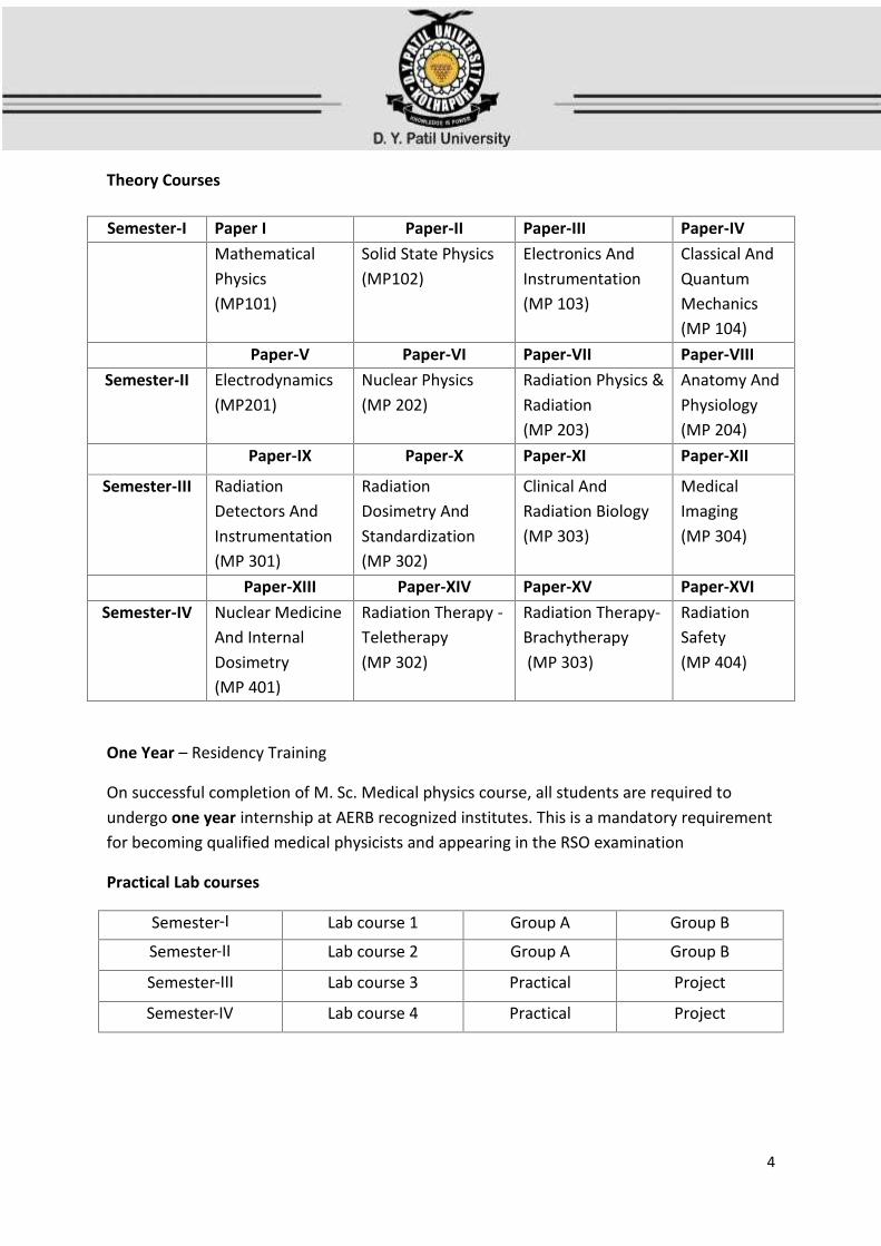
Theory Courses
Semester‐I Paper I Paper‐II Paper‐III Paper‐IVMathematicalPhysics(MP101)
Solid State Physics(MP102)
Electronics AndInstrumentation(MP 103)
Classical AndQuantumMechanics(MP 104)
Paper‐V Paper‐VI Paper‐VII Paper‐VIIISemester‐II Electrodynamics
(MP201)Nuclear Physics(MP 202)
Radiation Physics &Radiation(MP 203)
Anatomy AndPhysiology(MP 204)
Paper‐IX Paper‐X Paper‐XI Paper‐XII
Semester‐III RadiationDetectors AndInstrumentation(MP 301)
RadiationDosimetry AndStandardization(MP 302)
Clinical AndRadiation Biology(MP 303)
MedicalImaging(MP 304)
Paper‐XIII Paper‐XIV Paper‐XV Paper‐XVISemester‐IV Nuclear Medicine
And InternalDosimetry(MP 401)
Radiation Therapy -Teletherapy(MP 302)
Radiation Therapy-Brachytherapy(MP 303)
RadiationSafety(MP 404)
One Year – Residency Training
On successful completion of M. Sc. Medical physics course, all students are required toundergo one year internship at AERB recognized institutes. This is a mandatory requirementfor becoming qualified medical physicists and appearing in the RSO examination
Practical Lab courses
Semester‐I Lab course 1 Group A Group B
Semester‐II Lab course 2 Group A Group B
Semester‐III Lab course 3 Practical Project
Semester‐IV Lab course 4 Practical Project

5
Semester IM.Sc. Medical Physics Program for Semester‐I consists of four theory courses and one
laboratory course consisting two groups of practical. The details are as follows:
Theory Courses (4): 16 hours per week (One lecture of one hour duration)
Theory Paper Subject Lectures (Hrs.) CreditsPaper I: MP101 Mathematical Physics 60 04Paper II: MP102 Solid State Physics 60 04
Paper II: MP103 Electronics AndInstrumentation
60 04
Paper IV: MP104 Classical And QuantumMechanics
60 04
Total 240 16
Practical lab courses (2): 16 hours per week
Practical Lab Course 1 Practical Lab Sessions (Hrs) CreditsMPP101 (Group A) 120 04MPP102 (Group B) 120 04
Total 240 08
Semester IIM.Sc. Medical Physics Program for Semester‐II consists of four theory courses and onelaboratory course consisting two groups of practical. The details are as follows:
Theory Courses (4): 16 hours per week (One lecture of one hour duration)
Theory Paper Subject Lectures (Hrs.) CreditsPaper V: MP201 Electrodynamics 60 04Paper VI: MP202 Nuclear Physics 60 04Paper VII: MP203 Radiation Physics And
Radiation Generators60 04
Paper VIII: MP204 Anatomy AndPhysiology
60 04
Total 240 16

6
Practical lab courses (2): 16 hours per week
Practical Lab Course 2 Practical Lab Sessions (Hrs) CreditsMPP201 (Group A) 120 04MPP202 (Group B) 120 04
Total 240 08
Semester III
M.Sc. Medical Physics Program for Semester‐III consists of four theory courses and one
laboratory course and a project equivalent to one laboratory course. The details are as
follows:
Theory Courses (4): 16 hours per week (One lecture of one hour duration)
Theory Paper Subject Lectures (Hrs.) CreditsPaper IX: MP301 Radiation Detectors And
Instrumentation60 04
Paper X: MP302 Radiation Dosimetry AndStandardization
60 04
Paper XI: MP303 Clinical And RadiationBiology
60 04
Paper XII: MP304 Medical Imaging 60 04Total 240 16
Practical lab courses (2): 16 hours per week
Practical Lab Course 3 Practical Lab Sessions (Hrs) CreditsMPP301( Practical) 120 04MPP302 (Project ) 120 04
Total 240 08
Semester IV
M.Sc. Medical Physics Program for Semester‐IV consists of four theory courses and one
laboratory course and a project equivalent to one laboratory course. The details are as
follows:

7
Theory Courses (4): 16 hours per week (One lecture of one hour duration)
Theory Paper Subject Lectures (Hrs.) CreditsPaper XIII: MP401 Nuclear Medicine And
Internal Dosimetry60 04
Paper XIV: MP402 Radiation Therapy-Teletherapy
60 04
Paper XV: MP403 Radiation Therapy-Brachytherapy
60 04
Paper XVI: MP404 Radiation Safety 60 04Total 240 16
Practical lab courses (2):16 hours per week
Practical Lab Course 4 Practical Lab Sessions (Hrs) CreditsMPP401 (Practical) 120 04MPP402 (Project) 120 04Total 240 08
The candidate shall be awarded the degree of Master of Science in Medical Physics after
completing the course and meeting all the evaluation criteria.
(On successful completion of M. Sc. Medical physics course, all students are required to
undergo one year internship at AERB recognized institutes. This is a mandatory requirement
for becoming qualified medical physicists and appearing in the RSO examination)

8
M.Sc. Medical Physics (Theory Courses)
Semester –I
Paper I: MP101: Mathematical Physics (60 lectures, 4 credits)
UNIT I: VECTOR SPACES AND MATRICES AND DIFFERENTIAL EQUATIONS (15 h)
Vector spaces and subspaces, linear dependence and independence, basis and dimensions,
linear operators, matrices, inverse, orthogonal and unitary matrices, independent elements
of a matrix, eigenvalues and eigenvectors, diagonalization, complete orthonormal sets of
functionssecond order linear ODEs with variable coefficients, solution by series expansion.
UNIT II: SPECIAL FUNCTIONS OFDIFFERENTIAL EQUATIONS AND INTEGRAL TRANSFORMS(15 h)
Legendre, bessel, hermite and lagaurre equations, physical applications, generating
functions, recursion relations, laplace transform, first and second shifting theorems, inverse
LT by partial fractions, LT of derivative and integral of function, fourier series, FS or arbitrary
period, half wave expansions, partial sums, fourier integral and transforms, FT of delta
function
UNIT III: PROBABILITY, STATISTICS AND ERRORS (15 h)
Probability: addition and multiplication laws of probability, conditional probability,
population, variates, collection, tabulation and graphical representation of data. basic ideas
of statistical distributions, frequency distributions, averages or measures of central
tendency, arithmetic mean, properties of arithmetic mean, median, mode, geometric mean,
harmonic mean, dispersion, standard deviation, root mean square deviation, standard error
and variance, moments, skewness and kurtosis, application to radiation detection:
uncertainty calculations, error propagation, time distribution between background and
sample, minimum detectable limit. binomial distribution, Poisson distribution, Gaussian
distribution, exponential distribution, additive property of normal variates, confidence
limits, bivariate distribution, correlation and regression, chi-Square distribution, t-
distribution, F-distribution. Statistics of nuclear counting: Application of Poisson's
statistics - goodness-of-fit tests -Lexie's divergence coefficients, Pearson's chi-square test
9
and its extension, random fluctuations,evaluation of equipment performance, Signal-to-
noise ratio, selection of operating voltage, preset of rate meters and recorders, efficiency
and sensitivity of radiation detectors, statistical aspects of gamma ray and beta ray
counting, special considerations in gas counting and counting with proportional counters,
statistical accuracy in double isotope technique,sampling and sampling distributions,
confidence intervals, clinical study designs and clinical trials,hypothesis testing and errors,
regression analysis.
UNIT IV: NUMERICAL METHODS, COMPUTATIONAL TOOLS & TECHNIQUES(15 h)
Need for numerical methods, accuracy and errors on calculations - round-off error,
evaluation of formulae, iteration for Solving x = g(x), initial approximation and convergence
criteria, Newton-Raphson Method. Taylor series, approximating the derivation, numerical
differentiation formulas, introduction to numerical quadrature, Trapezoidal rule, Simpson's
1/3rule, Simpson’s 3/8rule, Boole rule, Weddle rule, initial value problems, Picard’s method,
Taylor’s method, Euler’s method, the modified Euler’s method, Runge-Kutta method, Monte
Carlo: Random variables, discrete random variables, continuous random variables,
probability density function, discrete probability density function, continuous probability
distributions, cumulative distribution function, accuracy and precision, law of large number,
central limit theorem, random numbers and their generation, tests for randomness,
inversion random sampling technique including worked examples, integration of simple 1-D
integrals including worked examples.
Computational packages: Overview of programming in C++, MATLAB, Origin and SPSS in
data analysis and graphics.
BOOKS FOR STUDY AND REFERENCE:1. Pipes L.A. & L.R. Harvil, Applied Mathematics for Engineers and Physicists (3rd Edition),
Mc Graw-Hill Book Co., New York, 1970.2. Mary.L.Boas, Mathematical methods in the Physical Sciences (2nd edition), John Wiley
& Sons., New York, 1983.3. E. Butkov, Mathematical Physics, Addison Wesley, New York, 1973.4. E. Walpole, R.M. Myers, S.L. Myers, K. Ye, “Probability & Statistics for Engineers and
Scientists (9thedition)”, Pearson Education, 2012.

10
5. SathyaPrakash, Mathematical Physics, Sultan Chand & Co., New Delhi, 2004.6. M.K. Venkatraman, Advanced Mathematics for Engineers & Scientists, National
Publishing co., Madras, 1994.7. G. Arfken and H.H. Weber, Mathematical Methods for Physicists (4th edition), Prism
Books, Bangalore, 1995.
Paper II: MP102: Solid State Physics (60 Lectures, 4 credits)
UNIT I: CRYSTAL STRUCTURE (15 h)
Crystalline and amorphous solids, translational symmetry.Elementary ideas about crystal
structure, lattice and bases, unit cell, reciprocal lattice, fundamental types of lattices, Miller
indices, lattice planes, simple cubic, fcc. and bcc, lattices, Laue and Bragg equations.
determination of crystal structure with X-rays.
Different types of bonding- ionic, covalent, metallic, van-der Waals and hydrogen. band
theory of solids, periodic potential and Bloch theorem, energy band structure.
UNIT II: STRUCTURE OF SOLIDS AND DIELECTRIC AND MAGNETIC PROPERTIES OFMATERIALS (15 h)
Band structure in conductors, direct and indirect semiconductors and insulators (qualitative
discussions); free electron theory of metals, effective mass, drift current, mobility and
conductivity, Wiedemann-Franz law. Hall effect in metals: Phenomenology and implication.
Electronic, ionic and dipolar polarizability, local fields, induced and oriented polarization,
molecular field in a dielectric; Clausius-Mosotti relation,dia, para and ferro-magnetic
properties of solids, Langevin’s theory of diamagnetism and paramagnetism,quantum
theory of paramagnetism, Curie’s law,ferromagnetism: spontaneous magnetization and
domain structure; temperature dependence of spontaneous magnetisation; Curie-Weiss
law, explanation of hysteresis.

11
UNIT III: LATTICE VIBRATIONS (15 h)
Elastic and atomic force constants; dynamics of a chain of similar atoms and chain of two
types of atoms; optical and acoustic modes; interaction of light with ionic crystals. Einstein's
and Debye's theories of specific heats of solids.
Lattice vacancies, diffusion, colour centres: F centres, other centres in alkali halides.
UNIT IV: SUPERCONDUCTIVITY ANDLUMINESCENCE IN SOLIDS (15 h)
Introduction (Kamerlingh-Onnes experiment), effect of magnetic field, type-I and type-II
superconductors, Isotope effect, Meissner effect, BCS pairing mechanisms, Ideas about
High-Tc superconductors
Types of Luminescence, Fluorescence and phosphorescence, Thermo luminescence,
Electroluminescence, LASER.
BOOKS FOR STUDY AND REFERENCE:1. C. Kittel, Introduction to Solid State Physics (8th edition), John Wiley and Sons, New
York, 2004.2. M. A. Omar, Elementary Solid State Physics: Principles and Applications, Addison-
Wesley Publishing Company, Inc, USA, 1975.3. A. J. Dekker, Solid State Physics, Macmillan India, 20004. S. O. Pillai, Solid State Physics, New Age International, India, 2006.5. J. P. Srivastava, Elements of Solid State Physics, Prentice Hall India Pvt., Limited, India,
2004.6. R.J. Elliot and A.F. Gibson, An Introduction to Solid State Physics and Applications,
McMillan, London, 1928.7. D.W. Snoke, Solid State Physics: Essential Concepts, Person Education, 2009

12
Paper III: MP103: Electronics and Instrumentation (60 lectures, 4 credits)
UNIT I: SEMICONDUCTOR DEVICES (15 h)Characteristic curves and physics of p-n junction; Schottky, tunnel and MOS diodes; bipolar
junction transistors(BJT), junction field effect transistor (JFET), metal oxide semiconductor
field effect transistor (MOSFET), uni-junction transistor (UJT) and silicon controlled rectifier
(SCR), optoelectronic devices (photo-diode, solar cell, LED, LCD and photo transistors)
diffusion of impurities in Si, growth of oxide.
Op-amp: introduction, input modes andop-amps with negative feedback, open-loop
response - mathematical operations, analog simulation, OTAs, CFOAs, active filters,
UNIT II: ANALOG ELECTRONICS (15 h)
Oscillators- principles, types, frequency stability, response, the phase shift oscillator, Wein
bridge oscillator, oscillator with RC feedback circuits (RC and LC) , relaxation oscillators,
linear and nonlinear oscillators, 555 timer as an oscillator, IC voltage regulators, evolution of
ICs, CCDs, multi-vibrators, classification, selection of a transducer, strain gauge,
displacement transducer (capacitive, inductive, differential transformer, photo electric and
piezoelectric transducers), strain flow measurements, thermistor and thermo couple based
thermometers for measuring temperature.
UNIT III: DIGITAL ELECTRONICS (15 h)
Introductory digital concepts, overview of logic functions , fixed function integrated circuits,
programmable logic devices , digital integrated circuits, NAND and NOR gates building block,
X-OR gate, simple combinational circuits, half and full address, functions of combinational
logic, flip flops and related devices, counters, shift registers, memory and storage (ROM,
RAM and EPROM), microprocessor and microcontroller basics (Intel 8085).
UNIT IV: ELECTRONICS FOR NUCLEAR DEVICES (15 h)
Preamplifier, AC-DC converter , Pulse shaper, Isolator, High range gamma survey meter
circuit, scintillation dose rate meter, scintillator photodiode X-ray detector, pocket monitor,

13
generalpurpose contamination monitor, discriminator single channel analyzer, linear gate,
time to amplitude converter.
BOOKS FOR STUDY AND REFERENCE:
1. S. M. Sze, K.K. Ng, Physics of semiconductor devices (3rd edition), Wiley-Interscience,
New York, 1969.
2. P.Horowitz and W.Hill, “The art of electronics’, (2nd edition), Cambridge university
press, Cambridge, 1995.
3. A.P.Malvino, “Electronic principles’, (6th edition), Tata McGraw Hill Publ.Co. Ltd., New
Delhi, 1999.
4. T.L.Floyd, Electronic devices’, (6th edition), Pearson Education Inc., New Delhi, 2003
5. R.F.Coughlin and F.F.Driscoll,’Operational amplifiers and linear integrated circuits’, (6th
edition), Pearson Education Inc., New Delhi, 2001.
6. M.Lakshmanan and K.Murali, Chaos, ‘Chaos in nonlinear Oscillators’, World Scientific,
Singapore, 1996.
7. T. L. Floyd, Digital Fundamentals, (8th edition), pearson education Inc., New Delhi,
2003.
8. S.Brown and Z.Vranesic,’Fundamentals of digital logic with Verilog design’, Tata
McGraw Hill Publ Co.Ltd., New Delhi, 2003.
9. H.Skalsi, “Electronic instrumentation (2nd edition), Tata McGraw Hill Publ. Co. Ltd.,
New Delhi,2004.

14
Paper IV: MP104 Classical and Quantum Mechanics
UNIT I: CENTRAL FORCE PROBLEM AND HAMILTONIAN FORMULATIO (15 h)
Two body problem, the equation of motion and first integral, equation of orbit, Kepler’s
laws, Kepler’s problem, general analysis of orbits, stability of orbits, artificial satellites,
Rutherford scattering: differential scattering crosssection, Rutherford Formulae for
scattering.
Hamilton’s principle, Hamiltonian, generalized momentum, constant of motion, Hamilton’s
canonical equations of motion, deduction of canonical equations from Variational principle.
UNIT II: APPLICATIONS OF HAMILTONIAN EQUATIONS OF MOTION, CANONICAL
TRANSFORMATIONS AND HAMILTONS - JACOBI THEORY (15 h)
Applications of Hamilton’s equations of motion. principle of least action, proof of principle
of least action.
Generating functions, illustrations of canonical transformations, condition for
transformation to be canonical, examples. Poisson’s brackets, Poisson’s theorem, properties
of Poisson’s Brackets, Hamilton’s canonical equations in terms of Poisson’s brackets,
Hamilton’s-Jacobi theory, solution of harmonic oscillator problem by HJ Method, problems.
UNIT III: FUNDAMENTAL CONCEPTS AND FORMALISM (15 h)
Need for Quantum mechanics, revision; inadequacy of classical mechanics; Sequential Stern-
Gerlach experiment, analogy with polarization of light, Ket and Bra spaces and inner
products, operators, the associative axiom base kets and matrix representations,
measurements, observables and the uncertainty relations, change of basis, position,
momentum and translation; wave function in position and momentum space.
Time evolution and Schrödinger equation; the Schrödinger versus the Heisenberg picture,
simple Harmonic oscillation, Schrödinger wave equation,

15
UNIT IV: QUANTUM DYNAMICS (15 h)
One-dimensional problems, wells and barriers; Harmonic oscillator by Schrödinger equation
and by operator method. Uncertainty relation of x and p, states with minimum uncertainty
product; General formalism of wave mechanics; Commutation relations.
Rotations and angular momentum commutation relations, spin ½ systems and finite
rotations; SO(3), SU(2) and Euler rotations, eigenvalues and eigenstates of angular
momentum, orbital angular momentum, addition of angular moment.
BOOKS FOR STUDY AND REFERENCE:
1. H. Goldstein,C. Poole, J. Safko, Classical Mechanics (3rd edition), Addison Wesley,
Cambridge, 1980.
2. N.C. Rana and P. S. Joag, Classical Mechanics, Tata McGraw Hill, New Delhi, 1991.
3. R. G. Takwale and P. S. Puranik, Introduction to Classical Mechanics, Tata McGraw Hill
Education, New Delhi, 1999.
4. S. L. Gupta, V. Kumar and R. C. Sharma, Classical Mechanics, Pragati Prakashan Meerut,
2000.
5. A. Ghatak, S. Lokanathan, Quantum Mechanics: Theory and Applications, Kluwer
Academic Publishers, London, 2004.
6. J. J. Sakurai, Modem Quantum Mechanics, Addison Wesley Publication Company
Inc.USA, 1933.
7. L. I. Schiff, Quantum Mechanics, Tata McGraw Hill Education, New Delhi, 1949.
8. M. Mathews, K. Venkatesan, Quantum Mechanics,Tata McGraw Hill Education, New
Delhi, 1978.

16
Semester –II
Paper V: MP201: Electrodynamics (60 lectures, 4 credits)
UNIT I: MAXWELL’S EQUATIONS AND E.M. WAVES (15 h)
Review of four-vector and lorentz transformation in four-dimentional space,
electromagnetic field tensor in four dimensions and Maxwell’s equations: microscopic and
macroscopic forms (revision), conservation of the bound charge and current densities, E.M.
waveequations in waveguide of the arbitrary cross section: TE and TM modes; Rectangular
and circular waveguides, hybrid modes, concept of LP modes.
UNIT II: TIME –DEPENDENT POTENTIALS AND FIELDS (15 h)
Scalar and vector potentials: coupled differential equations, Gauge transformations:
Lorentz and Coulomb Gauges, Retarded Potentials, Lienard –Wiechert Potentials, Fields due
to a charge in the arbitrary motion.
UNIT III: RADIATION FROM ACCELERATED CHARGES AND RADIATION REACTION (15 h)
Fields of charge in uniform motion, applications to linear and circular motions: cyclotron and
synchrotron radiations, Power radiated by point charge: Larmour’s formula, angular
distribution of radiated power, Cerenkov radiation and Bremsstrah lung (qualitative
treatments), radiation reaction: criteria for validity, Abraham–Lorentz formula, physical
basis of radiation reaction, self force.
UNIT IV: FORMULATION OF COVARIANTELECTRODYNAMICS (15 h)
Contravariant and co-variant four-vectors and their products, tensors of rank two and their
differentiation, co-variant form of Maxwell’s equations: four–potential and four current,
E.M. field tensor: its curl and divergence.
17
BOOKS FOR STUDY AND REFERENCE:
1. D.J. Griffiths, Introduction to Electrodynamics (3rd edition),Prentice Hall, New Jersey,
1999.
2. J.R. Reitz, F.J. Milford & R.W. Christy, Foundation of E.M. Theory (3rd edition), Addison
Wesley, New Jersey, 1979.
3. J. D. Jackson, Classical Electrodynamics (3rd edition), Wiley Eastern, New York, 1975.
4. S.P. Puri, Classical Electrodynamics, Tata McGraw Hill Education, New Delhi, 1990.
Paper VI: MP202: Nuclear Physics (60 lectures, 4 credits)
UNIT I: BULK PROPERTIES OF NUCLEI (15 h)
Nuclear mass, charge, size, binding energy, spin and magnetic moment, Isobars, isotopes
and isotones; mass spectrometer (Bainbridge), Spin and parity.
Nature of forces between nucleons, nuclear stability and nuclear binding.
UNIT II: NUCLEAR STRUCTURE AND UNSTABLE NUCLEI (15 h)
The liquid drop model (descriptive) and the Bethe-Weizsacker mass formula,application to
stability considerations, extreme single particle shell model (qualitative discussion
withemphasis on phenomenology with examples).
(a) Alpha decay: alpha particle spectra – velocity and energy of alpha particles. Geiger-Nuttal
law. (b) Beta decay : nature of beta ray spectra, the neutrino, energy levels and decay
schemes, positron emission and electron capture, selection rules, beta absorption and range
of beta particles, Kurie plot. (c)Gamma decay : gamma ray spectra and nuclear energy levels,
isomeric states. Gamma absorption in matter – photoelectric process, Compton scattering,
pair production (qualitative).
UNIT III: NUCLEAR REACTIONS (15 h)
Conservation principles in nuclear reactions. Q-values and thresholds, nuclear reaction
cross-sections, examples of different types of reactions and their characteristics. Bohr’s
postulate of compound nuclear reaction, Ghoshal’s experiment.

18
Discovery and characteristics, explanation in terms of liquid drop model, fission products
and energy release, spontaneous and induced fission, transuranic elements. Chain reaction
and basic principle of nuclear reactors. Nuclear fusion: energetics in terms of liquid drop
model.
UNIT IV: ELEMENTARY PARTICLES (15 h)
(a) Four basic interactions in nature and their relative strengths, examples of different types
of interactions. Quantum numbers – mass, charge, spin, isotopic spin, intrinsic parity,
hypercharge. Charge conjugation. Conservation laws. (b) Classifications of elementary
particles – hadrons and leptons, baryons and mesons, elementary ideas about quark
structure of hadrons – octet and decuplet families.
BOOKS FOR STUDY AND REFERENCE:
1. W.N. Cottingham and D. A. Greenwood,An Introduction to Nuclear Physics,CambridgeUniversity Press, 1986.
2. B. L. Cohen, Concepts of Nuclear Physcics,Tata McGraw Hill Education, New Delhi, 1971.3. S. N. Ghoshal, Atomic and Nuclear Physics, S. Chand, New Delhi, 1997.4. S. B. Patel, Nuclear Physics: An Introduction, New Age International, New Delhi, 1991.5. E. Segre, Nuclei and Particles (2nd edition), W.A. Benjamin Inc., 1977.6. J.S. Lilley, Nuclear Physics: Principles and applications (1st edition), John Willey and Sons
(Asia) Pvt. Ltd., 2001.7. J. Basdevant, J. Rich and M. Spiro, Fundamentals in Nuclear Physics: from Nuclear
Structure to Cosmology, Springer-Verlag New York, 2005.8. A. Seiden, Particle Physics: A Comprehensive Introduction, Persian Education, 2004.

19
Paper VII: MP203: Radiation Physics And Radiation Generators (60 lectures, 4 credits)
UNIT I: RADIOACTIVITY (15 h)
Radioactivity, general properties of alpha, beta and gamma rays, laws of radioactivity, laws
of successive transformations, natural radioactive series, radioactive equilibrium, alpha ray
spectra, beta ray spectra, theory of beta decay, gamma emission, electron capture, internal
conversion, nuclear isomerism, artificial radioactivity, nuclear cross sections, elementary
ideas of fission and reactors, fusion.
UNIT II: PARTICLE ACCELERATORS (15 h)
Particle accelerators for industrial, medical and research applications: the resonant
transformer, Cascade generator, Van De Graff Generator, Pelletron, Cyclotron, Betatron,
Synchro-Cyclotron linear accelerator, Klystron and magnetron, travelling and standing wave
acceleration, Microtron, electron synchrotron, proton synchrotron, details of accelerator
facilities in India.
UNIT III: X-RAY GENERATORS (15 h)
Discovery, production, properties of X-rays, characteristics and continuous spectra, design
of hot cathode X-ray tube, basic requirements of medical diagnostic, therapeutic and
industrial radiographic tubes, rotating anode tubes, hooded anode tubes, industrial X-ray
tubes, X-ray tubes for crystallography, rating of tubes, safety devices in X-ray tubes, ray
proof and shockproof tubes, insulation and cooling of X-ray tubes, mobile and dental units,
faults in X-ray tubes, limitations on loading, electric accessories for X-ray tubes, filament and
high voltage transformers, high voltage circuits, half-wave and full-wave rectifiers,
condenser discharge apparatus, three phase apparatus, voltage doubling circuits, current
and voltage stabilizers, automatic exposure control, automatic brightness control,
measuring instruments: Measurement of kV and mA, timers, control panels, complete X-ray
circuit, image intensifiers and closed circuit TV systems, modern trends.

20
UNIT IV: INTERACTION OF RADIATION WITH MATTER (ORIENTED TOWARDS RADIOLOGY)
(15 h)
Interaction of electromagnetic radiation with matter, exponential attenuation, Thomson
scattering, photoelectric and Compton process and energy absorption, pair production,
attenuation and mass energy absorption coefficients, relative importance of various
processes. interaction of charged particles with matter, classical theory of inelastic collisions
with atomic electrons, energy loss per ion pair by primary and secondary ionization,
dependence of collision energy losses on the physical and chemical state of the absorber,
Cerenkov radiation, electron absorption process, scattering excitation and ionization,
radiative collision, Bremmstrahlung: range energy relation, continuous slowing down
approximation (CSDA), straight ahead approximation and detour factors, transmission and
depth dependence methods for determination of particle penetration, empirical relations
between range and energy, back scattering, passage of heavy charged particles through
matter, energy loss by collision, range energy relation, Bragg curve, specific ionization,
stopping power, Bethe Bloch Formula, interaction of neutrons with matter, scattering,
capture, neutron induced nuclear reactions.
BOOKS FOR STUDY AND REFERENCE:
1. E.B. Podgorsak, Radiation Oncology Physics, IAEA Publication, Austria, 2005.2. F. M. Khan, The Physics of Radiation Therapy (3rd edition), LIPPINCOTT WILLIAMS &
WILKINS, USA, 2003.3. H. E. Jones, J. R. Cunnighum, Physics of Radiology (4th edition), Charles C Thimas, USA,
1983.4. W. J. Meredith & J. B. Massey, Fundamental Physics of Radiology (3rd edition), John
Wright & Sons Ltd. 1977.5. W. R. Handee, Medical Radiation Physics, Year Book Medical Publishers Inc., London,
2003.

21
Paper VIII: MP204: Anatomy and Physiology (60 lectures, 4 credits)
UNIT I: DEFINITIONS (15 h)
Applications, History: cells, structure and functions, sex cells, early development, the tissues,
the systems, skin, cartilage and bone, bacteria, inflammation, injection, ulceration,
neoplasm, bones, the skeleton, joints, the skeletal system, the skull, vertebral column,
thorax etc., the muscular system, the thoracic cage, the mediastinum, the diaphragm the
abdominal cavity and abdominal regions, anatomy of the heart.
UNIT II: DIGESTIVE SYSTEM (15 h)
Functions of mouth, tongue, teeth, esophagus, stomach, small intestine, large intestine,
digestion and assimilation of carbohydrates, fats and proteins, gastric juice, pancreatic juice,
function of liver and spleen, blood and circulatory system, blood and its composition, RBC
and WBC, blood grouping, coagulation of blood, artery, vein, capillaries and heart structure
and functions: Physiological properties of heart muscle, cardiac dynamics: EEG, blood
pressure and its regulation.
UNIT III: RESPIRATORY, REPRODUCTION AND EXCRETORY SYSTEMS (15 h)
Physical laws of respiration: trachea, lungs and its functions, oxygen transport, nervous
regulation of respiration, hormonal control over reproduction, kidney and its functions,
water and electrolyte metabolism.
UNIT IV: ENDOCRINE SYSTEM AND NERVOUS SYSTEM (15 h)
Pituitary glands and its functions: functions of adrenal, thyroid etc, secretion chemistry,
physiological actions, effect on removal effect on administration, hormonal assay detailed
molecular mechanism of hormone action.
Brain and spinal cord: its functions, central nervous system and autonomic nervous system
functions, physiology of special senses of hearing, taste vision etc.

22
BOOKS FOR STUDY AND REFERENCE:
1. C. H. Best and N. B. Taylor, A Test in Applied Physiology, Williams and Wilkins Company,Baltimore, 1999.
2. C. K. Warrick, Anatomy and Physiology for Radiographers, Oxford University Press,2001.
3. J. R. Brobek, Physiological Basis of Medical Practice, Williams and Wilkins, London,1995.
Semester –III
Paper IX: MP301: Radiation Detectors and Instrumentation (60 hours, 4 credits)
UNIT I: MEDICAL ELECTRONICS (15h)
Semiconductor diodes, JFET, MOSFET, integrated Circuits, operational amplifiers (OPAM)
and their characteristics, differential amplifier, operational amplifier systems, OPAM
applications -addition, subtraction, integration and differentiation, active amplifiers, pulse
amplifiers, decoders and encoders, microprocessors and associated peripherals, power
supplies - regulated power supplies using IC's - DC-DC converter and RF power supplies,
switching mode power supplies, AC regulators.
UNIT II: PRINCIPLES OF RADIATION DETECTION (15h)
Principles of radiation detection and measurement, basic principles of radiation detection,
gas filled detectors, ionisation chambers, theory and design, construction of condenser
typechambers and thimble chambers, gas multiplication, proportional and GM counters,
characteristics of organic and inorganic counters, dead time and recovery time, scintillation
detectors, semiconductor detectors, chemical systems, radiographic and radiochromic films,
thermoluminescent dosimeters (TLD), optically stimulated luminescence dosimeters (OSLD),
radiophotoluminescent dosimeters, neutron detectors, nuclear track emulsions for fast
neutrons, solid state nuclear track (SSNTD) detectors, calorimeters, new developments.

23
UNIT III: RADIATION MEASURING & MONITORING INSTRUMENTS I (15h)
Dosimeters based on condenser chambers, pocket chambers, dosimeters based on current
measurement, different types of electrometers, MOSFET, vibrating condenser andvaractor
bridge types, secondary standard therapy level dosimeters, farmer dosimeters radiation
field analyser (RFA), radioisotope calibrator, multipurpose dosimeter, water-phantom
dosimetry systems, brachytherapy dosimeters, thermoluminescent dosimeter readers for
medical applications, calibration and maintenance of dosimeters. instruments for personnel
monitoring, TLD badge readers, PM film densitometers, glass dosimeter readers, digital
pocket dosimeters using solid state devices and GM counters.
UNIT IV: RADIATION MEASURING & MONITORING INSTRUMENTS II (15h)
Teletector, industrial gamma radiography survey meter, gamma area (Zone) alarm monitors,
contamination monitors for alpha, beta and gamma radiation, hand and foot monitors,
laundry and portal monitors, scintillation monitors for X and gamma radiations, neutron
monitors, tissue equivalent survey meters, flux meter and dose equivalent monitors, pocket
neutron monitors, teledose systems. instruments for counting and spectrometry, portable
counting systems for alpha and beta radiation, gamma ray spectrometers,multichannel
analyser, liquid scintillation counting system, RIA counters, whole body counters, air
monitors for radioactive particulates and gases. details of commercially available
instruments and systems.
BOOKS FOR STUDY AND REFERENCE:
1. W.J. Price, Nuclear Radiation Detection (2nd edition), McGraw-Hill, New York, 19642. B.I Stepanor., Theory Of Luminescence (1st edition). Print ISSN,19683. Glenn F Knoll. Radiation Detection & Measurement(4th edition), John Wiley & Sons,
august-20104. Albert Paul Malvino, Electronics Principles.McGraw-Hill Higher Education; 7th edition,
May 1, 20065. Robert L. Boylestad, Electronics Devices and Circuit Theory, Prentice Hall,6th edition
19966. Paul-Horowitz, Art of Electronics(3rd edition), Cambridge University Press, April 9, 2015

24
7. R.A Greiner, Semiconductor Devices & Application (1st edition), McGraw-Hill Inc.,US,December 1961
8. R.H. Crawford, MOSFET in Circuit Design (1st edition),McGraw-Hill Education ,1967
Paper X: MP302: Radiation Dosimetry and Standardization (60 hours, 4 credits)
UNIT I: RADIATION QUANTITIES AND UNITS AND RADIATION SOURCES (15h)
Radiation quantities and units, Radiometry, Particle flux and fluence, energy flux and
fluence, cross section, linear and mass attenuation coefficients, mass energy transfer and
mass energy absorption coefficients, stopping power, LET, radiation chemical yield, W value
- dosimetry - energy imparted, absorbed dose, kerma, exposure, air kerma rate constant,
charged particle equilibrium (CPE), relationship between Kerma, absorbed dose and
exposure under CPE, dose equivalent, ambient and directional dose equivalents [(H*(d) and
H'(d)], individual dose equivalent penetrating Hp(d), individual dose equivalent superficial
Hs(d)
Radiation sources, natural and artificial radioactive sources, large scale production of
isotopes, reactor produced isotopes, cyclotron produced isotopes, fission products,
industrial uses, telecobalt and brachy Caesium sources, Gold seeds, tantalum wire, 125 I
sources, beta ray applicators, thermal and fast neutron sources, preparation of tracers and
labelled compounds, preparation of radio colloids.
UNIT II: DOSIMETRY & STANDARDIZATION OF X AND GAMMA RAYS BEAMS (15h)
Standards - primary and secondary standards, traceability, uncertainty in measurement.
charged particle equilibrium (CPE), free Air Ion Chamber (FAIC), design of parallel plate FAIC,
measurement of air kerma/ exposure. limitations of FAIC. bragg-gray theory, mathematical
expression describing bragg-gray principle and its derivation. burlin and spencer attix cavity
theories. transient Charged Particle Equilibrium (TCPE), concept of Dgas, Cavity ion
chambers, derivation of an expression for sensitivity of a cavity ion chamber.
general definition of calibration factor - NX, NK, ND, air, ND, W. IAEA TRS277: various steps
to arrive at the expression for DW starting from NX. TRS398: ND, W, Q : ND, W :KQ,Q0 :KQ ,
derivation of an expression for KQ,Q0. calorimetric standards – intercomparison of standard

25
measurement of DW for external beams from 60Co teletherapy machines: reference
conditions for measurement, type of ion chambers, phantom, waterproof sleeve, derivation
of an expression for machine Timing error, procedure for evaluation of temperature and
pressure correction: thermometers and pressure gauges. measurement of temperature and
pressure. saturation correction: derivation of expression for charge collection efficiency of
an ion chamber based on Mie theory. parallel plate, cylindrical and spherical ion chambers,
Ksat , Two voltage method for continuous and pulsed beams, polarity correction.
measurement of DW for high-energy photon beams from linear accelerators: Beam quality,
beam quality index, beam quality correction coefficient, cross calibration. measurement of
DW for high energy electron beams from linear accelerators: Beam quality, beam quality
index, beam quality correction coefficient, cross calibration using intermediate beam
quality. quality audit programmes in reference and non-reference conditions.
standardization of brachytherapy sources - apparent activity - reference air kerma rate – air
kerma strength - standards for HDR 192 Ir and 60 Co sources - standardization of 125 I and
beta sources - IAEA TECDOC 1274 - room scatter correction. calibration of protection level
instruments and monitors.
UNIT III: NEUTRON STANDARDS & DOSIMETRY AND STANDARDIZATION OF
RADIONUCLIDES (15h)
Neutron classification, neutron sources, neutron standards - primary standards, secondary
standards, Neutron yield and fluence rate measurements, manganese sulphate bath system,
precision long counter, activation method. neutron spectrometry, threshold detectors,
scintillation detectors & multispheres, neutron dosimetry, neutron survey meters,
calibration, neutron field around medical accelerators.
Methods of measurement of radioactivity, defined solid angle and 4π counting, Beta gamma
coincidence counting, standardization of beta emitters and electron capture nuclides with
proportional, GM and scintillation counters, standardization of gamma emitters with
scintillation spectrometers, ionization chamber methods, extrapolation chamber, routine
sample measurements, liquid counter, windowless counting of liquid samples, scintillation
counting methods for alpha, beta and gamma emitter, reentrant ionization chamber

26
methods, methods using (n, γ) and (n, p) reactions, determination of yield of neutron
sources, space integration methods, solid state detectors.
UNIT IV: RADIATION CHEMISTRY AND CHEMICAL DOSIMETRY (15h)
Definitions of free radicals and G-value-Kinetics of radiation chemical transformations, LET
and dose-rate effects, radiation chemistry of water and aqueous solutions, peroxy radicals,
pH effects, radiation chemistry of gases and reactions of dosimetry interest, radiation
polymerisation, effects of radiation on polymers and their applications in dosimetry -
formation of free radicals in solids and their applications in dosimetry, description of
irradiators from dosimetric view point, dosimetry principles, definitions of optical density,
molar absorption coefficient, Beer- lambert's law, spectrophotometry, dose calculations,
laboratory techniques, Reagents and procedures, requirements for an ideal chemical
dosimeter, fricke dosimeter, FBX dosimeter, free radical dosimeter, Ceric sulphate
dosimeter, other high and low level dosimeters, applications of chemical dosimeters in
radiotherapy and industrial irradiators.
BOOKS FOR STUDY AND REFERENCE:
1. Joseph Magill and Jean Galy. Radioactivity Radionuclides Radiation, European
Commission Joint Research Centre, Institute for Transuranium Elements, P. O. Box 2340,
76125 Karlsruhe, Germany(1st edition), Springer-Verlag Berlin Heidelberg, 2005
2. IAEA TRS 374, Calibration of Dosimeters used in Radiation Therapy.
3. F. H. Attix. Introduction to Radiological Physics and Radiation Dosimetry, Viley-VCH,
Verlog, 2004.
4. William H Beierwaltes Clinical Use of Radioisotopes, Philadelphia, Saunders, 1957

27
Paper XI: MP303: Clinical and Radiation Biology (60 hours, 4 credits)
UNIT I: CELL BIOLOGYAND INTERACTION OF RADIATION WITH CELLS (15h)
Cell physiology and biochemistry, structure of the cell, types of cells and tissue, their
structures and functions, organic constituents of cells, carbohydrates, fats, proteins and
nucleic acids, enzymes and their functions, functions of mitochondria, ribosomes, golgi
bodies and lysosomes, cell metabolism, DNA as concepts of gene and gene action, Mitotic
and meiotic cell division, semi conservative DNA synthesis, genetic variation crossing over,
mutation, chromosome segregation, heredity and its mechanisms.
Action of radiation on living cells, radiolytic products of water and their interaction with
biomolecules, Nucleic acids, proteins, enzymes, fats, influence of oxygen, temperature,
cellular effects of radiation, Mitotic delay, chromosome aberrations, mutations and
recombinations, giant cell formation, cell death recovery from radiation damage-potentially
lethal damage and sublethal damage recovery, pathways for repair of radiation damage. law
of Bergonie and Tribondeau.
survival curve parameters, model for radiation action, target theory - multihit, multitarget -
repair misrepair hypothesis, dual action hypothesis, modification of radiation damage, LET,
RBE, dose rate, dose fractionation, oxygen and other chemical sensitizers, anoxic, hypoxic,
base analogs, folic acid, and energy metabolism inhibitors, hyperthermic sensitization,
radio-protective agents.
UNIT II:BIOLOGICAL EFFECTS OF RADIATION (15h)
Somatic effects of radiation, physical factors influencing somatic effects, dependence on
dose, dose rate, type and energy of radiation, temperature, anoxia, Acute radiation sickness
-LD 50 dose,effect of radiation on skin and blood forming organs, digestive tract – sterility
and cataract formation, effects of chronic exposure to radiation, induction of leukaemia,
radiation carcinogenesis, risk of carcinogenesis, animal and human data, shortening of life
span, in-utero exposure, genetic effects of radiation, factors affecting frequency of
radiation induced mutations, dose-effect relationship, first generation effects, effects due to
mutation of recessive characteristic, genetic burden, prevalence of hereditary diseases and
defects, spontaneous mutation rate, concept of doubling dose and genetic risk estimate.

28
UNITIII: CLINICAL ASPECTS OF MEDICAL IMAGING & RADIATION ONCOLOGY (15h)
Radiation therapy, surgery, chemotherapy, hormone therapy, immunotherapy &
radionuclide therapy, benign and malignant disease, methods of spread of malignant
disease, staging and grading systems, treatment intent, curative & palliative, cancer
prevention and public education and early detection & screening.
site specific signs, symptoms, diagnosis and management: head and neck, breast,
gynaecological, gastro-Intestinal tract, genito-Urinary, lung & thorax, lymphomas &
leukemias & other cancers including AIDS related cancers.
patient management on treatment - side effects related to radiation and dose, acute & Late
–monitoring and common management of side effects, information and communication.
professional aspects and role of medical physicists: general patient care, principles of
professional practice, medical terminology, research & professional writing, patient privacy
- ethical & cultural issues. legal aspects - confidentiality, informed consent, health and
safety.
UNIT IV: BIOLOGICAL BASIS OF RADIOTHERAPY AND TIME DOSE FRACTIONATION (15h)
Physical and biological factors affecting cell survival, tumour regrowth and normal tissue
response -non-conventional fractionation scheme and their effect of reoxygenation, repair,
redistribution in the cell cycle - high LET radiation therapy.
Time dose fractionation, basis for dose fractionation in beam therapy, concepts for nominal
standard dose (NSD), roentgen equivalent therapy (RET), time dose fractionation (TDF)
factors and cumulative radiation effects (CRE), gap correction, linear and linear quadratic
models.
BOOKS FOR STUDY AND REFERENCE:
1. Meschan. Normal Radiation Anatomy, Philadelphia-London Saunders, 1951
2. Hollinshead W.H. Text Book Of Anatomy (2nd edition), Harper and Row, New York, NY,
USA, 1967

29
Paper XII: MP304: Medical Imaging (60 hours, 4 credits)
UNIT I: X- RAY GENERATORS AND ADVANCED X-RAY IMAGING SYSTEMS (15h)
Construction and working principals of stationary and rotating anode X- Ray tube, line focus
principle, heel effect, filters, beam limiting devices–grids-rectifiers-filament circuits, types of
generators–exposure switches–exposure timers. bremsstralung-characteristic line
spectrum- factors affecting the x-ray spectrum-attenuation of heterogeneous and
homogenous x-rays-sttenuation coefficients- attenuation mechanisms
radiographic image quality-factors affecting image quality-Intensifying screens diagnostic
applications of X-rays-Skeletal system-soft tissues-the chest mammography– digital
radiography, types of DR: image processing and documentation of Image: wet and dry
image, PACS, CT: basic principle, generation of CT, helical CT, single slice and multi slice CT
scan system, image reconstruction, CT artifacts, QA tests
UNIT II: MAGNETIC RESONANCE IMAGING (15h)
Basic principles – spin – processing – relaxation time – free induction decay – T1, T2
proton density weighted image – pulse sequences - basic and advance pulse sequences –
MR instrumentation –– image formation–localisation of the signal - factors influencing
signal intensity- contrast and resolution - types of magnets – super conductors– RF
transmitters –RF receivers – gradiant coils – RF shielding -MR spectroscopy – MR artifacts –
safety aspects in MRI – QA test .
UNIT III:DIAGNOSTIC ULTRASOUND (15h)
Ultrasonic waves, generation and detection of ultrasound-Beam characteristics, attenuation
of ultrasound, specific acoustic impedance, reflection at body interfaces-coupling medium-
interaction ultrasound with tissues, A scan B scan and M mode-real time scanners image
clarity - resolution, axial and lateral resolution, artifacts-pulse echo imaging-obsterics
abdominal investigations echo cardiograph (UCG), The doppler effect-doppler shift,
continuous wave doppler system-pulsed wave doppler systems, duplex scanning - display
devices for ultrasonic imaging.

30
UNIT IV: DIAGNOSTICS THERMOGRAPHY AND RADIO ISOTOPES IN DIAGNOSIS (15 h)
Physics of thermography, infrared detectors, thermographic equipments, quantitative
medical thermography, pyroelectric video camera, applications of thermography, radiation
detectors , production of artificial radio nucleides- radio pharmaceuticals, radio nucleid
imaging-image quality-radionucleide applications-radioactive tracers-uptake-dilution
analysis -gamma camera.
BOOKS FOR STUDY AND REFERENCE:
1. Thomas S Curry, III ,Christensen,s Physics of Diagnostic Radiology (3rd edition), LWW
1990
2. Noren Chesney & Muriel Chesney, X-ray equipment forradiographers (2nd edition),
Blackwell Scientific Publications, 1971
3. Jerrold T Bushberg, The Essential Physics for Medical Imaging,; 3rd revised
internationaled edition, Lippincott Williams & Wilkins, November 1, 2011
4. Catherine Westbrrok, MRI in Practice, 4th edition, Wiley-Blackwell; July 5, 2011
5. Perry Sprawls ,MRI – Medical Physics Publishing, Madison, Wisconsin-2000.
6. Hylton B Meire and Pat Farrant , Basic Ultrasound –John Wiley & Sons –NY-1994..
7. G S Pant ,Advances in Diagnositc Medical Physics – Himalaya Publishing House-2006.

31
Semester –IVPaper XIII: MP401: Nuclear Medicine & Internal Dosimetry (60 hours, 4 credits)
UNIT I: PHYSICS OF NUCLEAR MEDICINE (15h)
Introduction to nuclear medicine, unsealed sources, production of radionuclide used in
nuclear medicine; reactor based radionuclides, accelerator based radionuclides,
photonuclear activation, equations for radionuclide production, radionuclide generators and
their operation principles. various usages of radiopharmaceuticals.
In-vivo non-imaging procedures; thyroid uptake measurements, renogram, life span of RBC,
blood volume studies, Life Span of RBC etc. general concept of radionuclide imaging and
historical developments.
UNIT II:RADIONUCLIDE IMAGING AND DIFFERENT IMAGING TECHNIQUES (15h)
Other techniques and instruments; the rectilinear scanner and its operational principle,
basic principles and design of the anger camera / scintillation camera; system components,
detector system and electronics, different types of collimators, design and performance
characteristics of the converging, diverging and pin hole collimator, image display and
recording systems, digital image processing systems, scanning cameralimitation of the
detector system and electronics.
basic principles, two dimensional imaging techniques, three dimensional imaging techniques
- basic principles and problem, focal plane tomography, emission computed tomography,
single photon emission computed tomography, positron emission tomography. various
image reconstruction techniques during Image formation such as back projection and
fourier based techniques, iterativere construction method and their drawbacks. attenuation
correction, scatter correction, resolution correction, other requirements or sources of error.
UNIT III:IMAGE QUALITY PARAMETERS (15h)
Spatial resolution, factor affecting spatial resolution, methods of evaluation of spatial
Resolution, contrast, noise. NEMA protocols followed for quality assurance / quality control
of imaging instruments. in-vitro technique: RIA/IRMA techniques and its principles. physics
of PET and cyclotron: principles of PET, PET instrumentations, annihilation coincidence

32
detection, PET detector ad scanner design, data acquisition for PET, data corrections and
quantitative aspect of PET, working of medical cyclotron, radioisotopes produced and their
characteristics. treatment of thyrotoxicosis, thyroid cancer with I-131, use of P-32 and Y-90
for palliative treatment, radiation synovectomy and the isotopes used. concept of delay
tank and various waste disposal methods used in nuclear medicine. planning and shielding
calculations during the installation of SPECT, PET/CT and medical cyclotron in the nuclear
medicine department.
UNIT IV: INTERNAL DOSIMETRY (15h)
Internal radiation dosimetry: different compartmental model; single compartmental model,
two compartmental model with back transference, two compartmental model without back
transference. classical methods of dose evaluation; beta particle dosimetry; equilibrium
dose rate equation, beta dose calculation specific gamma ray constant, gamma ray
dosimetry, geometrical factor calculation, dosimetry of low energy electromagnetic
radiation. MIRD technique for dose calculations; basic procedure and some practical
problems, cumulative activity, equilibrium dose constant, absorbed fraction, specific
absorbed fraction, dose reciprocity theorem, mean dose per unit cumulative activity and
problems related to the dose calculations. limitation of MIRD technique.
BOOKS FOR STUDY AND REFERENCE:
1. W. H. Blahd, Nuclear Medicine, McGraw Hill Co., New Delhi, 2002.
2. W. N. Wagner, Principles of Nuclear Medicine, W. B. Saunders Co., London, 1990.
3. J. Herbert and D. A. Rocha, Text Book of Nuclear Medicine, Vol. 2 and 6, Lea and
Febiger Co., Philadelphia, 2002.
4. S. Webb, The Physics of Medical Imaging Medical Science Series Adam Hilger
Publications, Bristol, 1990.

33
Paper XIV: MP402: Radiation Therapy-Teletherapy (60 hours, 4 credits)
UNIT I: BEAM THERAPY (15h)
Description of low kV therapy x-ray units, spectral distribution of kV x-rays and effect of
filtration, thoraeus filter, output calibration procedure. construction and working of
telecobalt units, source design, beam collimation and penumbra, trimmers and breast
cones. design and working of medical electron linear accelerators, beam collimation,
asymmetric collimator, multileaf collimator, dose monitoring, electron contamination.
output calibration of 60Co gamma rays, high energy x-rays and electron beams using IAEA
TRS 398, AAPM TG 51 and other dosimetry protocols. Relative merits and demerits of kV x-
rays, gamma rays, MV x-rays and electron beams, radiotherapy simulator and its
applications. CT and virtual simulations.
UNIT II:CENTRAL AXIS DOSIMETRY PARAMETERS AND BEAM MODIFYING AND SHAPING
DEVICES (15h)
Tissue air ratio (TAR) back scatter/ peak scatter factor, (BSF/PSF) - percentage depth doses
(PDD) - tissue phantom ratio (TPR) - tissue maximum ratio (TMR) - collimator, phantom and
total scatter factors. relation between TAR and PDD and its applications - relation between
TMR and PDD and its applications. SAR, SMR, Off axis ratio and field factor. build-up region
and surface dose. tissue equivalent phantoms. radiation filed analyzer (RFA). description
and measurement of isodose curves/charts. dosimetry data resources.
wedge filters - universal, motorized and dynamic wedges- shielding blocks and
compensators. treatment planning in teletherapy, target volume definition and dose
prescription criteria- ICRU 50 and 62 - SSD and SAD set ups - two and three dimensional
localization techniques - contouring - simulation of treatment techniques - field
arrangements - single, parallel opposed and multiple fields - corrections for tissue
inhomogeneity, contour shapes and beam obliquity - integral dose. Arc/ rotation therapy
and clarkson technique for irregular fields - mantle and inverted Y fields. conventional and
conformal radiotherapy. treatment time and monitor unit calculations.

34
UNIT III:CLINICAL ELECTRON BEAMS (15h)
Energy specification - electron energy selection for patient treatment - depth dose
characteristics (Ds, Dx, R100, R90, R50, Rp etc.) - beam flatness and symmetry, penumbra,
isodose plots - monitor unit calculations, output factor formalisms, effect of air gap on beam
dosimetry - effective SSD. particulate beam therapy,relative merits of electron, neutron, x-
ray and gamma ray beams - neutron capture therapy - heavy ion therapy.
UNIT IV:QUALITY ASSURANCE IN RADIATION THERAPY (15h)
Precision and accuracy in clinical dosimetry, quality assurance protocols for telecobalt,
medical linear accelerator and radiotherapy simulators, IEC requirements, acceptance,
commissioning and. quality control of telecobalt, medical linear accelerator and
radiotherapy simulators. portal and in-vivo dosimetry. electronic portal imaging devices.
BOOKS FOR STUDY AND REFERENCE:1. H. E. Johns and Cunningham. The Physics of Radiology (4th edition),Thomas, Springfield,
Ill, USA, 19832. Faiz M. Khan, The Physics of Radiation Therapy (3rd edition), Lippincott Williams &
Wilkins, Philadelphia, , 2003.3. Faiz M. Khan, Roger A. Potish, Treatment Planning in Radiation Oncology, Williams &
Wilkins, Baltimore, 1998.4. S. Webb. The physics of three dimensional radiation therapy, Institute of Physics
publishing, Philadelphia, 1993.5. S. Webb. The physics of conformal radiotherapy, Institute of Physics publishing,
Philadelphia, 1997.6. S. Webb. Intensity Modulated radiation therapy, Institute of Physics publishing,
Philadelphia, 2001.7. S.K. Jani. CT simulation for radiotherapy, Medical Physics Publishing, Madison, WI, 19938. J. Van Dyk. The Modern Technology of Radiation Oncology, Medical Physics Publishing,
Madison, WI, 1999.9. S.C. Klevenhagen Physics and dosimetry of therapy Electron beams, Medical Physics
Publishing, Madison, WI, 1996.10. Thomas Bortfeld · Rupert Schmidt-Ullrich, Wilfried De Neve · David E.Wazer (Editors).
Image-Guided IMRT. Springer Berlin Heidelberg, 2006.

35
11. D. Baltas, L. Sakelliou and N. Zamboglou The Physics of Modern Brachytherapy forOncology CRC Press, Taylor and Francis Group, 6000 Brooken Sound Parkway NW Suite300, Boca Raton – FL 33487-2742.
12. S. H. Levitt, J. A. Purdy, C. A. Perez and S. Vijayakumar (Editors). Technical Basis ofRadiation Therapy Practical Clinical Applications (4th Revised Edition) Springer BerlinHeidelberg New York
Paper XV: MP403: Radiation Therapy-Brachytherapy (60 hours, 4 credits)
UNIT I: BASICS OF BRACHYTHERAPY (15h)
Definition and classification of brachytherapy techniques, surface mould, intracavitary,
interstitial and intraluminal techniques. Requirement for brachytherapy sources –
Description of radium and radium substitutes -137 Cs, 60 Co, 192 Ir, 125I and other
commonly used brachytherapy sources. Dose rate considerations and classification of
brachytherapy techniques - low dose rate (LDR), high dose rate (HDR) and pulsed dose rate
(PDR).
paterson parker and manchester dosage systems. ICRU 38 and 58 protocols. specification
and calibration of brachytherapy sources - RAKR and AKS - IAEA TECDOC 1274 and ICRU 72
recommendations. point and line source dosimetry formalisms - sievert Integral - AAPM TG-
43/43U1 and other dosimetry formalisms.
UNIT II: BRACHYTHERAPY TREATMENT PLANNING (15h)
Afterloading techniques, advantages and disadvantages of manual and remote afterloading
techniques. AAPM and IEC requirements for remote afterloading brachytherapy equipment.
acceptance, commissioning and quality assurance of remote after loading brachytherapy
equipment. ISO requirements and QA of brachytherapy sources. integrated brachytherapy
unit. brachytherapy treatment planning, CT/MR based brachytherapy planning - forward
and inverse planning - DICOM image import / export from OT - record & verification.
brachytherapy treatment for prostate cancer. ocular brachytherapy using photon and beta
sources. intravascular brachytherapy - classification - sources - dosimetry procedures -
AAPM TG 60 protocol. electronic brachytherapy (Axxent, Mammosite, etc.).

36
UNIT III:COMPUTERS IN TREATMENT PLANNING (15h)
Scope of computers in radiation treatment planning - review of algorithms used for
treatment planning computations - pencil beam, double pencil beam, clarkson method,
convolution superposition, lung interface algorithm, fast Fourier transform, inverse planning
algorithm, monte carlo based algorithms. treatment planning calculations for photon beam,
electron beam, and brachytherapy - factors to be incorporated in computational algorithms.
plan optimization, direct aperture optimization, beamlet optimization, simulated annealing,
dose volume histograms,indices used for plan comparisons,hardware and software
requirements, beam & source library generation. networking, DICOM and PACS. acceptance,
commissioning and quality assurance of radiotherapy treatment planning systems using
IAEA TRS 430 and other protocols.
UNIT IV: SPECIAL AND ADVANCED TECHNIQUES OF RADIOTHERAPY (15h)
Special techniques in radiation therapy, total body irradiation (TBI), large field dosimetry,
total skin electron therapy (TSET), electron arc treatment and dosimetry, intra-operative
radiotherapy. stereotactic radiosurgery/radiotherapy (SRS/SRT) - cone and mMLC based X-
Knife, gamma knife, immobilization devices for SRS/SRT, dosimetry and planning
procedures,evaluation of SRS/SRT treatment plans, QA protocols and procedures for X- and
gamma knife units - patient specific QA. physical, planning, clinical aspects and quality
assurance of stereotactic body radiotherapy (SBRT) and cyber knife based therapy. intensity
modulated radiation therapy (IMRT), principles, MLC based IMRT, step and shoot and sliding
window techniques,compensator based IMRT - planning process – inverse treatment
planning - immobilization for IMRT, dose verification phantoms, dosimeters, protocols and
procedures, machine and patient specific QA. intensity modulated arc therapy (IMAT e.g.
Rapid Arc). image guided radiotherapy (IGRT), concept, imaging modality, kV cone beam CT
(kVCT), MV cone beam CT (MVCT), image registration, plan adaptation, QA protocol and
procedures - special phantom, 4DCT. tomotherapy, principle, commissioning, imaging,
planning and dosimetry, delivery, plan adaptation, QA protocol and procedures.

37
BOOKS FOR STUDY AND REFERENCE:
1. S. Webb. Intensity Modulated radiation therapy, Institute of Physics publishing,
Philadelphia, 2001.
2. S.K. Jani. CT simulation for radiotherapy, Medical Physics Publishing, Madison, WI,
1993.
3. J. Van Dyk. The Modern Technology of Radiation Oncology, Medical Physics Publishing,
Madison, WI, 1999.
4. Faiz M. Khan, The Physics of Radiation Therapy (3rd edition), Lippincott Williams &
Wilkins, Philadelphia, 2003.
5. Faiz M. Khan, Roger A. Potish, Treatment Planning in Radiation Oncology, Williams &
Wilkins, Baltimore, 1998.
Paper XVI: MP404: Radiation Safet y (60 hours, 4 credits)
UNIT I: RADIATION PROTECTION STANDARDS AND PRINCIPLES OF MONITORING (15h)
Radiation dose to individuals from natural radioactivity in the environment and man-made
sources. basic concepts of radiation protection standards - historical background -
international commission on radiological protection and its recommendations – The system
of radiological protection-justification of practice, optimisation of protection and individual
dose limits-adiation and tissue weighting factors, equivalent dose, effective dose,
committed equivalent dose, committed effective dose – concepts of collective dose-
potential exposures, dose and dose constraints – system of protection for intervention -
categories of exposures – occupational, public and medical exposures - permissible levels
for neutron flux - factors governing internal exposure - radionuclide concentrations in air
and water - ALI, DAC and contamination levels.
evaluation of external radiation hazards - effects of distance, time and shielding – shielding
calculations - personnel and area monitoring - internal radiation hazards – radio toxicity of
different radionuclides and the classification of laboratories – control of contamination –
bioassay and air monitoring – chemical protection – radiation accidents – disaster
monitoring

38
UNIT II: SAFETY IN THE MEDICAL,INDUSTRIAL, AGRICULTURAL AND RESEARCH USES OF
RADIATION (15h)
Planning of medical radiation installations – general considerations – design of diagnostic,
deep therapy, telegamma and accelerator installations, brachytherapy facilities and medical
radioisotope laboratories.Evaluation of radiation hazards in medical diagnostic therapeutic
installations – radiation monitoring procedures - protective measures to reduce radiation
exposure to staff and patients - radiation hazards in brachytherapy departments and
teletherapy departments and radioisotope laboratories - particle accelerators protective
equipment - handling of patients waste disposal facilities - radiation safety during source
transfer operations special safety features in accelerators, reactors.
Use of ionising radiation in irradiator, industrial radiography, nucleonic gauging, well logging
and research such as medical research, industrial research and agricultural research.
UNIT III: RADIOACTIVE WASTE DISPOSAL AND TRANSPORTOF RADIOISOTOPES (15h)
Radioactive wastes – sources of radioactive wastes - classification of waste – treatment
techniques for solid, liquid and gaseous effluents – permissible limits for disposal of waste -
sampling techniques for air, water and solids – geological, hydrological and meteorological
parameters – ecological considerations.Disposal of radioactive wastes - general methods of
disposal - management of radioactive waste in medical, industrial, agricultural and research
establishments.
transportation of radioactive substances - historical background - general packing
requirements - transport documents - labeling and marking of packages – regulations
applicable for different modes of transport - transport by post - transport emergencies -
special requirements for transport of large radioactive sources and fissile materials -
exemptions from regulations – shipment approval – shipment under exclusive use –
transport under special arrangement – consignor’s and carrier’s responsibilities.
39
UNIT IV: LEGISLATION,RADIATION EMERGENCIES AND THEIR MEDICAL MANAGEMENT
(15h)
Physical protection of sources - safety and security of sources during storage, use, transport
and disposal – security provisions: administrative and technical – security threat and graded
approach in security provision national legislation – regulatory framework – atomic energy
act – atomic energy (radiation protection) rules – applicable safety codes, standards, guides
and manuals – regulatory control – licensing, inspection and enforcement – responsibilities
of employers, licensees, radiological safety officers and radiation workers – national
inventories of radiation sources – import, export procedures
Radiation accidents and emergencies in the use of radiation sources and equipment in
industry and medicine - radiographic cameras and teletherapy units - loading and unloading
of sources - loss of radiation sources and their tracing - typical accident cases. radiation
injuries, their treatment and medical management - case histories.
BOOKS FOR STUDY AND REFERENCE:
1. Herman Cember. Introduction to Health Physics (4th edition) McGraw-Hill Professional
Publishing New York, USA, 2009
2. United States. Congress. Joint Committee on Atomic Energy Atomic Energy Act 1962,
Washington, Govt. Print. Off., 1962.
3. AERB Radiation Protection Rules 2004.
4. ICRP 1990 Recommendations.
5. ICRP 2007 Recommendations.
6. IAEA Basic Safety Standards 115, 1997.
7. Shapiro J. Radiation Protection, Harvard University Press,1990
8. Mckenzie. Radiation Protection in Radiotherapy, Institute of Physical Sciences in
Medicine, ©1986
40
PRACTICAL LAB COURSE 1
SEM-I MPP1( Group A)
1) Write a C program to find the roots of quadratic equations.
2) C program for addition, subtraction and multiplication, division of two numbers.
3) Present your data by using MS-Office excel.
a. Pie chart
b. Polygon
c. Histogram
d. Scatter diagram
4) Present your data using Origin software.
a. Pie diagram
b. Scatter diagram
c. Polygon
d. Histogram
5) To verify Simpsons and trapezoidal rule.
6) Determination of crystal structure by X-ray diffraction(XRD) technique.
7) Simple measurement of the band gap in Silicon and Germanium.
8) To study the seven crystal structure (Bravais lattices).
9) To determine the resistivity of semiconductors by Fourprobe Method.
10) Determination of the size of lycopodium particles using XRD pattern.
11) To determine crystal structure of the material of thin film from given XRD pattern
a. FCC b.BCC c.HCP

41
SEM-I (MPP102 Group-B)
1) To study the stair case ramp generator.
2) To find the Ripple factor and regulation of a Full-wave Rectifier with and without filter.
3) To obtain the load regulation and ripple factor of a half- wave rectifier.
a. with Filter b. without Filter
4) To study astable multitvibrator with variable duty cycle using IC-555.
5) To construct a Zener diode voltage regulator and measure its line and load regulation.
6) To observe the characteristics of UJT and calculate the intrinsic stand of ratio ( ).
7) Laboratory Experiments Manual for 8085 Microprocessor
a. Write 8085 assembly language program for addition of two 8-bit numbers and
sum is 8 bit.
b. Write 8085 assembly language program for addition of two16-bit numbers and
sum is 16 bit.
8) To verify De-Morgan’s theorem using logic gates.
9) To verify the characteristic tables of D-type, R-S (Reset -Set) type T type and J-K type
Flip-Flops.
10) Using Strain gauge to find Poisson’s ratio and Young’s modulus.
11) To plot B-H curve in ferromagnetic material.
12) To study the current series negative feedback amplifier and determine frequency
response with and without feedback.
13) 1) Demonstrate the concept of Millikan’s oil drop experiment.
a)To find the terminal velocity of the drop b) To find the charge on a drop.
14) To study photoelectric effect and calculate Planck’s constant using five different
colored LEDs and photoelectric cell.
15) Measure the ratio of the electron charge-to-mass (e/m) by studying the electron
trajectories in a uniform magnetic field.
16) To determine Lande splitting factor (g) by ESR spectrometer.

42
PRACTICAL LAB COURSE 2
SEM II (MPP201 Group-A)
1) Construction and study of mode properties of planer wave guides.
2) To study the phenomena of magnetic hysteresis and calculate the retentivity,
coercivity and saturation magnetization of a material using a hysteresis loop tracer.
3) Measurement of inductance using impedance at different frequency.
4) To study the Hall effect and to find out Hall coefficient and determine carrier
concentration.
5) To determine the absolute activity of an alpha source.
6) To determine the absolute activity of americium source using Radlab software.
7) To determine the Decay ratio of 230-Th alpha source.
8) To measure the Percentage Energy resolution of NaI (TL) detector for C-60 source and
137-Cs.
9) Gamma spectroscopy and linear attenuation coefficient of the (Al) using gamma
radiation having energy (661.65KeV).
10) To determine the thermal neutron flux distribution for Am-Be source and source
strength for same source with BF3 counter.
11) Study of absorption of alpha and bêta rays.
12) Study of statistics in radioactive measurement.
SEM II (MPP202 Group-B)
1) To study the operating plateau of the Geiger Muller tube.
2) To study natural radioactivity series and its application in medical field.
3) Study of bones (Skeleton system).
4) To determine total WBC count in human blood.
5) To determine percentage distribution of different types of WBC’s in stained film.
6) To determine total RBC count.
7) Determination of Blood group.
8) To determine Bleeding time and coagulation time.
9) Estimation of Hemoglobin content of Blood.

43
PRACTICAL LAB COURSE 3SEM-III (MPP301 Lab-III)
1) Measurement of absorbed dose using an ionization chamber.
2) Radiation detection and dose measurement.
3) Study of voltage and current characteristics of an ion chamber.
4) Verification of inverse square law.
5) Characteristics of GM tube.
6) Gamma spectroscopy using NaI scintillation detector.
7) To study the use of TLD in environment monitoring.
8) Study of TLD in medical exposure.
9) To study the working of X-ray generator.
10) To study the cyclotron and hence study its application in medical field.
11) To assess annual effective dose for indoor using survey meter.
12) To assess annual effective dose for outdoor using survey meter.
13) Construction and calibration of GM monitor.
14) Calibration of radiation survey instruments.
15) Calibration of a TLDs for personnel monitoring and dose evaluation.
16) To study the Mitosis and meiosis stages of cell division.
17) To study the effect of hypertonic and hypotonic saline on RBC’s.
18) Osmotic fragility of RBC’s.
19) Determination of arterial blood pressure.
20) To determine optimum temperature of enzyme invertase.
21) X-ray radiography & film processing.
22) Quality assurance in X-ray radiography.
23) Quality assurance in CT.
24) Quality assurance in MRI machines.
25) Quality assurance in ultrasound scanners.
26) Performance of an ultrasound scanner.
27) Production of Characteristics of radiographic image (Image processing).
28) Quality assurance in therapy equipment.
29) Measurement of HVL of kV radiation beams.

44
MPP302 (Project)
PRACTICAL LAB COURSE 4
SEM IV ( MPP4 LAB IV)
1) Attenuation experiment.
a. Effect of atomic number.
b. Effect of Density.
c. Effect of Thickness.
d. Effect of Gamma ray energy.
2) Translocation of radio-phosphorus in plants.
1) Quality assurance (QA) tests procedures of teletherapy machines.
2) Dose output measurement of photon (Co-60gamma rays and high energy x-rays)
Beams used in a radiotherapy treatment.
3) Dose output measurement of electron beams used in a radiotherapy treatment.
4) Determination of percentage depth dose (PDD) of photon and electron beams.
5) Comparison of manual treatment planning and computerized treatment planning
irregular fields (Using Clarkson’s method.).
6) IMRT planning using treatment Planning System.
7) Measurement of symmetry and flatness of therapy beam using radiation field
analyzer (RFA).
10) Percentage depth dose (PDD) and penumbra measurement of therapy beam using
radiation field analyzer (RFA).
11) Quality-assurance (QA) test procedures of brachytherapy machine.
12) Source reconstruction and dose calculation for brachytherapy.
13) Integrity check and calibration of low activity brachytherapy sources.
14) AKS/ RAKR measurement of an HDR brachytherapy source using well type and
cylindrical ionization chamber.
15) In-phantom dosimetry of a brachytherapy source.
16) Intra-cavitary planning of carcinoma of cervix and dose prescription using treatment
planning system.
17) Survey of radioisotope laboratory and study of surface and air contamination.

45
18) Radiation protection survey of teletherapy installation.
19) Radiation protection survey of diagnostic radiology installation.
20) Radiation survey of linear accelerator to find the adequacy of shielding on safety
point of view.
21) Radiation exposure: Effect of distance, Shielding and time.
22) Familiarization with treatment planning procedure using a computerized
radiotherapy treatment planning system.
23) Room lay out planning of linear accelerator teletherapy unit.
24) Room lay out planning and radiation survey of a HDR brachytherapy unit.
MPP402 : project
BL-MP-05- Evaluation System
The candidate shall be awarded the degree of Master of Science in Medical Physics after
completing the course and meeting all the evaluation criteria.
A. Scheme of Examination and Passing
1. This course will have 20 % Term Work (TW)/ Internal Assessment (IA) and 80% external
(University written examination of 3 hours duration for each course paper and practical
examination of 3 hours duration for each practical). All external examinations will be
held at the end of each semester and will be conducted by the University as per the
existing norms.
2. Term work/ Internal assessment- IA (20%) and University examination (80%) - shall have
separate heads of passing (i.e. 8 Marks for passing in IA and 32 Marks for passing in
University examination). For Theory courses, internal assessment shall carry 20 marks
and semester-end examination shall carry 80 marks for each theory course.
3. To pass, a student has to obtain minimum grade point E, and above separately in the IA
and external examination.
4. The University (external) examination for Theory and Practical shall be conducted at the
end of each Semester.

46
5. The candidates shall appear for the external examination of 4 Theory courses each
carrying 80 marks of 3 hours duration and 2 practical courses each carrying 100 marks
at the end of each semester.
6. The candidate shall prepare and submit for the practical examination a certified journal
based on the practical course carried out under the guidance of a faculty member with
minimum number of experiments as specified in the syllabus for each group.
7. In order to appear for the examination, 80% attendance of the candidate is mandatory.
8. The candidate shall prepare the dissertation based on the Research Project for the
fulfillment of Master’s Degree.
B. Standard of Passing for University Examinations:
As per ordinances and regulations prescribed by the University for semester based credit an
d grading system.
C. Standard point scale for grading:
Grade Marks Grade PointsO 70 & above 7A 60-69.99 6B 55-59.99 5C 50-54.99 4D 45-49.99 3E 40-44.99 2
F(Fail) 39.99 & below 1
Grade Point Average (GPA) calculation:
1. GPA is calculated at the end of each semester after grades have been processed and after any
grade have been updated or changed. Individual assignments / quizzes /surprise tests / unit
tests / tutorials / practicals / project / seminars etc. as prescribed by University are all based
on the same criteria as given above. The teacher should convert his marking into the Quality‐P
oints and Letter‐Grade.

47
1. Performance of a student in a semester is indicated by a number called Semester Grade
Point Average (SGPA). It is the weighted average of the grade points obtained in all the
subjects registered by the students during the semester.
2. The Final remark will be decided on the basis of Cumulative Grade Point Average (CGPA
) which is weighted average of the grade point obtained in all the semesters registered
by the 1 earner.


![Untitled-3 [dypatilunikop.org]dypatilunikop.org/wp-content/uploads/2020/01/Emergency-Medical-Technician-Course.pdfThe training program will be structured in 3 parts: Training (OJT)"](https://img.dokumen.tips/doc/110x75/5e6770b13ac9a535551f7907/untitled-3-the-training-program-will-be-structured-in-3-parts-training-ojt.jpg)