Embed Size (px)
Citation preview

Drug Metabolism and more TDM
Michael E. Hodsdon
Departments of Laboratory Medicine and Pharmacology

Important Effects of Drug Metabolism
• Functional inactivation
• Increased water solubility– Enhanced excretion
– Redistribution away from hydrophobic tissue sites
• Occasionally, functional activation– The metabolites of some drugs are also active
– “Pro‐drugs” are activated by metabolic reactions

Sites of Drug Metabolism
• Liver is the most important organ.– Heavily perfused.
– Highest level of drug‐metabolizing enzymes.
• Others sites include skin, lungs, G.I. tract, and the kidneys.
• “First‐pass metabolism” applies to orally administered drugs.

Two Major Classes of Enzymatic Reactions
Phase I reactions chemically modify the drug.
Phase II reactions conjugate the drug to hydrophilic small molecules.

Phase I ReactionsConvert the drug to a more polar compound via enzymatic reactions that add small functional groups such as a hydroxyl, sulfhydryl or amino.
1. Oxidation‐ primarily occur via Cytochrome P450 oxidases(e.g. hydroxylation of barbiturates), but there are also a few P450‐independent oxidations (e.g. dehydrogenation of alcohols).
2. Reduction‐ for a few drugs such as chloramphenicol, methadone, halothane and naloxone .
3. Hydrolysis‐ Occurs with esterases and amidases (e.g. succinylcholine and indomethicin, respectively).

Cytochrome P450 Oxidases Primary Phase I enzyme system and the largest metabolizer of lipid soluble drugs or xenobiotics.
Close to 50 family members of P450 enzymes.
Membrane bound enzymes in the smooth ER (“microsomal”).
Each consists of 2 components: an oxidase and a reductase, which require molecular oxygen and NADPH as electron donor.

Phase II ReactionsConjugation or addition of polar molecules to a drug to greatly enhance polarity and water solubility in order to facilitate excretion in feces, bile or urine. Important examples include
(1) glucuronidation of Tylenol, (2) sulfation of estrogens, (3) acetylation of sulfonamide antibiotics, and (4) glutathione conjugation of Tylenol.

Inter‐individual Variability in Drug Metabolism
• Age and gender (and Race?)
• Diet and drug interactions
• Co‐morbidity (i.e. other diseases)
• Pharmacogenetics

Age and Gender (Race?)• Infants: decreased phase I and II reactions
– mature slowly over first two weeks of life
– bilirubin glucuronidation
– chloramphenicol toxicity due to deficient phase II reaction (gray baby syndrome)
• Elderly: general decrease in hepatic capacity – enzymatic metabolism is often reduced; however, phase II reactions
are preferentially spared. The biggest effect is seen for cytochrome P450 metabolism.
• Gender: hormones regulate enzyme levels
• Race: clear differences, part of pharmacogenetics

Cytochrome P450 System Induction and Inhibition
Although all enzyme systems are likely susceptible to both induction and inhibition, this has classically been defined generically for the Cytochrome P450 System given its widespread importance in drug metabolism.
Expect to see broader and more precise descriptions of these effects enter into routine clinical practice in the future.
Inducers InhibitorsBarbiturates CimetidinePhenytoin OmeprazoleCarbamazepine Acute Ethanol ToxicityChronic Ethanol Toxicity Valproic AcidRifampin ErythromycinRitonavir DisulframGriseofulvin Isoniazid(St. John’s Wort) Ciprofloxacin

Comorbidity (other diseases)
• Altered hepatic function– When hepatic function is compromised, so may be drug metabolism.
– However, this often requires extensive damage before having an effect
• Altered hepatic perfusion– When hepatic tissue is fully intact but not effectively perfused (e.g. from
heart failure) drug metabolism may be slowed.
• Nutritional deficiency– Malnutrition depletes sulfation stores, glutathione, and reductive
potential (NADH/NADPH levels), hindering drug metabolism.
– Often “malnutrition” is associated with a chronic illness instead of a restricted diet, a result of catabolic syndromes (e.g. “cancer cachexia”).

Pharmacogenetics
• Most drug metabolizing enzymes are genetically polymorphic in humans (gene frequencies generally range from 1 – 10%).
• Probably confers an evolutionary advantage– Diversity promotes adequate response to a new environmental toxin
(xenobiotic).
– Good correlation of P450 genetics with diet
• Complicates drug therapy.
• Pharmacogenetic Testing (Molecular Diagnostics Labs)– Take a look at “The Pharmacogenetics and Pharmacogenomics
Database”: http://www.pharmgkb.org

Important Pharmacogenetic Examples
Enzyme Medication
Thiopurine S‐methyltransferase (TPMT) 6‐thioguanine, mercaptopurine, azathioprine
Plasma (pseudo)cholinesterase Succinylcholine
N‐acetyl transferase (NAT1) Isoniazid
UDP glucuronosyltransferase 1A (UGT1A1) Irinotecan
Dihydropyrimidine Dehydrogenase 5‐Fluorouracil
CYP2D6 (cytochrome P450 isoenzyme) Codeine

Resistance to Warfarin
A 42‐year‐old man was admitted to hospital with chills and progressive shortness of breath on exertion. He had received an aortic valve replacement 12 years before presentation and had been taking warfarin (5.5 mg/day) since that time with an INR maintained between 2 and 3.
A diagnosis of pneumonia caused by Pneumocystis jiroveci was made. Serologic testing revealed a positive HIV status with a CD4 count of 150 cells/μL.
After successful treatment for his pneumonia, the patient was discharged from the hospital and prescribed aggressive antiretroviral therapy (zidovudine, lamivudine and lopinavir/ritonavir).

Resistance to WarfarinAt a follow‐up visit one month after discharge, the patient’s INR had declined to 1.1 (normal). Patient non‐adherence and changes and diet were ruled out as a possible causes of the apparent warfarin resistance.
Over a period of six months his warfarin dose was slowly titrated from the initial 5.5 mg/day to a final 13 mg/day in order to maintain an INR between 2 and 3.
This most likely represented a drug interaction between the protease inhibitor combination lopinavir/ritonavir and warfarin. Lopinavir/ritonavir both inhibit and powerfully induce the CYP3A4 enzyme complex, as well as, induce other P450 enzymes such as CYP2C9 and CYP1A2, which are both responsible for metabolism of warfarin.

Tylenol Overdose Case #1
HR is a 27 y.o. man with a history of depression who took approximately 40 325 mg (standard release) acetaminophen tablets around 2 hours ago. His wife found him with the empty pill bottle and brought him into the ED. He is very emotional, describes minor stomach upset, but otherwise has no significant signs or symptoms.

Tylenol Overdose Case #1
Past Medical History: 5+ year history of major depression with 2 previous suicide attempts. Currently under the care of a psychiatrist.
Medications: Zoloft 100 mg QD, Wellbutrin XL 300 mg QD.
Allergies: none
Physical Examination: VSS, 70 kg, otherwise non‐contributory (please note that this is a terrible presentation of a PE; I am trying to move quickly)

What do you think?
Approach to assessment:1) Is this a toxic exposure to acetaminophen? Are any other drugs involved?
2) Are the lack of signs and symptoms and physical exam findings appropriate?
3) What laboratory studies are necessary for evaluation?
4) How should this patient be treated medically (will also need psychiatric support, but not concerned with that here).

How much acetaminophen is too much?
For an acute exposure, this is well established:
Toxicity is considered possible if maximum potential exposure is > 7.5 g in adults or 150 mg/kg in children.
Based on history, our patient’s maximum exposure is 40 x 325 mg = 13,000 mg or 13 g. Note that this is also 13,000mg/70kg = 186 “mg/kg”.

Why is Acetaminophen Toxic?
Acetaminophen is a strong analgesic and antipyretic with weak anti‐inflammatory properties.
Its therapeutic effects are attributed to central inhibition of prostaglandin synthetase.
However, its toxic effects are unrelated to its therapeutic effects (i.e. for many drugs toxicity is simply “too much therapy”).
The toxic effects of acetaminophen are the consequence of an undesired metabolite. This is another common mechanism of drug toxicity.

Normally, only 5 – 15% is converted into NAPQI and a vast majority of that is conjugated to glutathione and eliminated. Hence, under normal dosages the toxic effects of NAPQI are avoided.
(NAPQI)

Phases of Acetaminophen Poisoning
Phase I (0.5 – 24 h): Consequences of G.I. distress – anorexia, nausea, malaise, pallor, vomiting and diaphoresis. Little or NO signs of hepatotoxicity. The patient may appear normal.
Phase II (24 – 72 h): Initial phase of hepatoxicity. RUQ pain may be evident. G.I. distress lessens. Hepatitis – elevated liver enzymes. Decreased hepatic function – elevated PT/INR, elevated unconjugated bilirubin. Possible decreased renal function.
Phase III (72 – 96 h): Sequelae of hepatic necrosis – massively elevated liver enzymes, coagulation defects, encephalopathy, jaundice. Renal failure and myocardial dysfunction may be present (multi‐system organ failure).
Phase IV (4 d – 2 wk): Either death (result of multi‐system organ failure) or if hepatic damage is reversible, complete resolution may occur. Liver transplant is another possibility.

Back to our patient…
Potential toxic exposure (13 g) and consistent with phase I of aceteminophen toxicity (G.I. distress).
No physical signs of other drugs (but cannot safely rule them out).
What to do next?
1) Laboratory Studies
2) Treatment
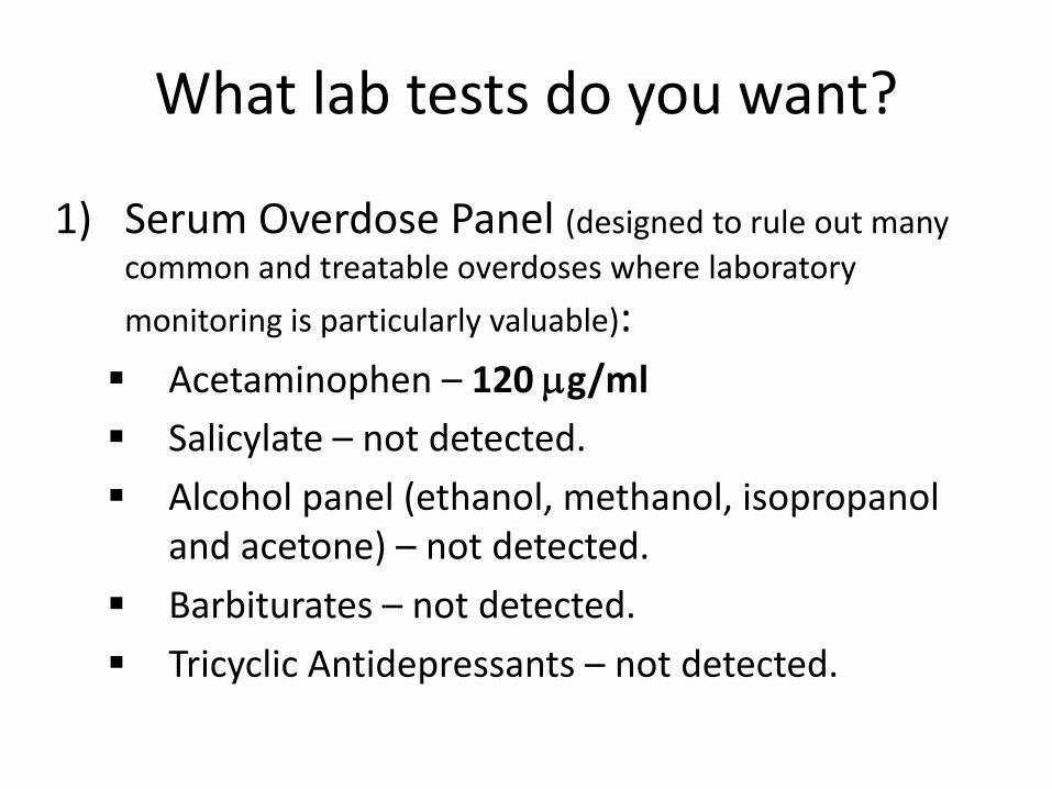
What lab tests do you want?
1) Serum Overdose Panel (designed to rule out many common and treatable overdoses where laboratory
monitoring is particularly valuable):Acetaminophen – 120 μg/mlSalicylate – not detected.
Alcohol panel (ethanol, methanol, isopropanol and acetone) – not detected.
Barbiturates – not detected.
Tricyclic Antidepressants – not detected.
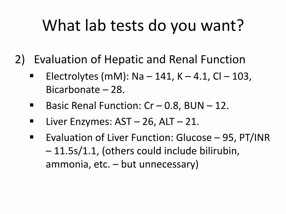
What lab tests do you want?
2) Evaluation of Hepatic and Renal FunctionElectrolytes (mM): Na – 141, K – 4.1, Cl – 103, Bicarbonate – 28.
Basic Renal Function: Cr – 0.8, BUN – 12.
Liver Enzymes: AST – 26, ALT – 21.
Evaluation of Liver Function: Glucose – 95, PT/INR – 11.5s/1.1, (others could include bilirubin, ammonia, etc. – but unnecessary)

What about the acetaminophen level of 120 μg/ml?
Therapeutic Range is 10 – 20 μg/ml.
Toxicity is expected for “acute” peak levels greater than 150 – 200 μg/ml.
But, you can’t expect to catch the peak drug level for overdoses. How do you deal with this?

What is his expected peak level?1) What information do you need?
Dose (13g), Patient Weight (70kg), Volume of distribution (~1 L/kg), Bioavailability (~60 – 98%, but in overdose tends to be on the higher side as metabolism reaches saturation).
2) How do you calculate a maximum peak level?95% of 13 grams is ~ 12 grams.Volume of distribution = 1 L/kg * 70 kg = 70 Liters.Peak Level = dose/Vd = 12g/70L = 171 μg/ml (multiplied by 1000 to convert g/L into μg/ml).
3) What factors might prevent the patient from actually achieving this theoretical maximum?
Delayed absorption due to delayed gastric emptying, formation of “concretions” and intestinal irritation.Ongoing elimination during delayed absorption (peak later and lower).Actual dose less than reported/anticipated.Unfortunate inter‐individual variability (often as much as 50 – 100%) in Volume of Distribution, Bioavailability, Elimination Half‐life, etc.Hence, although rough PK calculations useful, monitoring of serum drug levels remains essential.

Rumack‐Matthew Nomogram

What happens with our patient?
Treatment is initiated as a follow-up level at ~ 4 hours post-ingestion was > 150 mg/ml.
A series of levels measured every 4 hours is consistent with successful treatment as the half-life is less than 4 hours (green line represents ~ 2.5 – 3 hr half-life).

Treatment1) Generic
Decontamination: gastric lavage (if early) and activated charcoal (PO or via NG tube)Supportive: not indicated during Phase I, but relevant for other phases.
2) Antidote: N‐acetylcysteine (NAC)Maximal effectiveness if given in first 8 hours.
Given P.O. in the US ; may require an anti‐emetic as reputedly very noxious.
However, also demonstrates effectiveness is given after hepatotoxicity begins (i.e. Phase II).
Two mechanisms likely. First, replenishes reductive sulfation stores for detoxification of APAP and NAPQI. Second, may have a direct protective and/or regenerative effect on tissue, sometimes used in multi‐system organ failure due to other causes.
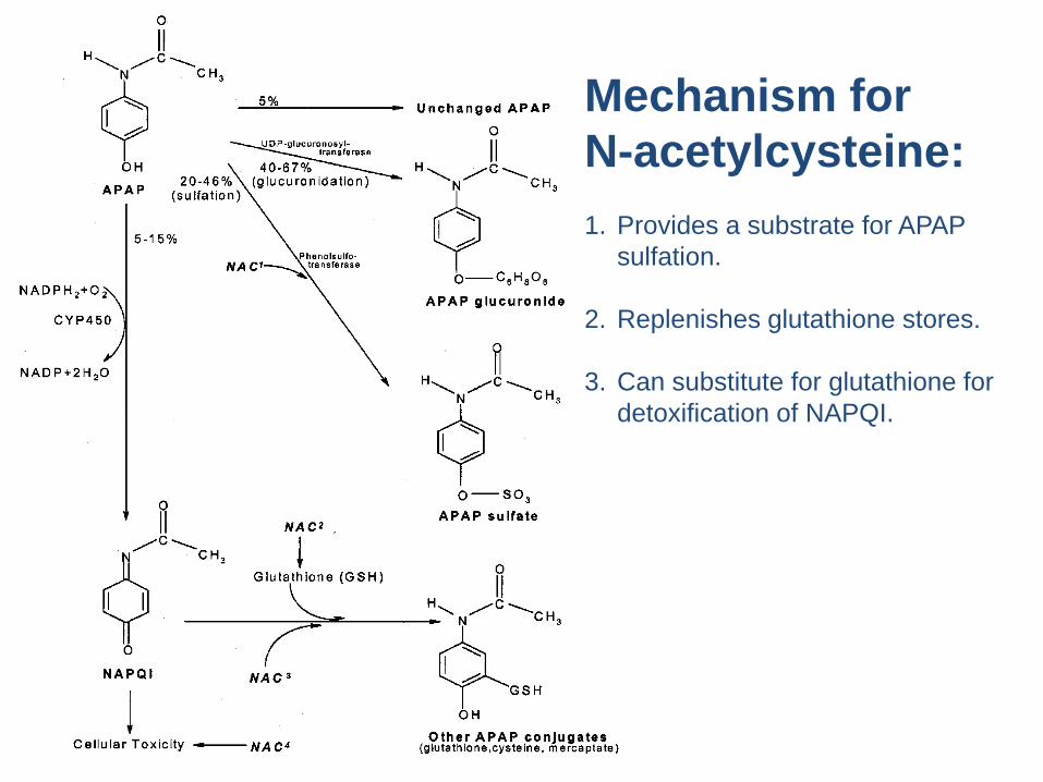
Mechanism for N-acetylcysteine:1. Provides a substrate for APAP
sulfation.
2. Replenishes glutathione stores.
3. Can substitute for glutathione for detoxification of NAPQI.

Tylenol Case #2
R.W. is a 31 y.o. woman with a PMH of major depression and alcohol abuse. She is currently in the process of divorcing an abusive husband and is separated from 8 y.o. son. Approximately 24 hours prior to presenting to the YNHH ED, the patient took approximately 48 tablets containing 500 mg Acetaminophen for a total dose of 24 grams. These were taken after approximately 8‐10 hours of heavy “binge” drinking by the patient.
She subsequently lost consciousness and upon awakening complained of nausea, vomiting and right upper quadrant abdominal pain.
Tylenol Case #2
Past Medical History: Significant only for major depression and alcohol abuse. She has had two previous suicide attempts and has been under treatment of a psychiatrist. However, she last visited her psychiatrist about one year ago and stopped taking her psychiatric medications at that time.
Medications: none
Allergies: none
Social History: Drinks ½ pint of hard alcohol per day and smokes tobacco occasionally. She has been a Jehovah’s Witness since 1986 and refused all blood products during the admission.

Physical Examination
• Vital Signs: Afebrile, BP 156/93, HR 90, RR 20, SaO2 98% (on room air)
• She is alert and oriented x 4 and in no acute distress.
• HEENT: PERRL, EOMI, no scleral icterus, oropharynx clear
• Neck: Supple, no lymphadenopathy, no carotid bruits, no JVD
• Heart: sinus tachycardia, regular rhythm, no G/M/R
• Lungs: clear to auscultation bilaterally
• Abdomen: soft, nondistended, mild RUQ tenderness
• Extremities: no asterexis, no C/C/E, no petechiae/ecchymosis
• Neuro: non‐focal
• Rectal: normal tone, heme negative
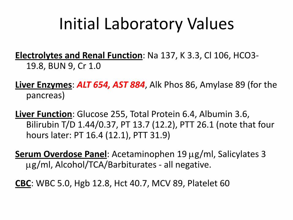
Initial Laboratory Values
Electrolytes and Renal Function: Na 137, K 3.3, Cl 106, HCO3‐19.8, BUN 9, Cr 1.0
Liver Enzymes: ALT 654, AST 884, Alk Phos 86, Amylase 89 (for the pancreas)
Liver Function: Glucose 255, Total Protein 6.4, Albumin 3.6, Bilirubin T/D 1.44/0.37, PT 13.7 (12.2), PTT 26.1 (note that four hours later: PT 16.4 (12.1), PTT 31.9)
Serum Overdose Panel: Acetaminophen 19 μg/ml, Salicylates 3 μg/ml, Alcohol/TCA/Barbiturates ‐ all negative.
CBC: WBC 5.0, Hgb 12.8, Hct 40.7, MCV 89, Platelet 60

One more example of using the Rumack-Matthew Nomogram…
A level measured ~ 24 hours after the overdose was 19 μg/ml.
Again, the red bar represents a degree of uncertainty in the timing (arbitrary choice of ± 4 hours).
The placement of the level in the region of “probable hepatic toxicity” is clear and also unnecessary as the patient has already elevated liver enzymes and early signs of hepatic failure.

Hospital Course
The patient did well and never became encephalopathic.
Of course, she received a complete treatment of N‐acetylcysteine (beginning ~24 hours after the overdose).
Her peak liver enzymes occurred about 36 hours after admission: AST 5870, ALT 5440.
She never developed signs of multi‐organ failure and maintained good ABGs, cardiac function, Cr, urine output, etc.
She was discharged on hospital day #4 with normal PT/PTT, AST 219, ALT 1860 and Bilirubin T/D of 0.64/0.26.

What was the role of ethanol?
• Chronic ethanol exposure induces the cytochrome P450 system, which increases the risk of toxicity. – Also, malnutrition depletes sulfation stores, further increasing risk.
• However, acute ethanol exposure (“binge drinking”) inhibits the cytochrome P450 system, which decreases the risk of toxicity.
• Overall consequence depends on the net balance of these two opposing effects.


All alcohols and glycols act on the CNS as a sedative‐hypnotic
• Respiratory depression (bradypnea)
• CNS depression (inebriation)
• Hypothermia
• Tachycardia and Hypotension
• Isopropanol is a much stronger CNS depressant than the others and induces coma at around 100 mg/dl.
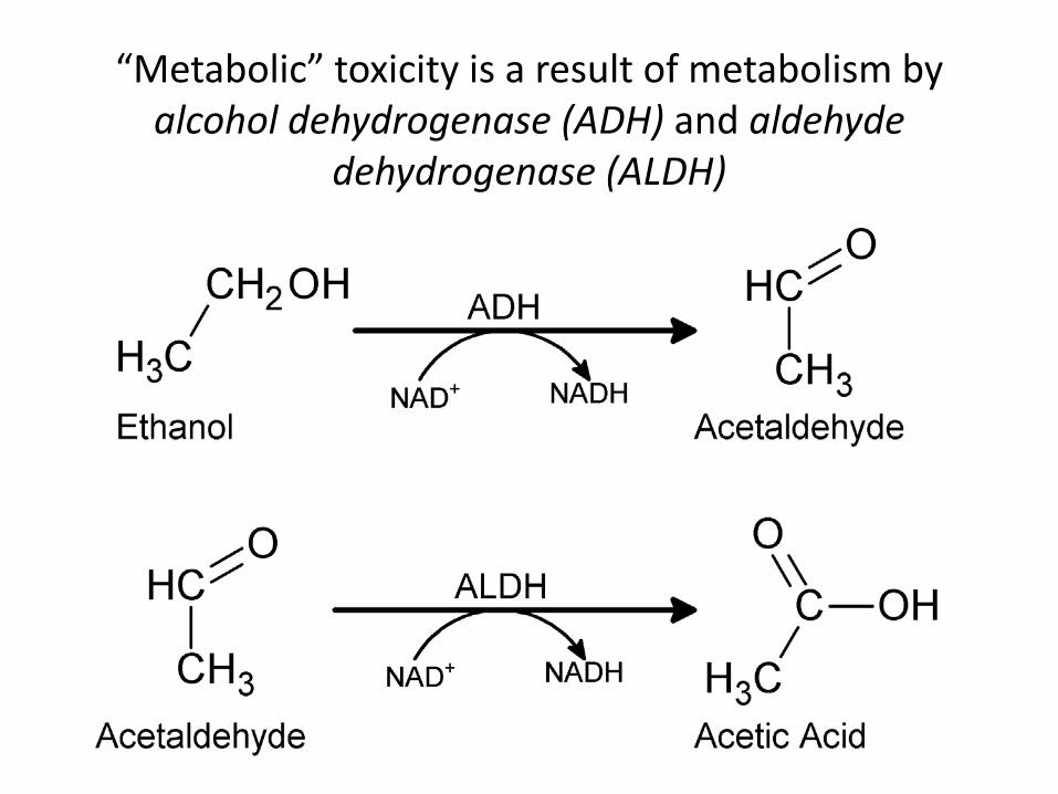
“Metabolic” toxicity is a result of metabolism by alcohol dehydrogenase (ADH) and aldehyde
dehydrogenase (ALDH)

Two Alternative Pathways for Alcohol Metabolism

Metabolism of Other Alcohols
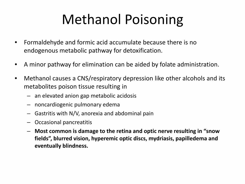
Methanol Poisoning• Formaldehyde and formic acid accumulate because there is no
endogenous metabolic pathway for detoxification.
• A minor pathway for elimination can be aided by folate administration.
• Methanol causes a CNS/respiratory depression like other alcohols and its metabolites poison tissue resulting in – an elevated anion gap metabolic acidosis
– noncardiogenic pulmonary edema
– Gastritis with N/V, anorexia and abdominal pain
– Occasional pancreatitis
– Most common is damage to the retina and optic nerve resulting in “snow fields”, blurred vision, hyperemic optic discs, mydriasis, papilledema and eventually blindness.

Ethylene Glycol MetabolismNote that the primary toxic metabolites are glycolic acid, glyoxylic acid, oxalic acid.
Precipitation of oxalic acid in tissues causes multisystem organ failure in untreated ingestions
Primary treatment is blockade of alcohol dehydrogenase with ethanol or fomepizole. Thiamine and pyridoxine are given therapeutically to detoxify glyoxylic acid and prevent its conversion into oxalic acid.

Ethylene Glycol Poisoning
• Three phases of toxicity: (1) CNS depression, (2) metabolic and cardiopulmonary compromise, and (3) renal damage.
1. CNS depression with persistent nausea, vomiting common and gradual onset of inebriation, lethargy and coma over the first 4 – 8 hours.
2. A profound metabolic acidosis develops after 8 – 12 hours often complicated by cardiopulmonary compromise (tachycardia, dysrhythmias, myocarditis, hyperventilation, pneumonitis and pulmonary edema common).
3. Although calcium oxalate crystals can be observed as early as 6 – 12 hours after ingestion, renal failure due to acute tubular necrosis is not common until after 24 – 48 hours.

Metabolism of Alcohols and Glycols Promotes Ketosis
• Alcohols and glycols are highly reduced molecules and their metabolism converts a LOT of NAD+ (ox.) to NADH (red.).
• A consequence of the resulting very high NADH/NAD+ ratio is promotion of ketosis.– Glycolysis and the Krebs cycle cannot proceed normally without available
NAD+.– Lactic acid production is stimulated by the high NADH/NAD+ ratio and,
thus, allows anaerobic metabolism of glucose through glycolysis.– Ultimately, hypoglycemia results from further limiting the Krebs cycle due
to consumption of carbohydrate intermediates such as oxaloacetate for gluconeogenesis.
– The combination of carbohydrate deficiency and the excess availability of acetyl CoA (from alcohol metabolism) promotes synthesis of acetoacetate and its conversion to β‐3‐hydroxybutyrate (which also driven by the high NADH/NAD+ ratio).

Acidosis from Alcohol/Glycol Metabolism
• Any sufficiently large alcohol/glycol consumption, especially in the face of pre‐existing malnourishment or glucose dysregulation can cause a mild lactic acidosis or ketoacidosis.
• Ethylene glycol and methanol ingestion result in the most serious acidosis because, besides the more general mechanisms for lactic acidosis and ketoacidosis,– their acidic metabolites (oxalic acid and formic acid, respectively)
accumulate to a high degree
– and these same metabolites are directly toxic to oxidative phosphorylation, which worsens the acidosis.
• Finally, note that as isopropanol is only metabolized to relatively non‐toxic acetone, it does not generate any acidic intermediates and only consumes ½ as much NAD+; thus, it is least likely to generate an acidosis, although mild ketosis is still seen.

Stuporous with blurred vision…
• An 84‐year‐old woman weighing 121 lb (55 kg) with no previous history of alcoholism was stuporous on presentation at the emergency department. Her family had found her obtunded and reported that she had complained earlier of blurred vision and had had one episode of emesis.
• On physical examination, blood pressure was 107/54 mm Hg, pulse rate 60 beats per minute, and respirations 16 per minute. The lungs were clear and heart rate was regular, with occasional premature beats. Funduscopic examination was unremarkable, and neurologic examination showed no evidence of focal deficits. The remainder of the examination was unremarkable.

Laboratory Values
• Na 146, K 4.2, Cl 107, HCO3‐ 14 (low)
• Anion Gap = 146 – (107 + 14) = 25• BUN 10, Cr 1.4, Glucose 148, Lactate 2.1• Liver Enzymes normal• Serum Ethanol < 10 mg/dl (enzymatic method)• ABG: pH 7.12, PaO2 71, PCO2 30• Measured serum osmolality = 354• Calculated Osmolality = 307.7• Osmolal Gap = 46.2• Urinalysis revealed calcium oxalate crystals.• CT scan of the head and lumbar puncture normal

Summary and Differential Diagnosis
• 84 y.o. woman with severe CNS depression, blurred vision, a metabolic acidosis with an elevated anion gap (possible respiratory contribution), an osmolal gap of 46.2 and a finding of calcium oxalate crystals in the urine.
• A toxic alcohol or glycol ingestion is highly suspected. Although blurred vision could suggest methanol poisoning, oxalate crystals in the urine is consistent with ethylene glycol.
• Note that with an osmolal gap of 46.2, expect a high ethylene glycol level (46.2 * 5.8 = 268).

An explanation…
• On further questioning, the family reports keeping a 2 L soda bottle containing antifreeze diluted with water in the kitchen.
• Suspect that patient’s poor baseline eyesight may have led to mistaken ingestion.
• Confirmed when a sample from the patient’s bedside drinking glass contained antifreeze and an ethylene glycol level was confirmed in the patient’s blood (217 mg/dl).

Treatment• Gastric lavage and oral activated charcoal.• 5% dextrose and sodium bicarbonate administered IV.• An intravenous IV loading dose of 10% ethanol given for a target of
100 mg/dl EtOH:– Vd = 0.54 L/kg * 55 kg = 29.7 L (297 dl)– (100 mg/dl * 297 dl)/(1000 mg/g * 0.7939 g/ml)– 37.4 ml of 100% EtOH or 374 ml of 10% EtOH
• A maintenance dose of 75 ml/hr was required to maintain a constant 100 mg/dl (frequently monitoring of EtOH level necessary).– (20 mg/dl/hr * 297 dl) / (1000 mg/g * 0.7939 g/ml)– Requires 7.5 ml/hr of 100% EtOH or 75 ml/hr of 10% EtOH
• Patient also given IV thiamine and pyridoxine to encourage non‐toxic metabolism of ethylene glycol.
• Because of extremely high ethylene glycol level, hemodialysis also performed to speed removal.

Pharmacogenetic Case ExampleHPI: 56 y.o. female, 140 lbs., presents with bleeding
gums, epistaxis, and bloody stools. Also describes excessive fatigue and mild chest pain on exertion.
PMH: Previously diagnosed with psoriasis and put on azathioprine (100 mg/day PO) about one month ago.
Lab: Hct 18%, Hb 6, WBC 800, Platelets 1,000: “Pancytopenia” – all blood cell levels strongly suppressed.
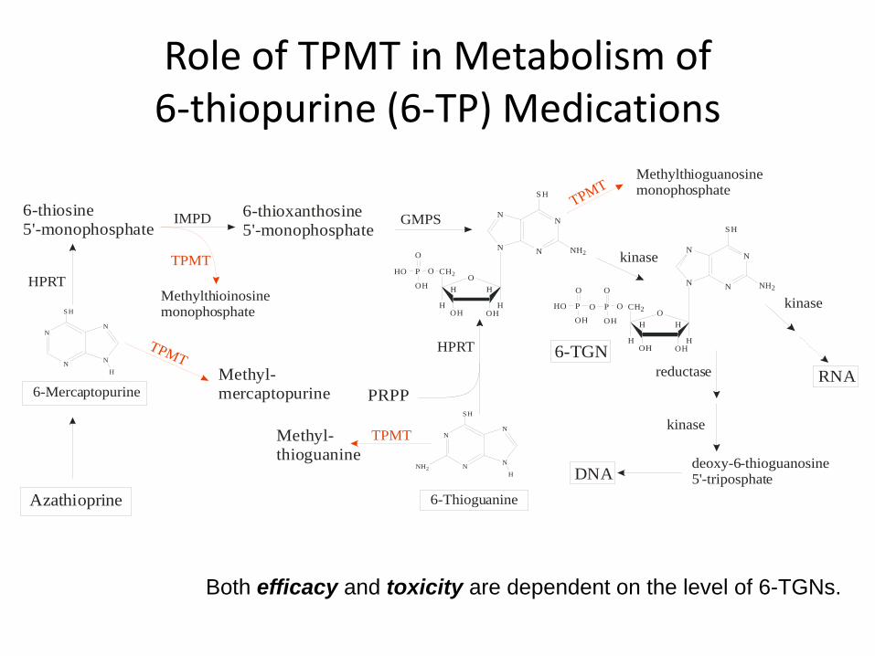
Role of TPMT in Metabolism of 6‐thiopurine (6‐TP) Medications
N
N
S H
N
N
H
N
N
S H
N
N
HNH2
N
N
N
S H
NH2N
O
OH
HH
H
CH2
HOH
OPHO
O
OH
N
N
N
S H
NH2N
O
OH
HH
H
CH2
HOH
OPO
O
OH
PO
OH
O
H
6-Mercaptopurine
6-ThioguanineAzathioprine
TPMT
TPMT
TPMT
TPMT
Methyl-mercaptopurine
HPRT
HPRT
IMPD GMPS
Methyl-thioguanine
PRPP
kinase
kinase
RNAreductase
deoxy-6-thioguanosine5'-triposphateDNA
Methylthioguanosinemonophosphate
Methylthioinosinemonophosphate
6-thiosine5'-monophosphate
6-thioxanthosine5'-monophosphate
kinase
6-TGN
Both efficacy and toxicity are dependent on the level of 6-TGNs.

Inter‐individual Heterogeneity in TPMT Activity Recognized by Weinshilboum over 20 Years Ago.

Relationship between Inherited Variations in TPMT Actvity and Serum Levels of (active) 6‐TGNs

6‐TP TDM and TPMT Testing
• Before 6‐TP drugs are started, TPMT genotype and/or RBC TPMT activity can be assessed and used to guide 6‐TP dosage.
• During treatment, RBC 6‐MP, 6‐MMP and 6‐TGN levels can be measured to guide therapy.

Pharmacogenetics
• Interindividual genetic variation in either– Drug Response (receptors, targets, etc.)
– Drug Metabolism
• Important current examples include, 6‐thiopurines, irinotecan, warfarin, succinylcholine and 5‐fluorouracil.
• Good Reference Sites– http://www.nigms.nih.gov/Initiatives/PGRN
– http://www.pharmgkb.org/

The Osmolal Gap• Osmolality should be measured using freezing point
depression (normal range: 275 – 295 mOsm/kg H2O).
• Calculated osmolality estimated usingOsmestimated = 2*[Na] + [BUN]/2.8 + [Glucose]/18
• Osmolal Gap = Osmmeasured – Osmestimated
• Normal range ~ ‐5 to + 10 mOsm/kg H2O
• The gap is a small difference between two large numbers and hence is very sensitive to any variations or inaccuracies in the measurements.
– Must be based upon measurements from a single blood draw

Causes of an Increased Serum Osmolal Gap
I. Real• Requires a large amount of a small (i.e. not a macromolecule),
neutral molecule. • There are really only three classes of molecules that cause an
elevated osmolal gap and can achieve a significant circulating level (without causing instant death):
i. Alcohols (and acetone)ii. Glycolsiii. Neutral Sugars (e.g. mannitol, sorbitol, glycerol)
(very rarely DMSO – rather toxic)
II. Artifact• Not from a single blood draw!• Artifactual hyponatremia due decreased serum free water from
hyperproteinemia or hypertriglyceridemia (can be corrected by measuring a “whole blood ionized” Na).
• Other unexpectedly high cations such as hypermagnesemia.

Relationship between osmolality and alcohol/glycol level
• Osmolality is in units of milliOsmoles per kg (or liter) of “free” water (note that serum and plasma contain ~ 93% free water).
• Alcohols and glycols are measured as milligrams per deciliter (mg/dL) of serum or plasma.
• Empirical conversion factors can be estimated as (Mol.Wt. / 10) * 0.93 {mg/dL per each 1 mOsm/kg H2O}
– Methanol 3.0
– Ethanol 4.3
– Ethylene Glycol 5.8
– Acetone 5.4
– Isopropanol 5.6
– Propylene Glycol 7.1

Caution: The osmolal gap is NOT a sensitive screen for toxic exposure!
• Anything over 25 mg/dl of ethylene glycol is considered severely toxic and hemodialysis is recommended.
• An ethylene glycol of 25 mg/dl contributes only ~4 mOsm/kg to the osmolal gap.
• Most people have a osmolal gap around 1 – 3 mOsm/kg (although there is a wide spread of normal values, up to 10 mOsm/kg).
• Hence, most people ingesting 25 mg/dl of ethylene glycol will still have a normal osmolal gap (3 + 4 = 7).
• For this reason, a normal osmolal gap does NOT rule out a toxic alcohol/glycol ingestion. On the other hand, a high gap does raise the suspicion of a toxic ingestion.
• Bottom line is that the BEST thing to do is to test for both alcohols and glycols whenever a reasonable clinical suspicion exist.
• Similarly, if a patient has clearly tested negative for both alcohols and glycols, yet still appears to have an elevated osmolal gap, it is probably due to a non‐toxic cause (DKA is an example).
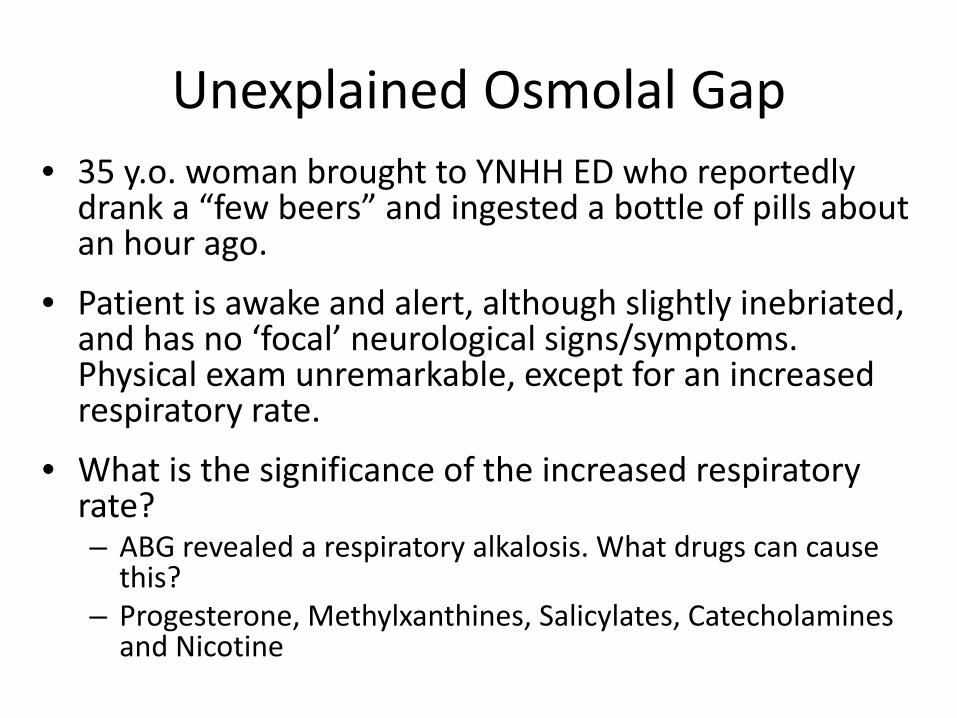
Unexplained Osmolal Gap• 35 y.o. woman brought to YNHH ED who reportedly drank a “few beers” and ingested a bottle of pills about an hour ago.
• Patient is awake and alert, although slightly inebriated, and has no ‘focal’ neurological signs/symptoms. Physical exam unremarkable, except for an increased respiratory rate.
• What is the significance of the increased respiratory rate?– ABG revealed a respiratory alkalosis. What drugs can cause this?
– Progesterone, Methylxanthines, Salicylates, Catecholamines and Nicotine

Immunoassay Results
Urine DAU panel• Barbiturate - NEG• Opiates - NEG• Methadone - NEG• Benzodiazepine - NEG• Cocaine - NEG• Amphetamine - NEG• PCP - NEG
“Serum” Overdose panel• Salicylates- POS
• 48 mg/dl
• Acetaminophen - NEG• Barbiturates - N/P• Alcohol - POS
• Ethanol @ 146 mg/dl
• TCA - N/P

Osmolal Gap
• Measured serum osmolality = 341 mOsm/kg H2O.
• Calculated serum osmolality =(2 * 140) + (8/2.8) + (115/18) = 289 mOsm/kg H2O.
• Osmolal Gap = 341 – 289 = 52 mOsm/kg H2O.
• Contribution of Ethanol to the osmolal gap = 146 / 4.3 = 34 mOsm/kg H2O.
• Unexplained osmolal gap = 52 – 34 = 18 mOsm/kg H2O.
• What should they do?

Test for Glycols by GC
• Ethylene glycol undetected.
• Propylene glycol = 120 mg/dl (high)
• Explains Osmolal Gap of 18 mOsm/Kg H2O:
– 120 mg/dl / 7.1 = 17 mOsm/kg H2O.
• Source?
– “Pet‐safe” antifreeze? – patient denies
– Iatrogenic: IV, PO medications (including oral activated charcoal!)
• Dangerous?
– Not normally. Converted to lactic acid and liver has a large metabolic capacity to convert lactic acid into alanine via the Cori cycle.
– Follow serum lactic acid levels for caution.

Propylene Glycol Metabolism

Iatrogenic Intoxication• 54 y.o. man admitted to a CT hospital for Ethanol withdrawal.
Given 1 g Ativan IV q6 hrs.
• Became unresponsive over next ~10 hours and required intubation due to respiratory depression. Transferred to ICU.
• During evaluation detected an osmolal gap of 144.
• Sample sent to YNHH for glycol testing.– Propylene glycol = 810 mg/dl
• Cause? – IV Ativan contains 4 mg/ml lorazepam in 80% propylene glycol. 1 g Ativan
would require 250 ml * 0.8 = 200 ml propylene glycol exposure over 6 hours.
– (200 ml*1.04 g/ml*103 mg/g) / (Vd=70kg*0.58 L/kg*10 dl/L)=512 mg/dl

DKA or alcohol ingestion?
• 35 year old woman with a history of poorly‐controlled type 1 diabetes and alcohol dependence presents with severe stupor, lethargy and smell of acetone on breath.
• On physical examination, the patient was – afebrile, with BP 125/65, HR 85 bpm, and RR 16,– lungs CTA, cardiac RRR no G/M/Rs,– funduscopic examination was unremarkable,– and a neurologic examination showed no evidence of focal deficits.

Initial Laboratory Values
• Na 138, K 4.0, Cl 108, HCO3‐ 22
• Anion Gap = 138 – (108 + 22) = 8 (normal)
• BUN 10, Cr 1.2, Glucose 350, Lactate 1.6
• Serum Ethanol < 10 mg/dl (enzymatic method)
• ABG: pH 7.35, PaO2 81, PCO2 35– (meant to represent a very mild acidosis)
• Urinalysis revealed positive “ketones”.
• CT scan of the head and lumbar puncture normal.

Additional Laboratory Values
• Serum Osmolality = 326 mOsm/kg H2O• Calculated Osmolality (Na, BUN, Glc)
= 299 mOsm/kg H2O.
• Osmolal Gap = 326 – 299 = 27 mOsm/kg H2O.
• Blood sample sent to outside lab for comprehensive alcohol/glycol panel by GC.

What to do?• Diabetic ketoacidosis not likely with glucose of 350 and lack of acidosis.
• Ketosis without significant acidosis argues against a toxic ingestion with either methanol or ethylene glycol.
• Degree of stupor cannot be explained by ethanol ingestion and considering the presence of an elevated osmolal gap and ketosis, must suspect isopropanol ingestion.
• However, very important to distinguish isopropanol from ethylene glycol or methanol ingestion (if possible) due to important question of whether or not to give ethanol drip or fomepizole (contraindicated with isopropanol).
• Isopropanol treatment is purely supportive with possible use of hemodialysis.
• If a comprehensive alcohol/glycol panel is not available, patient should be followed closely for signs of acidosis. Can also consider presumptive treatment for ethylene glycol/methanol poisoning (fomepizole or ethanol drip).
• Eventually, an isopropanol of 55 mg/dl and an acetone of 45 mg/dl was reported.