Embed Size (px)
Citation preview
© 2007 Nature Publishing Group
NEWS & VIEWS
nature nanotechnology | VOL 2 | DECEMBER 2007 | www.nature.com/naturenanotechnology 745
adam de la Zerda1,2 and Sanjiv S. Gambhir1,3
are in the 1Molecular Imaging Program at Stanford, Department of Radiology and Bio-X Program, the 2Department of Electrical Engineering and the 3Department of Bioengineering, Stanford University, Palo Alto, California 94305, USA.
e-mail: [email protected]
T he unique geometry of carbon nanotubes allows them to be used as drug-delivery vehicles or ‘nanocarriers’
in cancer therapy and other areas of medicine1. Nanocarriers work by bringing drugs directly to diseased areas of the body, thereby minimizing the exposure of healthy tissues while increasing the accumulation of the drug in the tumour area. This reduces both the dose necessary for treatment and the damage that can be caused to healthy tissue by powerful (and expensive) pharmaceuticals.
To convert a nanotube into a nanocarrier, it must be able to target tumours, and this ability could be introduced by attaching a peptide or an antibody to its outer surface — an approach that is already widely used in nanomedicine2. On administration of the nanocarrier to the body, the peptide or the antibody would bind to its target, and the drug — which could be inside the nanotube, or attached to it — would then be released. The release of drug molecules from a nanotube can be triggered by either a change in pH or by enzymes produced by the tumour that ‘cut’ the drug molecules from the nanotube.
These techniques would ensure that the nanotube reaches its target, but it is also important to know where the nanocarriers are in the body. Writing in Advanced Materials, Donglu Shi and co-workers at the University of Cincinnati, Shanghai Jiao Tong University, the University of Michigan and the Argonne National Laboratory report a significant step forward in this direction3. Shi and co-workers used a process called plasma surface polymerization to make novel nanostructures by combining quantum dots and multiwalled carbon nanotubes (Fig. 1). As carbon nanotubes are
not chemically reactive, this technique treats them with a variety of chemical compounds so that the quantum dots can attach. The new nanostructures combine the drug-delivery potential of the nanotubes with the fluorescence properties of quantum dots, which allow the position of the nanocarrier in the body to be monitored.
To demonstrate the utility of their approach, Shi and co-workers injected mice with varying amounts of their new nanostructures and found that the fluorescence signal was qualitatively
correlated to the amount injected. Moreover, they showed that the new nanoparticles were bright enough to be seen even when injected into the liver, kidney and leg muscle of a mouse. In other words, the fluorescence signal was strong enough to travel through several millimetres of tissue to reach the detector. The team focused on two types of quantum dots: CdSe/ZnS, which emit in the visible range, and InGaP/ZnS, which emit in the near-infrared and hence are better suited for deep-tissue imaging.
Combining the optical properties of quantum dots with the ability of carbon nanotubes to carry pharmaceutical cargos could prove highly beneficial in the field of drug delivery.
DruG DelIvery
Keeping tabs on nanocarriers
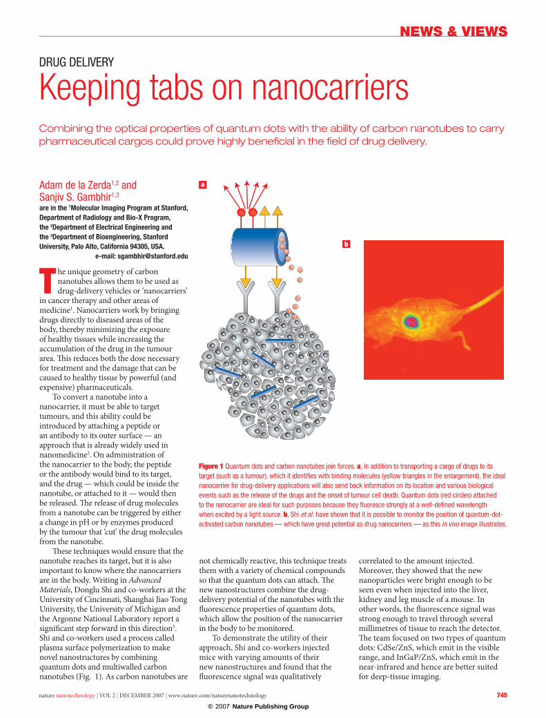
Figure 1 Quantum dots and carbon nanotubes join forces. a, In addition to transporting a cargo of drugs to its target (such as a tumour), which it identifies with binding molecules (yellow triangles in the enlargement), the ideal nanocarrier for drug-delivery applications will also send back information on its location and various biological events such as the release of the drugs and the onset of tumour cell death. Quantum dots (red circles) attached to the nanocarrier are ideal for such purposes because they fluoresce strongly at a well-defined wavelength when excited by a light source. b, Shi et al. have shown that it is possible to monitor the position of quantum-dot-activated carbon nanotubes — which have great potential as drug nanocarriers — as this in vivo image illustrates.

© 2007 Nature Publishing Group
NEWS & VIEWS
746� nature nanotechnology | VOL 2 | DECEMBER 2007 | www.nature.com/naturenanotechnology
Quantum dots have many advantages over organic fluorescent dyes: their fluorescence does not bleach over time and they can be accurately synthesized to emit light at any given colour in the visible and near-infrared regions. In drug-delivery applications, the physical location of the nanotubes is not the only parameter that would be useful to monitor — the binding of the nanotubes to the tumour, the release of the drug to the tumour environment and the biological response of the tumour to the drug would also be of interest.
Quantum dots are ideal for such multiplexing tasks. In the future, for example, a drug-loaded nanotube could be attached to three quantum dots, each emitting a different colour. The first dot would always be ‘on’, providing information on the physical location of the nanotube. The other two dots would initially be ‘off ’ (that is, quenched using standard techniques). However, the second dot could be designed so that the fluorescence turns on in the presence of a chemotherapeutic drug, so detection of the second colour would indicate that the drug had been released. Similarly, the third dot would be turned on by the presence of a protein that is excreted by the tumour during cell death, so detection of the third colour would indicate that the tumour is ‘responding’ to the drug. The possibility of one nanoparticle reporting on many biologically relevant events in parallel is a major goal in nanomedicine.
Although this work is a significant achievement, there are several issues that have to be addressed. It is usually desirable, for in vivo settings, to inject the nanoparticles intravenously, allowing them to find their way through the blood vessels to tumours. However, if the nanoparticles are too large, they are very likely to be removed from the blood pool by the reticuloendothelial system (for example, in the liver or spleen). The exact conditions under which this happens are not well characterized, but recent studies show that quantum dots smaller than 5.5 nm in diameter are unlikely to be removed in this way4.
The nanocarriers studied by Shi and co-workers are more than 200 nm in diameter and a few micrometres in length, and are therefore likely to encounter difficulties targeting tumours in a living subject. However, there are several options for decreasing the overall size of the nanoparticles to make them more useful in vivo. Whereas the multiwalled nanotubes used by the group had diameters between 70 and 150 nm, it might be possible to use single-walled nanotubes, which are only 1–3 nm across. It has recently been shown that single-walled nanotubes can carry drugs and target tumours5. It should also be possible to reduce the size of the quantum dots from about 40 nm to just a few nanometres across. Alternatively, commercially available organic fluorophores — which are less than 1 nm
in size — could replace the quantum dots, although their luminescence is much less bright and tends to get weaker with time.
Monitoring nanotubes without attaching any additional molecules (that is, with no quantum dots or fluorophores) is another possibility. Nanotubes have a very unique Raman signature, so they could be tracked with a Raman microscope. Nanotubes also exhibit high optical absorption, so monitoring them with a photoacoustic imaging instrument could also be an option. However, as with quantum dots and fluorophores, these approaches are limited to depths of a few centimetres. For whole-body imaging it would probably be necessary to attach radioactive labels to the nanotubes6 and use a positron emission tomography technique to monitor them.
Although some challenges still have to be overcome before nanotubes can be monitored optically in small living subjects, the work of Shi and co-workers is a good demonstration of this principle. The ability to image nanotubes this way could well be exploited in the next generation of biomedical research and nanomedicine.
references1. Bianco, A., Kostarelos, K. & Prato, M. Curr. Opin. Chem. Biol. 9,
674–679 (2005).2. Peer, D. et al. Nature Nanotech. 2, 751–760 (2007).3. Shi, D. et al. Adv. Mater. 19, 4033–4037.4. Soo Choi, H. et al. Nat. Biotechnol. 25, 1165–1170 (2007).5. Liu, Z., Sun, X., Nakayama-Ratchford, N. & Dai, H. ACS Nano
1, 50–56 (2007).6. Liu, Z. et al. Nature Nanotech. 2, 47–52 (2007).
adarsh Sandhuis at the Quantum Nanoelectronics Research Center, Tokyo Institute of Technology, and Nature Nanotechnology, Tokyo, Japan.
e-mail: [email protected]
Many biosensors take hours or even days to produce results, so there is a demand for devices that operate
on timescales of minutes. Such devices could, for example, have a major impact on the diagnosis of heart diseases by allowing at-risk patients to check for tell-tale signs of proteins by simply pricking a
finger and testing a small droplet of blood. A combination of ultrasensitive magnetic field sensors and magnetic nanoparticles is now showing potential as a perfect team to achieve these goals.
In these biosensors the nanoparticles are coated with chemical groups that will bind to the biomolecules that need to be detected. The nanoparticle–biomolecule complexes then react with ‘probe’ molecules that are fixed onto the surface of the magnetic sensors. Finally, the presence of the magnetic nanoparticles on the surface of the sensor produces an output signal.
Activity in this field began in 1998 when David Basalt and colleagues at the Naval Research Laboratory in Washington DC, showed that magnetic sensors could detect specific biomolecules1. This work triggered worldwide activity to develop magnetic biosensors that were faster and less expensive than existing devices. The current state-of-the-art in the field was on display at a recent conference organized by the European Science Foundation and the European Molecular Biology Organization2.
Next-generation magnetic biosensors will be able to detect extremely low concentrations of proteins and other biomolecules in very small samples within just a few minutes.
BIoSeNSING
new probes offer much faster results