Embed Size (px)
Citation preview

ORIGINAL RESEARCH ARTICLEpublished: 30 July 2013
doi: 10.3389/fnhum.2013.00412
Dreaming as mind wandering: evidence from functionalneuroimaging and first-person content reportsKieran C. R. Fox1*, Savannah Nijeboer1, Elizaveta Solomonova2,3, G. William Domhoff 4 andKalina Christoff1,5
1 Department of Psychology, University of British Columbia, Vancouver, BC, Canada2 Dream and Nightmare Laboratory, Center for Advanced Research in Sleep Medicine, Hôpital du Sacré-Coeur de Montréal, Montréal, QC, Canada3 Individual Studies, Université de Montréal, Montreal, QC, Canada4 Department of Psychology, University of California at Santa Cruz, Santa Cruz, CA, USA5 Brain Research Centre, University of British Columbia, Vancouver, BC, Canada
Edited by:
Wendy Hasenkamp, Mind and LifeInstitute, USA
Reviewed by:
Dimitrios Kourtis, Ghent University,BelgiumJennifer M. Windt, JohannesGutenberg-University of Mainz,Germany
*Correspondence:
Kieran C. R. Fox, Department ofPsychology, University of BritishColumbia, 2136 West Mall,Vancouver, BC V6T 1Z4, Canadae-mail: [email protected]
Isolated reports have long suggested a similarity in content and thought processes acrossmind wandering (MW) during waking, and dream mentation during sleep. This overlaphas encouraged speculation that both “daydreaming” and dreaming may engage similarbrain mechanisms. To explore this possibility, we systematically examined publishedfirst-person experiential reports of MW and dreaming and found many similarities: inboth states, content is largely audiovisual and emotional, follows loose narratives tingedwith fantasy, is strongly related to current concerns, draws on long-term memory,and simulates social interactions. Both states are also characterized by a relative lackof meta-awareness. To relate first-person reports to neural evidence, we comparedmeta-analytic data from numerous functional neuroimaging (PET, fMRI) studies of thedefault mode network (DMN, with high chances of MW) and rapid eye movement (REM)sleep (with high chances of dreaming). Our findings show large overlaps in activationpatterns of cortical regions: similar to MW/DMN activity, dreaming and REM sleep activateregions implicated in self-referential thought and memory, including medial prefrontalcortex (PFC), medial temporal lobe structures, and posterior cingulate. Conversely, inREM sleep numerous PFC executive regions are deactivated, even beyond levels seenduring waking MW. We argue that dreaming can be understood as an “intensified”version of waking MW: though the two share many similarities, dreams tend to belonger, more visual and immersive, and to more strongly recruit numerous key hubs ofthe DMN. Further, whereas MW recruits fewer PFC regions than goal-directed thought,dreaming appears to be characterized by an even deeper quiescence of PFC regionsinvolved in cognitive control and metacognition, with a corresponding lack of insightand meta-awareness. We suggest, then, that dreaming amplifies the same features thatdistinguish MW from goal-directed waking thought.
Keywords: dreaming, mind wandering, default mode network, first-person report, spontaneous thought,
neurophenomenology, memory consolidation, introspection
“The implication is that fantasy and dreams are part of a single con-tinuing fantasy process which is subject to certain transformationsimposed by physiological and stimulus events. It is unnecessary tosleep in order to generate dream-like ideation, and, apparently, itis unnecessary to be awake in order to produce relatively coherent,undream-like ideation”
–Eric Klinger (1971, p. 57).
INTRODUCTIONDreaming and daydreaming (or “mind wandering”) seem to havehad an enormous influence on human civilization through theages: they are alleged to have inspired René Descartes’s revolu-tionary view of the mathematical unity of nature (Baillet, 1691;Browne, 1977) and major scientific breakthroughs includingdiscovery of the Benzene ring by Kekulé (Benfey, 1958),formulation of the periodic table by Mendeleev (Strathern, 2000),
and Nobel prize-winning research on the chemical basis of neu-rotransmission by Loewi (1960)—to cite only a few examples.Psychological research into the subjective content of these stateshas revealed an intriguing, if less sensational, picture of dreamingand mind wandering (MW) as complex integrations of senso-rimotor imagery, emotions, memories, and future planning, inwhich problem-solving can also occur (Domhoff, 2003). Yet thesesimultaneously mundane and exceptional mental states remaindifficult to understand and study, in part because they are sub-jective and “spontaneous” in nature: undirected, unpredictable,and poorly characterized from both the personal and scientificperspectives.
Even after decades of scientific research, both behavioral andneurophysiological (reviewed in Hobson et al., 2000; Smallwoodand Schooler, 2006; Klinger, 2008; Kussé et al., 2010; Schredl,2010; Christoff et al., 2011; Gruberger et al., 2011; Zadra and
Frontiers in Human Neuroscience www.frontiersin.org July 2013 | Volume 7 | Article 412 | 1
HUMAN NEUROSCIENCE

Fox et al. Dreaming as mind wandering
Domhoff, 2011; Christoff, 2012), the sheer diversity of findingsand perspectives on dreaming and MW can be overwhelming.MW has been characterized as an unwelcome detriment to pro-fessional (Smallwood et al., 2011) and educational (Smallwoodet al., 2007) performance, as well as personal affect (Killingsworthand Gilbert, 2010), but has also been suggested to have anadaptive role in goal-directed planning (Christoff et al., 2009;Baird et al., 2011; Andrews-Hanna, 2012), deliberation on cur-rent concerns (Klinger, 1971, 2008), and creative insight (Bairdet al., 2012). Views on dreaming similarly span a broad spec-trum: dream mentation is considered by various researchers to beequivalent to brain delirium (Hobson, 1997) and schizophrenicpsychosis (Solms and Turnbull, 2002), or to be entirely epiphe-nomenal (Flanagan, 1995). Others, however, have seen in dream-ing a wellspring of individual growth and inspiration (Bulkeley,2010), a source of creativity, insight and problem-solving (Schredland Erlacher, 2007), an opportunity for emotional adapta-tion (Cartwright et al., 1998; Lara-Carrasco et al., 2009), andan expression and potential means of memory consolidation(Nielsen and Stenstrom, 2005; Wamsley and Stickgold, 2011).
Whereas specific beneficial (or conversely, disruptive) rolesremain largely speculative, however, similarities between both thesubjective and neurophysiological aspects of MW and dreaminghave recently been explored in some detail (Pace-Schott, 2007,2011; Christoff et al., 2011; Domhoff, 2011). In order to fur-ther address this question, we outline the general understandingof what dreaming and MW are, then discuss similarities in thesubjective experience and neural basis of both states. Finally, weconduct and compare meta-analyses of positron emission tomog-raphy (PET) and functional magnetic resonance imaging (fMRI)studies of dreaming (rapid eye movement or “REM” sleep) andMW (DMN activity) in order to examine potentially overlappingneural substrates.
WHAT ARE DREAMING AND MIND WANDERING?“Dreaming” is usually understood as subjective mental experi-ences during sleep. Although most famously (and strongly) asso-ciated with REM sleep (Aserinsky and Kleitman, 1953; Dementand Kleitman, 1957), dream-like thought is also reported duringother sleep stages (see Methods).
For several reasons, by “dreaming” we will generally be refer-ring to subjective reports drawn from REM sleep: for one thing,the majority of “dream” reports have been elicited from REMsleep-stage laboratory awakenings; further, only REM sleep showsa particularly strong correlation with dream mentation (∼80%of awakenings from REM sleep result in dream reports: Hobsonet al., 2000). For the purposes of the present paper, then, “dream-ing” refers to mentation reports from REM sleep.
“Undirected” thought is a similarly complex construct, andcan be divided into several different categories (Christoff, 2012).“Mind wandering” (MW) and “stimulus-independent thought”(SIT), for instance, are typically defined as thinking that devi-ates from a particular task a subject is meant to be completing(McGuire et al., 1996; Mason et al., 2007; Christoff et al., 2009).“Spontaneous thought,” on the other hand, is characterized ratherby its undirected, effortless nature—more akin to the everydayconcept of “daydreaming” (Singer, 1966; Klinger, 1990; Christoff,
2012); no particular task, or deviation from it, is required. Subtledifferences are apparent: MW, for example, might be initiateddeliberately (as when a subject decides to “tune out” during aboring task) rather than being “spontaneous.” Nonetheless, theseterms are often used interchangeably or with only minimal def-inition. Fluidity of terminology seems inevitable, however, in arelatively young field of inquiry (Christoff, 2012); moreover, thesubjective content and neural basis of these states appear highlysimilar (compare, e.g., Singer and McCraven, 1961; Christoffet al., 2004, 2009; Stawarczyk et al., 2011). We therefore use theseterms relatively interchangeably throughout this paper. MW,spontaneous thought, or daydreaming, then, all refer to subjec-tive reports of undirected thoughts during wakefulness (whetherdeviating from, or in the complete absence of, a task).
THE DEFAULT MODE NETWORK (DMN) AND REM SLEEPThough specific neural correlates of both daydreaming anddreaming remain somewhat elusive, these mental states, andtheir associated subjective content, are strongly correlated withthe “resting state” and REM sleep, respectively (Aserinsky andKleitman, 1953; Dement and Kleitman, 1957; Maquet et al., 1996;Mason et al., 2007; Christoff et al., 2009; Andrews-Hanna et al.,2010; Vanhaudenhuyse et al., 2010; Christoff, 2012; Hasenkampet al., 2012).
The default mode network (DMN) was discovered some-what serendipitously as a pattern of brain deactivations associatedwith the difference between brain activity during a quiet, rest-ing state (the typical baseline condition for early fMRI studies)and a goal-oriented, directed task (Raichle et al., 2001). Particularregions were consistently more active during “rest” than duringgoal-directed tasks of many kinds, suggesting a “default mode”network of regions active when a subject was “doing nothing”(Raichle et al., 2001; see Table 3 and Figure 2 for core regions ofthe DMN). It quickly became clear, however, that physical “rest”by no means implied mental inactivity. With no explicit task,subjects almost immediately engaged in spontaneous thought,including daydreaming, planning for the future, recalling mem-ories, and so on (Gusnard et al., 2001). Subsequent researchhas tied the subjective experience of MW to core DMN regions(Christoff et al., 2004, 2009; Mason et al., 2007; Andrews-Hannaet al., 2010; Vanhaudenhuyse et al., 2010; Hasenkamp et al., 2012).Although regions beyond the DMN appear to also be recruitedduring MW (e.g., Christoff et al., 2009), the DMN still remainsthe most commonly used neural proxy for spontaneous thought(see also Methods).
REM sleep is initiated by a network of cells in the pons andnearby portions of the midbrain (Siegel, 2011), but involves awidespread recruitment of higher cortical brain regions (see ourmeta-analytic results, below, for regions of this theoretical REMnetwork: Table 2 and Figure 1). REM sleep recurs, in increas-ingly lengthy periods, approximately every 90 mins throughoutthe sleep cycle, overall constituting about 1.5–2 h of an aver-age night of sleep. Whereas non-REM (NREM) sleep stages aregenerally characterized by deactivation of many regions as com-pared to wakefulness (e.g., Kaufmann et al., 2006), REM is uniquein that many brain regions are clearly more active than dur-ing wakefulness (Table 2, Figure 1). REM also appears to be the
Frontiers in Human Neuroscience www.frontiersin.org July 2013 | Volume 7 | Article 412 | 2

Fox et al. Dreaming as mind wandering
most active state from the subjective point of view, with longer,more emotional, and more frequent dream mentation in REMthan any other sleep stage (Hobson et al., 2000). REM thereforeappears to be by far the best neural marker of dreaming, thoughit nonetheless remains problematic (see Methods).
SUBJECTIVE AND NEURAL SIMILARITIES BETWEEN DREAMING ANDMIND WANDERINGA number of similarities in the subjective experience of dreamingand MW have previously been noted (see Section First-personReports of Content from Mind Wandering and Dreaming for adetailed overview). The possibility that the neural substrate ofthe DMN might be involved in, overlap with that of dream-ing/REM sleep has also been raised (Fosse and Domhoff, 2007;Pace-Schott, 2007, 2011; Ioannides et al., 2009; Nir and Tononi,2010), but these comparisons too have remained qualitative: aquantitative meta-analysis has yet to be applied to the question ofthe similarity in neural substrates between DMN/MW and REMsleep/dreaming. While major reviews and meta-analyses of theDMN have allowed for a tentative consensus regarding its neu-ral basis (e.g., Buckner et al., 2008), a meta-analytic evaluation ofbrain activity during REM sleep has yet to be undertaken, mak-ing a direct comparison between brain activity in the two statesdifficult. The execution of such a meta-analysis of REM sleep wastherefore a major goal of the present review.
AIMS OF THE PRESENT REVIEW AND META-ANALYSISHere we aim to build on prior qualitative comparisons of boththe experiential and brain basis of dreaming and spontaneousthought with a more definitive, quantitative assessment of thesimilarity in brain activity. A strong reliance on first-personreports of subjective experience has guided much research onboth MW and dreaming, and led to breakthroughs in the under-standing of their respective neural correlates. Accordingly, wepresent a detailed discussion of first-person content reports fromboth states in Section First-person Reports of Content from MindWandering and Dreaming. We outline our methods of meta-analysis of functional neuroimaging data in Section Methods.In Section Neuroimaging of Mind Wandering and Dreaming:Meta-analytic Results, we meta-analyze results from functionalneuroimaging (PET) studies of REM sleep (see Methods). Wecompare these results to an authoritative meta-analysis of DMNregions (Buckner et al., 2008) to determine to what extent theneural substrate of REM sleep overlaps with that of the DMN.Finally, we present a discussion of findings, limitations, and futuredirections, and propose a preliminary model of dreaming as“intensified” mind wandering.
FIRST-PERSON REPORTS OF CONTENT FROM MINDWANDERING AND DREAMINGSimilarities in subjective content have been noted since the begin-ning of such research. For instance, the dreamlike nature ofrelaxed waking thought was documented in two early studiesof what is now called MW, which were carried out in a sleeplaboratory using EEG to monitor wakefulness. In both studies,participants were randomly asked to report anything that wasgoing through their minds at the time of the probe. In the first
study, Foulkes and Scott (1973) found that 24% of thoughts couldbe categorized as visual, dramatic, and dreamlike. In a replicationstudy, Foulkes and Fleisher (1975) discovered that 19% of reportswere dreamlike.
The qualitative characteristics of dreaming have been inten-sively studied over the past century, yielding a considerable bodyof research from which some firm conclusions can be drawnregarding subjective content. Though qualitative data on the con-tent of MW is not nearly as comprehensive, a tentative overview isnonetheless possible. Although a comprehensive review of the lit-erature is beyond the scope of this article, we highlight consistentfindings regarding the subjective content of dreaming and MW.We focus on similarities in subject matter across several key areas,including sensory, emotional, fanciful, mnemonic, motivational,and social aspects, as well as addressing the presence or absenceof cognitive control and metacognition. Various disparities andinconsistencies are addressed here, as well as in the Discussion.
SENSORY ASPECTSThe broadest similarity between dreaming and MW is perhapsalso the most basic: the sensory building blocks of spontaneousthought in both waking and dreaming are overwhelmingly visualand auditory (though experiences in other sensory modalities areby no means precluded).
DreamingThe largely audiovisual nature of dreaming was noted overtwo millennia ago by Artemidorus in his Oneirocritica (Harris-McCoy, 2012) and has been often replicated in contemporaryresearch. For instance, a recent review of dream content (Schredl,2010), based on more than 4000 dream reports from both lab-oratory awakenings and home dream diaries, found that visualcontent was present in 100%, and auditory content in ∼57%,of all reports (Table 1). Other sense modalities (tactile, olfactory,gustatory, and nociceptive experiences), by contrast, were presentin ∼1% or less of all reports. Indeed, the next most prominentmodality after vision and audition was the vestibular sense: ∼8%of reports contained experiences of flying, floating, acceleration,etc. (Schredl, 2010). Intriguingly, a comparison with studies ofdream reports from more than a century ago shows a very similartrend: in the late nineteenth century, dream reports also almostalways featured visual elements, followed by auditory imagery asthe next most dominant aspect, and with the remaining senses
Table 1 | Sensory perception in dreaming.
Modality Frequency (% of reports)
Visual 100
Auditory 57
Vestibular ∼8
Tactile ∼1
Gustatory ∼1
Olfactory ∼1
Pain ∼1
Based on Schredl (2010).
Frontiers in Human Neuroscience www.frontiersin.org July 2013 | Volume 7 | Article 412 | 3

Fox et al. Dreaming as mind wandering
accounting for very small percentages (∼1–7%) (Schwartz, 2000).This suggests that the sensory aspects of dreaming may be consis-tent cross-culturally (or at least, cross-temporally).
The apparent predominance of audio-visual content in dreamsmay underestimate other sensory modalities, however. A num-ber of studies sampling other sensory data revealed that, whenprompted specifically for sensations such as pain (Nielsen et al.,1993; Raymond et al., 2002; Solomonova et al., 2008) or bod-ily orienting movements (Solomonova et al., 2008), participantsoften reported more information. To our knowledge, similartargeted sensory-content probes have not yet been undertakenduring MW, precluding a more detailed comparison.
Mind wanderingContent findings from mind wandering are not usually directlycomparable, since MW researchers have tended to focus on theintensity (rather than the prevalence) of audiovisual imagery,but available evidence suggests similar trends. For example, fac-tor analysis of nearly 1500 experience reports found that visualand auditory intensity are two of eight dimensions significantlycharacterizing spontaneous thoughts (Klinger and Cox, 1987).A more recent study similarly found a very high prevalence ofself-reported visual and auditory imagery during spontaneousthoughts (mean ratings of 4.22 and 4.02, respectively, on a 7-point Likert scale) (Stawarczyk et al., 2011). Along these lines, arecent review concluded that the average spontaneous thought ismoderately visual, contains at least some sound, and is very likely(74% of reports) to contain some form of interior monolog or“self-talk” (Klinger, 2008).
POSITIVE AND NEGATIVE EMOTIONALITYDreamingIt appears that most dreams (∼70–75% or more in adults) con-tain some emotion, though affect in dreams may not always beparticularly strong, or appropriate to the context (see Domhoff,2011, for a discussion). A number of studies have found a rela-tive predominance of negative emotions in dreams, particularlywhen dreams are scored by judges rather than by dreamers (seeSchredl, 2010, for a review). Other studies, however, have found abalance of emotions in REM sleep dream reports, and one study(Fosse et al., 2001) found that joy/elation was in fact the most fre-quently reported emotion. An interesting study directly comparedself-reports of dreaming vs. waking events, finding that negativeemotion (particularly fear) was more prevalent during dreaming,and positive emotions more common in waking (Nielsen et al.,1991).
It may be, however, that more intense and negativelytoned dreams are better remembered, and thus over-reported.Additionally, sampling techniques (e.g., laboratory awakeningsvs. home dream journals) may contribute to differences infindings. Irrespective of these differences and methodologicallimitations, however, it is evident that both positive and negativeemotions are ubiquitous during dreaming.
Mind-wanderingThough not yet extensively studied, emotion appears to be sim-ilarly ubiquitous during MW. One recent study, for instance,
involving thousands of reports, found that the majority (69%)of spontaneous thought reports involved emotion (positive emo-tion in 42.5% of reports, negative emotion in 26.5%), whereasonly 31% of reports were reported to be emotionally neutral(Killingsworth and Gilbert, 2010). Though data are generallylacking, it is interesting to note that, in contrast to dream-ing, positive emotion appears to predominate during wakingMW, and that many more waking spontaneous thoughts appearto be characterized by relatively flat (neutral) affect. Also ofinterest is that the temporal focus of MW content appearsto be more directed toward the past when negative moodhas been experimentally induced (Smallwood and O’Connor,2011).
IMPLAUSIBILITY AND BIZARRENESSThough the typical spontaneous thought or dream is a rel-atively plausible simulation or elucidation of past memories,current events, or future plans, generally in line with the cur-rent concerns of the subject (see “Motivational Aspects,” below),nonetheless implausible and bizarre elements are common toboth states—though their precise frequency remains disputed(Snyder, 1970; Dorus et al., 1971; Zadra and Domhoff, 2011).Examples are physically impossible or socially unlikely situa-tions, fanciful locales and characters, large discontinuities of timeand/or space, and so on.
DreamingDepending on scoring criteria, it has been estimated that between32% (Schredl, 2010) and 71% (Stenstrom, 2006) of dream reportsfeature bizarre or impossible elements. Despite widely varyingestimates, however, there is general agreement that bizarre, incon-gruous or impossible elements are features of at least a substantialproportion of dreams. Differences in precise estimates are likelydue to differing scoring procedures, as well as differences betweendreamer- or judge-rated scores.
Mind-wanderingThough many MW episodes contain relatively realistic simula-tions of plausible events in the external world, nonetheless asubstantial number (∼20% of reports) contain elements thatare bizarre, implausible, or fanciful (defined as “departing sub-stantially from physical or social reality”) (Klinger and Cox,1987; Kroll-Mensing, 1992; Klinger, 2008). A more recent studyhas provided a general replication of earlier results: analyzingthousands of thoughts reported by 124 subjects, Kane et al.(2007) found that the average thought during MW containeda moderate level of fantasy (a mean of 3.77 on a 7-pointscale).
In a rare study examining both waking fantasy and dreamreports in the same 12 subjects, Williams et al. (1992) foundthat bizarre elements were about twice as prevalent in dreamsvs. waking spontaneous thought. In a similar vein, dream anddaydream bizarreness have been studied in relation to “thick” vs.“thin” boundaries (Kunzendorf et al., 1997): though thin bound-ary personality was associated with more bizarre dreams anddaydreams than thick boundary, dreams were scored more bizarrethan daydreams across both personality types.
Frontiers in Human Neuroscience www.frontiersin.org July 2013 | Volume 7 | Article 412 | 4

Fox et al. Dreaming as mind wandering
MNEMONIC FEATURES: CONTRIBUTIONS OF EPISODIC ANDSEMANTIC MEMORYBoth dreaming and MW draw on episodic and semantic mem-ory sources as building blocks for novel subjective experiences. Inthis section we discuss the prevalence of past-oriented thoughtsduring both wakefulness and dreaming, and the potential contri-butions of both episodic and semantic memory to these states.
DreamingThere is an intriguing literature suggesting that sleep, especiallyNREM sleep, may have a role in memory consolidation (Walkerand Stickgold, 2006; Born and Wilhelm, 2012), including specificroles for REM sleep in consolidation of procedural (Smith et al.,2004) and emotional episodic (Nishida et al., 2009; Groch et al.,2013) memories. A dynamic model of sleep-dependent memoryconsolidation and reconsolidation has recently been proposed,suggesting a complex relationship between sleep stages, memorytypes and their contribution to cognitive stability, flexibility andbrain plasticity (Walker and Stickgold, 2006, 2010).
It is now well documented that dream content borrows fromboth temporally proximal and distal memories (Nielsen andStenstrom, 2005). The most proximal memories (those fromthe previous day) are generally known as “day residue” (Freud,1908), whereas the recurrence of elements 5–7 days followingan experience is referred to as the “dream-lag” effect (Nielsenand Powell, 1989). Personally relevant and emotionally salientevents appear to manifest themselves in dream content as dayresidue and dream lag effects, but can also surface many yearsafter initial encoding (Grenier et al., 2005). The presence of emo-tional and personally relevant content in dreams may be relatedto the fact that emotional and impactful events are preferentiallyconsolidated in memory (McGaugh et al., 2002; Nishida et al.,2009). While dreaming contains clear episodic autobiographicalelements, memories only rarely get “replayed” in dream content(∼1–2% of reports: Fosse et al., 2003).
Mind-wanderingMW appears to involve roughly equal percentages of thoughtsabout the past and future (Fransson, 2006), though some stud-ies suggest a “prospective bias” toward future-oriented thoughts(Smallwood et al., 2009; Andrews-Hanna et al., 2010; Stawarczyket al., 2011), and also a past-bias inducible by negative mood(Smallwood and O’Connor, 2011). Overall, however, it is clearthat memories, particularly episodic ones, play a large role inspontaneous thought. Many studies have reported a high preva-lence (∼20% or more of reports) of past-focused MW (Fransson,2006; Smallwood et al., 2009; Andrews-Hanna et al., 2010;Smallwood et al., 2011). Indeed, one of the first studies to explore“resting state” activity using PET noted the similarities betweensuch activity and episodic memory recall, as well as the fact thatsubjective reports of “rest” actually involved a large amount ofpast recollection and future planning (Andreasen et al., 1995).Similar to dreaming, memories incorporated in waking MW tendto be of emotional and personally relevant material, and areoften related to people’s current concerns (see section below on“Motivational Aspects”).
In summary, dreaming and MW both contain specific trace-able episodic and semantic memory sources, but very rarely
reproduce memories in their entirety. Rather, memories tendto reappear in novel, re-contextualized thoughts and scenarios(Nielsen and Stenstrom, 2005).
MOTIVATIONAL ASPECTS: CURRENT CONCERNSReports from both dreaming and MW show a strong proclivityto reflect the ongoing concerns of subjects, as well as elements ofanticipating and planning for the future.
DreamingA wealth of data supports the notion that dreaming reflects ongo-ing waking concerns, desires, and experiences, in line with the“continuity hypothesis” of dreaming and waking mental activity(see, e.g., Domhoff, 1996, Ch. 8). For example, transient stress-ful situations, such as divorce (Cartwright et al., 1984) and grief(Kuiken et al., 2008) are also often present in dream reports in ageneral form.
Although dream content is often found to be thematically andemotionally consistent with the waking state of the dreamer, cer-tain activities prevalent in waking are only rarely found in dreams.These include cognitive activities such as reading, writing, andusing a phone or a computer (Schredl, 2000).
Mind-wanderingSimilar to dreaming, the content of waking MW also centers heav-ily on subjects’ current concerns (Klinger and Cox, 1987; Klinger,2008; Andrews-Hanna, 2012).
Further, when the temporal focus of MW is examined, a largepercentage (∼40% in one recent study: Andrews-Hanna et al.,2010) of spontaneous thoughts center around the present time±1 day, supporting the notion that MW strongly involves currentconcerns and experiences. Future-oriented thought is also incred-ibly common during MW (Smallwood et al., 2009; Andrews-Hanna et al., 2010; Stawarczyk et al., 2011), further supporting arole for MW in future-planning and potentially problem-solving.Intriguingly, in one of the few neuroimaging studies to directlyexamine periods of MW, MW was associated with activationsnot only in the DMN but also in key executive prefrontal areas,including the dorsal anterior cingulate cortex and dorsolateralprefrontal cortex (Christoff et al., 2009). Such results are con-sistent with the prevalence of current concerns and unresolvedissues in first-person content reports, and may reflect an ongoing(if unconscious) effort to address them (Christoff et al., 2009; seealso Discussion).
IMAGINED SOCIAL INTERACTIONDreamingSimilar to waking life, dreaming is nearly always organizedaround interactions with others. Most dreams include othercharacters in some kind of relationship with the dreamer, ora generalized social situation (Hall and Van de Castle, 1966;Nielsen et al., 2003; Schredl et al., 2004; Zadra and Domhoff,2011). Social interactions in dreams follow a multitude ofpatterns, including threatening (Valli et al., 2005) and other-wise emotionally-charged situations (Cartwright et al., 1984).Occasionally, recognizable dream characters may change appear-ance or appear as a generalized entity, fused with features of
Frontiers in Human Neuroscience www.frontiersin.org July 2013 | Volume 7 | Article 412 | 5

Fox et al. Dreaming as mind wandering
other individuals. Also of interest is the prevalence of “mental-izing” or use of “theory of mind” in dreaming—i.e., thinkingabout others’ thoughts, emotions and motivations (even thoughthe “others” are of course merely imagined) (McNamara et al.,2007). In general, meaningful interactions with others may beone of the key factors guiding the progression of the dreamnarrative.
Mind wanderingFirst-person reports of MW often involve imagined social inter-actions with others, as well as thoughts about the intentions andbeliefs of other people (Klinger, 2008). This has led to the generalnotion that “mentalizing” (i.e., thinking about the thoughts andminds of others) and the consideration of hypothetical social situ-ations may be key components of spontaneous thought (Buckneret al., 2008; Andrews-Hanna, 2012). Supporting this idea, numer-ous studies have found that brain activity underlying “theory ofmind” and mentalizing overlaps significantly with DMN regions(see Buckner et al., 2008, for a review).
COGNITIVE CONTROL AND METACOGNITIONDreamingA singular aspect of dreams is the seemingly total lack of metacog-nitive awareness in the dream state. One experiences a com-plex simulation of oft-bizarre experiences, but without the overt
capacity to reflect on the bizarre state of affairs the mind andbody are actually in see, e.g., Rechtschaffen (1978). Intriguingly,it appears that well-trained, or talented, individuals can developmetacognitive awareness of the dream state, becoming “lucid” inthe dream and sometimes even directing its course and content(Dresler et al., 2012). The exceptional nature of “lucid” dream-ing, however, serves to prove the rule of the general lack of controland metacognitive awareness in ordinary dreaming, a character-istic likely attributable to the deactivation of numerous prefrontalcortical regions during REM sleep (see our results in Table 2 andFigure 1; also Hobson et al., 2000; Muzur et al., 2002).
Mind wanderingA lack of explicit goals, and an unawareness that one is evendaydreaming or has deviated from the task at hand, are typi-cal of MW (Schooler et al., 2011). But although MW tends tobe less characterized by intentional thought and self-reflectiveawareness, this is not always the case. A recent study from ourgroup, for instance, found that subjects who were probed at ran-dom intervals reported being unaware that they had been mindwandering about half (45%) of the time. One’s impression ofthe “controllability” of a segment of MW also varies widely,from a sense of being able to end it at any time, to being com-pletely absorbed in and swept along by a daydream (Klinger, 1978,2008; Klinger and Cox, 1987; Kroll-Mensing, 1992; Klinger and
Table 2 | Core cortical components of the neural network underlying REM sleep.
Region Cluster size (mm3) Talairach coordinates (x, y, z) [BA]
ACTIVATIONS (REM > WAKING REST)
Cortical regions
Medial prefrontal cortex 368 2, 32, 2 [Area 24]
Posterior cingulate cortex/lingual gyrus 656 28, −66, 4 [Areas 19, 30]
Parahippocampal cortex 1088 24, −40, −10 [Areas 36, 37]
416 −16, −26, −18 [Area 35]
Parahippocampal/entorhinal cortex 104 18, −30, −6 [Areas 28, 35]
Posterior parahippocampus/lingual gyrus 496 −18, −50, −8 [Area 19]
352 22, −58, −6 [Areas 19, 36]
Entorhinal cortex/hippocampus 360 22, −18, −14 [Areas 28, 35]
Subcortical regions
Pons/midbrain 688 8, −14, −18
Caudate nucleus 472 −6, 16, 10
DEACTIVATIONS (REM < WAKING REST)
Cortical regions
Mid/posterior cingulate 752 −8, −34, 28 [Area 23]
Rostrolateral prefrontal cortex 456 32, 44, 20 [Area 10]
Inferior frontal gyrus 296 −46, 26, −2 [Areas 47, 45]
Orbitofrontal cortex 256 −32, 38, −10 [Area 11]
224 38, 36, −12 [Area 11]
120 18, 46, −14 [Area 11]
Superior longitudinal fasciculus 176 28, −42, 20
Peak cortical foci of likely activation and deactivation from a meta-analysis of all functional neuroimaging (PET) studies of REM sleep compared to a baseline of
waking rest. Notably, every cortical cluster of activation overlaps (convergences in bold font) with a core component of the DMN, except for one cluster in left lingual
gyrus [Area 19] (compare with Table 3 and Figure 2). Conversely, significant clusters of deactivation overlap with DMN regions in only one case out of seven. The
cluster labeled as in superior longitudinal fasciculus is approximate only. BA, Brodmann area; DMN, default mode network; PET, positron emission tomography;
REM, rapid eye movement.
Frontiers in Human Neuroscience www.frontiersin.org July 2013 | Volume 7 | Article 412 | 6

Fox et al. Dreaming as mind wandering
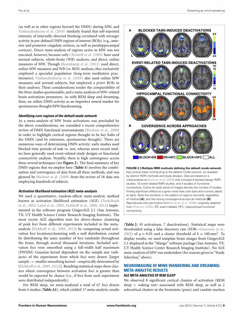
FIGURE 1 | Neural substrate of REM sleep vs. waking rest. Significantmeta-analytic clusters contributing to the neural substrate of REM sleep (asa proxy for dream mentation). Axial slices are displayed in Talairach space,with 3 mm skip. Color bars indicate likelihood that peaks represent actualpeaks of difference at a given voxel. Activations (REM > waking rest) are inred-yellow, deactivations (REM < waking rest) in blue-green.
Kroll-Mensing, 1995). Collectively, these results suggest that cog-nitive control and metacognitive awareness in MW lie somewherebetween the relative lucidity and self-reflectiveness of normalwaking thought and behavior, and the near-total lack of con-trol and metacognitive nescience characteristic of regular (i.e.,non-lucid) dreams. See the Discussion for an elaboration of thistheme.
METHODSSTUDY SELECTION FOR NEUROIMAGING META-ANALYSISDreamingThough the two phenomena have often been seen as synony-mous since Aserinsky and Kleitman’s discovery of the associ-ation between the REM sleep and dreaming (Aserinsky andKleitman, 1953), dream-like mental activity occurs in all sleepstages (Nielsen, 2000), including briefly at sleep onset (NREM1)(Mavromatis, 1987; Hori et al., 1994; Nielsen et al., 2005) aswell as in NREM2 (Antrobus et al., 1995; Fosse et al., 2004),particularly later in the night (Cavallero et al., 1992). Mentationfrom NREM3/4 sleep, also known as Slow Wave Sleep (SWS), hasalso been reported, albeit more rarely (Cavallero et al., 1992).In line with the markedly different patterns of brain activitythroughout the sleep cycle (Kaufmann et al., 2006), the length,
bizarreness, and emotionality of dream reports from varioussleep stages appear to differ significantly, though these dispar-ities remain controversial, with some researchers arguing thatthe important issue is level of cortical activation, not sleep stage(Antrobus et al., 1995; Cicogna et al., 1998; Foulkes, 1999).
There is also strong evidence from neuropsychological lesionwork that the neural mechanisms underlying REM sleep anddreaming are doubly dissociable (Solms, 1997, 2000, 2011;Oudiette et al., 2012). Nonetheless, we use neuroimaging stud-ies of REM sleep only as a neural proxy for the brain basis ofdreaming in the present study, for several reasons: (1) NREM1, farfrom being a uniform state, can be subdivided into at least 8 sub-stages (Hori et al., 1994). Hallucinatory, dream-like mentationis only strongly associated with particular sub-stages, especiallythose with strong EEG theta rhythms (Hori et al., 1994). To ourknowledge, however, no neuroimaging study has yet examinedthese brief epochs in isolation. Data collapsed across all phasesof NREM1, then, is an unsuitable neural marker for dream-likementation. (2) Of the few functional neuroimaging studies ofsleep, NREM2 sleep is rarely explicitly divided into early and latestages based on the ultradian changes in its EEG microarchitec-ture (Roth and Roehrs, 2000). Since only late NREM2 is evenmoderately correlated (∼0.40) with dream mentation (Nielsen,2000), data collapsed across all phases of NREM2 (which is allthat is currently available) is likewise unsuitable. (3) Despitethe apparent dissociability of REM sleep and dreaming, the tworemain extremely highly correlated, with roughly 70–90% ofawakenings from REM sleep yielding dream reports (∼83% onaverage: Nielsen, 2000). So while other sleep stages clearly give riseto dream-like mentation, we contend that REM is “the best andmost frequent trigger” for dreaming (Domhoff, 2005; p. 5) andis therefore the best objective neural indicator of strong dreammentation at the present time.
We therefore reviewed all functional neuroimaging (PET orfMRI) studies of REM sleep to date (14 studies; Table A1). Inorder to minimize the confounding effects of various tasks andbaseline conditions, only studies employing a baseline of restingwakefulness (either pre- or post-sleep) were included. A total of6 studies were included, and 8 excluded, from the meta-analysis(detailed in Table A1). Other reasons for exclusion included theaddition of extraneous factors (e.g., auditory stimulation dur-ing REM sleep), inclusion of clinical populations, failure toprovide information for peak foci of activation, or lack of anappropriate baseline (e.g., studies comparing REM sleep dur-ing phasic rapid-eye-movement events with regular tonic REMsleep).
Mind wanderingVery few papers to date directly examine periods of mindwandering vs. non-mind wandering (Christoff et al., 2009;Vanhaudenhuyse et al., 2010; Hasenkamp et al., 2012). Thoughnumerous other studies have addressed mind wanderingindirectly, they tend to assume an a priori link between DMNactivity and MW (e.g., Mason et al., 2007; Andrews-Hanna et al.,2010). It appears, however, that this assumption is at least some-what warranted: Christoff et al. (2009) and Hasenkamp et al.(2012) indeed found stronger activity in major hubs of the DMN
Frontiers in Human Neuroscience www.frontiersin.org July 2013 | Volume 7 | Article 412 | 7
Fox et al. Dreaming as mind wandering
(as well as in other regions beyond the DMN) during MW, andVanhaudenhuyse et al. (2010) similarly found that self-reportedintensity of internally-directed thinking correlated with strongeractivity in pre-defined DMN regions of interest (ROIs) (e.g., ante-rior and posterior cingulate cortices, as well as parahippocampalcortices). Direct meta-analysis of regions active in MW was notexecuted, however, because only Christoff et al. (2009) have usednormal subjects, whole-brain (WB) analyses, and direct, onlinemeasures of MW. Though Hasenkamp et al. (2012) used direct,online MW measures and WB (vs. ROI) analyses, they exclusivelyemployed a specialist population (long-term meditation prac-titioners); Vanhaudenhuyse et al. (2010) also used online MWmeasures and normal subjects, but employed a priori ROIs intheir analyses. These considerations render the comparability ofthe three studies questionable, and a meta-analysis of MW-relatedbrain activations premature. As with REM sleep and dreaming,then, we utilize DMN activity as an imperfect neural marker forspontaneous thought/MW/daydreaming.
Identifying core regions of the default mode networkAs a meta-analysis of MW brain activations was precluded bythe above considerations, we consulted a recent comprehensivereview of DMN functional neuroanatomy (Buckner et al., 2008)in order to highlight cortical regions thought to be key hubs ofthe DMN (and by extension, spontaneous thought). There arenumerous ways of determining DMN activity: early studies usedblocked time periods of task vs. rest, whereas more recent stud-ies have generally used event-related study designs or functionalconnectivity analysis. Notably, there is high convergence acrossthese several techniques (see Figure 2). The final summary of keyDMN regions that we employ here (Table 3) involves the combi-nation and convergence of data from all three methods, and wasgleaned by Buckner et al. (2008) from the review of 18 data setsemploying hundreds of subjects.
Activation likelihood estimation (ALE) meta-analysisWe used a quantitative, random-effects meta-analytic methodknown as activation likelihood estimation (ALE) (Turkeltaubet al., 2002; Laird et al., 2005; Eickhoff et al., 2009, 2012) imple-mented in the software program GingerALE 2.1 (San Antonio,TX: UT Health Science Center Research Imaging Institute). Themost recent ALE algorithm tests for above-chance clusteringof peak foci from different experiments included in the meta-analysis (Eickhoff et al., 2009, 2012) by comparing actual acti-vation foci locations/clustering with a null distribution createdby distributing the same number of foci randomly throughoutthe brain, through several thousand iterations. Included acti-vation foci were smoothed using a full-width half maximum(FWHM) Gaussian kernel dependent on the sample size (sub-jects) of the experiment from which foci were drawn [largersample -> smaller smoothing kernel—empirically determined by(Eickhoff et al., 2009, 2012)]. Resulting statistical maps show clus-ters where convergence between activation foci is greater thanwould be expected by chance (i.e., if foci from each experimentwere distributed independently).
For REM sleep, we meta-analyzed a total of 67 foci drawnfrom 6 studies (Table A1), which yielded 17 meta-analytic results
FIGURE 2 | Multiple fMRI methods defining the default mode network.
Key cortical areas contributing to the default mode network, as revealedby distinct fMRI methods and study designs. Data are based on ameta-analysis by Buckner et al. (2008) that included 4 blocked design fMRIstudies, 10 event-related fMRI studies, and 4 studies of functionalconnectivity. Colors for each panel of images denote the number of studiesfinding significant effects at a given voxel (see color bars and numeric labelsat right). Note the similarity in the pattern of regions recruited, regardlessof method (A), and the strong convergence across all methods (B).Reproduced with permission from Buckner et al. (2008); originally adaptedfrom Shannon (2006). ER, event-related; HFC, hippocampal functionalconnectivity.
(Table 2; 10 activations, 7 deactivations). Statistical maps werethresholded using a false discovery rate (FDR—Genovese et al.,2002) of q = 0.05 and a cluster threshold of k = 100 mm3. Todisplay results, we used template brain images from GingerALE2.1 displayed in the “Mango” software package (San Antonio, TX:UT Health Science Center Research Imaging Institute). No ALEmeta-analysis of MW was undertaken (for reasons given in “StudySelection,” above).
NEUROIMAGING OF MIND WANDERING AND DREAMING:META-ANALYTIC RESULTSALE META-ANALYSIS OF REM SLEEPWe observed 8 significant cortical clusters of activation (REMsleep > waking rest) associated with REM sleep, as well as 2subcortical clusters in the brainstem (pons) and caudate nucleus
Frontiers in Human Neuroscience www.frontiersin.org July 2013 | Volume 7 | Article 412 | 8

Fox et al. Dreaming as mind wandering
Table 3 | Core cortical components active in the default mode
network.
Region Approximate brain areas (BA)
Ventromedial prefrontal cortex 24, 10 m/10 r/10 p, 32 ac
Dorsal medial prefrontal cortex 24, 32ac, 10p, 9
Posterior cingulate/retrosplenial cortex 29/30, 23/31
Inferior parietal lobule 39, 40
Lateral temporal cortex 21
Hippocampus –
Parahippocampus 35, 36
Entorhinal cortex 28, 34
Key cortical brain structures contributing to human default mode network activ-
ity. Note that most (6 of 8) components of the DMN overlap with regions that
are activated during REM sleep (Table 2), with the exceptions of the inferior pari-
etal lobule and lateral temporal cortex. Adapted from Buckner et al. (2008). BA,
Brodmann area.
(Table 2 and Figure 1). Of the 8 cortical clusters, 7 overlappedwith key regions of the DMN (Table 2: convergences in bold font;compare with Table 3). We also observed 7 significant corticalclusters of deactivation (REM sleep < waking rest). Except forone area of overlap with the DMN in the mid/posterior cingulatecortex, almost all deactivations were in prefrontal areas.
CORE CORTICAL COMPONENTS OF THE DEFAULT MODE NETWORKWe identified 8 core cortical regions of the default mode network(Table 3 and Figure 2) based on a recent authoritative review(Buckner et al., 2008; see also Methods).
DISCUSSION: THE STREAM OF (SPONTANEOUS) THOUGHTAND ITS FUNCTIONS
“Imagery [i.e., spontaneous thoughts and fantasies] thus needs to beseen within this context–it is not simply produced under conditionsof demand by tasks of learning or recall, but it almost continuouslyemerges into consciousness, probably as a feature of the very nature ofthe brain’s function and of man as a plan-making organism”—Singerand Antrobus (1972, p. 176–177).
The appellation “daydreaming,” often used interchangeably with“mind wandering,” highlights the folk psychological similaritybetween MW and dreaming evident even in our language. Herewe have provided evidence that both quantitative meta-analysisand qualitative comparisons support this ostensibly facile anal-ogy. Our results suggest significant similarities in both the sub-jective content and neurophysiological signatures of these twoapparently distinct states, amplifying observations and theoreti-cal accounts of our own (Christoff et al., 2011; Domhoff, 2011)and others (Pace-Schott, 2007, 2011).
The idea that dreaming and MW may lie on a single con-tinuum has a number of precedents. Freud (1908), for instance,saw dreams, daydreams and creative endeavors as reflections ofthe same underlying processes. More recently, we have exploredthe idea that dreaming may share the same associative mech-anisms and recruit the same neural networks (particularly theDMN) as daydreaming (Christoff et al., 2011; Domhoff, 2011).
Others have also proposed an uninterrupted mental continuumbetween very focused waking thought, waking MW, and fullyimmersive dreaming (Hartmann, 1996; see also Windt, 2010).Below, we expand on this idea of a continuum in our discus-sion of our qualitative and meta-analytic results. We also addresslimitations of the present meta-analysis, potential functions ofspontaneous thought in both waking and dreaming, and futuredirections.
META-ANALYSIS OF CORTICAL ACTIVITY DURING REM SLEEPTo our knowledge, the present paper is the first to conducta quantitative meta-analysis of functional neuroimaging stud-ies of REM sleep. Based on data from six studies of essentially“pure” REM sleep (no extraneous stimuli or tasks, healthy non-clinical populations, comparison to waking baseline), we found10 meta-analytic clusters of significant activation (REM > wak-ing rest). As noted by the authors of the original studies, activatedregions are highly consistent with the subjective aspects of dream-ing. Clusters were observed in numerous high-level visual areas,such as the parahippocampal place area, fusiform gyrus, andlingual gyrus, consistent with the ubiquitous, immersive visualimagery characteristic of dreams. Regions implicated in long-term and episodic memory, as well as in imagining future scenesand situations (Schacter et al., 2007), are also active, includingparahippocampal cortex, hippocampus, and entorhinal cortex.Finally, multiple clusters were observed in mPFC regions, which,most relevant to the present results, have been strongly impli-cated in self-referential thought and affective decisions (Raichleet al., 2001; Buckner et al., 2008; Andrews-Hanna et al., 2010).We also found several (7) clusters of deactivation, mostly in thefrontal lobe—consistent with prior accounts (e.g., Muzur et al.,2002).
OVERLAPPING AND NON-OVERLAPPING PATTERNS OF BRAINACTIVITY IN THE DMN AND REM SLEEPWhen we compared our meta-analytic results for REM sleepto core regions of the DMN, we found substantial overlap.Specifically, of the 8 significant cortical clusters of activationidentified in our ALE meta-analysis of REM sleep, all but oneoverlapped to at least some extent with core regions of the DMN.The most complete overlap is apparent in regions of mPFCand medial temporal lobe (MTL) structures, including parahip-pocampal, hippocampal, and entorhinal cortices (Table 2 andFigure 1). Importantly, other sleep stages show mostly deacti-vations compared to waking baselines, and generally in regionsoutside the DMN (e.g., Kaufmann et al., 2006). This suggeststhat the observed overlap with the DMN is not common to allsleep stages, but specific to REM sleep—the only sleep stage trulyreliably associated with dream mentation.
The overlap is of course far from perfect (compare Table 2 andFigure 1 with Table 3 and Figure 2). Several regions beyond theDMN are evident in our results (fusiform gyrus, parahippocam-pal place area [PPA], and lingual gyrus). Conversely, severalregions of the DMN are represented poorly (posterior cingulatecortex [PCC]) or not at all (inferior parietal lobule [IPL], lat-eral temporal cortex [LTC]) in our tentative REM sleep map.The most easily explained discrepancy is that numerous REM
Frontiers in Human Neuroscience www.frontiersin.org July 2013 | Volume 7 | Article 412 | 9

Fox et al. Dreaming as mind wandering
clusters extend beyond DMN regions to include cortical regionswell known to be involved in high-level visual processing, such asthe fusiform gyrus, PPA, and lingual gyrus. Such results are con-sistent with the highly visual nature of dreaming, and with ourhypothesis (see below) that dreaming can be considered an inten-sified version of spontaneous waking thoughts (which are onlymoderately visual in nature—see Section First-person Reportsof Content from Mind Wandering and Dreaming). Another dis-crepancy is in PCC. REM sleep meta-analysis revealed a large(656 mm3) cluster of activation in the area of the right PCC,but this cluster extended largely into the lingual gyrus (BA 19),and was more lateral than typical activations in DMN (e.g.,Buckner et al., 2008) and during MW (e.g., Christoff et al.,2009). We also found a large (752 mm3) cluster of deactivationin the area of the mid/posterior cingulate cortex. Further, theIPL was not observed at all in our meta-analytic REM sleepresults.
Due to their rich reciprocal anatomical connections andstrong functional connectivity with MTL structures, the PCCand IPL have been hypothesized to be involved in access-ing episodic/autobiographical memories during spontaneousthought (Andrews-Hanna et al., 2010). These parietal regionsmay direct attention to such memories and make them availableto higher cortical (e.g., prefrontal) regions, whereby they reachconscious awareness. If tenable, this putative role for PCC andIPL in spontaneous thought may in part explain the observeddiscrepancies in brain activation between REM sleep and theDMN. Though dreaming clearly draws on both long-term andrecent memories (Section First-person Reports of Content fromMind Wandering and Dreaming), dream mentation almost neverinvolves replay of particular episodic memories (Fosse et al.,2003). Moreover, the general lack of self-knowledge in dreamsand the frequent failure to note abnormalities that an intactmemory might easily notice (such as the appearance of deceasedrelatives), are well known phenomena. Finally, dreams are notori-ously difficult to recall, even with regular practice, and especiallyafter any significant delay. All the above considerations are con-sistent with a general disconnect during dreaming between highlyactive memory centers in the MTL and relatively quiescent hubsin the PCC and IPL of the parietal lobe.
Another possibility is that DMN regions we failed to detect inour meta-analysis (in particular, IPL and LTC) are indeed activeduring REM sleep, but are simply no more active than duringwaking rest—the baseline condition with which REM sleep wascompared. Because studies of REM sleep have relied on sim-ple contrasts (REM > waking rest), these regions could be justas active during REM sleep as during waking rest (and there-fore, presumably, spontaneous thought). The lack of significantlygreater activity, however, would prevent their detection either inthe original REM sleep studies or in our meta-analysis (thoughthis would not explain the cluster of deactivation we observed inmid-PCC). At present, available data cannot address this possi-bility, but one option for future research would be to carefullyexamine functional connectivity between regions active in REMsleep, to determine whether other areas (possibly IPL and LTC)are implicated. This strategy has been used to further exploreregions involved in the DMN, and has led, e.g., to the conviction
that, despite earlier ambiguity, medial temporal lobe structuresare indeed a critical component (Buckner et al., 2008).
Aside from comparing overlap between regions key to bothREM sleep and DMN activity, also of interest is the conversecomparison: examining brain regions unrelated to dreaming andtheir potential overlap (or lack thereof) with DMN areas. Inan exhaustive study relating brain lesion locus to dreaming in332 neuropsychological patients, Solms (1997) found that 200patients reported no changes in dream mentation. In support ofour central hypothesis, lesions among these patients were primar-ily in sensorimotor cortices and dorsolateral PFC (Solms, 1997),none of which appear to be key to REM sleep (Table 2) or DMNfunctioning (Table 3).
Finally, we observed a number of significant clusters of deac-tivation (REM < waking rest), nearly all of which were in lateralPFC regions, including left RLPFC and bilateral orbitofrontal cor-tex. These regions have been strongly implicated in top-downregulation of emotion, cognitive control, and metacognitive mon-itoring (e.g., Christoff and Gabrieli, 2000), and are among thekey areas that become more active with a variety of effortful, top-down tasks, as compared to the resting state. This suggests a trendof decreasing PFC activity from waking thought, through MW, todreaming (see below, and Figure 3).
DREAMING AS INTENSIFIED MIND WANDERING: EVIDENCE FROMFIRST-PERSON REPORTSIn many ways, first-person experiences in both states are simi-lar: dreams and spontaneous thoughts are both likely (∼20–30%
FIGURE 3 | Tentative model of dreaming as intensified mind
wandering. A preliminary model of dreaming as an intensified version ofwaking mind wandering. Intensity of audiovisual imagery, number of bizarreor implausible elements, and activity in DMN regions all appear to increasefrom waking, goal-directed thought, through waking spontaneous thoughts,to dream mentation. The opposite trend may hold for activity levels inprefrontal executive regions such as anterior cingulate cortex anddorsolateral prefrontal cortex, which are highly active in goal-directedwaking thoughts and tasks, only somewhat active during mind wandering(Christoff et al., 2009), and mostly quiescent during dreaming/REM sleep(Table 2). Solid lines represent subjective, experiential elements; dashedlines represent brain activity levels as measured by regional cerebral bloodflow using PET, or BOLD (blood-oxygen-level-dependent) signal using fMRI.DMN, default mode network; PFC, prefrontal cortex.
Frontiers in Human Neuroscience www.frontiersin.org July 2013 | Volume 7 | Article 412 | 10

Fox et al. Dreaming as mind wandering
of reports) to contain bizarre or implausible elements, to con-tain positive or negative emotion (∼60–80% of reports), to drawon proximal and distal memory sources, to relate strongly tosubjects’ current concerns, and to involve simulated social inter-action. Differences are apparent in other respects, however, andwe argue that each difference suggests a greater preponderanceor “intensity” of a given element in dreaming. First, the sensoryaspects of dreams are far more immersive and intense than dur-ing waking spontaneous thought. Waking spontaneous thoughtstend to be tinged with audiovisual aspects, which typically coin-cide with some level of awareness of the external environment andsensory inputs. In dreaming, conversely, external sensory inputsare almost entirely blocked, and the audiovisual content can takeon the aspect of an immersive, three-dimensional simulated real-ity. Second, the potential for bizarre or impossible content seemsnot only more common but more intense in dreams, though thedebate over how to measure “bizarreness” makes strong claimsimpossible in this respect. Third, dreams appear to be temporallyextended, fairly cohesive narratives spanning several minutes orlonger, whereas waking MW thoughts typically only last for sev-eral seconds (Klinger, 1978). Fourth, a recent study examining thememory sources of dreams found that a substantial amount ofdream content that was traceable to waking experience (∼39% ofmemory sources) was in fact “replay” or recall of waking thoughts,as opposed to perceptions or other experiences (Fosse et al.,2003), further suggesting that dreaming amplifies and intensi-fies waking thoughts. Taken together, these findings (as well asour meta-analytic neuroimaging results—see next section) sup-port the idea that dreaming can be seen as an intensified versionof waking spontaneous thought—or conversely, that MW dur-ing wakefulness could be seen as an attenuated, waking form ofdreaming (or, as its colloquial moniker suggests, “daydreaming”)(see Figure 3).
NEURAL EVIDENCE FOR DREAMING AS INTENSIFIED MINDWANDERINGTo ensure a consistent picture of REM sleep brain activity, we onlyincluded in our meta-analysis studies that used relaxed wakeful-ness (instead of, e.g., other sleep stages) as a baseline condition.Thus the activations observed in REM sleep (Table 2) are in con-trast to quiet, waking rest, which—though not directly examinedin the studies in question—would very likely have resulted inspontaneous thought/MW at the subjective level, and recruitedDMN brain regions. Since the observed foci of activation gen-erally represent t-tests contrasting REM sleep > waking rest, itseems probable that our meta-analytic results actually representregions showing greater activity during REM sleep than duringDMN activation/MW. Because so many significant clusters forREM sleep activation overlapped with DMN regions, these resultssuggest that brain activity in REM sleep does not simply par-allel DMN activity, but rather represents an intensified versionof it (Figure 3). The finding of greater cerebral blood flow inDMN regions during REM sleep vs. probable waking DMN activ-ity is consistent with the many qualitative, first-person resultsdiscussed above (Section First-person Reports of Content fromMind Wandering and Dreaming), which suggest that mentationduring REM sleep is in many ways a longer, immersive, more
intensive version of waking spontaneous thoughts and daydreams(Figure 3).
Also of interest are prefrontal cortical (PFC) regions, involvedin executive processes like cognitive control and goal-directedthought. It is well known that numerous such regions, particu-larly the anterior cingulate cortex (ACC) and dorsolateral PFC(DLPFC), are consistently engaged by effortful, goal-directedtasks (Duncan and Owen, 2000). Though executive PFC regionsare not part of the canonical DMN (Table 3; Buckner et al.,2008), more direct, online assessments of MW, using first-personreports combined with fMRI, show that executive PFC areas,alongside core DMN areas, may also be activated during MW(Christoff et al., 2009). Though MW-related activity was notobserved in some other PFC regions, robust activation was foundin dorsal ACC and DLPFC (Christoff et al., 2009), suggestingthat executive processes may to some degree be ongoing duringMW. REM sleep, in contrast, shows no such activations; indeed,we found numerous executive PFC regions to be deactivated(Table 2, Figure 1). We propose the tentative notion that wakingthought, waking MW, and dream mentation may lie along a con-tinuum of intensity with respect to executive function, as well:executive regions are most active during waking goal-directedthought, undergo a large (but probably not total) diminutionduring waking rest/MW, and become relatively quiescent, per-haps even actively suppressed, during REM sleep (Figure 3; seealso Christoff et al., 2011).
PUTATIVE FUNCTIONS OF SPONTANEOUS THOUGHT DURINGWAKEFULNESS AND SLEEPNumerous reviews have recently examined potential functionsof spontaneous thought/DMN (Buckner et al., 2008; Klinger,2008; Christoff et al., 2011; Andrews-Hanna, 2012) and dream-ing/REM sleep (e.g., Domhoff, 2003; Deseilles et al., 2011, Ch.6), so we offer only a brief overview of key ideas here. Despitesparse empirical data overall, discussion of functionality seemsto us necessary because brains, particularly those as large as theones possessed by homo sapiens, are very metabolically expen-sive organs to maintain, consuming an egregiously dispropor-tionate share of the body’s energy when compared to theirrelative mass (roughly 15–20% of the body’s basal metabolicenergy expenditure for a mere ∼2% of its body mass: Aielloand Wheeler, 1995). The large amount of time spent in REMsleep (∼1.5–2 h per night) and the prevalence of MW duringwakefulness (∼30–50% of waking thought) collectively suggestthat a non-trivial proportion of this metabolic energy is dedi-cated to spontaneous thought in one form or another, invitingthe question of what biological-evolutionary function the lattermight serve.
One major theory is that spontaneous thought involves goal-oriented (if still somewhat “undirected”) processing of currentconcerns and planning for the future (Buckner et al., 2008;Klinger, 2008; Baird et al., 2011; Stawarczyk et al., 2011; Andrews-Hanna, 2012; Mooneyham and Schooler, 2013). This notion isconsonant with the large amount of subjective content focusedon imagined future scenarios and with the high prevalence ofsubjects’ current concerns in content reports of both dreamsand waking MW (Section First-person Reports of Content from
Frontiers in Human Neuroscience www.frontiersin.org July 2013 | Volume 7 | Article 412 | 11

Fox et al. Dreaming as mind wandering
Mind Wandering and Dreaming). On this view, a major func-tion of the brain when not strongly occupied by external stimuliis to address current issues and plan for future events, bothexpected and hypothetical. This process would likely involve therecombination of episodic and semantic memories to yield plau-sible future scenarios, explaining in part the large proportionof past-oriented thought evident in both waking and sleepingspontaneous thought.
A second possibility, complementary to the first, is that ofoffline memory consolidation and reconsolidation (Christoffet al., 2011). Memory traces are known to be reactivated duringREM sleep, both in terms of replay of neural activity sequencesas observed with single-cell recordings in rats (Wilson andMcNaughton, 1994), and reactivation of regions shown to beactive during learning, as revealed by fMRI in human subjects(Maquet et al., 2000). Intriguingly, very similar results have beenfound during periods of wakefulness after training, again at thesingle-cell level in rats (Sutherland and McNaughton, 2000; Fosterand Wilson, 2006) and at the regional level with fMRI in humans(Peigneux et al., 2006). Collectively, these results suggest thatthe subjective experiences of wakeful MW and dream menta-tion may represent, at least in part, the phenomenal side of anunderlying brain process involving memory consolidation andreconsolidation (see Christoff et al., 2011, for a more detaileddiscussion).
A third idea, oft-reported anecdotally but difficult to demon-strate experimentally, is that dreams and daydreams serveto facilitate creativity, insight, and problem-solving, some-times explained via the mechanism of “incubation” (Schredland Erlacher, 2007; Baird et al., 2012). Though intrigu-ing, support for this idea remains mostly anecdotal (seeIntroduction; also Csikszentmihalyi, 1996). Experimental stud-ies have begun to address this question, however (e.g.,Baird et al., 2012), and a recent fMRI study from ourgroup found higher activity in DMN regions (hippocam-pus, parahippocampus, and inferior parietal lobule—all bilater-ally) during the generation of creative artwork (Ellamil et al.,2012).
LIMITATIONSSeveral limitations of the present review and meta-analysis shouldbe acknowledged. First and most important is the use of DMNand REM sleep brain activity as neuromarkers for MW anddreaming, respectively. Though both pairs of states are tightlycoupled, as outlined in detail above, it should be stressed thatthey are by no means identical. We therefore anticipate that inthe future, more specific neuroimaging work will directly targetMW and dreaming, as distinct from DMN and REM sleep activ-ity, respectively, both extending and improving upon the presentpreliminary results. This issue is considered in greater detail in theMethods section above.
Second, though the meta-analytic neural substrate of dream-ing overlaps considerably with that of the DMN, many activationclusters extend beyond DMN hubs. These discrepancies may bedue in part to noise attributable to the small sample size of REMstudies (6 reports), but they likely also reflect real brain sub-strate differences between these two states. In our view, some of
these differences are consonant with the aforementioned “inten-sity” hypothesis; others, however (e.g., minimal PCC, and totallack of IPL and LTC activations in REM sleep) may point towardeither genuine differences in neural substrate, or possibly verysimilar levels of activity indistinguishable by subtraction contrasts(REM > waking rest, or waking rest > REM).
Third, despite many experiential and neural similarities,dreaming is predictably engaged in for long periods eachnight (several minutes to over an hour) throughout thesleep cycle, particularly during REM sleep (Aserinsky andKleitman, 1953; Dement and Kleitman, 1957), whereas day-dreaming tends to be more sporadic and short-lived, and ismost commonly occasioned by low external task demands(Antrobus et al., 1966), among other factors (Smallwood andSchooler, 2006). The mechanisms of initiation, and/or impe-tus for the content reaching conscious awareness, may thereforebe distinct.
Fourth, the neurochemical dynamics of REM sleep differmarkedly from those of normal waking (Solms, 2002). It maybe that the neurochemistry of MW and/or quiet waking restdiffers from that of normal waking, and might even resemblethat of REM sleep—or lie somewhere between the two. To ourknowledge, this remains a largely unexplored question (thoughsee Christoff et al., 2011, for a discussion), but it seems probablethat the neurochemical basis of waking rest and/or MW will differin important ways from the exceptional neurochemistry of REMsleep.
Fifth, all the studies in our meta-analysis of REM sleepemployed PET imaging (the incredible noise created by fMRIscanners make sleep studies difficult), whereas all the studiesincluded in the meta-analysis of Buckner et al. (2008) to iden-tify core regions of the DMN (Table 3) employed fMRI. Althoughthis presents the possibility of systematic confounding differ-ences, data from both modalities is routinely pooled together inmeta-analyses and reviews. Further, for the DMN at least, studieshave been conducted using both modalities with similar results.Indeed, the early work (e.g., Andreasen et al., 1995; Raichle et al.,2001) upon which all subsequent investigation of the DMN hasbeen based used exclusively PET imaging; much subsequent workwith fMRI, however, has largely confirmed these early PET results(see Buckner et al., 2008), reinforcing the idea of a certain degreeof comparability across these two modalities.
FUTURE DIRECTIONSThe relation between brain activity across sleep stages, theassociated subjective content related to each stage, and thepotential involvement of the DMN remain open and intrigu-ing questions for future research. Interestingly, other sleep stages(beyond REM) show evidence for dream-like mentation tovarying degrees, with late-night NREM2 and a brief epochat sleep onset (NREM1) of particular interest, whereas otherstages (early-night NREM2 and NREM3/4 or SWS) are associ-ated with little subjective experience. Intriguingly, some studieshave shown decreased DMN functional connectivity across var-ious NREM sleep stages (Horovitz et al., 2009; Sämann et al.,2011), consistent with our central hypothesis; others, however,find more complex relationships among subsets of the DMN
Frontiers in Human Neuroscience www.frontiersin.org July 2013 | Volume 7 | Article 412 | 12

Fox et al. Dreaming as mind wandering
(Koike et al., 2011). Research also continues apace into functionalconnectivity among various other brain regions and networks(other than the DMN) throughout the sleep cycle (e.g., Horovitzet al., 2008; Larson-Prior et al., 2009), further complicating thepicture.
One particular case allows for a fairly straightforward pre-diction, however: late-night/early-morning NREM2 laboratoryawakenings give rise to more, and more dream-like, menta-tion reports than awakenings from NREM2 cycles early in thenight (Antrobus et al., 1995; Cicogna et al., 1998). ThoughREM is the predominant sleep stage later in the night, present-ing a potential confound, these qualitative results nonethelesssuggest that late- vs. early-night NREM2 sleep may show dis-tinctive patterns of brain activity, though to our knowledge thishas yet to be examined with functional neuroimaging. Basedon first-person reports and the present meta-analytic results, wehypothesize that late-night NREM2, if isolable, may show brainactivity similar to the DMN and REM sleep (see also Domhoff,2011).
Comprehensive testing of the various theoretical accounts ofputative functionality for spontaneous thought and dreaming isalso important. Though at least some spontaneous thoughts seemof undeniable value to individuals, there appear too to be manyless-than-useful thoughts, and incoherent dreams. Future workcan address this issue by exploring differential neural correlatesand subjective qualities of dreams and spontaneous thoughtsrelated to any number of factors of interest, such as creativityand planning for the future (see, e.g., Andrews-Hanna et al., 2010;Stawarczyk et al., 2011).
Clearly, much work remains to be done in elucidatingthe connections between MW, dreaming, REM sleep andthe DMN. In particular, future work should further exam-ine spontaneous thoughts from both wakefulness and sleepstages in the same subjects to allow for direct comparisons offirst-person reports. Ideally, such studies could also compareDMN activity and REM sleep activation in the same subjects,as well.
Though subjective content reports have long suggested sim-ilar neural processes underlying dreaming and waking MW,here we have presented the first strong neuroimaging evi-dence that this is indeed the case. We hope that subse-quent behavioral and neuroimaging research, ideally con-ducted in conjunction with detailed first-person reports, willincrease our knowledge of these still poorly understood men-tal states, and amplify the present finding of their shared neuralbasis.
ACKNOWLEDGMENTSThe authors would like to thank two independent reviewers forthorough, thoughtful, and constructive critiques of the originalmanuscript. We also thank Dr. Randy L. Buckner for his kind per-mission to reproduce an excellent figure (Figure 2 in this paper).Last but not least, we extend our gratitude to Dr. Keith Lohsefor a careful reading of, and conscientious comments upon, themanuscript. This work was supported in part by a grant fromthe Natural Sciences and Engineering Research Council (NSERC)
REFERENCESAiello, L. C., and Wheeler, P. (1995).
The expensive-tissue hypothesis: thebrain and the digestive systemin human and primate evolution.Curr. Anthropol. 36, 199–221. doi:10.1086/204350
Andreasen, N. C., O’Leary, D. S.,Cizadlo, T., Arndt, S., Rezai, K.,et al. (1995). Remembering thepast: two facets of episodic mem-ory explored with positron emissiontomography. Am. J. Psychiatry 152,1576–1585.
Andrews-Hanna, J. R. (2012). Thebrain’s default network and itsadaptive role in internal menta-tion. Neuroscientist 18, 251–270.doi: 10.1177/1073858411403316
Andrews-Hanna, J. R., Reidler, J. S.,Huang, C., and Buckner, R. L.(2010). Evidence for the default net-work’s role in spontaneous cogni-tion. J. Neurophysiol. 104, 322–335.doi: 10.1152/jn.00830.2009
Antrobus, J., Kondo, T., and Reinsel,R. (1995). Dreaming in the latemorning: summation of REMand diurnal cortical activation.Conscious. Cogn. 4, 275–299. doi:10.1006/ccog.1995.1039
Antrobus, J., Singer, J. L., andGreenberg, S. (1966). Studiesin the stream of consciousness:experimental enhancement andsuppression of spontaneouscognitive processes. Percept.Mot. Skills 23, 399–417. doi:10.2466/pms.1966.23.2.399
Aserinsky, E., and Kleitman, N.(1953). Regularly occurringperiods of eye motility, andconcomitant phenomena, duringsleep. Science 118, 273–274. doi:10.1126/science.118.3062.273
Baillet, A. (1691). La Vie de MonsieurDescartes (1970 reprint). Geneve:Slatkine Reprints.
Baird, B., Smallwood, J., Mrazek, M.D., Kam, J. W. Y., Franklin, M.S., and Schooler, J. W. (2012).Inspired by distraction: mindwandering facilitates creativeincubation. Psychol. Sci. 23,1117–1122. doi: 10.1177/0956797612446024
Baird, B., Smallwood, J., and Schooler,J. W. (2011). Back to the future:autobiographical planning and thefunctionality of mind-wandering.Conscious. Cogn. 20, 1604–1611.doi: 10.1016/j.concog.2011.08.007
Benfey, O. T. (1958). August Kekulé andthe birth of the structural theory oforganic chemistry in 1858. J. Chem.35, 21–23. doi: 10.1021/ed035p21
Born, J., and Wilhelm, I. (2012). Systemconsolidation of memory duringsleep. Psychol. Res. 76, 192–203. doi:10.1007/s00426-011-0335-6
Braun, A. R., Balkin, T. J., Wesensten,N. J., Carson, R. E., Varga, M.,Baldwin, P., et al. (1997). Regionalcerebral blood flow throughout thesleep-wake cycle: an H15
2 O PETstudy. Brain 120, 1173–1197. doi:10.1093/brain/120.7.1173
Braun, A. R., Balkin, T. J., Wesensten,N. J., Gwadry, F., Carson, R.E., Varga, M., et al. (1998).Dissociated pattern of activityin visual cortices and their pro-jections during human rapid eyemovement sleep. Science 279,91–95. doi: 10.1126/science.279.5347.91
Browne, A. (1977). Descartes’s dreams.J. Warburg Courtauld Instit. 40,256–273. doi: 10.2307/750999
Buckner, R. L., Andrews-Hanna, J. R.,and Schacter, D. L. (2008). Thebrain’s default network: anatomy,function, and relevance to disease.
Ann. N.Y. Acad. Sci. 1124, 1–38. doi:10.1196/annals.1440.011
Bulkeley, K. (2010). Dreaming as inspi-ration: evidence from religion, phi-losophy, literature, and film. Int.Rev. Neurobiol. 92, 31–46. doi:10.1016/S0074-7742(10)92002-3
Cartwright, R. D., Lloyd, S., Knight,S., and Trenholme, I. (1984).Broken dreams: a study of theeffects of divorce and depressionon dream content. Psychiatry 47,251–259.
Cartwright, R., Luten, A., Young, M.,Mercer, P., and Bears, M. (1998).Role of REM sleep and dreamaffect in overnight mood regula-tion: a study of normal volun-teers. Psychiatry Res. 81, 1–8. doi:10.1016/S0165-1781(98)00089-4
Cavallero, C., Cicogna, P., Natale, V.,and Occhionero, M. (1992). Slowwave sleep dreaming. Sleep 15,562–566.
Christoff, K. (2012). Undirectedthought: neural determinants andcorrelates. Brain Res. 1428, 51–59.doi: 10.1016/j.brainres.2011.09.060
Christoff, K., and Gabrieli, J. D. E.(2000). The frontopolar cortexand human cognition: evidence
Frontiers in Human Neuroscience www.frontiersin.org July 2013 | Volume 7 | Article 412 | 13
of Canada to Kalina Christoff, and an NSERC Vanier CanadaGraduate Scholarship to Kieran C. R. Fox.

Fox et al. Dreaming as mind wandering
for a rostrocaudal hierarchicalorganization within the humanprefrontal cortex. Psychobiology 28,168–186.
Christoff, K., Gordon, A. M.,Smallwood, J., Smith, R., andSchooler, J. W. (2009). Experiencesampling during fMRI revealsdefault network and executivesystem contributions to mindwandering. Proc. Natl. Acad.Sci. U.S.A. 106, 8719–8724. doi:10.1073/pnas.0900234106
Christoff, K., Gordon, A. M., andSmith, R. (2011). “The role ofspontaneous thought in humancognition,” in Neuroscienceof Decision Making, eds O.Vartanian and D. R. Mandel(New York, NY: Psychology Press),259–284.
Christoff, K., Ream, J. M., and Gabrieli,J. D. E. (2004). Neural basis of spon-taneous thought processes. Cortex40, 623–630. doi: 10.1016/S0010-9452(08)70158-8
Cicogna, P., Natale, V., Occhionero, M.,and Bosinelli, M. (1998). A compar-ison of mental activity during sleeponset and morning awakening. Sleep21, 462–470.
Csikszentmihalyi, M. (1996).Creativity. New York, NY:HarperCollins.
Dement, W., and Kleitman, N. (1957).Cyclic variations in EEG dur-ing sleep and their relation toeye movements, body motil-ity, and dreaming. EEG Clin.Neurophysiol. 9, 673–690. doi:10.1016/0013-4694(57)90088-3
Deseilles, M., Dang-Vu, T. T.,Sterpenich, V., and Schwartz, S.(2011). Cognitive and emotionalprocesses during dreaming: aneuroimaging view. Conscious.Cogn. 20, 998–1008. doi:10.1016/j.concog.2010.10.005
Domhoff, G. W. (1996). FindingMeaning in Dreams: A QuantitativeApproach. New York, NY: Plenum.doi: 10.1007/978-1-4899-0298-6
Domhoff, G. W. (2003). TheScientific Study of Dreams:Neural Networks, CognitiveDevelopment, and Content Analysis.Washington, DC: AmericanPsychological Association. doi:10.1037/10463-000
Domhoff, G. W. (2005). Refocusingthe neurocognitive approach todreams: a critique of the hobsonversus solms debate. Dreaming15, 3–20. doi: 10.1037/1053-0797.15.1.3
Domhoff, G. W. (2011). The neu-ral substrate for dreaming: is it asubsystem of the default network?
Conscious. Cogn. 20, 1163–1174.doi: 10.1016/j.concog.2011.03.001
Dorus, E., Dorus, W., andRechtschaffen, A. (1971).The incidence of novelty indreams. Arch. Gen. Psychiatry25, 364–368. doi: 10.1001/arch-psyc.1971.01750160076014
Dresler, M., Wehrle, R., Spoormaker, V.I., Koch, S. P., Holsboer, F., Steiger,A., et al. (2012). Neural correlates ofdream lucidity obtained from con-trasting lucid versus non-lucid REMsleep: a combined EEG/fMRI casestudy. Sleep 35, 1017–1020.
Duncan, J., and Owen, A. M. (2000).Common regions of the humanfrontal lobe recruited by diversecognitive demands. Trends Cogn.Sci. 23, 475–483.
Eickhoff, S. B., Bzdok, D., Laird,A. R., Kurth, F., and Fox, P. T.(2012). Activation likelihood esti-mation meta-analysis revisisted.Neuroimage 59, 2349–2361. doi:10.1016/j.neuroimage.2011.09.017
Eickhoff, S. B., Laird, A. R., Grefkes,C., Wang, L. E., Zilles, K., andFox, P. T. (2009). Coordinate-basedactivation likelihood estimationmeta-analysis of neuroimagingdata: a random-effects approachbased on empirical estimatesof spatial uncertainty. Hum.Brain Mapp. 30, 2907–2926. doi:10.1002/hbm.20718
Ellamil, M., Dobson, C., Beeman,M., and Christoff, K. (2012).Evaluative and generative modes ofthought during the creative process.Neuroimage 59, 1783–1794. doi:10.1016/j.neuroimage.2011.08.008
Flanagan, O. (1995). Deconstructingdreams: the spandrels ofsleep. J. Philos. 92, 5–27. doi:10.2307/2940806
Fosse, R., and Domhoff, G. W. (2007).“Dreaming as non-executive ori-enting: a conceptual frameworkfor consciousness during sleep,”in The New Science of Dreaming:Content, Recall, and PersonalityCorrelates, Vol. 2, eds D. Barrettand P. McNamara (Westport, CT:Praeger), 49–78.
Fosse, M. J., Fosse, R., Hobson, J.A., and Stickgold, R. J. (2003).Dreaming and episodic mem-ory: a functional dissociation?J. Cogn. Neurosci. 15, 1–9. doi:10.1162/089892903321107774
Fosse, R., Stickgold, R., and Hobson,J. A. (2001). The mind in REMsleep: reports of emotional experi-ence. Sleep 24, 947–955.
Fosse, R., Stickgold, R., and Hobson,J. A. (2004). Thinking and halluci-nating: reciprocal changes in sleep.
Psychophysiology 41, 298–305. doi:10.1111/j.1469-8986.2003.00146.x
Foster, D. J., and Wilson, M. A. (2006).Reverse replay of behaviouralsequences in hippocampalplace cells during the awakestate. Nature 440, 680–683. doi:10.1038/nature04587
Foulkes, D. (1999). Children’sDreaming and the Developmentof Consciousness. Cambridge, MA:Harvard University Press.
Foulkes, D., and Fleisher, S. (1975).Mental activity in relaxed wakeful-ness. J. Abnorm. Psychol. 84, 66–75.doi: 10.1037/h0076164
Foulkes, D., and Scott, E. (1973). Anabove-zero baseline for the inci-dence of momentarily hallucinatorymentation. Sleep Res. 2:108.
Fransson, P. (2006). How default is thedefault mode of brain function?Further evidence from intrin-sic BOLD signal fluctuations.Neuropsychologia 44, 2836–2845.doi: 10.1016/j.neuropsychologia.2006.06.017
Freud, S. (1908). Creative writers andday-dreaming. Standard Edn. 9,143–153.
Genovese, C. R., Lazar, N. A., andNichols, T. (2002). Thresholding ofstatistical maps in functional neu-roimaging using the false discoveryrate. Neuroimage 15, 870–878. doi:10.1006/nimg.2001.1037
Germain, A., James, J., Insana, R.J., Mammen, O., Price, J., andNofzinger, E. (2013). A window intothe invisible wound of war: func-tional neuroimaging of REM sleepin returning combat veterans withPTSD. Psychiatry Res. 211, 176–179.doi: 10.1016/j.pscychresns.2012.05.007
Grenier, J., Cappeliez, P., St-Onge, M.,Vachon, J., Vinette, S., Roussy, F.,et al. (2005). Temporal references indreams and autobiographical mem-ory. Mem. Cogn. 33, 280–288. doi:10.3758/BF03195317
Groch, S., Wilhelm, I., Diekelmann,S., and Born, J. (2013). The roleof REM sleep in the processing ofemotional memories: evidence frombehavior and event-related poten-tials. Neurobiol. Learn. Mem. 99,1–9. doi: 10.1016/j.nlm.2012.10.006
Gruberger, M., Ben-Simon, E.,Levkovitz, Y., Zangen, A., andHendler, T. (2011). Towards aneuroscience of mind-wandering.Front. Hum. Neurosci. 5:56. doi:10.3389/fnhum.2011.00056
Gusnard, D. A., Akbudak, E., Shulman,G. L., and Raichle, M. E. (2001).Medial prefrontal cortex and self-referential activity: relation to a
default mode of brain function.Proc. Natl. Acad. Sci. U.S.A. 98,4259–4264. doi: 10.1073/pnas.071043098
Hall, C., and Van de Castle, R. (1966).The Content Analysis of Dreams.New York, NY: Appleton-Century-Crofts.
Harris-McCoy, D. E. (2012).Artemidorus’ Oneirocritica: Text,Translation, and Commentary. NewYork, NY: Oxford University Press.
Hartmann, E. (1996). Outline for a the-ory on the nature and functionsof dreaming. Dreaming 6, 147–170.doi: 10.1037/h0094452
Hasenkamp, W., Wilson-Mendenhall,C. D., Duncan, E., and Barsalou,L. W. (2012). Mind wanderingand attention during focusedmeditation: a fine-grained tem-poral analysis of fluctuatingcognitive states. Neuroimage 59,750–760. doi: 10.1016/j.neuroimage.2011.07.008
Hobson, J. A. (1997). Dreaming asdelirium: a mental status analysis ofour nightly madness. Semin. Neurol.17, 121–128. doi: 10.1055/s-2008-1040921
Hobson, J. A., Pace-Schott, E. F., andStickgold, R. (2000). Dreamingand the brain: toward a cogni-tive neuroscience of consciousstates. Behav. Brain Sci. 23,793–842. doi: 10.1017/S0140525X00003976
Hong, C. C., Gillin, C., Dow, B. M., Wu,J., and Buchsbaum, M. S. (1995).Localized and lateralized cerebralglucose metabolism associated witheye movements during REM sleepand wakefulness: a positron emis-sion tomography (PET) study. Sleep18, 570–580.
Hong, C. C., Harris, J. C., Pearlson,G. D., Kim, J.-S., Calhoun, V.D., Fallon, J. H., et al. (2009).fMRI evidence for multisensoryrecruitment associated with rapideye movements during sleep. Hum.Brain Mapp. 30, 1705–1722. doi:10.1002/hbm.20635
Hori, T., Hayashi, M., and Morikawa,T. (1994). “Topographical EEGchanges and the hypnagogic expe-rience,” in Sleep Onset: Normaland Abnormal Processes, edsR. D. Ogilvie and J. R. Harsh(Washington, DC: AmericanPsychological Association),237–253.
Horovitz, S. G., Braun, A. R., Carr,W. S., Picchioni, D., Balkin, T.J., Fukunaga, M., et al. (2009).Decoupling of the brain’s defaultmode network during deep sleep.Proc. Natl. Acad. Sci. U.S.A. 106,
Frontiers in Human Neuroscience www.frontiersin.org July 2013 | Volume 7 | Article 412 | 14

Fox et al. Dreaming as mind wandering
11376–11381. doi: 10.1073/pnas.0901435106
Horovitz, S. G., Fukunaga, M., deZwart, J. A., van Gelderen, P.,Fulton, S. C., Balkin, T. J., et al.(2008). Low frequency BOLD fluc-tuations during resting wakeful-ness and light sleep: a simul-taneous EEG-fMRI study. Hum.Brain Mapp. 29, 671–682. doi:10.1002/hbm.20428
Ioannides, A., Kostopoulos, K.,Liu, L., and Fenwick, P. (2009).MEG identifies dorsal medialbrain activations during sleep.Neuroimage 44, 455–468. doi:10.1016/j.neuroimage.2008.09.030
Kane, M. J., Brown, L. H., McVay,J. C., Silvia, P. J., Myin-Germeys,I., and Kwapil, T. R. (2007). Forwhom the mind wanders, and when:an experience-sampling study ofworking memory and executivecontrol in daily life. Psychol. Sci.18, 614–621. doi: 10.1111/j.1467-9280.2007.01948.x
Kaufmann, C., Wehrle, R., Wetter,T. C., Holsboer, F., Auer, D. P.,Pollmächer, T., et al. (2006). Brainactivation and hypothalamic func-tional connectivity during humannon-rapid eye movement sleep:an EEG/fMRI study. Brain 129,655–667. doi: 10.1093/brain/awh686
Killingsworth, M. A., and Gilbert, D.T. (2010). A wandering mind is anunhappy mind. Science 330, 932.doi: 10.1126/science.1192439
Klinger, E. (1971). Structure andFunctions of Fantasy. New York, NY:Wiley-Interscience.
Klinger, E. (1977). Meaning and Void:Inner Experience and the Incentivesin People’s Lives. Minneapolis,MN: University of MinnesotaPress.
Klinger, E. (1978). “Modes of nor-mal conscious flow,” in TheStream of Consciousness: ScientificInvestigations into the Flow ofHuman Experience, eds K. S. Popeand J. L. Singer (New York, NY:Plenum), 225–258.
Klinger, E. (1990). Daydreaming. LosAngeles, CA: Tarcher.
Klinger, E. (2008). “Daydreaming andfantasizing: thought flow and moti-vation,” in Handbook of Imaginationand Mental Simulation, eds K. D.Markman, W. M. P. Klein, and J.A. Suhr (New York, NY: PsychologyPress), 225–239.
Klinger, E., and Cox, W. M. (1987).Dimensions of thought flow ineveryday life. Imagin. Cogn. Pers. 7,105–128. doi: 10.2190/7K24-G343-MTQW-115V
Klinger, E., and Kroll-Mensing, D.(1995). “Idiothetic assessment:experience sampling and moti-vational analysis,” in ClinicalPersonality Assessment: PracticalApproaches, ed J. N. Butcher (NewYork, NY: Oxford University Press),267–277.
Koike, T., Kan, S., Misaki, M.,and Miyauchi, S. (2011).Connectivity pattern changesin default-mode network withdeep non-REM and REM sleep.Neurosci. Res. 69, 322–330. doi:10.1016/j.neures.2010.12.018
Kroll-Mensing, D. (1992).Differentiating Anxiety andDepression: An ExperienceSampling Analysis. UnpublishedPh.D. dissertation, University ofMinnesota.
Kuiken, D., Dunn, S., and LoVerso,T. (2008). Expressive writing aboutdreams that follow trauma and loss.Dreaming 18, 77. doi: 10.1037/1053-0797.18.2.77
Kunzendorf, R. G., Hartmann, E.,Cohen, R., and Cutler, J. (1997).Bizarreness of the dreams anddaydreams reported by individualswith thin and thick bound-aries. Dreaming 7, 265–271. doi:10.1037/h0094482
Kussé, C., Muto, V., Mascetti, L.,Matarazzo, L., Foret, A., Shaffii-Le Bourdiec, A., et al. (2010).Neuroimaging of dreaming: stateof the art and limitations. Int.Rev. Neurobiol. 92, 87–99. doi:10.1016/S0074-7742(10)92005-9
Laird, A. R., Fox, M., Price, C. J.,Glahn, D. C., Uecker, A. M.,Lancaster, J. L., et al. (2005). ALEmeta-analysis: controlling the falsediscovery rate and performingstatistical contrasts. Hum. BrainMapp. 25, 155–164. doi: 10.1002/hbm.20136
Lara-Carrasco, J., Nielsen, T. A.,Solomonova, E., Levrier, K., andPopova, A. (2009). Overnightemotional adaptation to negativestimuli is altered by REM sleepdeprivation and is correlated withintervening dream emotions.J. Sleep Res. 18, 178–187. doi:10.1111/j.1365-2869.2008.00709.x
Larson-Prior, L. J., Zempel, J. M.,Nolan, T. S., Prior, F. W., Snyder,A. Z., and Raichle, M. E. (2009).Cortical network functionalconnectivity in the descent tosleep. Proc. Natl. Acad. Sci.U.S.A. 106, 4489–4494. doi:10.1073/pnas.0900924106
Loewi, O. (1960). An autobiographi-cal sketch. Perspect. Biol. Med. 4,1–25.
Lövblad, K.-O., Thomas, R., Jakob,P. M., Scammell, T., Bassetti, C.,Griswold, M., et al. (1999). Silentfunctional magnetic resonanceimaging demonstrates focal activa-tion in rapid eye movement sleep.Neurology 53, 2193–2195. doi:10.1212/WNL.53.9.2193
Maquet, P., Laureys, S., Peigneux,P., Fuchs, S., Petiau, C., Phillips,C., et al. (2000). Experience-dependent changes in cerebralactivation during human REMsleep. Nat. Neurosci. 3, 831–836.doi: 10.1038/77744
Maquet, P., Péters, J., Aerts, J., Delfiore,G., Deueldre, C., Luxen, A., andFranck, G. (1996). Functionalneuroanatomy of humanrapid-eye-movement sleep anddreaming. Nature 383, 163–166.doi: 10.1038/383163a0
Mason, M., Norton, M. I., Van Horn,J. D., Wegner, D. M., Grafton,S. T., and Macrae, C. N. (2007).Wandering minds: the default modenetwork and stimulus-independentthought. Science 315, 393–395. doi:10.1126/science.1131295
Mavromatis, A. (1987). Hypnagogia:The Unique State of ConsciousnessBetween Wakefulness and Sleep.London: Routledge & Kegan Paul.
McGaugh, J. L., McIntyre, C. K., andPower, A. E. (2002). Amygdalamodulation of memory con-solidation: interaction withother brain systems. Neurobiol.Learn. Mem. 78, 539–552. doi:10.1006/nlme.2002.4082
McGuire, P. K., Paulesu, E., Frackowiak,R. S. J., and Frith, C. D. (1996).Brain activity during stimulus inde-pendent thought. Neuroreport 7,2095–2099.
McNamara, P., McLaren, D.,Kowalczyk, S., and Pace-Schott,E. F. (2007). “‘Theory of mind’ inREM and NREM Dreams,” in TheNew Science of Dreaming, ContentRecall, Personality Correlates,Vol. 2, eds D. Barrett and P.McNamara (Westport, CT: PraegerPerspectives), 201–220.
Miyauchi, S., Misaki, M., Kan, S.,Fukunaga, T., and Koike, T. (2009).Human brain activity time-lockedto rapid eye movements duringREM sleep. Exp. Brain Res. 192,657–667. doi: 10.1007/s00221-008-1579-2
Mooneyham, B. W., and Schooler, J.W. (2013). Costs and benefits ofmind-wandering: a review. Can.J. Exp. Psychol. 67, 11–18. doi:10.1037/a0031569
Muzur, A., Pace-Schott, E. F., andHobson, J. A. (2002). The pre-
frontal cortex in sleep. Trends Cogn.Sci. 6, 475–481. doi: 10.1016/S1364-6613(02)01992-7
Nielsen, T. A. (2000). A review of men-tation in REM and NREM sleep:“covert” REM sleep as a possi-ble reconciliation of two oppos-ing models. Behav. Brain Sci. 23,851–866; discussion: 904–1121. doi:10.1017/S0140525X0000399X
Nielsen, T. A., Deslauriers, D., andBaylor, G. W. (1991). Emotionsin dream and waking eventreports. Dreaming 1, 287–300. doi:10.1037/h0094340
Nielsen, T. A., McGregor, D. L., Zadra,A., Ilnicki, D., and Ouellet, L.(1993). Pain in dreams. Sleep 16,490–498.
Nielsen, T. A., and Powell, R. A. (1989).The ‘dream-lag’ effect: a 6-day tem-poral delay in dream content incor-poration. Psychiatr. J. Univ. Ott. 14,561–565.
Nielsen, T. A., and Stenstrom, P. (2005).What are the memory sources ofdreaming? Nature 437, 1286–1289.doi: 10.1038/nature04288
Nielsen, T., Stenstrom, P., Takeuchi,T., Saucier, S., Lara-Carrasco, J.,Solomonova, E., et al. (2005).Partial REM-sleep deprivationincreases the dream-like quality ofmentation from REM sleep andsleep onset. Sleep 28, 1083–1089.
Nielsen, T., Zadra, A., Simard, V.,Saucier, S., Stenstrom, P., Smith,C., et al. (2003). The typicaldreams of Canadian universitystudents. Dreaming 13, 211–235.doi: 10.1023/B:DREM.0000003144.40929.0b
Nir, Y., and Tononi, G. (2010).Dreaming and the brain: from phe-nomenology to neurophysiology.Trends Cogn. Sci. 14, 88–100. doi:10.1016/j.tics.2009.12.001
Nishida, M., Pearsall, J., Buckner,R. L., and Walker, M. P. (2009).REM sleep, prefrontal theta, andthe consolidation of humanemotional memory. Cereb.Cortex 19, 1158–1166. doi:10.1093/cercor/bhn155
Nofzinger, E. A., Mintun, M. A.,Wiseman, M. B., Kupfer, D. J., andMoore, R. Y. (1997). Forebrainactivation in REM sleep: anFDG PET study. Brain Res. 770,192–201. doi: 10.1016/S0006-8993(97)00807-X
Oudiette, D., Dealberto, M. J.,Uguccioni, G., Golmard, J.L., Merino-Andreu, M., Tafti,M., et al. (2012). Dreamingwithout REM sleep. Conscious.Cogn. 21, 1129–1140. doi:10.1016/j.concog.2012.04.010
Frontiers in Human Neuroscience www.frontiersin.org July 2013 | Volume 7 | Article 412 | 15

Fox et al. Dreaming as mind wandering
Pace-Schott, E. (2007). “The frontallobes and dreaming,” in The NewScience of Dreaming: Content, Recall,and Personality Correlates, Vol. 1,eds D. Barrett and P. McNamara(Westport, CT: Praeger), 115–154.
Pace-Schott, E. (2011). “The neurobi-ology of dreaming,” in Principlesand Practice of Sleep Medicine,5th Edn, eds M. Kryger, T. Roth,and W. C. Dement (Philadelphia,PA: Elsevier Saunders), 563–575.doi: 10.1016/B978-1-4160-6645-3.00048-7
Peigneux, P., Laureys, S., Fuchs,S., Delbeuck, X., Degueldre, C.,Aerts, J., et al. (2001). Generationof rapid eye movements duringparadoxical sleep in humans.Neuroimage 14, 701–708. doi:10.1006/nimg.2001.0874
Peigneux, P., Orban, P., Balteau, E.,Degueldre, C., Luxen, A., Laureys,S., et al. (2006). Offline persis-tence of memory-related cerebralactivity during active wakefulness.PLoS Biol. 4:e100. doi: 10.1371/jour-nal.pbio.0040100
Raichle, M. E., MacLeod, A. M.,Snyder, A. Z., Powers, W. J.,Gusnard, D. A., and Shulman,G. L. (2001). A default mode ofbrain function. Proc. Natl. Acad.Sci. U.S.A. 98, 676–682. doi:10.1073/pnas.98.2.676
Raymond, I., Nielsen, T. A., Lavigne,G., and Choiniere, M. (2002).Incorporation of pain in dreams ofhospitalized burn victims. Sleep 25,765–770.
Rechtschaffen, A. (1978). The single-mindedness and isolation ofdreams. Sleep 1, 97–109.
Roth, T., and Roehrs, T. (2000).Sleep organization and regulation.Neurology 54(5 Suppl. 1), S2–S7.
Sämann, P. G., Wehrle, R., Hoehn,D., Spoormaker, V. I., Peters, H.,Tully, C., et al. (2011). Developmentof the brain’s default mode net-work from wakefulness to slow wavesleep. Cereb. Cortex 21, 2082–2093.doi: 10.1093/cercor/bhq295
Schacter, D. L., Addis, D. R.,and Buckner, R. L. (2007).Remembering the past to imaginethe future: the prospective brain.Nat. Rev. Neurosci. 8, 657–661. doi:10.1038/nrn2213
Schooler, J. W., Smallwood, J.,Christoff, K., Handy, T. C., Reichle,E. D., and Sayette, M. A. (2011).Meta-awareness, perceptual decou-pling and the wandering mind.Trends Cog. Sci. 15, 319–326. doi:10.1016/j.tics.2011.05.006
Schredl, M. (2000). Continuity betweenwaking life and dreaming: are allwaking activities reflected equally
often in dreams? Percept. Mot.Skills 90(3 Pt 1), 844–846. doi:10.2466/pms.2000.90.3.844
Schredl, M. (2010). Characteristicsand content of dreams. Int. Rev.Neurobiol. 92, 135–154. doi:10.1016/S0074-7742(10)92007-2
Schredl, M., Ciric, P., Gotz, S., andWittmann, L. (2004). Typicaldreams: stability and gender differ-ences. J. Psychol. 138, 485–494. doi:10.3200/JRLP.138.6.485-494
Schredl, M., and Erlacher, D. (2007).Self-reported effects of dreams onwaking-life creativity: an empiricalstudy. J. Psychol. 141, 35–46. doi:10.3200/JRLP.141.1.35-46
Schwartz, S. (2000). A historicalloop of one hundred years:similarities between 19th cen-tury and contemporary dreamresearch. Dreaming 10, 55–66. doi:10.1023/A:1009455807975
Shannon, B. J. (2006). FunctionalAnatomic Studies of MemoryRetrieval and the Default Mode. St.Louis, MO: Washington University,184.
Siegel, J. (2011). “REM sleep,” inPrinciples and Practices of SleepMedicine, 5th Edn., eds M. Kryger, T.Roth, and W. Dement (Phildephia,PA: Elsevier Saunders), 92–111.
Singer, J. L. (1966). Daydreaming: AnIntroduction to the ExperimentalStudy of Inner Experience. NewYork, NY: Crown PublishingGroup/Random House, 234.
Singer, J. L., and Antrobus, J. (1972).“Daydreaming, imaginal processes,and personality: a normativestudy,” in The Function and Natureof Imagery, ed P. W. Sheehan(New York, NY: Academic Press),175–202.
Singer, J. L., and McCraven, V.(1961). Some characteris-tics of adult daydreaming.J. Psychol. 51, 151–164. doi:10.1080/00223980.1961.9916467
Smallwood, J., Fishman, D. J., andSchooler, J. W. (2007). Counting thecost of an absent mind: mind wan-dering as an underrecognized influ-ence on educational performance.Psychonom. Bull. Rev. 14, 230–236.doi: 10.3758/BF03194057
Smallwood, J., Fitzgerald, A., Miles,L. K., and Phillips, L. H. (2009).Shifting moods, wandering minds:negative moods lead the mind towander. Emotion 9, 271–276. doi:10.1037/a0014855
Smallwood, J., Mrazek, M. D., andSchooler, J. W. (2011). Medicine forthe wandering mind: mind wander-ing in medical practice. Med. Edu.45, 1072–1080. doi: 10.1111/j.1365-2923.2011.04074.x
Smallwood, J., Nind, L., and O’Connor,R. C. (2009). When is your head at?An exploration of the factors associ-ated with the temporal focus of thewandering mind. Conscious. Cogn.18, 118–125.
Smallwood, J., and O’Connor, R.C. (2011). Imprisoned by thepast: unhappy moods lead toa retrospective bias to mindwandering. Cogn. Emot. 25,1481–1490.
Smallwood, J., and Schooler, J.W. (2006). The restless mind.Psychol. Bull. 132, 946–958. doi:10.1037/0033-2909.132.6.946
Smith, C. T., Nixon, M. R., and Nader,R. S. (2004). Posttraining increasesin REM sleep intensity implicateREM sleep in memory processingand provide a biological marker oflearning potential. Learn Mem. 11,714–719. doi: 10.1101/lm.74904
Snyder, F. (1970). “The phe-nomenology of dreaming,” inThe Psychodynamic Implicationsof the Physiological Studies onDreams, eds L. Madow and L.Snow (Springfield, IL: Thomas),124–151.
Solms, M. (1997). The Neuropsychologyof Dreams: A Clinico-AnatomicalStudy. Mathwah, NY: LawrenceErlbaum Associates.
Solms, M. (2000). Dreaming andREM sleep are controlled by dif-ferent brain mechanisms. Behav.Brain Sci. 23, 843–850. doi:10.1017/S0140525X00003988
Solms, M. (2002). “Dreaming:cholinergic and dopaminergichypotheses,” in Neurochemistryof Consciousness, eds E. Perry,H. Ashton, and A. Young,(Philadelphia, PA: John BenjaminsPublishing Company), 123–132.
Solms, M. (2011). Neurobiology andthe neurological basis of dreaming.Handb. Clin. Neurol. 98, 519–544.doi: 10.1016/B978-0-444-52006-7.00034-4
Solms, M., and Turnbull, O. (2002).The Brain and the Inner World. NewYork, NY: Other Press.
Solomonova, E., Nielsen, T., Stenstrom,P., Lara-Carrasco, J., Brochu, C., andRoss, M. (2008). Enhanced DreamReports and Better Identification ofDream Memory Sources FollowingTraining in an IntrospectiveTechnique. Paper presented atthe International Association forthe Study of Dreaming, Montreal.
Solomonova, E., Nielsen, T., Stenstrom,P., Simard, V., Frantova, E.,and Donderi, D. (2008). Sensedpresence as a correlate of sleepparalysis distress, social anxietyand waking state social imagery.
Conscious. Cogn. 17, 49–63. doi:10.1016/j.concog.2007.04.007
Stawarczyk, D., Majerus, S., Maj,M., Van der Linden, M., andD’Argembeau, A. (2011). Mind-wandering: phenomenology andfunction as assessed with a novelexperience sampling method. ActaPsychol. 136, 370–381.
Strathern, P. (2000). Mendeleyev’sDream: The Quest for the Elements.New York, NY: St. Martin’sPress.
Stenstrom, P. (2006). Dreamingas Madness? An Evaluation ofSchizophrenia-Like Cognition inStage REM and Stage 2 SleepMentation Reports. Unpublished M.Sc. Thesis, University of Montreal,Montreal, Canada.
Sutherland, G. R., and McNaughton,B. L. (2000). Memory trace reac-tivation in hippocampal andneocortical neuronal ensembles.Curr. Opin. Neurobiol. 10, 180–186.doi: 10.1016/S0959-4388(00)00079-9
Turkeltaub, P. E., Eden, G. F., Jones,K. M., and Zeffiro, T. A. (2002).Meta-analysis of the functionalneuroanatomy of single-wordreading: method and validation.Neuroimage 16, 765–780. doi:10.1006/nimg.2002.1131
Valli, K., Revonsuo, A., Palkas, O.,Ismail, K. H., Ali, K. J., andPunamaki, R. L. (2005). The threatsimulation theory of the evolu-tionary function of dreaming: evi-dence from dreams of trauma-tized children. Conscious. Cogn. 14,188–218. doi: 10.1016/S1053-8100(03)00019-9
Vanhaudenhuyse, A., Demertzi,A., Schabus, M., Noirhomme,Q., Bredart, S., Boly, M., et al.(2010). Two distinct neuronalnetworks mediate the aware-ness of environment and of self.J. Cogn. Neurosci. 23, 570–578. doi:10.1162/jocn.2010.21488
Walker, M. P., and Stickgold, R. (2006).Sleep, memory, and plasticity.Annu. Rev. Psychol. 57, 139–166.doi: 10.1146/annurev.psych.56.091103.070307
Walker, M. P., and Stickgold, R.(2010). Overnight alchemy: sleep-dependent memory evolution.Nat. Rev. Neurosci. 11:218. doi:10.1038/nrn2762-c1
Wamsley, E. J., and Stickgold, R.(2011). Memory, sleep and dream-ing: experiencing consolidation.Sleep Med. Clin. 6, 97–108. doi:10.1016/j.jsmc.2010.12.008
Wehrle, R., Czisch, M., Kaufmann,C., Wetter, T. C., Holsboer, F.,Auer, D. P., et al. (2005). Rapid eye
Frontiers in Human Neuroscience www.frontiersin.org July 2013 | Volume 7 | Article 412 | 16

Fox et al. Dreaming as mind wandering
movement-related brain activationin human sleep: a functionalmagnetic resonance imagingstudy. Neuroreport 16, 853–857.doi: 10.1097/00001756-200505310-00015
Wehrle, R., Kaufmann, C., Wetter, T. C.,Holsboer, F., Auer, D. P., Pollmacher,T., et al. (2007). Functionalmicrostates within human REMsleep: first evidence from fMRIof a thalamocortical networkspecific for phasic REM periods.Eur. J. Neurosci. 25, 863–871. doi:10.1111/j.1460-9568.2007.05314.x
Williams, J., Merritt, J., Rittenhouse,C., and Hobson, J. A. (1992).Bizarreness in dreams and
fantasies: implications for theactivation-synthesis hypothesis.Conscious. Cogn. 1, 172–185. doi:10.1016/1053-8100(92)90059-J
Wilson, M. A., and McNaughton,B. L. (1994). Reactivation ofhippocampal ensemble memoriesduring sleep. Science 265, 676–679.doi: 10.1126/science.8036517
Windt, J. M. (2010). The immersivespatiotemporal hallucination modelof dreaming. Phenomenol. Cogn. Sci.9, 295–316. doi: 10.1007/s11097-010-9163-1
Zadra, A., and Domhoff, G. W. (2011).“The content of dreams: meth-ods and findings,” in Principlesand Practices of Sleep Medicine,
5th Edn., eds M. Kryger, T. Roth,and W. Dement (Philadelphia,PA: Elsevier Saunders), 585–594.doi: 10.1016/B978-1-4160-6645-3.00050-5
Conflict of Interest Statement: Theauthors declare that the researchwas conducted in the absence of anycommercial or financial relationshipsthat could be construed as a potentialconflict of interest.
Received: 01 May 2013; paper pendingpublished: 24 May 2013; accepted: 11July 2013; published online: 30 July 2013.Citation: Fox KCR, Nijeboer S,Solomonova E, Domhoff GW and
Christoff K (2013) Dreaming as mindwandering: evidence from functionalneuroimaging and first-person contentreports. Front. Hum. Neurosci. 7:412.doi: 10.3389/fnhum.2013.00412Copyright © 2013 Fox, Nijeboer,Solomonova, Domhoff and Christoff.This is an open-access article dis-tributed under the terms of the CreativeCommons Attribution License (CC BY).The use, distribution or reproductionin other forums is permitted, providedthe original author(s) or licensor arecredited and that the original publicationin this journal is cited, in accordancewith accepted academic practice. No use,distribution or reproduction is permittedwhich does not comply with these terms.
Frontiers in Human Neuroscience www.frontiersin.org July 2013 | Volume 7 | Article 412 | 17

Fox et al. Dreaming as mind wandering
APPENDIX
Table A1 | Summary of included and excluded neuroimaging studies of REM sleep.
Study Modality Subjects Included? Reason for exclusion
Hong et al., 1995 PET 9 N Only epochs containing REMs/saccades analyzed
Maquet et al., 1996 PET 11 Y –
Nofzinger et al., 1997 PET 6 Y –
Braun et al., 1997 PET 37 Y –
Braun et al., 1998 PET 10 Y –
Lövblad et al., 1999 fMRI 5 N No peak foci reported
Maquet et al., 2000 PET 5 Y –
Peigneux et al., 2001 PET 12 Y –
Wehrle et al., 2005 fMRI 11 N Within-REM sleep comparisons only
Wehrle et al., 2007 fMRI 11 N Auditory stimulation during sleep
Hong et al., 2009 fMRI 11 N Only epochs containing REMs/saccades analyzed
Miyauchi et al., 2009 fMRI 17 N Within-REM sleep comparisons only
Dresler et al., 2012 fMRI 4 N Within-REM sleep comparisons only
Germain et al., 2013 PET 18 N Clinical population (combat veterans with PTSD)
fMRI, functional magnetic resonance imaging; N, no; PET, positron emission tomography; PTSD, post-traumatic stress disorder; REM, rapid eye movement; Y, yes.
Frontiers in Human Neuroscience www.frontiersin.org July 2013 | Volume 7 | Article 412 | 18