Embed Size (px)
Citation preview

General Embryology
Department
of
Anatomy and Histology
School of medicine
The University of Jordan
School of Medicine
2019

https://www.facebook.com/DrAmjad-Shatarat

Is the science that deals with
the development of the embryo from a single cell to a baby in 9 months
What is embryology ?
Development begins with
F E R T A L I Z A T I O N
What is fertilization?
Fertilization is the process by which the male
gamete the sperm and the female gamete
the oocyte unite to form the zygote

Why do we need the union of two cells to form the zygote?
According to the number of chromosomes in the nucleus of the human cells we
Have two types :
1- Somatic cells
2- Reproductive cells (also called sex cells)

A somatic cell (soma body) is any cell of the
body other than a germ cell.
Somatic cells : contain two sets of chromosomes:
first set contains 23 chromosomes coming from the mother called maternal
The second set contains 23 chromosomes coming from the father called paternal
Therefore, Somatic cells called
diploid
cells (dipl- double; -oid form), symbolized 2n
The two chromosomes that make up each pair are called homologous
chromosomes(homo- same) they
contain similar genes arranged in the same (or almost the same) order
A germ cell is a gamete
(sperm or oocyte)
or any precursor cell destined to become a gamete

When examined under a light microscope
generally they look very similar.
The exception to this rule is
one pair of chromosomes called the sex
chromosomes, designated
X and Y.
In females the homologous pair of sex
chromosomes consists
of two large X chromosomes;
in males the pair consists of an
X and a much smaller Y chromosome
HOMOLOGOUS CHROMOSOMES
Note : If the sex pair is XX the individual is
genetically female If the sex pair is XY the
individual is genetically male
Where can we find somatic cells?
All the cells in the human body are somatic except the sperm and the oocyte
How they divide?
Somatic cells divide by mitosis for growth and to replace cells that die from tear and wear
46 46 46+Somatic cell (2n) Daughter cells (2n) (2n)
Mitosis conserves chromosomes number
Mitosis

2- Reproductive cells (also called sex cells)
Reproductive cells develop in gonads (ovaries in female and testes in male)
They contain only 23 chromosomes that is why they are called haploid cells (1n)
Reproductive cells divide by meiosis
462n
231n
231n
+
Reproductive cell
Diploid 46
chromosomes
Gametes haploid cells
23
chromosomes
Meiosis does not conserve
Chromosomes number instead
It reduces it by half
precursor cell destined to
become a gamete

Thus,
It is impossible for a female to reproduce here self simply because here sex cells (the
oocytes are haploid (23, 1n)
It is impossible for a male to reproduce him self simply because his sex cells (the
sperms are haploid (23, 1n)
What to do?
Union
+Fertilization
Zygote
46 chromosomes
2n, diploid
Oocyte
23 chromosomes
1n, haploid
Sperm
23 chromosomes
1n, haploid

When a cell reproduces, it must replicate (duplicate) all
its
chromosomes to pass its genes to the next
generation of cells
1/25/2019 كلية الطب-امجد الشطرات االردنية .د 10
Cell Division


INTERPHASE

Interphase consists of three phases:
1-G1 phase
2-S phase
3-G2 phase
During interphase
1- The cell replicates its DNA
2-Produces additional organelles and
cytosolic components
The Interphase
Interphase is a state of high metabolic
activity; it is
during this time that the cell does most
of its growing.
1/25/2019 كلية الطب-امجد الشطرات االردنية .د13

proteins called histones
+
e
e
dc
b
a
Chromosome's structure
all cells before division undergo DNA synthesis during the Interphase

Inte
rp
hase
DN
A
rep
lica
tes
(du
pli
cate
s)
Can
co
nd
ense
d t
o f
orm
ch
rom
oso
me
1/25/2019 كلية الطب-امجد الشطرات االردنية .د 15

The chromatin of nucleus condense into a
chromosome
2-Centromere
1-Telomere
3-Telomere
Each chromosome has
the following parts:
(where spindle attaches)
(special structures at the ends)
1- The monad form consists of a single chromatid, a single piece of DNA containing
a centromere and telomeres at the ends.
2- The dyad form consists of 2 identical chromatids
(sister chromatids) attached together at the centromere
depending on the stage of the cell cycle chromosomes come in 2 forms:

The chromatin of nucleus condense into a chromosome
One chromosome coming
from the mother called
maternal
One chromosome coming
from the father called
paternal
HOMOLOGOUS CHROMOSOMES
1/25/2019 كلية الطب-امجد الشطرات االردنية .د 17

Notice that in this picture
There are two chromosomes
Numbered 1 and etc. These
Chromosomes are called homologous chromosomes; one
comes from the mother and the
other comes from the father
During fertilization Picture of the 46 chromosomes (23 pairs of chromosomes)
Notice that chromosomes number 23 are not homologous, what does this mean?
1/25/2019 كلية الطب-امجد الشطرات االردنية .د 18

The G1 phase is the interval between the mitotic phase and the S phase.
Because the G phases are periods when there is
no activity related to DNA duplication, they are
thought of as gaps or interruptions in DNA
duplication.
1/25/2019 كلية الطب-امجد الشطرات االردنية .د 19
During G1,
the cell replicates most of its
organelles and cytosolic components
but
not its DNA.
Replication of centrosomes also begins in the G1
phase.

For a cell with a total cell cycle time of 24 hours, G1 lasts 8 to
10 hours. However, the duration of this phase is quite variable. It
is very short in many embryonic cells or cancer cells. Cells that
remain in G1 for a very long time, perhaps destined never to divide
again, are said to be in the G0 phase.
Most nerve cellsare in
the G0 phase. Once a cell enters the S phase, however, it is committed
to go through the rest of the cell cycle.
phase is the interval between the S
phase and the mitotic
phase. It lasts 4 to 6 hours.
1/25/2019 كلية الطب-امجد الشطرات االردنية .د 20
During G2, cell growth continues, enzymes
and other proteins are synthesized in preparation for cell
division, and replication of centrosomes is completed.

MITOTIC (M) PHASE

Cell Division
Somatic cells
by
Mitosis
Reproductive cells
by
MeiosisCell Cycle
Interphase
(Cell is not dividing)
Mitotic phase
(Cell is dividing)
Consist of three phases:
1- The G1 phase
2- The S phase
3- The G2 phase
Consists of four stages:
1-Prophase
2-Metophase
3-Anaphase
4-Telophase
To be discussed later
1/25/2019 كلية الطب-امجد الشطرات االردنية .د 22

Mitotic spindle
microtubules
Kinetochore
Centromere (a constricted region holds the chromatid pair together)
Outside of each centromere is a protein complex called Kinetochore
Later in prophase tubulins in the pericentriolar material of the centrosomes start to
form
the mitotic spindlethe mitotic spindle attaches to the Kinetochore
As the mitotic spindle (microtubules) lengthen they push the centrosomes to the poles
Prophase
it has two sets of
Chromosomes (two copies)
One chromosome is
paternal and the other maternal
2n
1/25/2019 كلية الطب-امجد الشطرات االردنية .د 23

Mitotic spindlemicrotubules
Kinetochore
Centromere (a constricted region holds the chromatid pair together)
Outside of each centromere is a protein complex called Kinetochore
Later in prophase tubulins in the pericentriolar material of the centrosomes start to form the mitotic spindle
the mitotic spindle attaches to the Kinetochore
As the mitotic spindle (microtubules) lengthen they push the centrosomes to the poles
Prophase
it has two sets of Chromosomes (two copies)
One chromosome is paternal and the other maternal
2n
1/25/2019 كلية الطب-امجد الشطرات االردنية .د 24

Mitosis Prophase
1/25/2019 كلية الطب-امجد الشطرات االردنية .د 25

METAPHASE
The Kinetochore microtubules align the centromeres at the exact center of the mitotic
spindle
This midpoint region called metaphase plate
1/25/2019 كلية الطب-امجد الشطرات االردنية .د 26

Mitosis Metaphase
1/25/2019 كلية الطب-امجد الشطرات االردنية .د 27

ANAPHASE
The centromeres split leading to the separation of the two members of the
chromatid pair
once separated the chromatids are termed chromosomes
2n 2n
1/25/2019 كلية الطب-امجد الشطرات االردنية .د 28

TELOPHASE
The identical sets of chromosomes now at apposite poles of the cell
A nuclear envelope forms around each chromatin mass
The mitotic spindle disappears
1/25/2019 كلية الطب-امجد الشطرات االردنية .د 29

Mitosis Telophase
1/25/2019 كلية الطب-امجد الشطرات االردنية .د 30

Meiosis
Meiosis occurs in two successive stages :
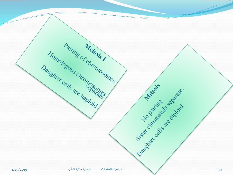
Meiosis Ι (also known as reductional meiosis) which deals with the number of
chromosomes it halves the number of chromosomes
Meiosis Ι Ι (also known as equational meiosis) which deals with the conditions of
chromosomes
2n
1n
1n
1n
Reproductive cell1n
Equational
Meiosis Ι Ι
Reductional
Meiosis Ι46
23
23
23
23
1n
1n
23
23
1/25/2019 كلية الطب-امجد الشطرات االردنية .د 31

Meiosis I is generally divided into four stages:
1-Propahse
2-metaphase
3-
Anophase
4-Telophase
1-Prophase is running into stages
A- LEPTOTEN stage, (lepto means long)
In this stage chromosomes are elongated and
extended and become gradually visible
1/25/2019 كلية الطب-امجد الشطرات االردنية .د 32
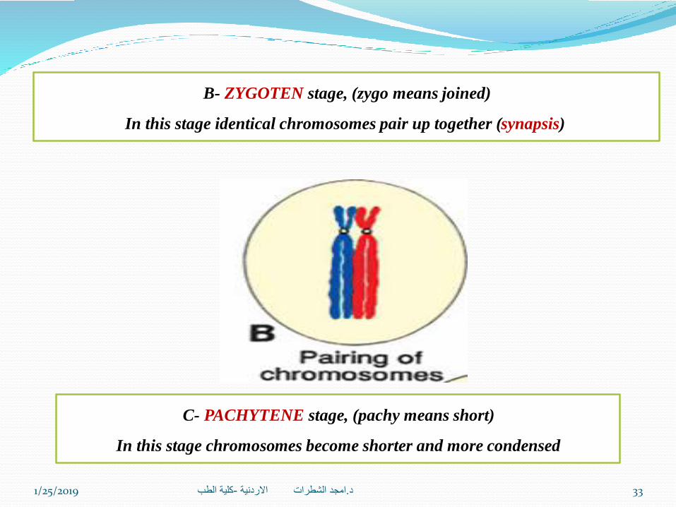
B- ZYGOTEN stage, (zygo means joined)
In this stage identical chromosomes pair up together (synapsis)
C- PACHYTENE stage, (pachy means short)
In this stage chromosomes become shorter and more condensed
1/25/2019 كلية الطب-امجد الشطرات االردنية .د 33

D- DIPLOTENE stage,
Chromosomes come together and cross each other by certain segments of their bodies
forming what we called CHIASMATA: X- shaped structure
Formed by the junction of two chromatids of the for chromatids (tetrad)
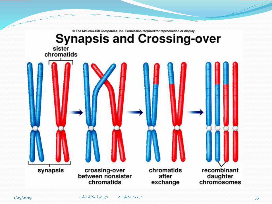
In Prophase I
Crossing over of non-sister
chromatids
During prophase I, non-sister
chromatids can undergo
synapsis, in which the
chromatids line up side-by-
side & exchange genetic
information between them
This allows new combination
of genetic material which will
become part of a new
offspring
1/25/2019 كلية الطب-امجد الشطرات االردنية .د 34
1/25/2019 كلية الطب-امجد الشطرات االردنية .د 35

2-Metaphase
1/25/2019 كلية الطب-امجد الشطرات االردنية .د 36
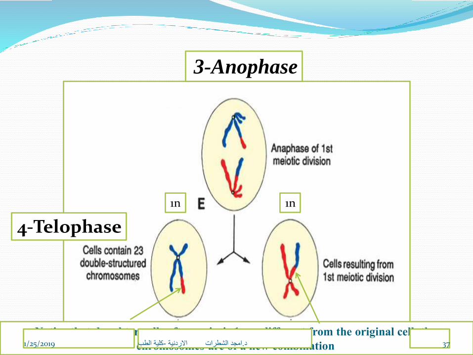
3-Anophase
4-Telophase
Notice that daughter cells after meiosis 1 are different from the original cell, the
chromosomes are of a new combination
1n 1n
1/25/2019 كلية الطب-امجد الشطرات االردنية .د 37

Meiosis II runs into 4 stages:
1-Prophase
2-Metophase
3-Anaphase
4-Telophase
1/25/2019 كلية الطب-امجد الشطرات االردنية .د 38
1/25/2019 كلية الطب-امجد الشطرات االردنية .د 39

Chromosomal abnormalities
numerical or structural
may be
Abnormalities in chromosome number may originateduring meiotic or mitotic divisions.
1/25/2019 كلية الطب-امجد الشطرات االردنية .د 40
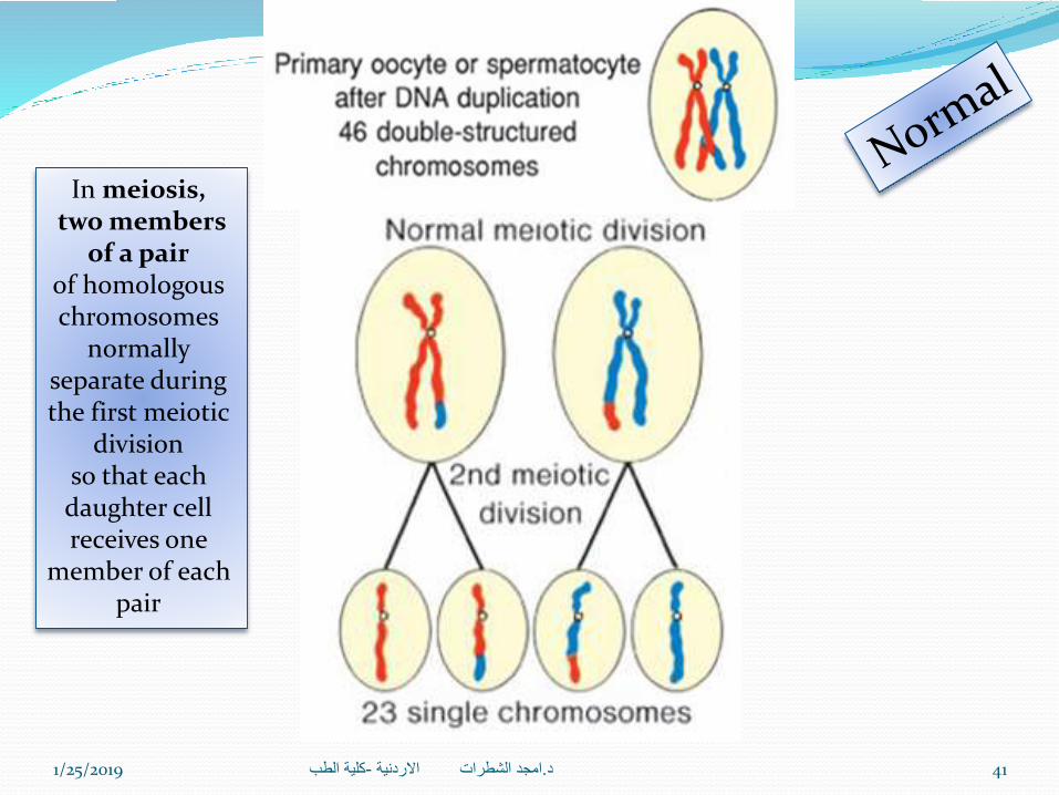
In meiosis,two members
of a pairof homologous chromosomes
normally separate during the first meiotic
divisionso that each daughter cell receives one
member of each pair
1/25/2019 كلية الطب-امجد الشطرات االردنية .د 41

Bothmembers of a pair move
into one cell .
As a result ofnondisjunction of the chromosomes, one
cell receives 24 chromosomes,and the other receives 22 instead of the
normal 23.
1/25/2019 كلية الطب-امجد الشطرات االردنية .د 42

1/25/2019 كلية الطب-امجد الشطرات االردنية .د 43

Sometimes chromosomes break, and pieces of one chromosome attach to another.
may be
1- Balanced, in which case breakage andreunion occur between two chromosomes
but no critical genetic material islost and individuals are normal
2-Unbalanced, in which casepart of one chromosome is lost and an
altered phenotype is produced.
An example, unbalanced translocations between the long arms
of chromosomes 14 and 21 during meiosis I or II
produce gametes with an extra copy of chromosome 21, one of the causes of Down syndrome
Translocations
1/25/2019 كلية الطب-امجد الشطرات االردنية .د 44

at fertilization,a gamete having 23 chromosomes fuses with
a gamete having 24 or22 chromosomes, the result is an individual
with either 47 chromosomes
Trisomyor 45 chromosomes
Monosomy
1/25/2019 كلية الطب-امجد الشطرات االردنية .د 45

Trisomy 21Maternal Age
Frequencyat birth
15-191/125020-241/140025-291/1100
301/900311/900311/750321/625331/500341/386351/300361/225371/175381/140391/100401/80411/65421/50431/40441/25
90%: Meiotic nondisjunction during meiosis II of oogenesis10%: Meiotic nondisjunction during meiosis I of spermatogenesis
1/25/2019 كلية الطب-امجد الشطرات االردنية .د 46

XXY – Phenotypically
male with an extra X
chromosome
1/25/2019 كلية الطب-امجد الشطرات االردنية .د 47

XO – Phenotypically
female missing an X
chromosome
Turner’s Syndrome
is the only monosomy compatiblewith life.
Even then, 98% of all fetuses with the syndrome are spontaneously
aborted. The few that survive are unmistakably
female in appearanceand are characterized by the absence of
ovaries (gonadal dysgenesis)
1/25/2019 كلية الطب-امجد الشطرات االردنية .د 48

1/25/2019 كلية الطب-امجد الشطرات االردنية .د 49

Thank you
1/25/2019 كلية الطب-امجد الشطرات االردنية .د 50