Embed Size (px)
Citation preview
ENV
1
Draft OECD Guidance Document on Histopathology for inhalation toxicity studies, Supporting TG
412 (Subacute Inhalation Toxicity: 28-Day) and TG 413 (Subchronic Inhalation Toxicity: 90-Day)
(Version 15 June 2009)
Contributors:
Roger A. Renne, DVM, DACVP (Chair)
Roger Renne ToxPath Consulting
Sumner, WA, USA
Jeffrey I. Everitt, DVM, DACVP
Director, Comparative Biology & Medicine
GlaxoSmithKline
Research Triangle Park, NC, USA
Jack R. Harkema, DVM, PhD, DACVP
University Distinguished Professor
Department of Pathobiology and Diagnostic Investigation
College of Veterinary Medicine
Michigan State University
East Lansing, MI, USA
Charles G. Plopper, PhD
Professor Emeritus
Anatomy Physiology and Cell Biology
University of California, Davis
Davis, CA, USA
Martin Rosenbruch, DVM, PhD
Toxicologic Pathology
Bayer Schering Pharma AG
Wuppertal, Germany

ENV
2
Table of Contents
Introduction…………………………………………………………………………………………………4
Tasks prior to Necropsy…………………………………………………………………………………… 4
Required Tasks…………………………………………………………………………………4
Review of pertinent study data……………………………………………………………..4
Clinical pathology………………………………………………………………………….4
Supplementary Tasks……………………………………………… ………………………….7
Ophthalmoscopic examination…………………………………………………………….7
Bronchoalveolar lavage……………………………………………………………………7
Labeling for Cytokinetic Studies………………………………………………………….8
Necropsy…………………………………………………………………………………………………..9
Required Tasks………………………………………………………………………………..9
Necropsy-related scheduling issues……………………....................................................9
Methods of euthanasia……………………………………………………………………9
Necropsy Technique……………………………………………………………………..9
Amplifications regarding necropsy tasks……………………………………………….12
Supplementary Tasks………………………………………………………………………..15
Tissue fixation via vascular perfusion…………………… …………………………….15
Tissue Trimming and Slide Preparation………………………………………………………………....15
Required Tasks……………………………………………………………………………...15
Supplementary Tasks for Trimming and slide preparation……………………………..17
Airway microdissection…………………………………………………………………17
Histopathology…………………………………………………………………………………………..17
Required Tasks………………………………………………………………………………17
Nasal Cavity/ Nasopharynx……………………………………………………………..18
Larynx…………………………………………………………………………………...19
Trachea and Bronchi…………………………………………………………………….19
Bronchioles, Alveolar Ducts, Alveoli, Pleura…………………………………………..19
Lung associated lymph nodes (LALN)………………………………………………….19
ENV
3
Supplementary Tasks for histopathology………………………………………………….20
Special stains…………………………………………………………………………..20
Immunohistochemistry…………………………………...............................................20
Morphometry ……………………………………………...........................................20
Ultrastructural examination……………………………................................................21
Reporting………………………………………………………………………………………………21
Required Tasks ………………………………………………………………………..21
Report preparation and internal review ………………………………………………21
Statistics……………………………………………………………………..21
Quality assessment……………………………………................................................22
Data Review……………………………………………..............................................22
Supplementary Tasks……………………………………………………………………..22
Pathology working groups……………………………………………………………22
References…………………………………………………………………………………………….23
ENV
4
Introduction
1. OECD Guidelines for 28 or 90 day inhalation studies (TG 412 and 413) were adopted in 1981 and draft
updates of these two documents were published in 2009 (OECD, 2009a, b) The purpose of this document
is to provide pathologists, toxicologists, and associated scientists with guidelines for performing the
pathology tasks required by TG 412 and 413, and for optimizing the value of pathology-related tasks in
these studies. This draft document is structured to provide essential and supplementary information in the
temporal sequence one would encounter in planning, participating in, and reporting the results of pathology
tasks in a 28 or 90 day inhalation toxicology study utilizing rodents.
2. General guidelines and recommended practices for histopathological tasks in toxicology studies have
been published in the open literature (Hildebrandt, 1991; Crissman et al, 2004). Following these
published practices will serve the pathologist well in avoiding the potential pitfalls associated with
performing and reporting pathology tasks in inhalation studies. This document provides key information
on each aspect of the pathology tasks required for inhalation studies in rodents, and references to additional
publications with details on methods and anticipated results.
Tasks Prior to Necropsy
Required tasks prior to necropsy
Review of pertinent study data
3. All pertinent data generated on the study should be available prior to scheduled necropsies. Confirm
availability to the pathologist, toxicologist, and other appropriate staff of quality control data required by
the study protocol on:
Clinical signs, clinical pathology, pulmonary physiology, any other tasks performed during the
exposure or recovery phase of the study;
Data from unscheduled deaths or moribund sacrifices during the exposure or recovery phase of
the study;
Records indicating sufficient training in necropsy procedures by prosectors, technicians, and
other staff involved in the pathology/clinical pathology tasks.
Clinical Pathology:
4. Clinical pathology is a key tool in detection, quantitation, and interpretation of effects of inhaled
toxicants in rodent toxicology studies. An extensive database of clinical pathology data on laboratory
rodents generated from toxicology studies by pharmaceutical and chemical industries, regulatory agencies,
contract research organizations, and academic institutions is available in the open literature (Loeb and
Quimby, 1999; Moore, 2000; Car et al, 2006). These and similar literature sources should be utilized to
optimize the value of clinical pathology data for specific study protocols and for other criteria such as
gender, stock/strain of the animals and diet status (restricted or ad libitum). A thorough understanding of
data from traditional clinical pathology parameters is required to be able to accurately interpret data from
new biomarkers and systems biology tools such as proteomic, transcriptomic and metabolomic studies.

ENV
5
5. Clinical pathology assessments should be made for all (or a statistically valid subset) animals on study,
including control and satellite animals, at scheduled interim sampling times prior to and during the
exposure period, and/or prior to terminal sacrifice. The time interval between exposure and blood and/or
urine collection should be defined in the study protocol. Rapid sampling after exposure is indicated for
parameters with a short plasma half-time (e.g., COHb, CHE, and MetHb). Prior to collections of samples,
it should be confirmed that collection methods, fluids, containers, diluents, or vehicles to be used are
correct and consistent for the clinical pathology tasks required in the protocol. Standard procedures
should be followed for randomization of samples and periodic insertion of quality control sera as described
in the literature (Loeb and Quimby, 1999; Car et al, 2006) and in study-specific protocols. Overnight
fasting prior to blood sampling is routinely done to insure uniformity of clinical chemistry and hematology
data. Methods should be used to acclimate animals to specialized housing and equipment (Damon et al.,
1986).
6. In assessing the results of clinical pathology data, comparisons should be made to concurrent cage,
vehicle, excipient, or other control groups, and to a range (95% interval) of normal values based on
samples from a large number (~50) of animals of similar age, stock/strain,and receiving simlar feeding and
husbandry practices. Appropriate statistical tools should be used to compare data from exposed and
control groups. Interpretation of statistically significant differences among groups must be tempered by the
realization that many clinical pathology parameters have wide ranges and are affected by a number of
variables. Similarly, a lack of statistical significance does not imply a lack of effect of exposure, and
interpretation of the results requires careful evaluation and correlation of clinical pathology parameters
with all other available toxicology data.
7. Table 1 lists the clinical pathology parameters that are generally required for rodent toxicology studies.
The study director may choose to assess additional parameters to better characterize a test article’s toxicity
(e.g., cholinesterase, lipids, hormones, acid/base balance, methaemoglobin, creatine kinase,
myeloid/erythroid ratio, and blood gases).
8. Urinalysis is not required on a routine basis, but may be performed when deemed useful based on
expected or observed toxicity. The use of metabonomics tools on urine has proven useful to detect outliers
prior to initiation of the study (Car et al, 2006), and to detect nephrotoxicity earlier than standard
histopathologic methods (Boudonck et al, 2009). Collection of urine samples free of external
contamination and of volume adequate for testing may require special collection containers and caging and
an overnight sampling period.
Table 1. Standard Clinical Pathology Parameters
Haematology
Erythrocyte count (RBC)
Haematocrit (Hct)
Haemoglobin Concentration (Hgb)
Mean corpuscular haemoglobin (MCH)
Mean corpuscular volume (MCV)
Mean corpuscular haemoglobin
concentration (MCHC)
Heinz bodies
Total leukocyte count (WBC)
Differential leukocyte count (Diffs)
Platelet count (Plate)
Clotting potential (select one):
Prothrombin time (PT)
Clotting time (CT)

ENV
6
Reticulocytes (Retics) Partial thromboplastin time (PTT)
Clinical Chemistry
Fasting glucose (GLU)*
Total cholesterol (Chol)
Triglycerides (TRIG)
Blood urea nitrogen (BUN)
Total bilirubin (T. Bili)
Creatinine (Creat)
Lactate dehydrogenase (LDH)
Total protein (T. Prot)
Albumin (Alb)
Globulin (Glob)
Alanine aminotransferase (ALT)
Aspartate aminotransferase (AST)
Alkaline phosphatase (ALP) or
Sorbitol dehydrogenase (SDH)
Potassium (K)
Sodium (Na)
Calcium (Ca)
Phosphorus (P)
Chloride (Cl)
Urinalysis
Appearance (colour and turbidity)
Volume
Specific gravity or osmolality
pH
Total protein
Glucose
* The fasting period should be appropriate to the species used; for the rat this may be 16 h (overnight
fasting). Determination of fasting glucose may be carried out after overnight fasting during the last exposure
week, or after overnight fasting prior to necropsy (in the latter case together with all other clinical pathology
parameters).
9. Hematology data collected using automated instrumentation should be routinely confirmed by manual
counting and enumeration of cell types (Moore, 2000). Histologic examination of bone marrow, spleen,
and liver is recommended to evaluate hematopoietic elements and detect fibrosis or necrosis. Flow
cytometry can be used to differentiate hematopoietic lineages (Criswell et al., 1998), and may also be
utilized to quantify micronuclei (Weaver and Torous, 2000).
10. Evaluation of clinical pathology data in inhalation studies should consider the impact of stress related
to exposure to the test material and to handling and change of environment during cage transfer and
specimen collection. The most consistently affected parameters related to stress in rats are increased
lymphocytes, glucose, adrenalin, and ACTH and decreased eosinophils (Car et al, 2006).
ENV
7
Supplementary tasks prior to necropsy
Ophthalmoscopic Examination
11. Ophthalmic examination should be considered if there is evidence of potential ocular effects from
clinical observations during the exposure or from information in the published literature. Ophthalmological
examinations by an ophthalmologist or other qualified professional on the fundus, refractive media, iris,
and conjunctiva using an ophthalmoscope or an equivalent device should be performed on all animals (or a
specific subset) from each group prior to the administration of the test article, and for all high
concentration and control groups at termination. If changes in the eyes are detected, all animals in the other
groups should be examined including the satellite (reversibility) group.
Bronchoalveolar Lavage
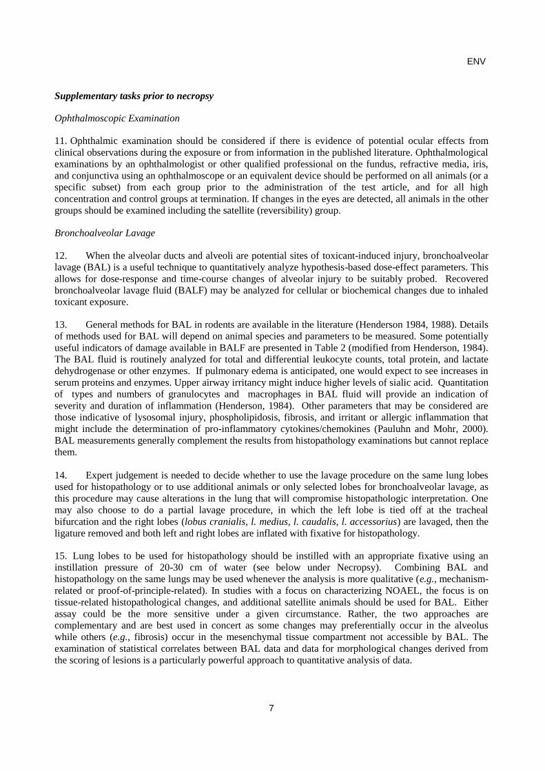
12. When the alveolar ducts and alveoli are potential sites of toxicant-induced injury, bronchoalveolar
lavage (BAL) is a useful technique to quantitatively analyze hypothesis-based dose-effect parameters. This
allows for dose-response and time-course changes of alveolar injury to be suitably probed. Recovered
bronchoalveolar lavage fluid (BALF) may be analyzed for cellular or biochemical changes due to inhaled
toxicant exposure.
13. General methods for BAL in rodents are available in the literature (Henderson 1984, 1988). Details
of methods used for BAL will depend on animal species and parameters to be measured. Some potentially
useful indicators of damage available in BALF are presented in Table 2 (modified from Henderson, 1984).
The BAL fluid is routinely analyzed for total and differential leukocyte counts, total protein, and lactate
dehydrogenase or other enzymes. If pulmonary edema is anticipated, one would expect to see increases in
serum proteins and enzymes. Upper airway irritancy might induce higher levels of sialic acid. Quantitation
of types and numbers of granulocytes and macrophages in BAL fluid will provide an indication of
severity and duration of inflammation (Henderson, 1984). Other parameters that may be considered are
those indicative of lysosomal injury, phospholipidosis, fibrosis, and irritant or allergic inflammation that
might include the determination of pro-inflammatory cytokines/chemokines (Pauluhn and Mohr, 2000).
BAL measurements generally complement the results from histopathology examinations but cannot replace
them.
14. Expert judgement is needed to decide whether to use the lavage procedure on the same lung lobes
used for histopathology or to use additional animals or only selected lobes for bronchoalveolar lavage, as
this procedure may cause alterations in the lung that will compromise histopathologic interpretation. One
may also choose to do a partial lavage procedure, in which the left lobe is tied off at the tracheal
bifurcation and the right lobes (lobus cranialis, l. medius, l. caudalis, l. accessorius) are lavaged, then the
ligature removed and both left and right lobes are inflated with fixative for histopathology.
15. Lung lobes to be used for histopathology should be instilled with an appropriate fixative using an
instillation pressure of 20-30 cm of water (see below under Necropsy). Combining BAL and
histopathology on the same lungs may be used whenever the analysis is more qualitative (e.g., mechanism-
related or proof-of-principle-related). In studies with a focus on characterizing NOAEL, the focus is on
tissue-related histopathological changes, and additional satellite animals should be used for BAL. Either
assay could be the more sensitive under a given circumstance. Rather, the two approaches are
complementary and are best used in concert as some changes may preferentially occur in the alveolus
while others (e.g., fibrosis) occur in the mesenchymal tissue compartment not accessible by BAL. The
examination of statistical correlates between BAL data and data for morphological changes derived from
the scoring of lesions is a particularly powerful approach to quantitative analysis of data.

ENV
8
Table 2. Indicators of Acute Injury in Bronchoalveolar Fluids (from Henderson, 1984)
Parameter Location Possible indication if elevated
Lactic Dehydrogenase Cytosol (glycolysis Cell damage(increased membrane permeability to
frank cell lysis)
Glucose-6-phosphate-
dehydrogenase
Cytosol (hexose
monophosphate shunt
Cell damage; leakage from cells undergoing repair
Lysosomal acid
hydrolases
Lysosomes Release during phagocytosis; PMN and/or macrophage
damage
Alkaline phosphatase Plasma membranes,
Type II cell lamellar
bodies, serum
Type II cell damage or increased secretions;
transudation of serum proteins
Glutathione peroxidase
Glutathione reductase
Cytosol Protection mechanism activated against
lipoperoxidation
Angiotensin converting
enzyme
Endothelial cells Endothelial cell damage
Total Protein Extracellular Transudation of proteins across alveolar-capillary
barrier
Sialic acid Mucus
Glycoproteins
Increased mucus secretion
Transudation of serum glycoproteins
Phagocytic cells
PMNs
Macrophages
Inflammation
Labeling for Cytokinetic Studies
16. Changes in cell turnover rate are a useful tool to detect and quantify short or long term changes in
tissues in response to toxicants, and may yield insight into their mechanism of action (Evans et al, 1991).
Cell proliferation has long been recognized as a key factor in carcinogenesis (Melnick et al, 1993), but is
also a useful tool to detect and quantify cellular changes in short term toxicology studies. Cell kinetics
studies are based on the use of compounds that label cellular DNA and allow the microscopic detection and
quantitation of cells undergoing DNA synthesis at the time of tissue fixation. Immunohistochemical
detection using a monoclonal antibody to bromodeoxyuridine (BrdU) is the most widely accepted tool for
this assay. Cells in the DNA synthesis (S) phase can be labeled over short (single pulse via intraperitoneal
or intravascular injection) or long (subcutaneously implanted osmotic pump or oral dosing in water)
periods of time (Goldsworthy et al, 1993). Endogenous markers of cell cycle status such as proliferating
cell nuclear antigen (Eldridge et al, 1993) and Ki67 (Gerdes et al, 1983; Ignatiadis and Sotiriou, 2008;
Challen et al, 2009;) may also be measured using immunohistochemical techniques. Cell proliferation
ENV
9
studies have provided much useful information in studying toxicity of nasal (Monticello et al, 1993) and
pulmonary (Haschek and Witschi, 1991) epithelium. However, analysis of these data requires caution and
sample size is an important factor (Morris, 1993).
Necropsy
Required Tasks
17. All study animals, including vehicle or excipient controls, sentinels and any animals that die during the
exposure or recovery period or are removed from the study for animal welfare reasons, should be subjected
to complete exsanguination (if feasible) and necropsy by well trained, experienced prosectors under the
direct supervision of a pathologist, closely following the requirements of a study protocol. The only
exception to a requirement for a complete necropsy would be animals found dead or sacrificed due to
moribund condition during the first few days of a study in which the study protocol specifically requires
replacement of animals accidentally killed or injured early in the study by replacement animals. In this case
the necropsy of the animal to be replaced may be a less complete necropsy to determine cause of death or
moribundity but not intended to generate tissues for histopathology related to the goals of the study.
18. Necropsies should be performed as soon as possible. If a necropsy cannot be performed immediately
after a dead animal is discovered, the animal should be refrigerated (not frozen) at temperatures low
enough to minimize autolysis and necropsied within 8 hours. All gross pathological changes should be
recorded for each animal with particular attention to any changes in the respiratory tract.
Necropsy- related scheduling issues
19. The optimal time from last exposure to removal of food (fasting), blood and/or urine collection,
euthanasia, BAL, and necropsy may vary with the physical or chemical characteristics of the test article
and available information on the effect of the exposure on study animals. Interval between last exposure
and blood or urine collection are discussed above under Clinical Pathology. In typical inhalation studies
animals are fasted for 16 hours (overnight) following the last exposure, although fasting prior to necropsy
is not done universally. If data available from clinical signs, clinical pathology, or other information
obtained during the study or from the literature indicate fasting may not be indicated, it may be omitted.
Not fasting prior to necropsy may impact body and organ weight ratios, histology of liver (glycogen in
hepatocytes), blood glucose, and other clinical pathology values when compared with data from the
literature.
Methods of euthanasia
20. Methods of euthanasia for laboratory rats are described in a recent publication (Everitt and Gross,
2006). AVMA Guidelines provide a general description of euthanasia methods and issues on a wide
variety of animal species (AVMA, 2007). Inhalation studies require increased attention paid to the effect
of the anesthetic and method of administration on the respiratory tract and associated tissues. The most
frequently used methods of euthanasia of laboratory rodents are inhalation of halogenated ether anesthetics
via a mask, inhalation of carbon dioxide/oxygen combinations in a closed chamber, and intravenous or
intraperitoneal injection of a barbiturate anesthetic. Exsanguination of the anesthetized animal via incision
of the abdominal vena cava or axillary vessels is used to terminate the animal’s life and concurrently
improve the quality of fixed tissues for examination by removal of a portion of the circulating blood.
Heartbeat must be completely stopped prior to start of the necropsy.
ENV
10
21. The choice of individual anesthetic/chemical agents for euthanasia may depend in part on the material
being tested and the clinical pathology parameters being measured. Barbiturate anesthetics may have an
effect on cytochrome P450 levels with particularly important effects in liver (Popp and Cattley, 1991) or
nasal epithelium (Dahl and Hadley, 1991). Halogenated ether anesthetics are commonly used in rodent
toxicology studies including inhalation studies, but have the potential for effects on the respiratory tract
since they are administered via inhalation, and care must be taken to avoid exposure to humans.
Recommended halogenated ether anesthetics for euthanasia of rodents are, halothane, enflurane, isoflurane,
sevoflurane, methoxyflurane, and desflurane (AVMA, 2007). Carbon dioxide/oxygen combinations are
useful for short term anesthesia to collect blood samples, and are used in combination with exsanguination
for euthanasia prior to necropsy. However, failure to maintain a ratio of 70% carbon dioxide/30% oxygen
may induce multiple small hemorrhages in lung parenchyma (Renne et al, 2007) which makes the use of
carbon dioxide inhalation unacceptable for euthanasia in inhalation toxicology studies.
Necropsy Technique
22. Well trained, conscientious prosectors thoroughly familiar with the anatomy and dissection procedures
of the animals to be necropsied and the requirements of the specific study protocol are an essential tool for
success in the necropsy task (Bucci, 1991). Training must emphasize attention to detail and consistency
combined with efficiency and appreciation of the importance of this critical task in which mistakes are
almost never correctable. Training of these staff members must be in the hands of an experienced
supervisor and/or pathologist who has sufficient bench/lab experience to appreciate the disastrous results of
mistakes made in necropsy. The supervising pathologist and other staff supervising the necropsy must be
active, informed participants in the generation and approval of the study protocol, and are responsible to
provide all protocol-specific necropsy, trimming, and histology information to the other staff involved in
the necropsy.
23. Table 3 lists the organs and tissues that should be preserved in a suitable fixative (see below) during
necropsy for histopathological examination. The preservation of other organs and tissues depends on the
test article being studied and is at the discretion of the study director. The lungs will be weighed after
removal of the lung associated lymph nodes (LALN; cervical, mediastinal, and tracheobronchial lymph
nodes) and heart and before inflation with fixative. Other organs to be weighed will be specified in the
individual study protocol. Tissues and organs should be fixed in 10% buffered formalin or another suitable
fixative (see below) as soon as necropsy is performed, and fixation should continue for no less than 24-48
hours prior to trimming depending on the fixative to be used. Special attention is required to insure that
tissues to be weighed are kept moist with saline-moistened gauze sponges, and that time from removal
from the carcass for weighing to placement in fixative is minimized to avoid autolysis.

ENV
11
Table 3. Organs and Tissues Preserved During Necropsy
Adrenals Pituitary
Aorta Prostate
Bone marrow aspirate (sternal or femoral) Rectum
Brain (including sections of cerebrum, cerebellum,
and medulla/pons)
Salivary glands
Caecum Seminal vesicles (with coagulating glands)
Cervix Skin
Colon Spinal cord (cervical, mid-thoracic, and lumbar
Duodenum
[Epididymides] Spleen
[Eyes (retina, optic nerve) and eyelids] Sternum with marrow
Femur and stifle joint Stomach
Gallbladder (where present) Teeth
Testes
Thymus
Thyroids
Harderian glands Tongue
Heart Trachea
Ileum Urinary bladder
Jejunum Uterus
Kidneys Zymbal’s glands
[Lacrimal glands (extraorbital)]
Larynx (see comments below)
Liver
Lungs (see comments below)
Lymph nodes
- lung associated (tracheobronchial, mediastinal,
, cervical),
- mandibular,
- mesenteric
- popliteal
Target organs specified in the study protocol
Mammary gland (both sexes) Gross lesions
Muscle (thigh)
Nasal cavity (see below)
Oesophagus
Ovaries
Pancreas
Parathyroids
Peripheral nerve (sciatic or tibial,
24. A document providing an excellent description of the background, preparation and planning,
equipment, and procedures for a general rodent necropsy as part of a toxicology study is available in the

ENV
12
literature (Everitt and Gross, 2006), and should be used as the basic text in preparing for necropsy of
rodents in toxicology studies.
Amplifications, additions and exceptions to the necropsy task
25. Examination and recording of gross observations and dissection and placement in fixative of all
protocol-required tissues is a critical part of every necropsy in toxicology studies. Standard protocols
require the pathologist supervising the necropsy to ensure by his/her signature on the necropsy form that all
protocol-required tissues were examined and placed in fixative by the prosector. During the necropsy a
responsible pathologist must be available to supervise the prosectors. In all cases of questionable gross
findings the responsible pathologist should confirm and/or improve upon the description of findings by the
prosector.
Figure 1
26. One item useful in assuring that all tissues were collected and examined is a plastic tray divided into
numerous compartments (Figure 1). The prosector can place tissues small enough to be contained in
individual compartments in preset compartments within this tray, which contains fixative solution that
allows the tissues to begin fixation within the tray. When the prosector has completed the necropsy he/she
can call the pathologist to examine the tissues within the tray to confirm that they have been collected.
Photographs of pertinent findings at necropsy are taken if required by the study protocol or if necessary for
the documentation of unusual findings.

ENV
13
27. The document cited (Everitt and Gross, 2006) lists dissection of the thoracic viscera following
dissection of the head and abdominal viscera. For inhalation studies the most crucial target organs are the
upper and lower respiratory tract; therefore dissection of the thoracic viscera should immediately follow
dissection of the head.
28. Special attention during dissection of the head and thoracic viscera must be paid to dissection, fixation,
and identification of the mandibular and the lung associated lymph nodes (cervical, mediastinal,
tracheobronchial LALN), since these are the terminal collection sites of lymphatic drainage for the
respiratory tract (Hebel and Stromberg, 1986). The mandibular LNs are usually easily visible craniolateral
to the mandibular salivary glands. The cervical LNs are less obvious; the superficial cervical LN is
attached to the ventral muscles of the neck; the deep cervical LN is just lateral to the trachea. The
mediastinal LNs are attached to the thymus and the precordial mediastinum. If the study protocol requires
thymus weights, the mediastinal LNs should be removed from the thymus prior to weighing the thymus.
The tracheobronchial LNs are located just cranial to the bifurcation of the trachea into mainstem bronchi
and between the mainstem bronchi. All the LNs described above can be placed in individually labeled
cassettes to insure they are available for microscopic examination.
29. After the thoracic viscera are removed from the carcass as a single unit, the cranial surface of the
larynx should be examined visually for the presence of foam, blood, or other material. The trachea should
be transected midway between the larynx and the tracheal bifurcation and its lumen examined for foam or
other fluids, and the larynx and attached tongue placed in fixative.
30. Most protocols of 28-day or 90-day inhalation toxicology studies require weighing of lungs, heart,
thymus, and other selected tissues at necropsy. Thus, the heart will be dissected free from the other
thoracic viscera prior to weighing. Special care must be taken when removing the heart from the thoracic
viscera to avoid cutting through the wall of the trachea or mainstem bronchi. Perforation of the tracheal or
bronchial wall will make it impossible to fully inflate the lungs with fixative or maintain the inflated state.
Due to problems such as this and other inconsistencies in trimming tissues for weighing, only thoroughly
trained and experienced necropsy staff should trim tissues for weighing (Bucci, 1991; Everitt and Gross,
2006,). In this method, prosectors dissect out tissues to be weighed but do not finely dissect surrounding
tissues. Tissues to be weighed are transferred to person/s trimming the tissues to be weighed , weighing
tissues and recording weights using a computerized balance system. This system has decreased the
problem of leakage due to accidentally punctured airways and concurrently improved consistency of organ
weight data, as well as decreased the time between removal of lungs from the carcass and instillation with
fixative.
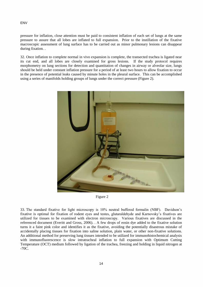
31. Lungs to be processed for histopathology should be inflated with fixative at a pressure of 20-30 cm of
water with a gravity flow apparatus such as that illustrated in Figure 2 (Renne et al, 2001; Everitt and
Gross, 2006). ). This apparatus consists of a clear glass bottle containing fixative solution and sealed at the
top with a rubber stopper containing a pipette. The tip of the pipette is set at a precise vertical distance
(20-30 cm; see below) above the level of the lungs to be inflated with fixative. Flexible tubing connected
to a spigot on the side of the bottle carries fixative to the tracheal opening . Proper operation of this
apparatus requires an airtight seal of the stopper and a patent pipette lumen. This apparatus should be
tested for accuracy and consistency of pressure prior to and at intervals during its use. The optimal
pressure for instillation varies with the species and size of the animal and could also vary with the effects
of exposure to the test material. Ideally, the set target pressure for inflation should be determined at the
start of the necropsy procedure by inflation of the lungs prior to their removal from the thoracic cavity
from a sentinel or other non-core study animal that has been exposed to the highest concentration of test
article. The degree of inflation should be closely observed, starting with a pressure of 20 cm and not
exceeding 30 cm of water, and continuing inflation until the lungs fill but do not protrude from the thoracic
cavity, i.e., until complete expansion but not overexpansion of all lobes. Regardless of the set target
ENV
14
pressure for inflation, close attention must be paid to consistent inflation of each set of lungs at the same
pressure to assure that all lobes are inflated to full expansion. Prior to the instillation of the fixative
macroscopic assessment of lung surface has to be carried out as minor pulmonary lesions can disappear
during fixation. .
32. Once inflation to complete normal in vivo expansion is complete, the transected trachea is ligated near
its cut end, and all lobes are closely examined for gross lesions. If the study protocol requires
morphometry on lung sections for detection and quantitation of changes in airway or alveolar size, lungs
should be held under constant inflation pressure for a period of at least two hours to allow fixation to occur
in the presence of potential leaks caused by minute holes in the pleural surface. This can be accomplished
using a series of manifolds holding groups of lungs under the correct pressure (Figure 2).
Figure 2
33. The standard fixative for light microscopy is 10% neutral buffered formalin (NBF). Davidson’s
fixative is optimal for fixation of rodent eyes and testes, glutaraldehyde and Karnovsky’s fixatives are
utilized for tissues to be examined with electron microscopy. Various fixatives are discussed in the
referenced document (Everitt and Gross, 2006). . A few drops of eosin dye added to the fixative solution
turns it a faint pink color and identifies it as the fixative, avoiding the potentially disastrous mistake of
accidentally placing tissues for fixation into saline solution, plain water, or other non-fixative solutions.
An additional method for preserving lung tissues intended to be utilized for immunohistochemical analysis
with immunofluorescence is slow intratracheal inflation to full expansion with Optimum Cutting
Temperature (OCT) medium followed by ligation of the trachea, freezing and holding in liquid nitrogen at
-70C.
ENV
15
Supplementary Tasks for Necropsy
Fixation of tissues by vascular perfusion
34. Vascular perfusion of fixative provides superior tissue fixation and is used when electron microscopic
examination is a primary goal. Whole body perfusion of glutaraldehyde or other EM fixative via the left
ventricle or abdominal aorta is the standard technique. A flushing solution is used to remove blood from
the circulation prior to infusion with fixative (Nyska et al, 2004). The time and technical expertise
required for vascular perfusion make it impractical for routine toxicology studies.
Tissue Trimming and Slide Preparation
Required Tasks
35. The task of trimming tissues obtained at necropsy has the same requirements for close attention to
detail, consistency, efficiency, and powers of observation that were described for the necropsy task (Bucci,
1991). Errors made during trimming are also seldom repairable and can also have an impact on the quality
of the pathology data. Training of staff for tissue trimming must be done by supervisory personnel
experienced in the procedure and very familiar with the protocols. Staff trimming tissues must pay close
attention to the necropsy findings as well as the study protocol, and close communication among the
necropsy and trimming technical staff, histology supervisor, and pathologist is essential for obtaining tissue
sections that accurately reflect the necropsy findings and provide the pathologist tissue sections need to
interpret the results.
36. Publications providing specific techniques for tissue trimming are available in the literature
(Young, 1981; Sminia et al, 1990; Uriah and Maronpot, 1990; Morgan,1991; Lewis, 1991; Renne et al,
1992; Sagartz et al, 1992; Bahnemann et al, 1995; Harkema and Morgan, 1996; Plopper, 1996; Germann et
al, 1998; Hardisty et al, 1999; LeBlanc, 2000; Kaufman et al, 2009) and on the internet
(http://reni.item.fraunhofer.de/reni/trimming/index.php) The information provided in this internet site is
the product of a large effort by a number of toxicologic pathologists and histologists with extensive
experience in performing toxicology studies for industry and regulatory agencies. It provides a detailed
introduction and excellent descriptions and graphics of specific techniques for trimming rodent tissues,
including the respiratory tract. The internet site document cited above should be used as a basic text for
trimming rodent respiratory tract tissues, although there are other methods utilized, some of which are
described below. The eight paragraphs below also provide amplifications, additions, and exceptions to the
methods presented in the internet document
37. The internet site trimming guide recommends four transverse nasal sections for rats and three for mice
and provides excellent text and graphics for obtaining sections at those sites. However, the appropriate
choice for number and location of nasal sections may depend on the physical and chemical characteristics
of the test article and previous knowledge of its effect on the respiratory tract. Highly water soluble or
caustic test compounds may have a profound effect on the squamous epithelium of the nasal vestibule,
requiring additional nasal sections through the nasal vestibule (Gross et al, 1994; Harkema et al, 2006).
Test compounds suspected or known to have an effect on olfaction, olfactory epithelium, or the central
nervous system may require additional sections through the caudal portions of the nasal cavity containing
olfactory epithelium and the olfactory bulb area of the brain. Excellent publications providing detailed
methods for preparation of multiple sections through the olfactory area of the nasal cavity and
nomenclature for describing lesions in olfactory epithelium are present in the literature (Morgan, 1991;
Brenneman et al, 2002; Mery et al, 1994).

ENV
16
38. Poor choices in methods and materials for decalcification of rodent nasal tissues may result in less
than ideal quality of stained slides due to overexposure to relatively strong acid chemicals. Use of formic
acid and a commercially available ion exchange resin to decalcify the skull prior to trimming nasal cavity
sections may provide optimal cellular detail as well as faster decalcification (Sheehan and Hrapchak,
1980).
39. Consistency in trimming and embedding nasal and laryngeal tissues is critical for interpretation of
mucosal changes related to exposure. The zones of transition from squamous to transitional to respiratory
to olfactory epithelium are relatively narrow in rodents and errors in trimming or embedding can easily
result in the pathologist being unable to examine these critical areas. Staff undertaking the trimming and
embedding of these important tissues must have the training and experience necessary to ensure that the
tissues are available to the pathologist, and excellent communication between the pathologist and histology
laboratory staff is required.
40. The internet site recommends three transverse laryngeal sections in rats and two in mice and
provides excellent graphics for obtaining these sections in both species. The internet site text indicates the
most sensitive laryngeal site for an effect of inhaled toxicants is the ventral pouch and the medial surface
of the arytenoid cartilages. However, according to several papers, the most sensitive site in rats or mice to
inhaled toxicants is the transitional epithelium at the base of the epiglottis (Renne et al, 1992, 2007; Renne
and Gideon, 2006; Kaufmann et al, 2009). This is important because it is somewhat challenging to
consistently provide a precise section through the base of the epiglottis and it is possible that only one
section of larynx (including the ventral pouch) will be available in some inhalation studies.
41. Most published reports utilize the multiple transverse sections method for larynx described in the
internet site publication for routine inhalation studies. An alternative method utilizing longitudinal
sections of larynx has been described (Germann et al, 1998). These authors found the longitudinal
sectioning method advantageous for detection of laryngeal or tracheal cartilage degeneration and
granulomatous inflammation in the oropharyngeal cavity of Fischer 344 rats in a chronic gavage study.
The longitudinal method provides a different perspective of the ventral pouch area at the expense of a full
transverse view of the base of the epiglottis and the medial surface of the arytenoid processes
42. The internet site publication provides an excellent description and graphics for preparing
longitudinal sections of tracheal bifurcation. In most studies in which the protocol also requires sections of
thyroid parathyroid, a transverse section of trachea at the level of the thyroid gland will also be available.
The internet site publication also correctly describes the requirement for careful microtoming until the
required section containing the carina is available for embedding. However, this procedure may result in
loss of the tissue containing one or more of the tracheobronchial lymph nodes present adjacent to the
tracheal bifurcation. For this reason, it is recommended (see in Necropsy section above) to remove all
tracheobronchial lymph nodes and place them in labeled cassettes for histologic processing either at
necropsy or trimming, prior to trimming and embedding the trachea.
43. The internet site publication provides an excellent description and graphics for preparing multiple
sections from all lobes of the lungs of rats and mice, resulting in five separate sections. An optional
method for mice is also described in which all lobes are detached from the trachea and embedded together
in one cassette. This method has been used successfully and is recommended for consideration as an
optional method, depending on the size of the lungs and the presumed degree of importance of the
peripheral lung tissues.
44. An alternative method utilized successfully in preparing lung sections from smaller (Fischer 344)
rats is separating the right and left lungs at the tracheal bifurcation and embedding each lung dorsal surface
ENV
17
down, in two separate cassettes. The microtomer cuts through each coronal longitudinal section until the
mainstem bronchus is clearly visible in the section, then takes a section for microscopy.
Supplementary Tasks for Trimming and Slide Preparation.
Airway Microdissection
45. A method for precise sampling of the bronchi and bronchioles of rodents by microdissection has been
developed which provides explants of intact airways to examine subpopulations of bronchial and
bronchiolar epithelium (Plopper et al, 1991; Hyde et al, 1991). This method utilizes fixation of the lungs
via intratracheal infusion of aldehydes or inflation of fresh lung with low temperature agarose, removing
adjacent vessels and connective tissue, and longitudinally opening bronchi and bronchioles and numbering
branches of airways as far as possible into the lung parenchyma. This valuable research tool provides the
opportunity to assess metabolic capabilities and do morphometry of airway cells from defined sites at the
light microscopy or electron microscopy level. It is especially useful for assessments of airway toxicity
when the aerodynamic properties of the inhaled material are such that it’s distribution within the lungs is
highly dependent on the three-dimensional architecture of the airway tree.
Histopathology
Required Tasks
46. A histopathological evaluation of all the organs and tissues listed in Table 3 should be performed for
the control and high concentration groups, and for all animals which die or are sacrificed due to moribund
condition during the study. Particular attention should be paid to the respiratory tract, other potential target
organs, and gross lesions. Nasal cavity, larynx, trachea, lungs, lung associated lymph nodes
(tracheobronchial, mediastinal, cervical) and mandibular lymph nodes, heart and gross lesions observed at
necropsy or tissue trimming should be examined microscopically in all exposed and control groups. The
organs and tissues in the high concentration groups that have microscopic lesions suspected of being
related to exposure should be examined in all lower concentration groups. The study director may choose
to perform histopathological evaluations for additional groups to demonstrate a clear concentration
response. When reversibility (recovery) groups are used, histopathological evaluation should be performed
in these groups using the same criteria as those listed for the groups necropsied immediately following the
last exposure to test article. If there are excessive early deaths or other problems in the high exposure group
that compromise the significance of the data, tissues from the next lower concentration should be examined
microscopically. An attempt should be made to correlate each gross observation with microscopic findings;
the results of this attempt should be recorded in the findings for individual animals.
47. Terminology used in histopathologic description, diagnosis, and reporting in rodent inhalation studies
should follow standard veterinary pathology nomenclature, utilizing the wealth of information available in
the published toxicologic pathology literature. The goRENI website (http://www.goreni.org) provides a
standard reference for nomenclature and diagnostic criteria in toxicologic pathology, including respiratory
tract tissues for rats and mice. This internet site is the product of a large effort by a number of toxicologic
pathologists and histologists with extensive experience in performing toxicology studies for industry and
regulatory agencies. It provides a detailed introduction and excellent descriptions and graphics of
degenerative, inflammatory, and proliferative respiratory tract lesions in rats and mice, including criteria
for diagnosis, differential diagnosis, comments on histogenesis and toxicologic significance, and an
extensive bibliography. This internet site document should be used as a basic text for describing,
diagnosing, and reporting lesions observed in the respiratory tract and elsewhere, supplemented by journal
articles and texts that provide information on subject matter relevant to specific study protocols.

ENV
18
48. Additional details on important aspects of microscopic examination of tissues from inhalation studies
are presented below:
Nasal Cavity/Nasopharynx
49. Adequate microscopic examination of respiratory tract tissues from rodent inhalation studies requires
close comparison of carefully selected tissue sections from control and exposed animals. As noted above,
areas of normal transition from various epithelial types are relatively small but are important to evaluate,
since these are often the site of subtle but meaningful lesions. An excellent example is the nasal cavity of
rodents, which is composed of a four distinct epithelial subtypes, with well mapped transition zones
(Harkema, 1991; Mery et al, 1994; Harkema et al, 2006). Recognition and documentation of metaplasia or
hyperplasia of mucosal epithelium in these areas is easily missed without precisely located tissue sections
and close comparison of control and exposed nasal sections by the pathologist. A useful tool to assist in
the evaluation of changes in goblet cell populations in the nasal cavity and elsewhere is the Alcian
blue/periodic acid Schiff stain, which stains the mucus in goblet cells a dark purple.
50. As mentioned above in the Trimming section, it is important to consider the physical and chemical
characteristics of the test article in deciding the number and location of nasal sections for histopathologic
examination. Test articles with high water solubility and caustic properties are most likely to induce
lesions in the squamous epithelium of the nasal vestibule, in which case a section through this site would
be mandatory. Induced lesions in the stratified squamous epithelium lining the nasal vestibule may include
hyperplasia, inflammation, and necrosis with ulceration. Examples of chemicals inducing lesions in this
area include glutaraldehyde (Gross et al, 1994) and ammonia (Bolon et al, 1991).
51. In the author’s experience (Renne et al, 2007), The areas of transitional and respiratory nasal
epithelium (distal third of the nasal and maxillary turbinates and adjacent lateral wall in the nasal section
just caudal to the upper incisors, level I as described by Young, 1981) are most sensitive to inhaled
toxicants. The most frequent and earliest change is an increase in thickness of the surface transitional
epithelium, often with a minimal suppurative inflammatory infiltrate in the adjacent submucosa and glands.
If this lesion progresses with increased dose or continued exposure, the affected epithelium will continue to
thicken and may also undergo squamous metaplasia.
52. Lesions induced in olfactory epithelium most frequently arise in the rostral extension of olfactory
epithelium lining the dorsal medial meatus in the section taken at the level of the incisive papilla (Young,
1981 section II). Induced lesions range from subtle degeneration of olfactory epithelium with regeneration
to necrosis and ulceration of mucosa followed by atrophy or respiratory metaplasia (Harkema et al, 2006).
Induced lesions may extend to similar areas of the dorsal meatus in more caudal nasal sections, depending
on the concentration and inherent toxicity of the test material and the duration of exposure.
53. Although the rostral extension of olfactory epithelium is the most frequent site of olfactory epithelial
lesions induced by inhaled toxicants, certain chemicals may initially induce olfactory lesions further
caudally. The mode of action of these chemicals is frequently related to metabolism via cytochrome P-450
enzymes (Pino et al, 1999; Harkema et al, 2006). Inhalation of finely divided metal particles may result in
deposition in the olfactory lobes of the brain via the olfactory neuroepithelium (Henrikkson et al,, 2000;
Dorman et al, 2002).
54. Olfactory lesions induced by a number of inhaled materials are described and illustrated in an excellent
paper by Hardisty et al (1999). This publication also provides valuable information on nomenclature and
criteria for diagnosis of induced olfactory lesions. Another valuable source of information on methods of
describing the location of induced nasal lesions in rodents is the publication by Mery et al (1994). These
ENV
19
authors designed a system for mapping lesions in the rodent nose using a nomenclature that identified and
numbered specific turbinate bones and cavities within the nose. Use of these two valuable publications
enables the pathologist to clearly locate and characterize lesions within the complex rodent nasal cavity.
55. Nasal-associated lymphoid tissue (NALT) is located in the ventral/lateral portion of the walls forming
the rostral opening of the nasopharyngeal duct, level III ( Young, 1981; Harkema, 1991). The efferent
lymph drainage from this tissue is to the posterior cervical lymph nodes (Kuper, 2003). Although less is
known about the function of this lymphoid tissue compared to other mucosal-associated lymphocenters, it
has been demonstrated to induce specific local immune responses and activate immune mucosal tissue
elsewhere as well as the systemic immune system (Kuper, 2003). There is much current interest in the
role of the immune system in toxic effects observed in laboratory animals. Examination of the NALT
should be included in standard inhalation toxicology studies, and special efforts made to provide adequate
sections through this tissue.
Larynx
56. Many of the issues critical for successful microscopic examination of nasal tissues described above
apply to histopathology of the rodent larynx. Areas requiring precisely located tissue sections and close
comparison with controls for diagnosis of squamous metaplasia and/or hyperplasia of mucosal epithelium
are the base of the epiglottis, ventral pouch, and medial surface of the arytenoid processes of the larynx
(Lewis, 1991; Renne and Gideon, 2006; Kaufmann et al, 2009)
57. As mentioned above, an alternate method of examining larynx is via longitudinal/parasaggital
sections, which provide a different perspective of the ventral pouch at the expense of a full transverse view
of the base of the epiglottis and the medial surface of the arytenoid processes. The paper by Germann et al
(1998) describes the technique and illustrates the advantages of longitudinal sections of larynx.
Trachea and mainstem bronchi
58. The trimming section above described methods that maximize the amount of longitudinal trachea
and mainstem bronchi available for microscopic examination. The carina is of particular interest because it
is a site of increased impact for inhaled test material and thus potentially a primary site for induced
mucosal lesions. The mainstem bronchi and their immediate distal bifurcations are a primary location for
goblet cell hypertrophy and hyperplasia induced by irritant aerosols such as acrolein or cigarette smoke
(see also comment below under Special Stains). Microdissection of bronchi and bronchioles (see above
under Trimming) is a useful research tool for morphometry and studying metabolism of test articles on
subpopulations of airway epithelium.
Bronchioles, alveolar ducts, alveoli, pleura
59. Evaluation of the effects of inhaled test materials on the lower airway and alveolar parenchyma
requires the same attention to detail and close comparison with concurrent control tissues as described for
upper respiratory tract. A number of excellent texts are available describing lesions induced in rodent
lungs from inhaled xenobiotics (Boorman et al, 1990; Haschek et al, 2001; Greaves, 2007), as well as
detailed information on the normal anatomy, physiology, and biology of rodent lungs (Parent, 1992).
Furthermore, the goRENI website (http://www.goreni.org) provides a standard reference for nomenclature
and diagnostic criteria for induced lesions in the respiratory tract tissues of rats and mice, with an extensive
bibliography.
60. As noted above, the response in the deep lung is dependent not only on the irritant/toxic properties of
the inhaled test material but of its physical characteristics. Amount of impact and/or deposition in airways
or alveoli and clearance from the lung are dependent on particle or aerosol droplet size and respiratory rate

ENV
20
and volume (Sweeney and Brain, 1991; Oberdorster, 1996; Pauluhn and Mohr, 2000; Pauluhn, 2009).
Knowledge of these and other physical/chemical characteristics of the inhaled material are useful for the
pathologist in determination of likely sites and types of effects in the airways and lung parenchyma.
Inhaled particulates will often induce lesions in alveolar ducts and adjacent proximal alveoli; extension
into more distal alveoli may depend on particle size and the toxic nature of the inhaled material. Exposure-
related lesions often occur at the bronchiolar-alveolar junction, including inflammation, septal thickening
and hyperplasia. Exposure-induced effects may be as subtle as minimal congestion, edema, acute
inflammatory cell infiltrates, or slight changes in populations of alveolar macrophages, mucosal
epithelium, or connective tissue in lung parenchyma. More severe inflammatory or degenerative
pulmonary lesions may occur in 28 or 90 day studies, whereas proliferative findings are more likely to
occur in chronic studies. In longer duration studies, differentiation of neoplastic from hyperplastic
epithelial lesions is often of critical importance.
Lung-associated lymph nodes (LALN)
61. The necropsy and tissue trimming procedures described steps to assure availability of lung-associated
lymph nodes (LALN) for microscopic examination. Examination of LALN provides useful information on
numbers and types of cells, fluids, or foreign materials infiltrating the lung parenchyma of exposed animals
(Moyer et al, 2002; Calderon-Garciduenas et al, 2002). Quantitative assay of LALN for inhaled material is
a useful tool for confirming and quantifying retained dose of inhaled material.
Supplementary Tasks for histopathology
Special Stains
62. Special stains of respiratory tract sections are often helpful in diagnosis and interpretation of induced
lesions. Goblet cell hyperplasia or metaplasia, frequently induced in nasal and bronchial tissue in
inhalation studies, are more easily detected and quantified using AB/PAS staining (Harkema et al, 1989).
Trichrome, Sirius-red and Van Gieson’s stains are helpful in evaluating alveolar fibrosis (Richards et al,
1991; Kamp et al, 1995). Congo Red and modified H&E stains are useful for quantifying eosinophils or
differentiating eosinophils and neutrophils in formalin-fixed lung sections (Meyerholz et al, 2009)
Immunohistochemistry
63. Immunohistochemistry is a powerful tool utilized in rodent toxicology studies for identifying specific
cells and/or tissues in respiratory tract tissues using antibodies linked to a chromogen (Burnett et al, 1997;
Schlage et al, 1998; Ghio et al, 2000). However care must be taken to avoid nonspecific staining related to
cross-reactive binding of the antibody to a non-target tissue, and to use appropriate controls (Johnson,
1999).
Morphometry
64. Morphometry provides the capability to quantify and thus more accurately evaluate and interpret a
number of lesions induced in respiratory tract by inhaled toxicants (Hyde et al, 2006). Labeling nuclei of
proliferating cells with a monoclonal antibody to 5-bromo-2’deoxyuridine (BrdU) enables the pathologist
to not only detect but quantify hyperplasia of respiratory tract epithelium in the nose (Goldsworthy et al,
1993; Rios-Blanco et al, 2003) or lung (Yokohira et al, 2008). Proliferating cell nuclear antigen (PCNA)
(Goldsworthy et al, 1993;Tuck et al, 2008) and Ki-67 (Tian et al, 2008) are two markers of cells in DNA
synthesis used to detect cell proliferation in rodent lungs. Measurement of total area of epithelium lining
the base of the epiglottis using image analysis is useful for quantifying laryngeal squamous metaplasia and
hyperplasia (Renne et al, 2007). Measurements of various parameters of pulmonary architecture (total
lung volume, mean alveolar size) are keys to detect and quantify alveolar emphysema or fibrosis (Hyde et
ENV
21
al, 1991). A recent document (Hsia, etal, 2008), available from the American Thoracic Society and the
European Respiratory Society, provided detailed guidelines of the currently accepted standards and
approaches for quantitative assessment of lung pathology.
65. Regardless of labeling or measurement method used, the pathologist must determine the appropriate
procedures for assessing morphometry and cell proliferation and application of statistical methods of
evaluation.
Ultrastructural Examination
66. Although not routinely utilized in toxicology studies, transmission electron microscopy (TEM) is
valuable for identification of induced lesions at the ultrastructural level. Scanning electron microscopy
(SEM) provides clear, photogenic images of the internal surface of the nasal cavity, airways, and lung
parenchyma.
Reporting
Required Tasks
Report preparation and internal review
67. Format and content of the pathology report will vary with the goals of the study and the interests and
wishes of the intended recipient and the sponsor of the study. The study protocol may provide specific
instructions on the content of the report and the relative depth and breadth of information presented. Most
sponsors will have a preferred format for presentation of the tabular data and the text. Tables are the
standard format for summarizing the pathology and related data, and individual animal pathology data are
usually presented in tables in an appendix. A text description of pertinent histopathology findings is
provided in narrative format, with references to the summary tables.
68. In the narrative part of the Pathology Report details and/or information on specific findings or
diagnoses, presented in the relevant incidence tables may be given.
69. The depth of discussion of the interpretation of the findings may vary widely, depending on the
wishes and needs of the study sponsor and the format of the entire study report. Current guidance from
regulatory agencies regarding format and content for the pathology report and the other portions of the
overall study report varies with the organization. Many organizations and sponsors require a separate
pathology report within the overall study report and an overall conclusion/discussion section that integrates
the pathology and other study data. Other sponsors prefer a completely separate pathology report. In any
case the input of the pathologist is clearly a critical part of the study report, and the pathologist must be an
active participant in the overall interpretation of the study results (Morton et al, 2006).
Statistics
70. Statistical analysis of histopathology data from 28 or 90 day studies needs to emphasize comparison of
severity grade as well as incidence of lesions between exposed groups and controls. Choosing appropriate
statistical tests may require assistance from a professional biostatistician. Fisher’s Exact and Kolmogorov-
Smirnov tests have proven to be appropriate and useful for evaluating and interpreting the incidence and
severity of respiratory tract lesions. In evaluating statistical data it is important to keep in mind the normal
range of biological variation, utilize concurrent as well as historical control data, and consider findings
that, although not statistically significant, may suggest an effect of exposure to the test material.

ENV
22
Quality Assessment
71. Laboratories performing toxicology studies under OECD TG 412 or 413 are in the usual case
generating data for submission to regulatory agencies under Good Laboratory Practices (GLP) or similar
regulatory guidelines. Quality Assurance (QA) is the basic task of a specific group of people responsible
for assessing and assuring the quality of all aspects of the study. The many facets of the pathology tasks
and the criticality of the data generated often present technical challenges to QA staff that require close
cooperation between QA, pathologists, and histology and necropsy staff (Hildebrandt, 1991). The most
effective way for pathology and histology staff to approach the issue of quality of pathology data is to
always keep in mind that these critical data will be thoroughly audited, reviewed, and often challenged by
knowledgeable and experienced pathologists and toxicologists within the regulatory agency receiving the
data, as well as any industrial sponsor whose product’s safety is being addressed by the study. Any
deficiencies found in the pathology data will reflect directly on the pathology staff, not on the QA staff.
Thus, it is clearly in the best interests of the pathologist and pathology staff to exercise vigorous and
thorough quality control for all procedures and on all data, and assist QA staff in any way possible in their
audit of the pathology data.
Data Review
72. Some form of at least informal peer review of pathology data should be a routine part of any
histopathologic evaluation in which data are to be used for regulatory decisions or safety evaluation. This
review of data (cross check, peer review) should be an established and reproducible process.
Histopathology data in which there was never any room for questions or suggested alternative diagnoses is
likely less valuable due to lack of effort. Formal peer review is an established process that is becoming
routine in toxicology studies, resulting in greater consistency and higher quality of the data (Ward et al,
1995).
Supplementary Tasks
Pathology Working Groups
73. A pathology working group (PWG) is a assemblage of toxicologic pathologists with experience in a
selected area or subject of toxicologic pathology meeting to examine selected specimens related to that
subject, usually tissue sections, from one or more studies in which differences of opinion are present
regarding the diagnoses, classification, and/or toxicologic significance of the findings on the sections
(Hildebrandt, 1991; Crissman et al, 2004). Standard procedure is for each pathologist to examine the slides
in question, usually without knowledge of the original diagnoses, and offer an opinion. Following
tabulation of the results an attempt is made at a consensus opinion on individual lesions and an overall
conclusion.The results are presented in a report to which all members of the PWG contribute. PWGs are
useful tools which provide a valuable contribution to resolution of disagreements, enhance the quality of
the pathology data, and in most cases speed up the process of acceptance of the study results by regulatory
agencies.

ENV
23
References
American Veterinary Medical Association [AVMA] (2001), 2000 report of the AVMA Panel on
Euthanasia. J. Am. Vet. Med. Assoc. 218, 669-696.
Bahnemann, R, Jacobs, M, Karbe E, et al (2009). RITA - Registry of Industrial Toxicology Animal-data -
Guides for organ sampling and trimming procedures in rats. Exp Toxic Pathol; 47: 247-266.
Bolon, B., Bonnefoi, M.S., Roberts, K.C., Marshall, M.W., and Morgan, K.T. (1991). Toxic interactions
in the rat nose: pollutants from soiled bedding and methyl bromide. Toxicol. Pathol.19: 571-579.
Boudonck, K.J., Mitchell, M.W., Nemet, L., Keresztes, L., Nyska, A., Shinar, D., and Rosenstock, M.
(2009). Discovery of metabolomics biomarkers for early detection of nephrotoxicity. Toxicol. Pathol. 37:
280-292.
Brenneman, K.A., Meleason, D.F., Sar, M., Marshall, M.W., James, A.R., Gross, E.A., Martin, J.T., and
Dorman, D. C. (2002). Olfactory mucosal necrosis in male CD rats following acute inhalation exposure to
hydrogen sulfide: reversibility and the possible role of hydrogen metabolism. Toxicol. Pathol. 30: 200-
208.
Bucci, T. J. (1991). Evaluation of altered morphology. In: Handbook of Toxicologic Pathology, 1st Edition,
W.M. Haschek and C.G. Rousseaux, eds. Academic Press, San Diego. Pp. 23-36.
Burnett, R., Guichard, Y., and Barale, E. (1997). Immmunohistochemistry for ligh microscopy in safety
evaluation of therapeutic agents: an overview. Toxicology 119: 83-93.
Calderon-Garciduenas, L., Azzarelli, B., Acuna, H., Garcia, R., Gambling, T.M., Osnaya, N., Monroy, S.,
Tizapantzi, M.D.L., Carson, J.L., Villarreal-Calderon, A., Rewcastle, B. (2002). Air pollution and brain
damage. Toxicol. Pathol. 30: 373-389.
Car, B.D., Eng, V. M., Everds, N.E., and Bounos, D. I. (2006). Clinical Pathology of the Rat. Chapter 5
pp. 128-146 in: The Laboratory Rat, 2nd
. Edition, M. A. Suckow, S. H. Weisbroth, and C. L. Franklin,
editors. Academic Press. San Diego.
Challen, G.A., Boles, N., Lin, K.K., Goodell, M.A. (2009). Mouse hepatopoietic stem cell identification
and analysis. Cytometry 75: 14-24.
Crissman, J.W., Goodman, D.G., Hildebrandt, P.K., Maronpot, R.R., Prater, D.A., Riley, J.H., Seaman,
W.J., and Thake, D.C. (2004). Best practice guideline: toxicologic pathology. Toxicol. Pathol. 32: 126-
131.
Criswell, K.A., Bleavins, M.R., Zielinski, D., Zandee, J.C. (1998a). Comparison of flow cytometric and
manual bone marrow differentials in Wistar rats. Cytometry 32: 9-17.
Dahl, A.R., and Hadley, W.M. (1991). Nasal cavity enzymes involved in xenobiotic metabolism: effects on
the toxicity of inhalants. Crit. Rev. Toxicol. 21: 345-372.
Damon, E.G., Eidson, A.F., Hobbs, C.H., Hahn, F.F. (1986). Effect of acclimation to caging on
nephrotoxic response of rats to uranium. Lab. Anim. Sci. 36: 24-27.

ENV
24
Dorman, D. C., Brenneman, K. A., McElveen, A. M., Lynch, S. E., Roberts,
K. C., and Wong, B. A. (2002). Olfactory transport: a direct route of delivery of inhaled manganese
phosphate to the rat brain. JToxicol Environ Health A 65, 1493–511.
Eldridge, S.R., Butterworth, B.E., and Goldsworthy, T.L. (1993). Proliferating cell nuclear antigen: a
marker for hepatocellular proliferation in rodents. Environ. Health Perspect. 101: 211-218.
Evans, M.J., Shami, S. G., and Wells, J. R. (1991). Quantitative Techniques for Morphological
Evaluation. Chapter 4 pp. 37-49 in Handbook of Toxicologic Pathology, W. M. Haschek and C. G.
Rousseaux, editors, Academic Press, San Diego.
Everitt, J. I. and Gross, E. A. (2006). Euthanasia and Necropsy. Chapter 20 pp. 665-678 in The
Laboratory Rat, 2nd
. Edition, M. A. Suckow, S. H. Weisbroth, and C. L. Franklin, editors. Academic
Press. San Diego.
Gerdes, J., Schwaub, U., Lemke, H., Stein, H. (1983). Production of a mouse monoclonal antibody reactive
with a human nuclear antigen associated with cell proliferation. Int. J. Cancer: 31:13.
Germann, P.G., Ockert, D., and Heinrichs, M. (1998). Pathology of the oropharyngeal cavity in six strains
of rats: predisposition of Fischer 344 rats for inflammatory and degenerative changes. Toxicol. Pathol. 26:
283-289.
Ghio, A.J., Richards, J. H., Carter, J.D., and Madden, M.C. (2000). Accumulation of iron in the rat lung
after tracheal instillation of diesel particles. Toxicol. Pathol. 28: 619-627.
Goldsworthy, T. L., Butterworth, B. E., and Maronpot, R. R. (1993). Concepts, labeling procedures, and
designs of cell proliferation studies relating to carcinogenesis. Environ. Health Perspectives 101(Suppl. 5):
59-66.
Greaves, P. (2007). Respiratory Tract. Chapter 6, pp. 215-269 in: Histopathology of Preclinical Toxicity
Studies. Elsevier, Amsterdam.
Gross, E.A., Mellick, P. W., Kari, F.W., Miller, F.J., and Morgan, K. T. (1994). Histopathology and cell
replication responses in the respiratory tract of rats and mice exposed by inhalation to glutaraldehyde for
up to 13 weeks. Fund. Appl. Toxicol. 23: 348-362.
Hardisty JF, Garman RH, Harkema JR, Lomax LG, Morgan KT (1999) Histopathology of nasal olfactory
mucosa from selected inhalation toxicity studies conducted with volatile chemicals. Toxicol Pathol 27:
618–627
Harkema, J.R., Hotchkiss, J.A., and Henderson, R.F. (1989). Effects of 0.12 and 0.80 ppm ozone on rat
nasal and nasopharyngeal epithelial mucosubstances: Quantitative histochemistry. Toxicol. Pathol. 17:
525-535.
Harkema J.R., Morgan K.T. (1996) Normal morphology of the nasal passages in laboratory rodents. In:
Jones TC, Dungworth DL, Mohr U (eds) Monographs on pathology of laboratory animals. Respiratory
system, 2nd edition. Springer, Berlin Heidelberg New York Tokyo, pp 3–17.
ENV
25
Harkema, J.R., Carey, S. A., and Wagner, J. G. (2006). The nose revisited: a brief review of the
comparative structure, function, and toxicologic pathology of the nasal epithelium. Toxicol. Pathol. 34:
252-269.
Haschek, W.M. and Witschi, H. R. (1991). Respiratory System. Chapter 22 pp. 761-828 in Handbook of
Toxicologic Pathology, W. M. Haschek and C. G. Rousseaux, editors, Academic Press, San Diego.
Haschek, W.M., Rousseaux, C., and Wallig, M. (2001). Handbook of Toxicologic Pathology, 2nd
Edition.
Academic Press, San Diego.
Hebel, R. and Stromberg, M. W. (1986). Anatomy and Embryology of the Laboratory Rat. Biomed
Verlag, Gunzberg.
Henderson, R. F. (1984). Use of bronchoalveolar lavage to detect lung damage. Environ. Health Perspect.
56: 115-129.
Henderson, R. F. (1988). Use of Bronchoalveolar Lavage to Detect Lung Damage. Pp. 239-268 in
Toxicology of the Lung, Gardner, D. E., Crapo, J.D., and Massaro, E. J., editors. Raven Press, New York.
Henriksson, J., and Tjalve, H. (2000). Manganese taken up into the CNS via the olfactory pathway in rats
affects astrocytes. Toxicol Sci 55, 392–8.
Hildebrandt, P.K. (1991). Regulatory aspects and potential pitfalls of toxicologic pathology. Chapter 9 in:
Handbook of Toxicologic Pathology, 1st Ed., W. M. Haschek and C.G. Rousseaux, editors. Academic
Press, San Diego. Pages 143-154.
Hsia, CW, Hyde, DM, Ochs, M, and Weibel, ER. 2008. Standards for Quantitative Assessment of Lung
Structure: An Official Research Policy statement of the ATS/ERS. American Thoracic Society. 96 pp.
Hyde, D.M., Magliano, D.J., and Plopper, C.G. (1991). Morphometric assessment of pulmonary toxicity
in the rodent lung. Toxicol. Pathol. 19: 428-446.
Hyde, D.M., Harkema, J.R., Tyler, N.K., and Plopper, C.G. (2006). Design-based sampling and
quantitation of the respiratory airways. Toxicol. Pathol. 34: 286-295.
Ignatidis, M., Sotiriou, C. (2008). Understanding the molecular basis of histologic grading. Pathobiology
75: 104-111.
Johnson, C. (1999). Issues in Immunohistochemistry. Toxicol. Pathol. 27: 246-248.
Kamp, D.W., Israbian, V.A., Yeldandi, A.V., Panos, R.J., Graceffa, P., and Weitzman, S.A. (1995). Phytic
acid, an iron chelator, attenuates pulmonary inflammation and fibrosis in rats after intratracheal instillation
of asbestos. Toxicol. Pathol. 23: 689-695.
Kaufmann, W., Bader, R., Ernst, H., Harada, T., Hardisty, J., Kittel, B., Kolling, A., Pino, M., Renne, R.,
Rittinghausen, S., Schulte, A., Wohrmann, T., Rosenbruch, M. ―Larynx squamous metaplasia‖: A re-
consideration of morphology and diagnostic approaches in rodent studies and its relevance for human risk
assessment. Exp. Toxicol. Pathol. , 2009
Kuper, C.F., Arts, J.H.E., and Feron, V.J. (2003). Toxicity to nasal-associated lymphoid tissue.
Toxicology Letters 140-141: 281-285.
ENV
26
LeBlanc, B. (2000). Pathology and tissue sampling protocols for rodent carcinogenicity studies; time for
revision. Toxicol Pathol 28: 628-633.
Lewis DJ (1991) Morphological assessment of pathological changes within the rat larynx. Toxicol Pathol
19: 352–358
Loeb, W.F., and Quimby, F. W. (1999). The Clinical Chemistry of Laboratory Animals. Taylor and
Francis, Ann Arbor.
Melnick, R.L., Huff, J., Barrett, J.C., Maronpot, R.R., Lucier, G., and Portier, C.J. (1993). Cell
proliferation and chemical carcinogenesis: symposium overview. Environ. Health Perspect. 101(Suppl. 5)
3-8.
Mery, S., Gross, E.A., Joyner, D.R., Godo, M., and Morgan, K.T. (1994). Nasal diagrams: a tool for
recording the distribution of nasal lesions in rats and mice. Toxicol. Pathol. 22: 353-372.
Meyerholz, D.K., Griffin, M.A., Castilow, E.M., and Varga, S.M. (2009). Comparison of histochemical
methods for murine eosinophil detection in an RSV vaccine-induced inflammation model. Toxicol. Pathol.
37: 249-255.
Moore, D.M. (2000). Hematology of the Rat (Rattus norvegicus). In Schalm’s Veterinary Hematology ,B.
F. Feldman, J.G. Zinkl, and N.C. Jain, editors, pp. 1210-1218, Williams and Wilkins, Philadelphia.
Morgan KT (1991) Approaches to the identification and recording of nasal lesions in toxicology studies.
Toxicol Pathol 19: 337–35.
Morris, R.W. (1993). Analysis of cell proliferation data. Environ. Health Perspect. 101: 73-78.
Morton, D.M., Kemp, R.K., Francke-Carroll, S., Jensen, K., McCartney, J., Monticello, T.M., Perry, R.,
Pulido, O., Roome, N., Schafer, K., Sellers, R., and Snyder, P.W. (2006). Best practices for reporting
pathology interpretations within GLP toxicology studies. Toxicol. Pathol. 34: 806-809.
Moyer, C.F., Kodavanti, U.P., Haseman, J.K., Costa, D.L., and Nyska, A. (2002). Systemic vascular
disease in male B6C3F1 mice exposed to particulate matter by inhalation: studies conducted by the
National Toxicology Program. Toxicol. Pathol. 30: 427-434.
Nyska, A., Cummings, C.A., Vainshtein, A., Nadler, J., Ezov, N., Grunfeld, Y., Gileadi, O., and Behar, V.
(2004). Electron microscopy of wet tissues: a case study in renal pathology. Toxicol. Pathol. 32: 357-363.
Oberdörster, G. (1996). Significance of particle parameters in the evaluation of exposure dose-response
relationships of inhaled particles. Inhal. Toxicol. 8 Suppl.: 73-89.
OECD(2009a). Draft Updated Test Guideline 412: Subacute Inhalation Toxicity-28 Day Study.
ENV/JM/TG(2009)5. Organisation for Economic Cooperation and Development.
OECD(2009b). Draft Updated Test Guideline 413: Subacute Inhalation Toxicity-90 Day Study.
ENV/JM/TG(2009)5. Organisation for Economic Cooperation and Development.

ENV
27
Pauluhn, J. (2009). Pulmonary toxicity and fate of agglomerated 10 nm and 40 nm aluminum
oxyhydroxides (AlOOH) followed by 4-week inhalation exposure of rats: toxic effects are determined by
agglomerated, not primary particle size. Toxicol. Sci. 109: 152-167.
Pauluhn, J., and Mohr, U. (2000). Inhalation studies in laboratory animals—current concepts and
alternatives. Toxicol. Pathol. 28: 734-753.
Pino, M.V., Valerio, M.G., Miller, G. K., Larson, J.L., Rosolia, D.L., Jayyosi, Z, Crouch, C.N.,
Trojanowski, J.Q., and Geiger, L.E. (1999). Toxicology and carcinogenic effects of the type IV
posphodiesterase inhibitor RP 73401 on the nasal olfactory tissue of rats. Toxicol. Pathol. 27: 383-394.
Plopper CG (1996) Structure and function of the lung. In: Jones TC, Dungworth DL, Mohr U (eds)
Monographs on pathology of laboratory animals. Respiratory system, 2nd edition. Springer, Berlin
Heidelberg New York Tokyo, pp 135–150.
Popp, J.A., and Cattley, R.C. (1991). Hepatobiliary System. In: Handbook of Toxicologic Pathology, 1st
Ed., W. M. Haschek and C.G. Rousseaux, editors. Academic Press, San Diego. Pages270-314.
Renne RA, Gideon KM, Miller RA, Mellick PW, Grumbein SL (1992) Histologic methods and
interspecies variations in the laryngeal histology of F344/N rats and B6C3F1 mice. Toxicol Pathol 20: 44–
51
Renne R, Fouillet X, Maurer J, Assaad A, Morgan K, Hahn F, Nikula K, Gillet N, Copley M (2001)
Recommendation of optimal method for formalin fixation of rodent lungs in routine toxicology studies.
Toxicol Pathol 29: 587–589.
Renne, R.A., Gideon, K.M., Harbo, S.J., Staska, L.M., and Grumbein, S.L. (2007). Upper respiratory tract
lesions in inhalation toxicology. Toxicol. Pathol 35: 163-169.
Richards, R.J., Maske, L.C., and Brown, R.F.R. (1991). Biochemical and cellular mechanisms of fibrosis.
Toxicol. Pathol. 19: 526-539.
Rios-Blanco, M.N., Yamaguchi, S., Dhawan-Robi, M., Kessler, W., Schoonhoven, R., Filser, J.G., and
Swenberg, J.A. (2003). Effects of propylene oxide exposure on rat nasal respiratory cell proliferation.
Toxicol. Sci. 75: 279-288.
Sagartz JW, Madarasz AJ, Forsell MA, Burger GT, Ayres PH, Coggins CR (1992) Histological sectioning
of the rodent larynx for toxicity testing. Toxicol Pathol 20: 118–121
Schlage, W.K., Bulles, H., Friedrichs, D., Kuhn, M., Teredesai, A., and Terpstra, P.M. (1998). Cytokeratin
expression patterns in the rat respiratory tract as markers of epithelial differentiation in inhalation
toxicology. I. Determination of normal cytokeratin expression patterns in nose, larynx, trachea, and lung.
Toxicol. Pathol. 26: 324-343.
Sheehan, D.C., and Hrapchak, B.B. (1980). Theory and Practice of Histotechnology, 2nd
edition, C.V.
Mosby, St. Louis, Pp. 92-93.
Sminia T, van der Brugge-Gamelkoorn G, van der Ende MB (1990) Bronchus-associated lymphoid tissue,
rat, normal structure. In: Jones TC, Ward JM, Mohr U, Hunt RD (eds) Monographs on pathology of
laboratory animals. Hemopoietic system. Springer, Berlin Heidelberg New York Tokyo, pp 300–307

ENV
28
Sweeney, T.D., and Brain, J.D. (1991). Pulmonary deposition: determinants and measurement techniques.
Toxicol. Pathol. 19: 384-397.
Tian, X., Tao, H., Brisolara, J., Chen, J., Rando, R.J., and Hoyle, G.W. (2008). Acute lung injury induced
by chlorine inhalation in C57Bl/6 and FVB/N mice. Inhal. Toxicol. 20: 783-793.
Tuck, S.A., Ramos-Barbon, D., Campbell, H., McGovern, T., Karmouty-Quintana, H., and Martin, J.G.
(2008). Time course of airway remodeling after an acute chlorine gase exposure in mice. Respir. Res. 14:
61.
Uraih LC, Maronpot RR (1990) Normal histology of the nasal cavity and application of special techniques.
Environ Health Perspect 85: 187–208
Ward, J.M., Hardisty, J.F., Hailey, J.R., and Street, S. (1995). Peer review in toxicologic pathology.
Toxicol. Pathol. 23: 226-234.
Weaver, J.L., Torous, D. (2000). Flow cytometry assay for counting micronucleated erythrocytes:
developmental process. Methods 21: 281-287.
Yokohira, M., Kuno, T., Yamakawa, K., Hosokawa, K., Matsuda, Y., Hashimoto, N., Suzuki, S., Saoo, K.,
and Imaida, K. (2008). Lung toxicity of 16 fine particles on intratracheal instillation in a bioassay model
using F344 male rats. Toxicol. Pathol. 36: 620-631
Young JT (1981) Histopathologic examination of the rat nasal cavity. Fund Appl Toxicol 1: 309–312