Embed Size (px)
Citation preview
C L I N I C A L O V E R V I E Wm i n i m a l l y i n v a s i v e s p i n e
E L L I Q U E N C E , L L C . F O R M E R L Y E L L M A N I N N O V A T I O N S , L L C .
Introduction
Since the introduction of interlaminary access for intervertebral
disc surgery by Mixter and Barr, and its modification by Love, there
has always been a demand for additional access paths to the
vertebral column. Besides the posterolateral open access
according to Wiltse, as early as 1964 the posterolateral access
was used by Smith for chemonucleolysis as one of the first
minimally invasive procedures. Additional minimally invasive
procedures, like percutaneous discectomy, as well as laser disc
decompression and nucleotomy, have become established
alongside endoscopic techniques as minimally invasive
procedures for intervertebral disc-related pain syndromes. In the
past few years, different applications based on radio-frequency
energy have been added to these procedures in vertebral
column surgery. Every technique, from thermocoagulation of the
fibrous annulus for "annuloplasty" to "coblation" for nucleoplasty,
must be considered a separate procedure with specific effects.
This results in special limitations and corresponding indications for
the different minimally invasive energy- based techniques of
intervertebral disc treatment.
Based on long experience with endoscopic transforaminal
intervertebral disc surgery and an overview of 10,000
non-endoscopic percutaneous intervertebral disc surgeries, an
analysis of the advantages and disadvantages of all these
methods was carried out. The result was a combination of
different techniques in a surgery for optimization of the quality of
results. An increase in result quality due to the combination of
surgeries was already demonstrated by Sang Ho Lee and M.
Mayer, with the use of laser and mechanical decompression.
The significant component of this newly developed surgery is the
use of high- frequency Radiowave application with a controllable
probe. While radio frequency techniques currently in use work at
frequencies between 300 and 500 kHz, we use a frequency of 1.7
MHz in the band of radiowave provided by the elliquence
Surgi-Max® Generator, with
correspondingly different modulations and consequently
different biophysical properties. This radiowave technology has
proved useful in endoscopic intervertebral disc surgery and is
today a significant component of these interventions. The
reliability and effectiveness of this technology has been
demonstrated in more than 50,000 endoscopic interventions
worldwide. The high ablation rate in Bipolar Turbo mode and the
modulation of the annulus in Bipolar Hemo mode, with a
decompression due to significant shrinkage, have been demon-
strated by examination of human cadaver intervertebral discs.
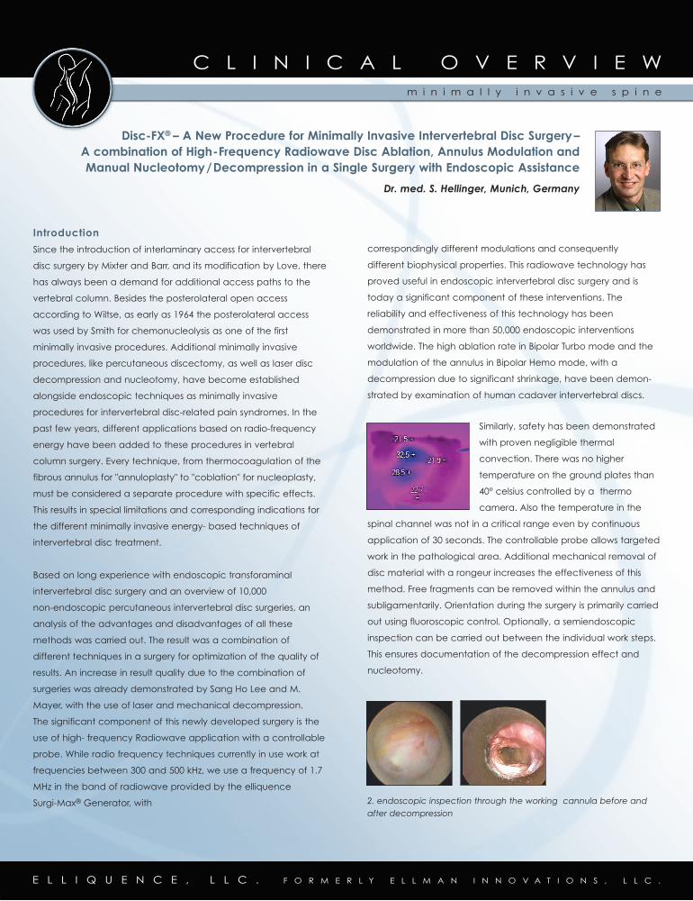
Similarly, safety has been demonstrated
with proven negligible thermal
convection. There was no higher
temperature on the ground plates than
40° celsius controlled by a thermo
camera. Also the temperature in the
spinal channel was not in a critical range even by continuous
application of 30 seconds. The controllable probe allows targeted
work in the pathological area. Additional mechanical removal of
disc material with a rongeur increases the effectiveness of this
method. Free fragments can be removed within the annulus and
subligamentarily. Orientation during the surgery is primarily carried
out using fluoroscopic control. Optionally, a semiendoscopic
inspection can be carried out between the individual work steps.
This ensures documentation of the decompression effect and
nucleotomy.
2. endoscopic inspection through the working cannula before andafter decompression
Disc-FX® – A New Procedure for Minimally Invasive Intervertebral Disc Surgery– A combination of High-Frequency Radiowave Disc Ablation, Annulus Modulation and Manual Nucleotomy/Decompression in a Single Surgery with Endoscopic Assistance
Dr. med. S. Hellinger, Munich, Germany
E L L I Q U E N C E , L L C . F O R M E R L Y E L L M A N I N N O V A T I O N S , L L C .
Indications
Degenerative changes of the intervertebral disc can lead under
certain conditions to pain in the vicinity of the lumbar portion of
the spine, with mono- or polysegmental radiation. Similarly,
neurological deficits, such as distinct dysesthesia or weakness in
the segment-indicating muscles. The intervertebral disc itself can
also lead to pain in the lumbar region, due to penetration of
nerves and vegetative blood vessels. It must be taken into
consideration that the greater part of the pain is derived through
the sinuvertebral nerve and from the intervertebral disc, as well as
the tissues surrounding the nerves. This often makes an
unambiguous mapping between radicular and pseudoradicular
symptoms. Moreover, the unambiguous pathoetiology of
intervertebrally related pain syndrome has yet to be clarified.
Besides mechanical compression, which can often be
demonstrated by imaging techniques, vascular and biochemical
changes in the segment can be discussed. In particular, venous
stasis seems to play a large role in the early phase of the pain
syndrome. The smallest changes in the epidural space can result
in significant changes in venous flow conditions, thus influencing
the course of the disease. Unfortunately, demonstration with
today's technical means is difficult.
The decision for an intervention should primarily be made based
on a thorough case history and clinical symptomology. The
recording of intervertebrally related symptoms is thoroughly
understood, and is the basis for evaluation of an imaged
morphological condition. Nowadays, an MRI of the vertebral
column is considered the standard. In exceptional cases, a
CT scan can also provide the information desired. When
evaluating the radiology, agreement with the clinical assignment
is important.
The chief application areas of the technique exhibited are
contained extrusions and symptomatic protrusions of the
intervertebral discs. Advanced degeneration with clear
intervertebral pain, just as with annuloplasty, can also be
involved. In exceptional cases, good results can also be
obtained with lateral and intraforaminal uncontained extrusions.
Before deciding on intervention, all options for conservative
therapy, including epidural injection as the gold standard of
intervertebral disc treatment, should have been exhausted.
These therapeutic options should also be allotted sufficient time,
at least six weeks. The minimization of posterolateral access using
a 2.5mm working cannula allows surgery with local anesthesia.
Possible complications in the area of the foramen and epidural
space can still be avoided with this method. In particular, the fre-
quent irritation of the ganglion within the foramen with postopera-
tive pain syndromes due to larger endoscopy cannulae to be
discussed have yet to occur. Due to the low level of operative
trauma, fast rehabilitation of the patient can be anticipated.
Instruments Required
Besides the usual surgical equipment, including suction and
irrigation, an image converter is necessary. As an option, a video
unit can be used. The core of the intervention is a complete
single-use instrument set by elliquence, with access cannulae,
guide wires, trephine, and controllable radio frequency probe.
A 16- or 18-gauge spinal needles are also used. Furthermore the
radiowave generator “Surgi-Max®” is necessary. The rongeur
required for mechanical decompression can also be resterilized
and was developed especially for this instrument set. For the
optional endoscopic inspection, there is a special lens with an
irrigation sheath available. The placement of the instruments
requires a fluoroscope with C-arm.
Performing the Intervention/Surgical Technique
The technical
performance of the
surgery is simple. Using a
posterolateral access, a
16- or 18-gauge spinal
needle is introduced
transforaminally into the
intervertebral disc. This is possible in side or prone position. The
placement is more or less medial with respect to the morphology
of the pathology. After an optional discography, a guide wire is
placed using the needle. A small skin incision of 3mm is sufficient
for introduction of the dilatator and working cannula. These can
either be placed on the annulus or an intervertebral disc
fragment. An initial endoscopic inspection is carried out along
with fluoroscopy, to ensure correct positioning. The three main
steps of the procedure will be done through the placed cannula.
E L L I Q U E N C E , L L C . F O R M E R L Y E L L M A N I N N O V A T I O N S , L L C .
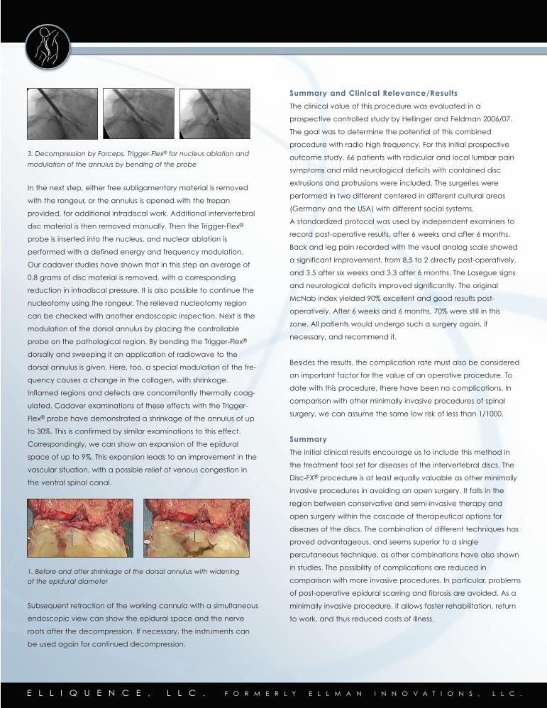
3. Decompression by Forceps, Trigger-Flex® for nucleus ablation and
modulation of the annulus by bending of the probe
In the next step, either free subligamentary material is removed
with the rongeur, or the annulus is opened with the trepan
provided, for additional intradiscal work. Additional intervertebral
disc material is then removed manually. Then the Trigger-Flex®
probe is inserted into the nucleus, and nuclear ablation is
performed with a defined energy and frequency modulation.
Our cadaver studies have shown that in this step an average of
0.8 grams of disc material is removed, with a corresponding
reduction in intradiscal pressure. It is also possible to continue the
nucleotomy using the rongeur. The relieved nucleotomy region
can be checked with another endoscopic inspection. Next is the
modulation of the dorsal annulus by placing the controllable
probe on the pathological region. By bending the Trigger-Flex®
dorsally and sweeping it an application of radiowave to the
dorsal annulus is given. Here, too, a special modulation of the fre-
quency causes a change in the collagen, with shrinkage.
Inflamed regions and defects are concomitantly thermally coag-
ulated. Cadaver examinations of these effects with the Trigger-
Flex® probe have demonstrated a shrinkage of the annulus of up
to 30%. This is confirmed by similar examinations to this effect.
Correspondingly, we can show an expansion of the epidural
space of up to 9%. This expansion leads to an improvement in the
vascular situation, with a possible relief of venous congestion in
the ventral spinal canal.
1. Before and after shrinkage of the dorsal annulus with widening of the epidural diameter
Subsequent retraction of the working cannula with a simultaneous
endoscopic view can show the epidural space and the nerve
roots after the decompression. If necessary, the instruments can
be used again for continued decompression.
Summary and Clinical Relevance/Results
The clinical value of this procedure was evaluated in a
prospective controlled study by Hellinger and Feldman 2006/07.
The goal was to determine the potential of this combined
procedure with radio high frequency. For this initial prospective
outcome study, 66 patients with radicular and local lumbar pain
symptoms and mild neurological deficits with contained disc
extrusions and protrusions were included. The surgeries were
performed in two different centered in different cultural areas
(Germany and the USA) with different social systems.
A standardized protocol was used by independent examiners to
record post-operative results, after 6 weeks and after 6 months.
Back and leg pain recorded with the visual analog scale showed
a significant improvement, from 8.5 to 2 directly post-operatively,
and 3.5 after six weeks and 3.3 after 6 months. The Lasegue signs
and neurological deficits improved significantly. The original
McNab index yielded 90% excellent and good results post-
operatively. After 6 weeks and 6 months, 70% were still in this
zone. All patients would undergo such a surgery again, if
necessary, and recommend it.
Besides the results, the complication rate must also be considered
an important factor for the value of an operative procedure. To
date with this procedure, there have been no complications. In
comparison with other minimally invasive procedures of spinal
surgery, we can assume the same low risk of less than 1/1000.
Summary
The initial clinical results encourage us to include this method in
the treatment tool set for diseases of the intervertebral discs. The
Disc-FX® procedure is at least equally valuable as other minimally
invasive procedures in avoiding an open surgery. It falls in the
region between conservative and semi-invasive therapy and
open surgery within the cascade of therapeutical options for
diseases of the discs. The combination of different techniques has
proved advantageous, and seems superior to a single
percutaneous technique, as other combinations have also shown
in studies. The possibility of complications are reduced in
comparison with more invasive procedures. In particular, problems
of post-operative epidural scarring and fibrosis are avoided. As a
minimally invasive procedure, it allows faster rehabilitation, return
to work, and thus reduced costs of illness.