Embed Size (px)
Citation preview

Biochemistry of
Neurotransmitters
Dr. Diala Abu-Hassan
CNS

References
Mark’s Basic Medical Biochemistry, 4th ed, pp. 908-
918
http://what-when-
how.com/neuroscience/neurotransmitters-the-
neuron-part-1/

What is a neurotransmitter?
A neurotransmitter is a chemical substance that is:
synthesized in a neuron,
Is released at a synapse following depolarization
of the nerve terminal (usually dependent on
influx of calcium ions),
binds to receptors on the postsynaptic cell
and/or presynaptic terminal
to elicit a specific response.
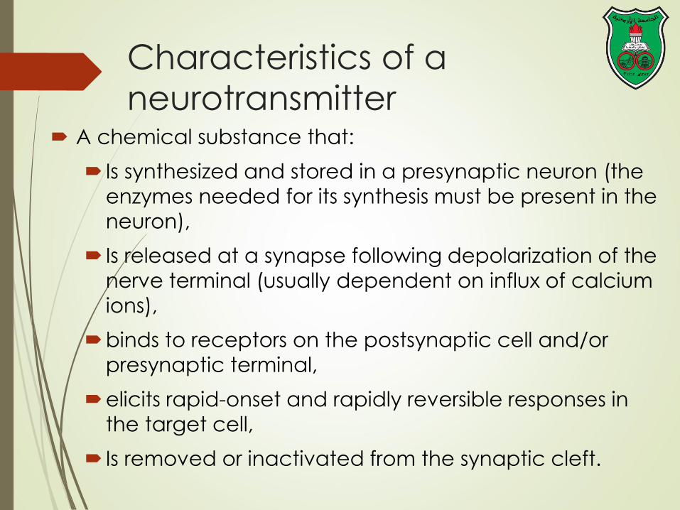
Characteristics of a
neurotransmitter A chemical substance that:
Is synthesized and stored in a presynaptic neuron (the
enzymes needed for its synthesis must be present in the
neuron),
Is released at a synapse following depolarization of the
nerve terminal (usually dependent on influx of calcium
ions),
binds to receptors on the postsynaptic cell and/or
presynaptic terminal,
elicits rapid-onset and rapidly reversible responses in
the target cell,
Is removed or inactivated from the synaptic cleft.

Types of neurotransmitters Small-molecule
Amines (acetylcholine, epinepherine, dopamine, histmaine,
serotonin, norepinephrine, etc.)
Amino acids (glutamate, aspartate, glycine)
Neuropeptides
Gases (nitric oxide)
Each neuron synthesizes only those neurotransmitters that it uses for
transmission through a synapse or to another cell.
The neuronal tracts are often identified by their neurotransmitter; e.g, a
dopaminergic tract synthesizes and releases the neurotransmitter
dopamine.
More than one transmitter (usually a small-molecule transmitter and a
neuroactive peptide) coexist in many mature neurons (e.g., most spinal
motor neurons contain acetylcholine and calcitonin gene-related
peptide).

Structure of neurotransmitters

Neuron may contain
(1) more than one small-molecule
neurotransmitter
(2) more than one neuropeptide
neurotransmitter,
or (3) both types of neurotransmitters.
The differential release of the various
neurotransmitters is the result of the neuron
altering its frequency and pattern of firing.
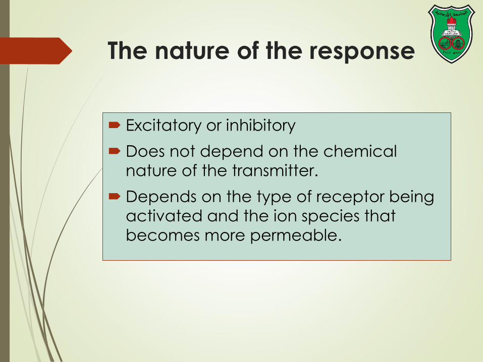
The nature of the response
Excitatory or inhibitory
Does not depend on the chemical
nature of the transmitter.
Depends on the type of receptor being
activated and the ion species that
becomes more permeable.

Neuropeptides

Introduction
Usually mediate slow, ongoing brain functions
More than 50 neuropeptides have been described
Behavior
Pain perception
Memory
Appetite
Thirst
Temperature
Homeostasis
Sleep
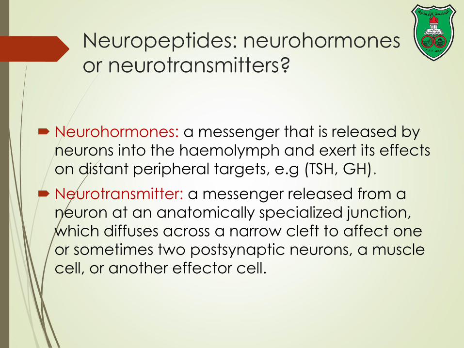
Neuropeptides: neurohormones
or neurotransmitters?
Neurohormones: a messenger that is released by
neurons into the haemolymph and exert its effects
on distant peripheral targets, e.g (TSH, GH).
Neurotransmitter: a messenger released from a
neuron at an anatomically specialized junction,
which diffuses across a narrow cleft to affect one
or sometimes two postsynaptic neurons, a muscle
cell, or another effector cell.
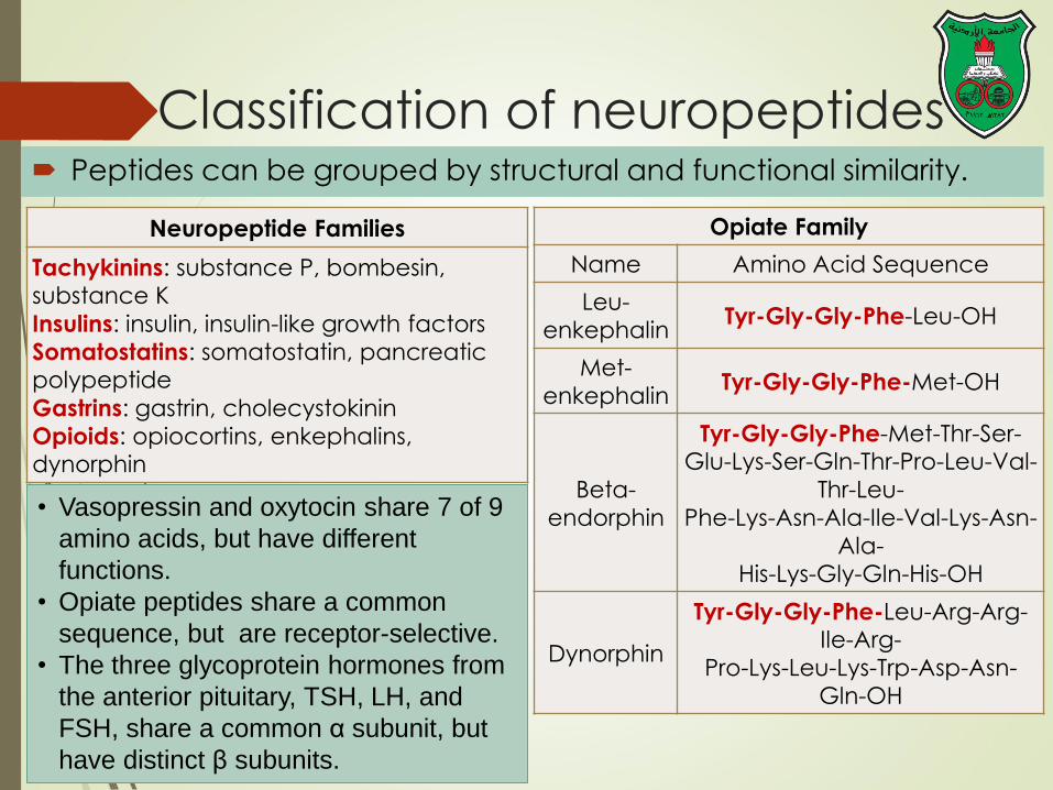
Classification of neuropeptides Peptides can be grouped by structural and functional similarity.
Neuropeptide Families
Tachykinins: substance P, bombesin, substance K
Insulins: insulin, insulin-like growth factors
Somatostatins: somatostatin, pancreatic polypeptide
Gastrins: gastrin, cholecystokinin
Opioids: opiocortins, enkephalins, dynorphin
Opiate Family
Name Amino Acid Sequence
Leu-
enkephalin Tyr-Gly-Gly-Phe-Leu-OH
Met-
enkephalin Tyr-Gly-Gly-Phe-Met-OH
Beta-
endorphin
Tyr-Gly-Gly-Phe-Met-Thr-Ser-Glu-Lys-Ser-Gln-Thr-Pro-Leu-Val-
Thr-Leu-
Phe-Lys-Asn-Ala-Ile-Val-Lys-Asn-
Ala-
His-Lys-Gly-Gln-His-OH
Dynorphin
Tyr-Gly-Gly-Phe-Leu-Arg-Arg-Ile-Arg-
Pro-Lys-Leu-Lys-Trp-Asp-Asn-
Gln-OH
• Vasopressin and oxytocin share 7 of 9
amino acids, but have different
functions.
• Opiate peptides share a common
sequence, but are receptor-selective.
• The three glycoprotein hormones from
the anterior pituitary, TSH, LH, and
FSH, share a common α subunit, but
have distinct β subunits.

Stages of action
Synthesis (ER and Golgi apparatus)
Packaging into large-dense core vesicles (with modifying enzymes)
Transport (fast-axonal transport)
During the transport, proteases cleave the precursor neuropeptide into the final mature form.
Release
They are released gradually over time in response to general increases in the level of intracellular calcium.
Action (prolonged)
Termination by diffusion and degradation (no reuptake)
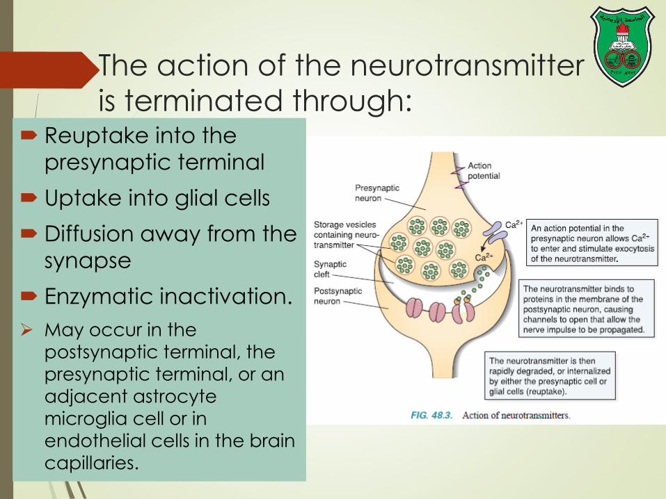
The action of the neurotransmitter
is terminated through: Reuptake into the
presynaptic terminal
Uptake into glial cells
Diffusion away from the
synapse
Enzymatic inactivation.
May occur in the postsynaptic terminal, the
presynaptic terminal, or an
adjacent astrocyte
microglia cell or in endothelial cells in the brain
capillaries.

Diversity: alternative splicing
Alternative splicing
of mRNA leads to
translation of
distinct precursors,
and subsequent
processing leads to
unique mature
peptides.
Example is the
substance P
mRNA that normally also
includes mRNA
encoding
substance K.
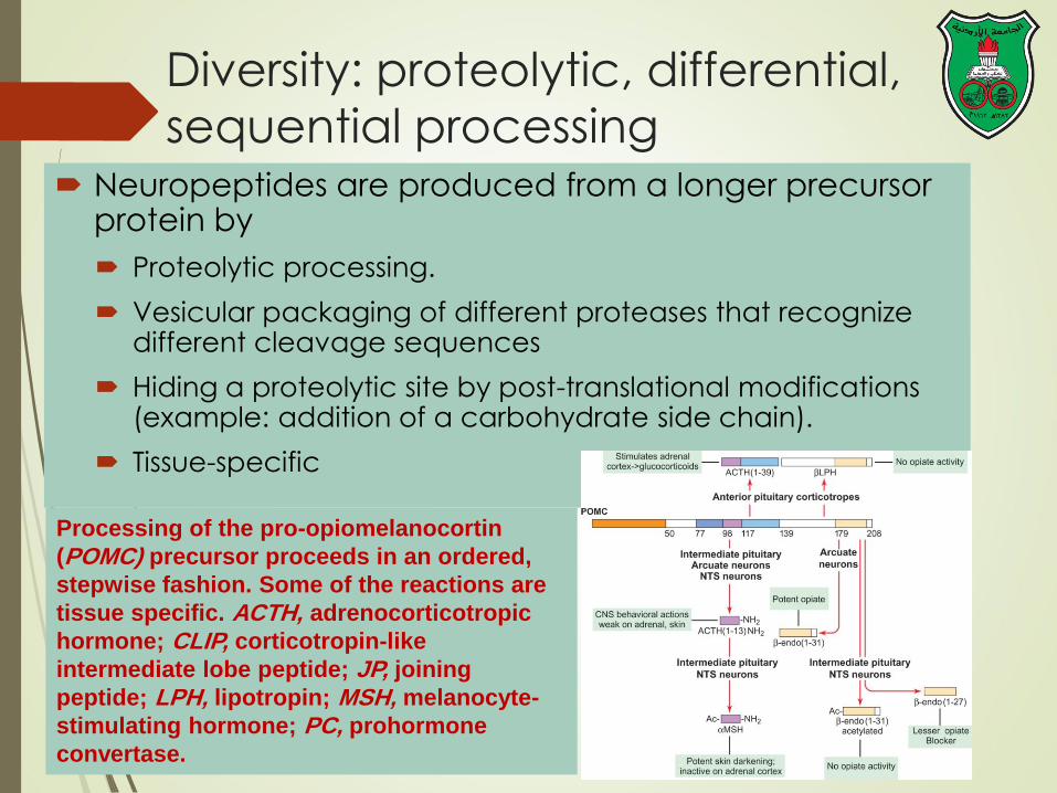
Diversity: proteolytic, differential,
sequential processing Neuropeptides are produced from a longer precursor
protein by
Proteolytic processing.
Vesicular packaging of different proteases that recognize different cleavage sequences
Hiding a proteolytic site by post-translational modifications (example: addition of a carbohydrate side chain).
Tissue-specific
Processing of the pro-opiomelanocortin
(POMC) precursor proceeds in an ordered,
stepwise fashion. Some of the reactions are
tissue specific. ACTH, adrenocorticotropic
hormone; CLIP, corticotropin-like
intermediate lobe peptide; JP, joining
peptide; LPH, lipotropin; MSH, melanocyte-
stimulating hormone; PC, prohormone
convertase.

The levels of regulation of
neuropeptide expression

Small-molecule
neurotransmitters

Types of small-molecule
neurotransmitters
Nitrogen-containing molecules
amino acids and their derivatives
intermediates of glycolysis and the
Krebs cycle (TCA cycle)
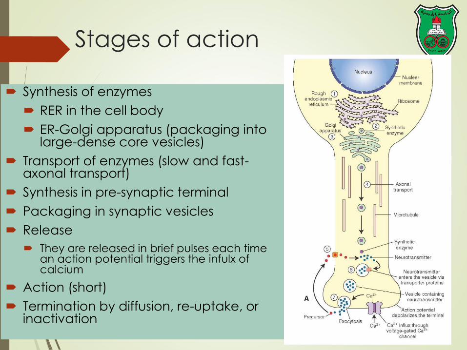
Stages of action
Synthesis of enzymes
RER in the cell body
ER-Golgi apparatus (packaging into large-dense core vesicles)
Transport of enzymes (slow and fast-axonal transport)
Synthesis in pre-synaptic terminal
Packaging in synaptic vesicles
Release
They are released in brief pulses each time an action potential triggers the infulx of calcium
Action (short)
Termination by diffusion, re-uptake, or inactivation

Role of calcium
• Vesicles are located further away from the presynaptic
membrane and away from area of Ca influx
• Ca influx can be from external or internal sources.

mM2 [Ca+] =
uM0.1 [Ca+] =
uM100 -50[Ca+] =

• The fused vesicular membrane is retrieved and
recycled within a minute by a complex process called
endocytotic budding.
• Several proteins, including clathrin, form a basket-like
lattice on the remnants of the fused vesicle giving the
appearance of a coated pit which is then pinched off
from the presynaptic membrane by another protein
called dynamin
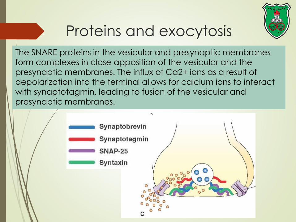
Proteins and exocytosis
The SNARE proteins in the vesicular and presynaptic membranes form complexes in close apposition of the vesicular and the
presynaptic membranes. The influx of Ca2+ ions as a result of
depolarization into the terminal allows for calcium ions to interact
with synaptotagmin, leading to fusion of the vesicular and presynaptic membranes.

Differences between
neuropeptides and small molecule
neurotransmitters
Onset and duration of action
Synthesis, transport, and packaging
Concentration for action and receptor
binding
Concentration of [Ca+] for release
Site of synthesis, modification
Fate

Synthesis of
neurotransmitters
Most are synthesized from amino acids,
intermediates of glycolysis and the TCA
cycle, and O2 in the cytoplasm of the
presynaptic terminal.
The rate of synthesis is generally
regulated to correspond to the rate of
firing of the neuron.

Tyrosine-Derived
Neurotransmitters Dopamine, norepinephrine, and epinephrine

Notes
Role of cofactors
S-adenosylmethionine (methyl
transfer)
Pyrodoxal phosphate (vitamin B6):
transamination, decarboxylation
Tetrahydrobiopterin (BH4)
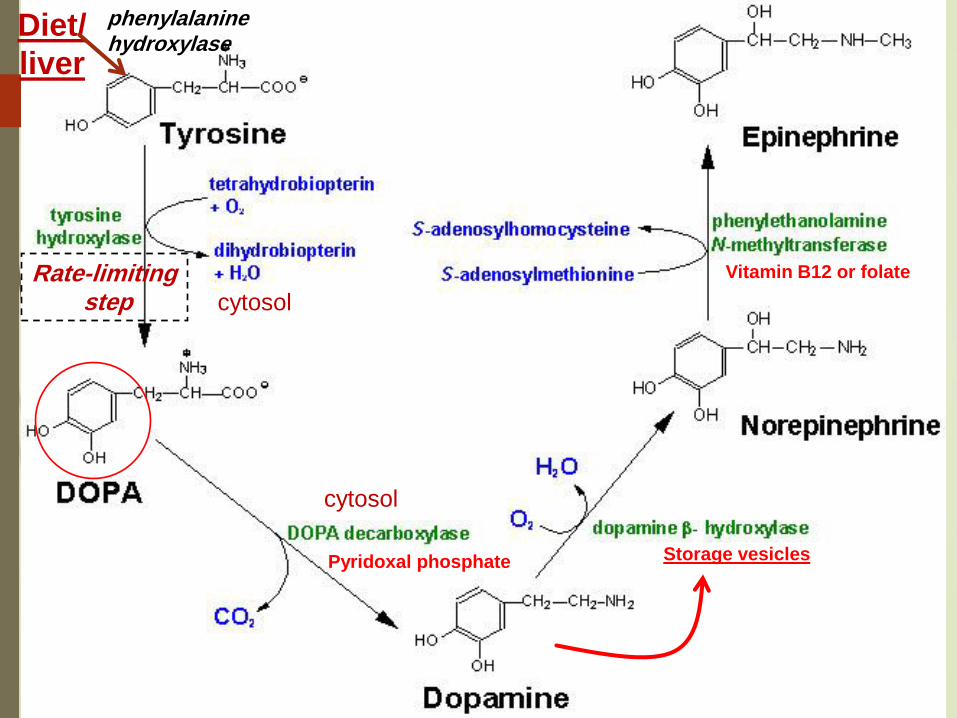
Rate-limiting step
Pyridoxal phosphate Storage vesicles
Vitamin B12 or folate
Diet/
liver
phenylalanine hydroxylase
cytosol
cytosol
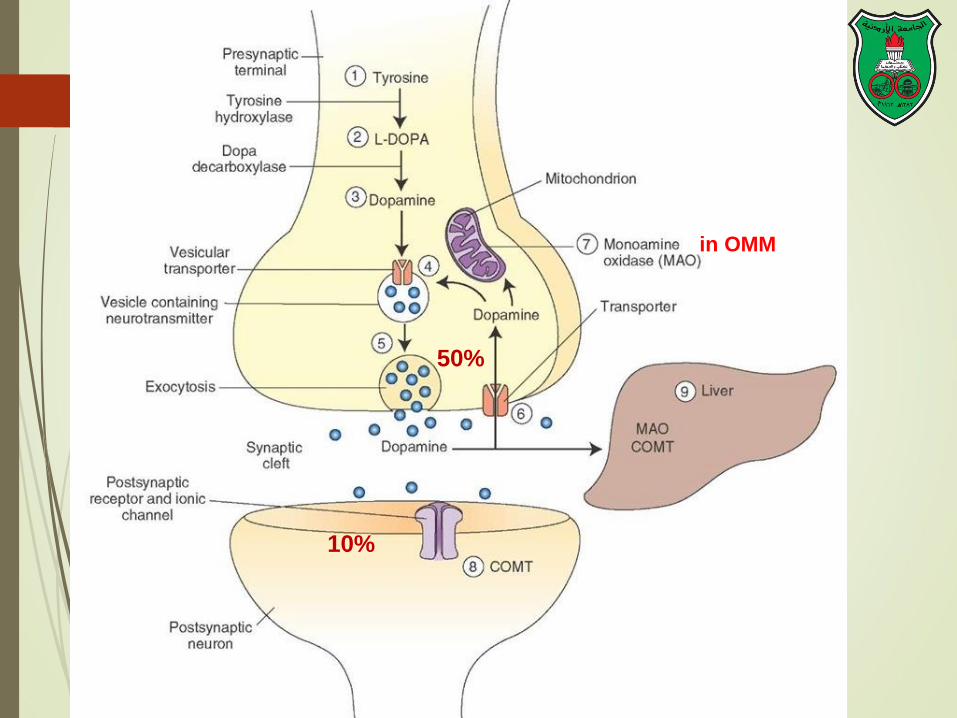
50%
10%
in OMM

LDCV large
dense-core
vesicles

Leaking

Packaging into vesicles The catecholamines
(dopamine an epinepherine) are transported into vesicles by an ATP-dependent process linked to a proton pump.
Protons are pumped into the vesicles by a vesicular ATPase (V-ATPase).
The protons then exchange for the positively charged catecholamine via the transporter VMAT2 (vesicle monoamine transporter 2).

COMT and MAO
Parkinson’s
disease
Inactivation is
dependent on SAM,
vitamin B12 and folate

Regulation
• Tyrosine hydroxylase
– Short term
• Inhibition by free cytosolic catecholamines
• Catecholamines compete with BH4 binding to
enzyme
• Activation by depolarization
– Tight binding of the enzyme to BH4 following
phosphorylation by PKA, CAM kinases, PKC
– Long-term (plus dopamine -hyroxylase)
Alterations (increase) in the enzyme amounts when sympathetic
neuronal activity is increased for a prolonged period

Tryptophan-Derived
Neurotransmitters Serotonin and melatonin
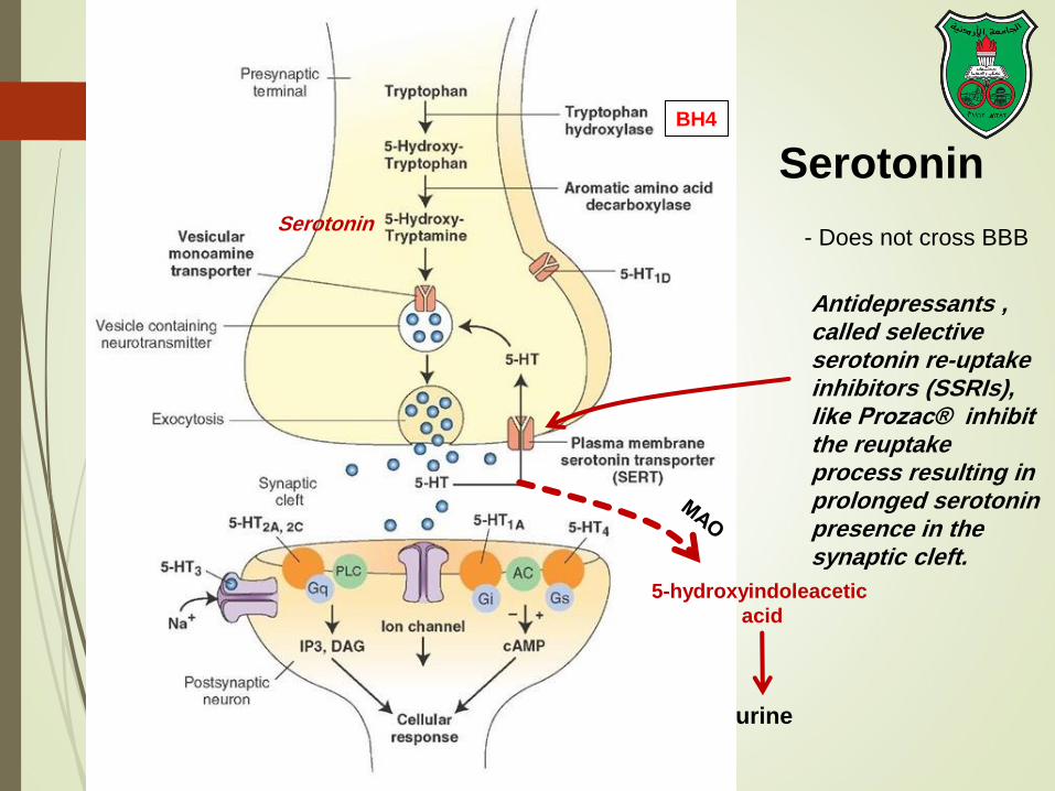
BH4
Serotonin
5-hydroxyindoleacetic
acid
urine
Antidepressants , called selective serotonin re-uptake inhibitors (SSRIs), like Prozac® inhibit the reuptake process resulting in prolonged serotonin presence in the synaptic cleft.
Serotonin
- Does not cross BBB

Melatonin
Serotonin synthesized in the pineal gland serves as a precursor
for the synthesis of melatonin, which is a neurohormone
involved in regulating:
sleep patterns
Seasonal and circadian (daily) rhythms
Dark-light cycle
+SAM +SAH

Glutamate and aspartate

Glutamate and aspartate
Nonessential amino acids
Do not cross BBB
must be synthesized in neurons de novo from glucose rather than taken up from the blood
Main synthetic compartments
neurons
glial cells
Both are excitatory neurotransmitters.

Synthesis of glutamate Sources:
1. Glycolysis Krebs cycle
dehydrogenation of -
ketoglutarate
2. Glutamine (deamination by
glutaminase)
3. Aspartate (transamination)
Is stored in vesicles, and its release is
Ca2-dependent.
Removal by high-affinity uptake
systems in nerve terminals and glial
cells.
excitatory amino acid carrier-1
(EAAC1)
glutamate transporter-1 (GLT-1) and
glutamate—aspartate transporter
(GLAST)
GABA
glutaminase Glutamine
synthetase
transaminase 1
2
3
-KG
Glu
Dehydro

Sources of glutamate
(supplementary)
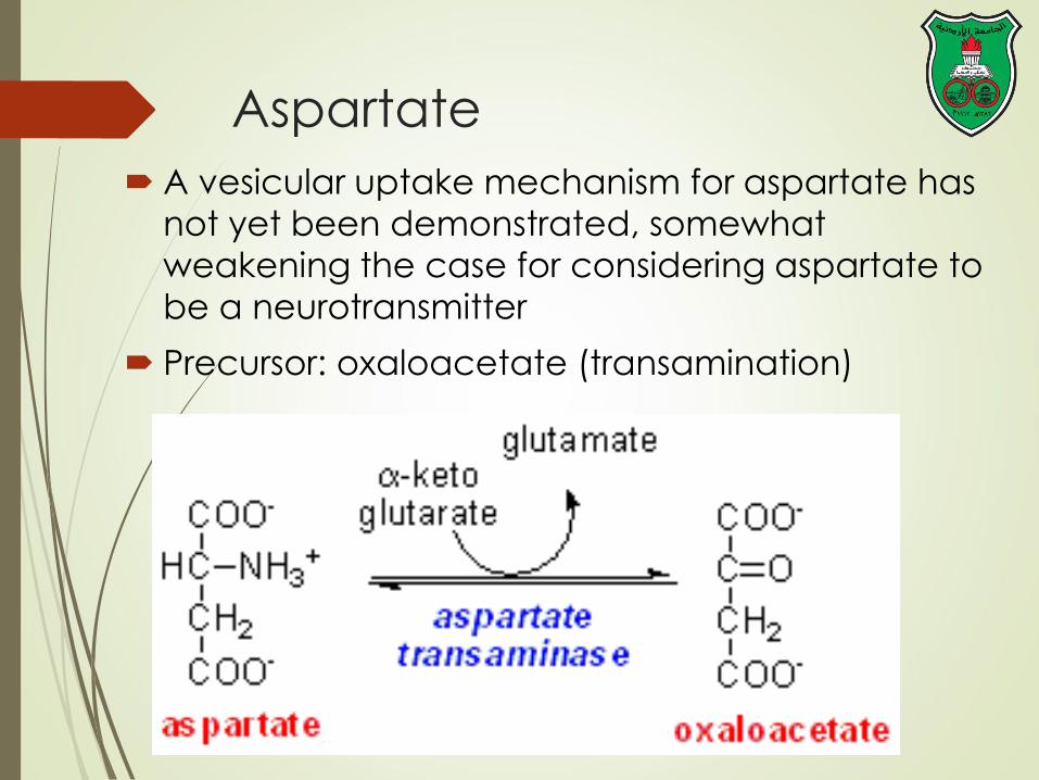
Aspartate
A vesicular uptake mechanism for aspartate has
not yet been demonstrated, somewhat
weakening the case for considering aspartate to
be a neurotransmitter
Precursor: oxaloacetate (transamination)
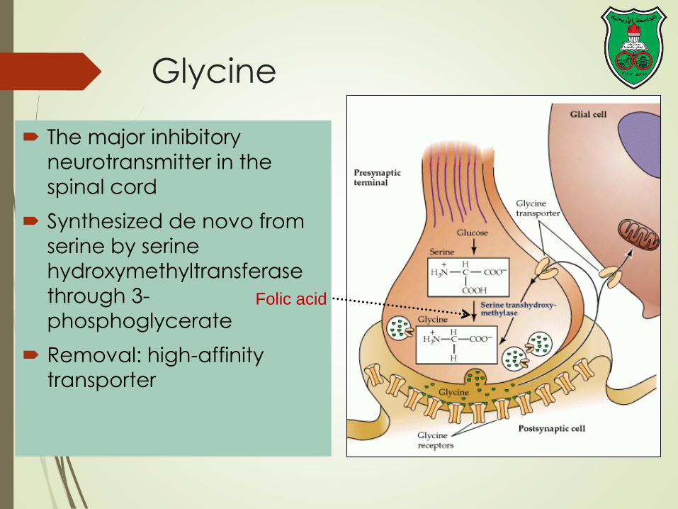
Glycine
The major inhibitory
neurotransmitter in the
spinal cord
Synthesized de novo from
serine by serine
hydroxymethyltransferase
through 3-
phosphoglycerate
Removal: high-affinity
transporter
Folic acid
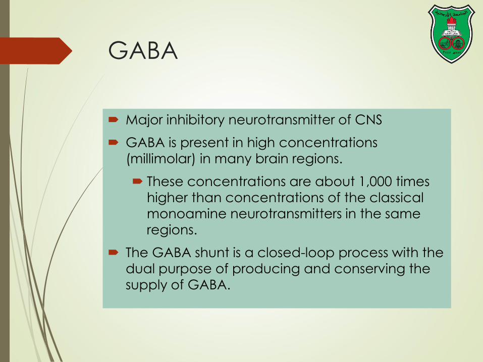
GABA
Major inhibitory neurotransmitter of CNS
GABA is present in high concentrations
(millimolar) in many brain regions.
These concentrations are about 1,000 times
higher than concentrations of the classical
monoamine neurotransmitters in the same
regions.
The GABA shunt is a closed-loop process with the
dual purpose of producing and conserving the
supply of GABA.
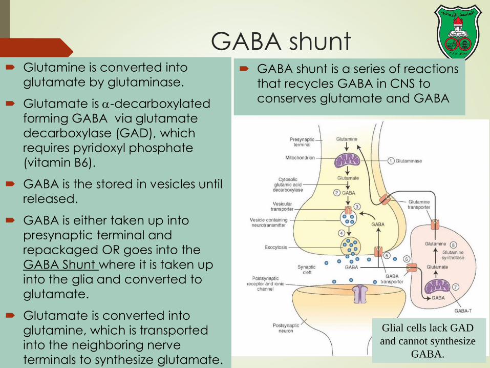
GABA shunt Glutamine is converted into
glutamate by glutaminase.
Glutamate is -decarboxylated
forming GABA via glutamate
decarboxylase (GAD), which
requires pyridoxyl phosphate
(vitamin B6).
GABA is the stored in vesicles until
released.
GABA is either taken up into
presynaptic terminal and
repackaged OR goes into the
GABA Shunt where it is taken up
into the glia and converted to
glutamate.
Glutamate is converted into
glutamine, which is transported
into the neighboring nerve
terminals to synthesize glutamate.
GABA shunt is a series of reactions
that recycles GABA in CNS to
conserves glutamate and GABA
Glial cells lack GAD
and cannot synthesize
GABA.

Acetylcholine
The acetyl group used for
acetylcholine synthesis is derived
principally from glucose oxidation
to pyruvate and decarboxylation of
pyruvate to form acetyl-CoA via
the pyruvate dehydrogenase
reaction.
serine
esterase
Sarin nerve
gas inhibits it
• Acetylcholine (AC) is the
major neurotransmitter at
the NMJ
• Inability to inactivate AC
leads to constant activation
of the nerve–muscle
synapses, leading to varying
degrees of paralysis.
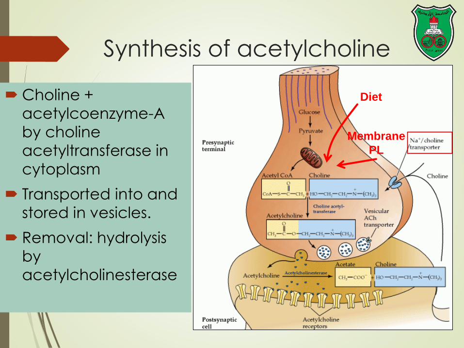
Diet
Membrane
PL
Synthesis of acetylcholine
Choline +
acetylcoenzyme-A
by choline
acetyltransferase in
cytoplasm
Transported into and
stored in vesicles.
Removal: hydrolysis
by
acetylcholinesterase

Histamine it does not penetrate the blood-brain barrier and, hence, must be
synthesized in the brain.
Histamine is inactivated by two enzymes—histamine methyltransferase then oxidation by MAO-B (brain) and diamine oxidase (histaminase) (peripheral tissues).
Pyridoxal phosphate
Astrocytes
(MAO) Neuron
X
No recycling into
presynaptic terminal
Newly synthesized
neuronal histamine is
stored in the nerve
terminal vesicles.
Once it is released
from neurons,
histamine is thought
to activate both
postsynaptic and
presynaptic receptors.
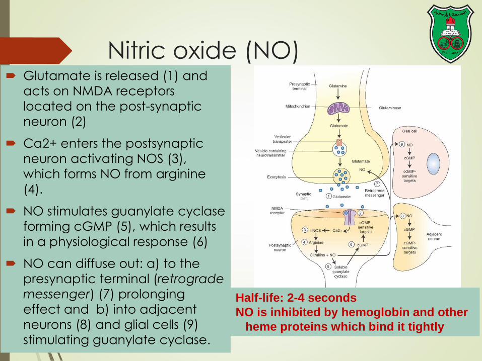
Nitric oxide (NO) Glutamate is released (1) and
acts on NMDA receptors
located on the post-synaptic
neuron (2)
Ca2+ enters the postsynaptic
neuron activating NOS (3),
which forms NO from arginine
(4).
NO stimulates guanylate cyclase
forming cGMP (5), which results
in a physiological response (6)
NO can diffuse out: a) to the
presynaptic terminal (retrograde
messenger) (7) prolonging
effect and b) into adjacent
neurons (8) and glial cells (9)
stimulating guanylate cyclase.
Half-life: 2-4 seconds
NO is inhibited by hemoglobin and other
heme proteins which bind it tightly

Is NO a neurotransmitter?
Yes, but:
It is not stored in vesicles
It is not released by calcium-dependent exocytosis (it
diffuses)
Its inactivation is passive (there is no active process that
terminates its action)
It decays spontaneously
It does not interact with receptors on target cells
Its sphere of action depends on the extent to which it diffuses, and its action is not confined to the
conventional presynaptic-postsynaptic direction.
NO acts as a retrograde messenger and regulates the function of axon terminals presynaptic to the neuron in
which it is synthesized.

NO synthase Isoform I (nNOS or cNOS)
Neurons and epithelial cells
activated by the influx of
extracellular calcium
isoform II (iNOS)
Macrophages and smooth
muscle cells
induced by cytokines
and isoform III (eNOS)
Endothelial cells lining blood vessels
activated by the influx of extracellular calcium
All three isoforms require BH2 as a cofactor and nicotinamide adenine
dinucleotide phosphate (NADPH) as a coenzyme

![Plasma l-[3H]Norepinephrine, d-['4C]Norepinephrine, › ... › JCI83111134.pdf · 2014-01-30 · Plasma l-[3H]Norepinephrine, d-['4C]Norepinephrine, and d,l-[3H]Isoproterenol Kinetics](https://img.dokumen.tips/doc/110x75/5f0f14b47e708231d44264fd/plasma-l-3hnorepinephrine-d-4cnorepinephrine-a-a-jci83111134pdf.jpg)