Embed Size (px)
Citation preview

DR. AMADIN A. OLOTU
LECTURER/CONSULTANT CLINICAL MICROBIOLOGIST
BOWEN UNIVERSITY/BOWEN UNIVERSITY TEACHING HOSPITAL OGBOMOSO
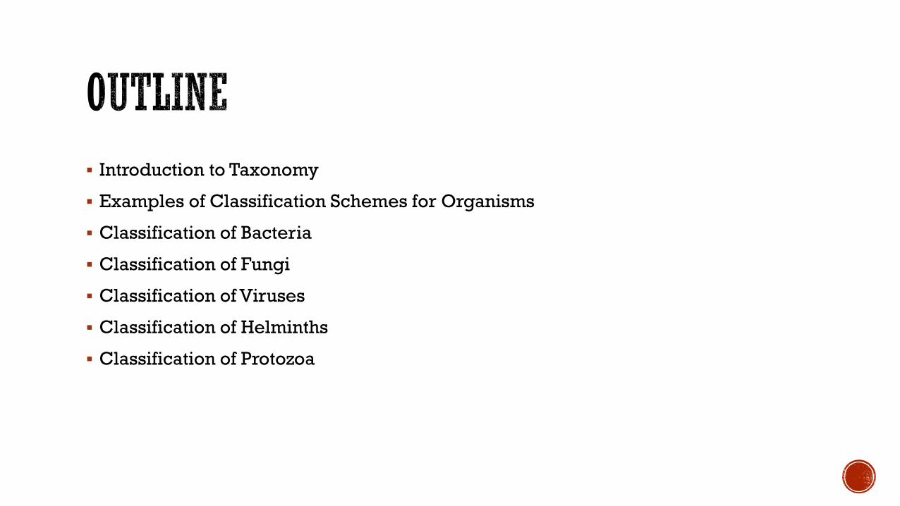
▪ Introduction to Taxonomy
▪ Examples of Classification Schemes for Organisms
▪ Classification of Bacteria
▪ Classification of Fungi
▪ Classification of Viruses
▪ Classification of Helminths
▪ Classification of Protozoa
▪ The science of taxonomy involves not just naming organisms, but grouping them with other organisms that share common properties.
▪ It is the classification of organisms in an ordered system that indicates a natural relationship
▪ A taxon is a collection of related organisms grouped together for purposes of classification. Thus, genus, family, etc. are taxons.
▪ The Swedish botanist Linnaeus introduced the ´ binomial systemof nomenclature, by which each organism was assigned a genus and a species.

▪ The ranks commonly used are kingdom, phylum, class, order, family, genus, and species
▪ family, genus, and species are the most useful to us.

▪ The first letter in the genus name is always written as a capital letter and the other letters as small letters while all the letters in the species name are written as smallletters.
▪ The genus and species name are italicised, or, if this isn’t possible, underlined
▪ Homo sapiens
▪ Escherichia coli
▪ In the past bacteria have been grouped and named primarily based on their morphology, staining reactions, biochemical properties, nutritional requirements and antigenic differences.
▪ So there was a reliance on phenotype or observable characteristics.
▪ More recently classification has relied more on genotype or genetic make up.

▪ Comparison of nucleic acid sequences, notably those of 16S ribosomal RNA genes, has led to a new, phylogenetically based scheme of classification, that is, one based on how closely, different groups of bacteria are thought to be related, rather than what morphological or physiological features they may share.
▪ This has led to reclassification of some organisms.
▪ Different schemes for classification of organisms have been proposed over the years
▪ One of the most widely accepted of these was the five kingdom system proposed by Robert Whittaker in 1969
▪ Monera
▪ Protista
▪ Fungi
▪ Plantae
▪ Animalia

Whittaker’s five-kingdom system of classification
Examples of Classification Schemes for Organisms
EXAMPLES OF CLASSIFICATION SCHEMES FOR ORGANISMS
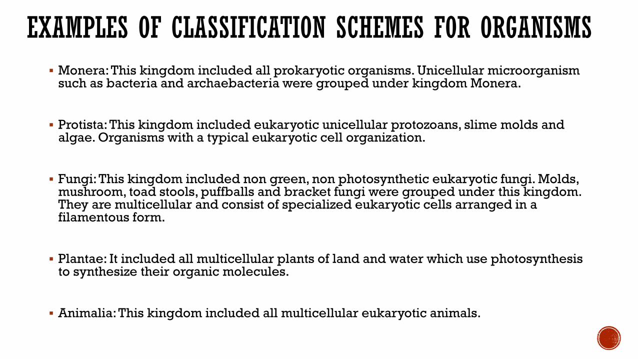
▪ Monera: This kingdom included all prokaryotic organisms. Unicellular microorganism such as bacteria and archaebacteria were grouped under kingdom Monera.
▪ Protista: This kingdom included eukaryotic unicellular protozoans, slime molds and algae. Organisms with a typical eukaryotic cell organization.
▪ Fungi: This kingdom included non green, non photosynthetic eukaryotic fungi. Molds, mushroom, toad stools, puffballs and bracket fungi were grouped under this kingdom. They are multicellular and consist of specialized eukaryotic cells arranged in a filamentous form.
▪ Plantae: It included all multicellular plants of land and water which use photosynthesis to synthesize their organic molecules.
▪ Animalia: This kingdom included all multicellular eukaryotic animals.
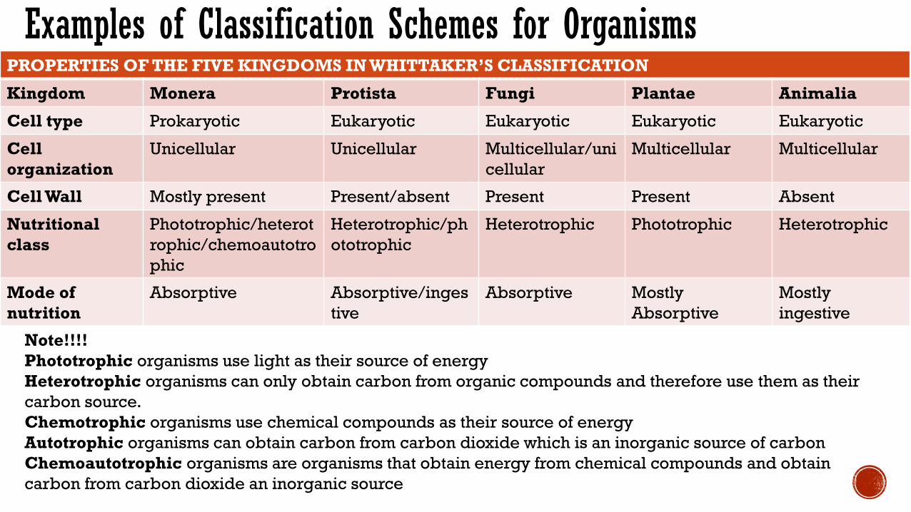
PROPERTIES OF THE FIVE KINGDOMS IN WHITTAKER’S CLASSIFICATION
Kingdom Monera Protista Fungi Plantae Animalia
Cell type Prokaryotic Eukaryotic Eukaryotic Eukaryotic Eukaryotic
Cell
organization
Unicellular Unicellular Multicellular/uni
cellular
Multicellular Multicellular
Cell Wall Mostly present Present/absent Present Present Absent
Nutritional
class
Phototrophic/heterot
rophic/chemoautotro
phic
Heterotrophic/ph
ototrophic
Heterotrophic Phototrophic Heterotrophic
Mode of
nutrition
Absorptive Absorptive/inges
tive
Absorptive Mostly
Absorptive
Mostly
ingestive
Examples of Classification Schemes for Organisms
Note!!!!
Phototrophic organisms use light as their source of energy
Heterotrophic organisms can only obtain carbon from organic compounds and therefore use them as their
carbon source.
Chemotrophic organisms use chemical compounds as their source of energy
Autotrophic organisms can obtain carbon from carbon dioxide which is an inorganic source of carbon
Chemoautotrophic organisms are organisms that obtain energy from chemical compounds and obtain
carbon from carbon dioxide an inorganic source

▪ However molecular studies in the 1970s revealed that the Archaea differed from all other bacteria in their 16S rRNA sequences, as well as in their cell wall structure, membrane lipids
▪ These differences were seen as sufficiently important for the recognition of a third basic cell type to add to the procaryotes and eucaryotes.
▪ This led to the proposal of a three-domain scheme of classification:
▪ Archaea, Bacteria and Eucarya

▪ The Three-Domain System was first proposed by Carl Woese in 1990.
▪ This classification system divides life forms into three domains and six kingdoms.
▪ The three-Domains are Archaea, Bacteria, Eukarya
▪ The six kingdoms are:
▪ under the Domain Archaea - Kingdom Archaebacteria
▪ under the Domain Bacteria – Kingdom Eubacteria
▪ under the Domain Eukarya – Kingdom Protista, Kingdom Fungi, Kingdom Plantae, Kingdom Animalia.
▪ This classification system divides the life based on the differences in the 16S ribosomal RNA (rRNA) structure and as well as the cell’s membrane lipid structure and its sensitivity to antibiotics.

▪Domain Archaea▪ The organisms in the Domain Archaea (Kingdom Archaebacteria) possess the following
characteristics:
▪ Archaea are prokaryotic cells.
▪ Unlike the Bacteria and the Eukarya, the Archaea have membranes composed of branched hydrocarbon chains (many also containing rings within the hydrocarbon chains) attached to glycerol by ether linkages.
▪ The cell walls of Archaea contain no peptidoglycan.
▪ Archaea are not sensitive to some antimicrobials that affect the Bacteria, but are sensitive to some antimicrobials that affect the Eukarya.
▪ Archaea contain rRNA that is unique to the Archaea this is demonstrated by the presence of molecular regions distinctly different from the rRNA of Bacteria and Eukarya.
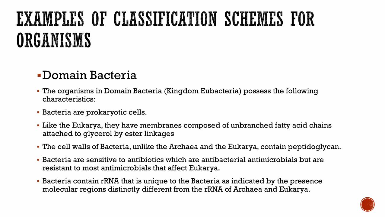
▪Domain Bacteria▪ The organisms in Domain Bacteria (Kingdom Eubacteria) possess the following
characteristics:
▪ Bacteria are prokaryotic cells.
▪ Like the Eukarya, they have membranes composed of unbranched fatty acid chains attached to glycerol by ester linkages
▪ The cell walls of Bacteria, unlike the Archaea and the Eukarya, contain peptidoglycan.
▪ Bacteria are sensitive to antibiotics which are antibacterial antimicrobials but are resistant to most antimicrobials that affect Eukarya.
▪ Bacteria contain rRNA that is unique to the Bacteria as indicated by the presence molecular regions distinctly different from the rRNA of Archaea and Eukarya.

▪Domain Eukarya▪ The organisms in the Domain Eukarya possess the following characteristics:
▪ Eukarya have eukaryotic cells.
▪ Like the Bacteria, they have membranes composed of unbranched fatty acid chains attached to glycerol by ester linkages
▪ Not all Eukarya possess cells with a cell wall, but for those Eukarya having a cell wall, that wall contains no peptidoglycan.
▪ Eukarya are mostly resistant to traditional antibacterial antimicrobials but are sensitive to various antimicrobials that affect eukaryotic cells.
▪ Eukarya contain rRNA that is unique to the Eukarya as indicated by the presence molecular regions distinctly different from the rRNA of Archaea and Bacteria.
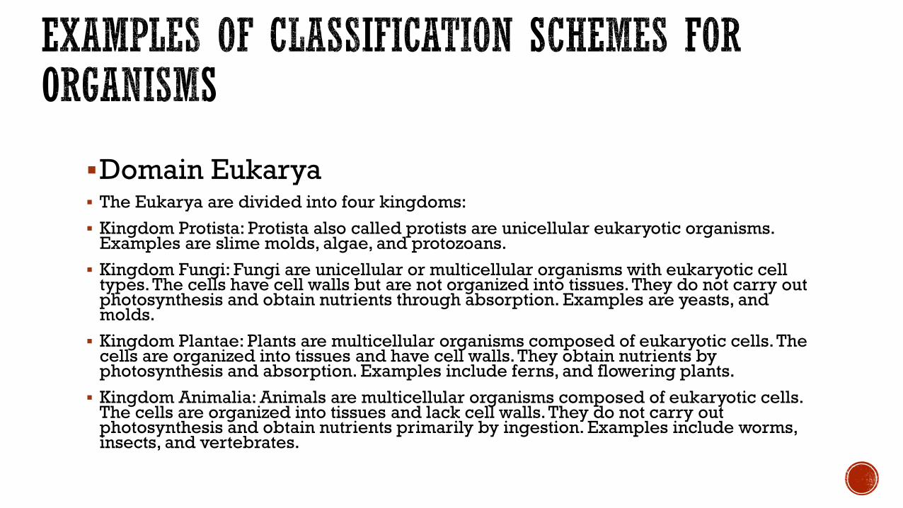
▪Domain Eukarya▪ The Eukarya are divided into four kingdoms:
▪ Kingdom Protista: Protista also called protists are unicellular eukaryotic organisms. Examples are slime molds, algae, and protozoans.
▪ Kingdom Fungi: Fungi are unicellular or multicellular organisms with eukaryotic cell types. The cells have cell walls but are not organized into tissues. They do not carry out photosynthesis and obtain nutrients through absorption. Examples are yeasts, and molds.
▪ Kingdom Plantae: Plants are multicellular organisms composed of eukaryotic cells. The cells are organized into tissues and have cell walls. They obtain nutrients by photosynthesis and absorption. Examples include ferns, and flowering plants.
▪ Kingdom Animalia: Animals are multicellular organisms composed of eukaryotic cells. The cells are organized into tissues and lack cell walls. They do not carry out photosynthesis and obtain nutrients primarily by ingestion. Examples include worms, insects, and vertebrates.

▪ Using the 3 domain system of classification the bacterium: Escherichia coli would be classified scientifically as;
▪ Domain: Bacteria
▪ Kingdom: Eubacteria
▪ Phylum: Proteobacteria
▪ Class: Gammaproteobacteria
▪ Order: Enterobacteriales
▪ Family: Enterobacteriaceae
▪ Genus: Escherichia
▪ Species: Escherichia coli
▪ However what is more useful to us clinically is a classification based on certain characteristics especially characteristics that can be used in identification

▪ The simplest and most useful classification of bacteria is based on their gram stain reaction and morphology
▪ Gram stain reaction
▪ Developed by Hans Christian Gram
▪ Based on cell wall differences
▪ Enables us view bacteria from specimens or culture under the microscope

▪ Gram stain Method:
▪ 1 Fix the dried smear with heat or alcohol
▪ 2 Cover the fixed smear with Crystal violet stain for 30–60 seconds.3 Rapidly wash off the stain with clean water.4 Tip off all the water, and cover the smear with Lugol’s iodine for 30–60 seconds.5 Wash off the iodine with clean water.6 Decolorize rapidly (few seconds) with acetone. Wash immediately with clean water.7 Cover the smear with Safranin stain for 1-2 minutes.8 Wash off the stain with clean water.9 Wipe the back of the slide clean, and place it in a draining rack for the smear to air-dry.10 Examine the smear microscopically, first with the 40x objective to check the staining and to see the distribution of material, and then with the oil immersion or 100x objective to report the bacteria and cells

Gram negative bacilli Gram positive cocci
Classification of Bacteria

▪ Principle of the gram stain
▪ – Gram positive bacteria have a thick layer of peptidoglycan and when acetone is applied in the decolorization step in the gram stain they retain the complex of crystal violet and lugols iodine because of the thick peptidoglycan layer and so do not take up the counterstain safranin and so look purple or dark blue under the microscope.
▪ Gram negative bacteria on the other hand when decolorized by acetone do not retain the crystal violet – lugols iodine complex because they have a thin layer of peptidoglycan so they take up the counterstain safranin and appear red or pink under the microscope.

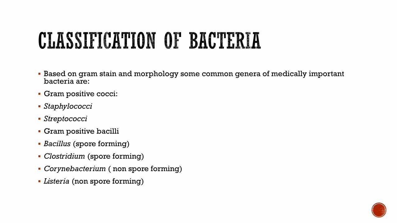
▪ Based on gram stain and morphology some common genera of medically important bacteria are:
▪ Gram positive cocci:
▪ Staphylococci
▪ Streptococci
▪ Gram positive bacilli
▪ Bacillus (spore forming)
▪ Clostridium (spore forming)
▪ Corynebacterium ( non spore forming)
▪ Listeria (non spore forming)

▪ Gram negative cocci
▪ Neisseria
▪ Moraxella
▪ Gram negative bacilli
▪ Escherichia
▪ Klebsiella
▪ Enterobacter
▪ Citrobacter
▪ Proteus
▪ Shigella
▪ Salmonella

Morphology Gram positive Gram negative
Coccus/cocci (spherical) Streptococcus
Staphylococcus
Neisseria
Bacillus/bacilli (rod shaped) Listeria
Corynebacterium
Bacillus
Clostridium
Escherichia coli
Klebsiella
Enterobacter
Citrobacter
Proteus
Salmonella
Shigella
Yersinia
Pseudomonas
Vibrio
Campylobacter
Helicobacter
Spiral Spirochetes
Treponema
Borrelia
Leptospira
Branching filamentous growth Actinomyces
Norcardia (partially acid fast)
Pleomorphic Chlamydia
Rickettsiae
Absence of a cell wall Mycoplasma ( cannot be classified as gram positive or gram negative)
Acid fast – Mycobacterium cannot be stained using the gram stain rather Ziehl Neelson is used
▪ One important property that can be used to classify bacteria is:
▪ How the organism deals with oxygen:
▪ Molecular oxygen is very reactive, and when it snatches up electrons, it can form hydrogen peroxide (H2O2),
▪ superoxide radicals (02-), and a
▪ hydroxyl radical (OH-).
▪ All of these are toxic unless broken down

▪ There are 3 enzymes that some bacteria possess to break downthese oxygen products:
▪ Catalase breaks down hydrogen peroxide
▪ Peroxidase also breaks down hydrogen peroxide
▪ Superoxide dismutase breaks down the superoxide radical

▪ Bacteria with these enzymes can survive in the presence of oxygen while bacteria without these enzymes will not survive in the presence of oxygen so based on this we can classify bacteria as:
▪ Obligate aerobes
▪ Facultative anaerobes
▪ Microaerophilic
▪ Obligate anaerobes

Staining
Reacttion
Obligate
aerobes
Facultative
Anaerobes
Microaerophill
ic
Obligate
anaerobes
Gram positive Nocardia
(weakly acid
fast)
Bacillus cereus
Staphycoccus
Bacillus anthracis
Corynebacterium
Listeria
Actinomyces
Clostridium
Gram negative Neisseria
Pseudomonas
Bordetella
Legionella
Brucella
Enterobacteriacea
e
Spirochaetes
Treponema
Borrelia
Leptospira
Campylobacter
Bacteroides
Acid fast Mycobacterium
Norcardia
No cell wall
Mycoplasma

▪ Fungi are
▪ Nonmotile eukaryotic organisms, with cell walls and which produce filamentous structures and spores.
▪ They exist as unicellular forms called yeasts or multicellular filamentous forms called moulds
▪ They are considered saprophytes
▪Two kinds of classification commonly used
▪Morphologic classification and Clinical classification
▪Morphologic Classification▪Yeasts (unicellular forms) eg Candida spp.
▪Moulds (multicellular filamentous forms) eg Aspergillus spp.
▪Dimorphic fungi – can exist as yeasts and moulds at different temperatures eg Histoplasmosis spp
▪Clinical Classification▪ Superficial mycoses –fungal infection of the superficial layers
of the skin eg
▪ Cutaneous mycoses –fungal infection of keratinized tissue (skin, hair and nails)
▪ Subcutaneous mycoses –fungal infection of subcutaneous tissue
▪ Systemic mycoses –fungal infections that are systemic in nature involving deep tissue and organs.
▪ Opportunistic mycoses –fungal infections that occur mainly in the immunocompromised

Type of Mycosis Disease Causative Fungal Agents
Superficial mycoses Pityriasis versicolor Malassezia species
Tinea nigra Hortaea werneckii
White piedra Trichosporon species
Black piedra Piedraia hortae
Cutaneous mycoses Dermatophytosis Microsporum species, Trichophyton species, and Epidermophyton floccosum
Candidiasis of skin, mucosa, or nails Candida albicans and other Candida species
Subcutaneous mycoses Sporotrichosis Sporothrix schenckii
Chromoblastomycosis Phialophora verrucosa, Fonsecaea pedrosoi, and others
Mycetoma Pseudallescheria boydii, Madurella mycetomatis, and others
Phaeohyphomycosis Exophiala, Bipolaris, Exserohilum, and other dematiaceous mold
Systemic mycoses Coccidioidomycosis Coccidioides posadasii and Coccidioides immitis
Histoplasmosis Histoplasma capsulatum
Blastomycosis Blastomyces dermatitidis
Paracoccidioidomycosis Paracoccidioides brasiliensis
Opportunistic mycoses Systemic candidiasis C. albicans and many other Candida species
Cryptococcosis Cryptococcus neoformans and Cryptococcus gattii
Aspergillosis Aspergillus fumigatus and other Aspergillus species
Hyalohyphomycosis Species of Fusarium, Paecilomyces, Trichosporon, and other hyaline molds
Phaeohyphomycosis Cladophialophora bantiana; species of Alternaria, Cladosporium, Bipolaris,
Exserohilum, and numerous other dematiaceous molds
Mucormycosis Species of Rhizopus, Lichtheimia, Cunninghamella, and other members of the Order
Mucorales
Pneumocystis pneumonia Pneumocystis jiroveci
Penicilliosis Talaromyces marneffei

▪ Viruses are
▪ obligate intracellular organisms that contain either RNA or DNA but never both. They are some of the smallest infective agents, which can affect man, animals or plants. Sizes range from 10 to 300nm in diameter.
▪ Viruses depend on the metabolic machinery of the host cell for their replication
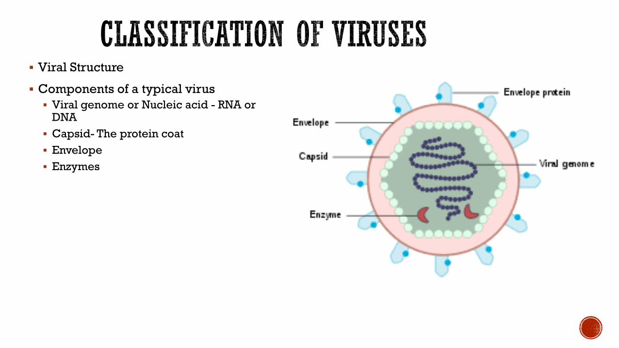
▪ Viral Structure
▪ Components of a typical virus
▪ Viral genome or Nucleic acid - RNA or DNA
▪ Capsid- The protein coat
▪ Envelope
▪ Enzymes
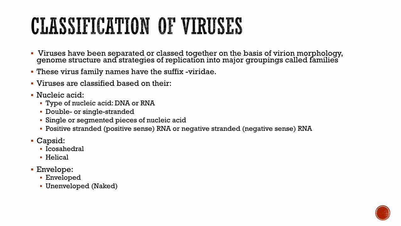
▪ Viruses have been separated or classed together on the basis of virion morphology, genome structure and strategies of replication into major groupings called families
▪ These virus family names have the suffix -viridae.
▪ Viruses are classified based on their:
▪ Nucleic acid:▪ Type of nucleic acid: DNA or RNA
▪ Double- or single-stranded
▪ Single or segmented pieces of nucleic acid
▪ Positive stranded (positive sense) RNA or negative stranded (negative sense) RNA
▪ Capsid:▪ Icosahedral
▪ Helical
▪ Envelope:▪ Enveloped
▪ Unenveloped (Naked)
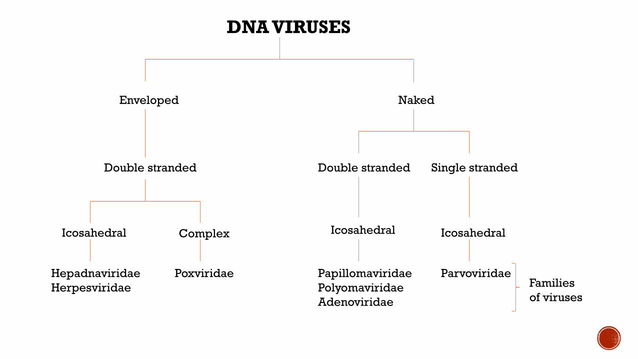
DNA VIRUSES
Enveloped Naked
Double stranded Double stranded Single stranded
Icosahedral Icosahedral IcosahedralComplex
ParvoviridaePapillomaviridae
Polyomaviridae
Adenoviridae
PoxviridaeHepadnaviridae
Herpesviridae Families
of viruses

RNA VIRUSES
Single strandedDouble stranded
Naked
Positive stranded Negative stranded
Reoviridae
Enveloped
Bunyaviridae
Orthomyxoviridae
Paramyxoviridae
Rhabdoviridae
Arenaviridae
Filoviridae
Naked Enveloped
Picornaviridae
CaliciviridaeTogaviridae
Flaviviridae
Icosahedral Icosahedral Helical
Coronaviridae
Icosahedral Helical
Families
of
viruses
Complex
Retroviridae

Classification of viruses that cause human disease
Nucleic Acid Virus Family Examples of Specific viruses or viral diseases
DNA Hepadnaviridae Hepatitis B virus
Herpesviridae Herpes simplex viruses, Varicella zoster virus
Adenoviridae Epidemic keratoconjuctivitis, Respiratory illness of childhood
Poxviridae Small pox, vaccinia virus
Parvoviridae Erythema infectiosum, Transient aplastic anaemia crisis
Papillomaviridae Human papilloma viruses
Polyomaviridae BK polyoma virus, JC polyoma virus
RNA Reoviridae Rotavirus
Picornaviridae Poliovirus
Caliciviridae Noroviruses
Togaviridae Alphaviruses, Rubella(rubivirus)
Flaviviridae Yellow fever virus, Dengue virus
Coronaviridae Common cold, severe acute respiratory syndrome coronavirus 2 (SARS-CoV-2), SARS-
CoV
Retroviridae HIV-1, HIV-2
Bunyaviridae Hanta virus, Rift valley fever virus
Orthomyxoviridae Influenza viruses
Paramyxoviridae Parainfluenza virus, measles virus, mumps virus
Rhabdoviridae Rabiesvirus
Arenaviridae Lassa fever virus
Filoviridae Ebolavirus, Marburg virus

▪ The word helminth is derived from the Greek word for worm
▪ Helminths are multicellular organisms, they worms and range in size from microscopic to large enough to be seen by the naked eye.
▪ Those of medical importance can be classified based on their morphology into –
▪ Platyhelminths (flatworms) – these include:
▪ Trematodes (flukes)
▪ Cestodes (tapeworms)
▪ Nematodes (roundworms)
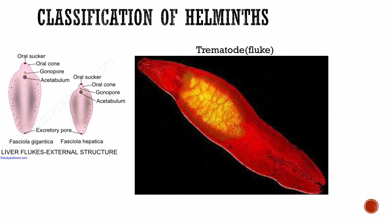
Trematode(fluke)

Cestode(Tapeworm)

Nematode(roundworm)
▪ Either
▪ The adult forms of these worms may reside in the gastrointestinal tract, blood, lymphatic system or subcutaneous tissues.
▪ Or
▪ The immature forms or larva may infect or invade various body tissues to cause disease.
▪ Thus we can also classify the helminths based on where in the body they reside
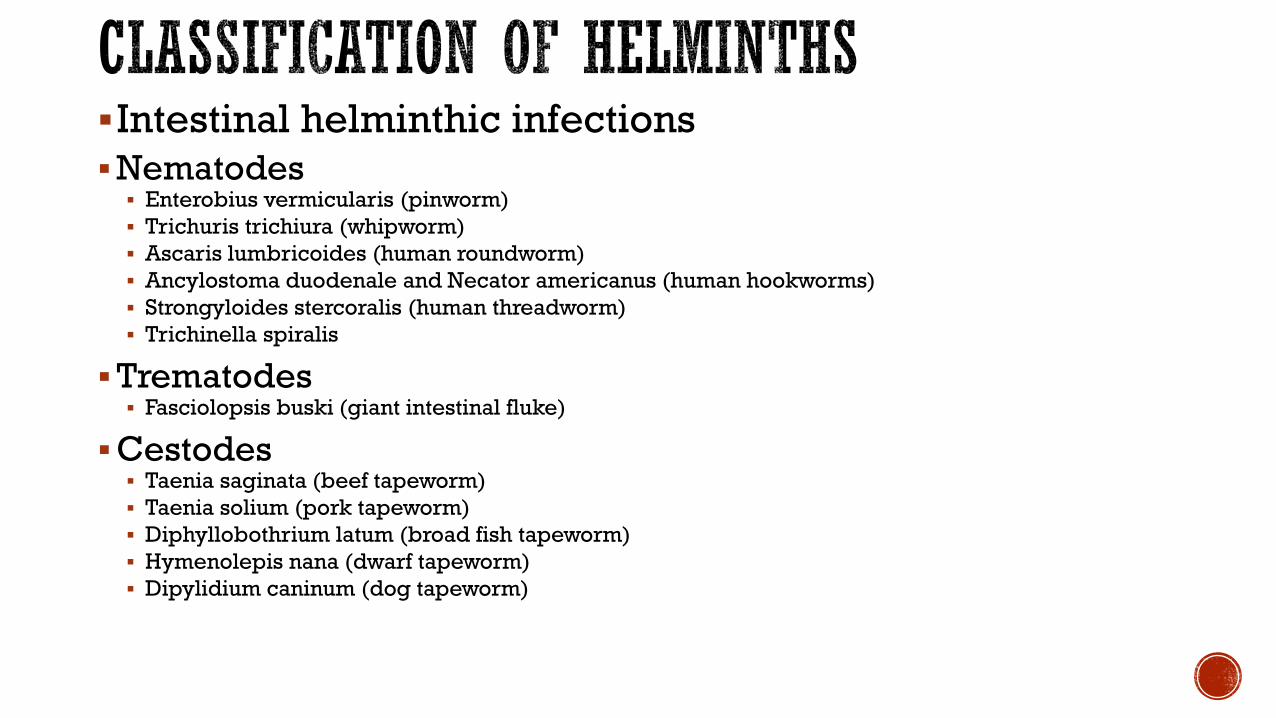
▪Intestinal helminthic infections
▪Nematodes▪ Enterobius vermicularis (pinworm)
▪ Trichuris trichiura (whipworm)
▪ Ascaris lumbricoides (human roundworm)
▪ Ancylostoma duodenale and Necator americanus (human hookworms)
▪ Strongyloides stercoralis (human threadworm)
▪ Trichinella spiralis
▪Trematodes▪ Fasciolopsis buski (giant intestinal fluke)
▪Cestodes▪ Taenia saginata (beef tapeworm)
▪ Taenia solium (pork tapeworm)
▪ Diphyllobothrium latum (broad fish tapeworm)
▪ Hymenolepis nana (dwarf tapeworm)
▪ Dipylidium caninum (dog tapeworm)
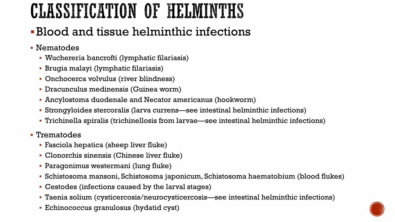
▪Blood and tissue helminthic infections
▪ Nematodes
▪ Wuchereria bancrofti (lymphatic filariasis)
▪ Brugia malayi (lymphatic filariasis)
▪ Onchocerca volvulus (river blindness)
▪ Dracunculus medinensis (Guinea worm)
▪ Ancylostoma duodenale and Necator americanus (hookworm)
▪ Strongyloides stercoralis (larva currens—see intestinal helminthic infections)
▪ Trichinella spiralis (trichinellosis from larvae—see intestinal helminthic infections)
▪ Trematodes
▪ Fasciola hepatica (sheep liver fluke)
▪ Clonorchis sinensis (Chinese liver fluke)
▪ Paragonimus westermani (lung fluke)
▪ Schistosoma mansoni, Schistosoma japonicum, Schistosoma haematobium (blood flukes)
▪ Cestodes (infections caused by the larval stages)
▪ Taenia solium (cysticercosis/neurocysticercosis—see intestinal helminthic infections)
▪ Echinococcus granulosus (hydatid cyst)

▪ Protozoa are microscopic, one-celled organisms
▪ The protozoa that infect humans can be classified into four groups based on their mode of movement:
▪ Sarcodina –(the amoeba) move with pseudopodia e.g. Entamoeba
▪ Mastigophora – (the flagellates) move with flagella e.g. Giardia, Leishmania
▪ Ciliophora – (the ciliates) move with cilia e.g. Balantidium
▪ Apicomplexa (Sporozoa) – organisms that have an apical complex and do not have flagella, cilia or pseudopods e.g. Plasmodium, Cryptosporidium
▪ They may also be classified based on where in the body they infect

Sarcodina(amoe
ba)
Mastigophora(flagellat
es)
Ciliophora(ciliat
es)
Apicomplexa(Sporoz
oa)

▪ Intestinal protozoa▪ G. lamblia (flagellate)
▪ Entamoeba histolytica (ameba)
▪ Balantidium coli (ciliate)
▪ Cryptosporidium hominis (sporozoa)
▪ Cyclospora cayetanensis (sporozoa)
▪ Sexually transmitted protozoan infection▪ Trichomonas vaginalis (flagellate)
▪ Blood and tissue protozoan infections
▪ Flagellates▪ T. brucei rhodesiense and T. brucei gambiense
▪ T. cruzi
▪ Leishmania donovani, Leishmania tropica, Leishmania mexicana
▪ Amebae▪ Entamoeba histolytica (see intestinal protozoa)
▪ Naegleria fowleri and Acanthamoeba castellanii
▪ Sporozoa▪ Plasmodium vivax, Plasmodium falciparum, Plasmodium ovale, and Plasmodium malariae
▪ Babesia microti
▪ Toxoplasma gondii
▪ Microsporidia

▪ Mandel, Douglas and Bennetts’ principles and practice of infectious diseases. 9th
ed.Philadelphia. Churchhill Livingstone Elsevier
▪ Jawetz, Melnink and Adelberg’s medical microbiology. 28th edition
▪ Clinical Microbiology Made Ridiculously Simple. Mark Gladwin Bill Trattler. Edition 3