Embed Size (px)
Citation preview
Dr. Ahmed Fathalla Ibrahim
LIVERLIVER
LIVERLIVER
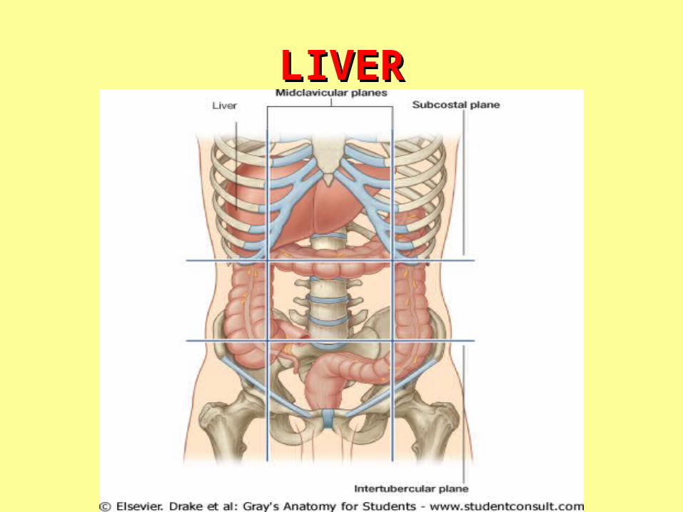
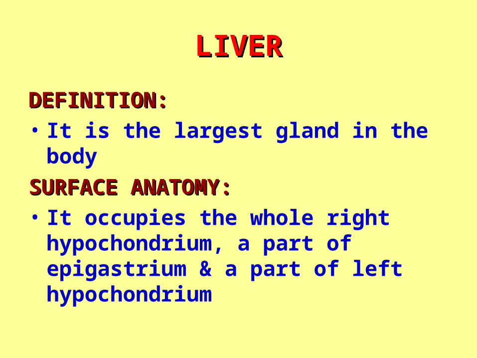
DEFINITION:DEFINITION:
• It is the largest gland in the body
SURFACE ANATOMY:SURFACE ANATOMY:
• It occupies the whole right hypochondrium, a part of epigastrium & a part of left hypochondrium

ANTERIOR SURFACE OF LIVERANTERIOR SURFACE OF LIVER
LIVERLIVER
SURFACES:SURFACES:• ANTERIOR: ANTERIOR: related to diaphragm &
anterior abdominal wall. The diaphragm separates it from pleurae & lungs
• SUPERIOR: SUPERIOR: related to diaphragm separating it from pericardium & heart (in the middle) & pleurae & lungs (on each side)

POSTERIOR SURFACE OF POSTERIOR SURFACE OF LIVERLIVER

LIVERLIVER
SURFACES:SURFACES:• POSTERIOR: POSTERIOR: formed of (from right to left):1.1. Bare area:Bare area: triangular area related to diaphragm. Its
base is formed by groove for IVC; its sides are formed by upper & lower layers of coronary ligaments that meet at the apex to form the right triangular ligament
2.2. Groove for IVCGroove for IVC3.3. Caudate lobe:Caudate lobe: related to diaphragm, projects
downwards to form a process separating IVC from porta hepatis & forming upper boundary of epiploic foramen
4.4. Fissure for ligamentum venosum (obliterated ductus Fissure for ligamentum venosum (obliterated ductus venosus)venosus)
5.5. Esophageal notchEsophageal notch

INFERIOR SURFACE OF LIVERINFERIOR SURFACE OF LIVER

INFERIOR SURFACE OF LIVERINFERIOR SURFACE OF LIVER

LIVERLIVERSURFACES:SURFACES:• INFERIOR: INFERIOR: 1. Related to the following visceraviscera: right kidney,
right suprarenal gland, right colic flexure, 2nd part of duodenum, fundus & body of stomach
2. Formed of (from right to left): • Gall bladder fossaGall bladder fossa • Quadrate lobe:Quadrate lobe: related to pylorus, 1st part of
duodenum & transverse colon• Fissure for ligamentum teresFissure for ligamentum teres (obliterated left
umbilical vein)
LIVERLIVER
• ACCORDING TO ITS RELATIONS, ACCORDING TO ITS RELATIONS, THE LIVER IS DIVIDED INTO:THE LIVER IS DIVIDED INTO:
1.1. DIAPHRAGMATIC SURFACE: DIAPHRAGMATIC SURFACE: includes superior, anterior & most of posterior surface
2.2. VISCERAL SURFACE: VISCERAL SURFACE: includes inferior & a part of posterior surface

LIVERLIVER
PORTA HEPATIS:PORTA HEPATIS:
• It is the hilumhilum of liver
• It lies in the inferior surfacein the inferior surface of liver
• Structures passing through it: right & left hepatic ducts, right & left branches of hepatic artery & portal vein (DAVDAV, from before backwardsfrom before backwards)+ lymphatics, lymph nodes & autonomic fibers

LOBES OF LIVERLOBES OF LIVER
ANATOMICAL DIVISION:ANATOMICAL DIVISION:• The liver is divided into a smaller left a smaller left
& a larger right lobe& a larger right lobe by:
1. Falciform ligament: anteriorly
2. Fissure for ligamentum venosum: posteriorly
3. Fisssure for ligametnum teres: inferiorly

LOBES OF LIVERLOBES OF LIVER
FUNCTIONAL DIVISION:FUNCTIONAL DIVISION:• The liver is divided into a nearly equal a nearly equal
left & right lobesleft & right lobes by a plane passing through groove for IVC & gall bladder fossa.
• The caudate & quadrate lobes are The caudate & quadrate lobes are included in the left lobeincluded in the left lobe (because they are supplied by left branches of hepatic artery & portal vein)

LIGAMENTS OF LIVERLIGAMENTS OF LIVER
PERITONEAL LIGAMENTSPERITONEAL LIGAMENTS• FALCIFORM LIGAMENT:FALCIFORM LIGAMENT: a triangular
fold formed of two layers (right & left) with:
1.1. Anterior border:Anterior border: attached to diaphragm
2.2. Posterior border:Posterior border: attached to anterior & superior surfaces of liver
3.3. Lower free border:Lower free border: extending from liver to umbilicus & enclosing ligamentum teres & paraumbilical veins
LIGAMENTS OF LIVERLIGAMENTS OF LIVER
PERITONEAL LIGMENTSPERITONEAL LIGMENTS• LEFT TRIANGULAR LIGAMENT:LEFT TRIANGULAR LIGAMENT: to the to the
left of falciform ligament,left of falciform ligament, the peritoneum is reflected from the diaphragm to the upper surface of the liver to form the anterior layer the anterior layer of left triangular ligamentof left triangular ligament (continuous with left layer of falciform ligament). The peritoneum then covers anterior, inferior & posterior surfaces & is reflected to the diaphragm to form the posterior layer of left the posterior layer of left triangular ligament triangular ligament

LIGAMENTS OF LIVERLIGAMENTS OF LIVER
PERITONEAL LIGMENTSPERITONEAL LIGMENTS• RIGHT TRIANGULAR LIGAMENT:RIGHT TRIANGULAR LIGAMENT: to the to the
right of falciform ligament,right of falciform ligament, the peritoneum is reflected from the diaphragm to the upper surface of the liver to form the upper layer of the upper layer of coronary ligament coronary ligament (continuous with right layer of falciform ligament). The peritoneum then covers anterior & inferior surfaces then becomes reflected to the front of right kidney to form the lower layer of coronary ligament. the lower layer of coronary ligament. The right extremity of both layers meet to form the right triangular ligament the right triangular ligament

LIGAMENTS OF LIVERLIGAMENTS OF LIVER
PERITONEAL LIGMENTSPERITONEAL LIGMENTS• LESSER OMENTUM:LESSER OMENTUM:
1. It is attached to the margins of porta the margins of porta hepatis & fissure for ligamentum hepatis & fissure for ligamentum venosumvenosum
2. It extends to lesser curvature of stomach & upper border of 1st inch of 1st part of duodenum

LIGAMENTS OF LIVERLIGAMENTS OF LIVER
LIGMENTUM M TERES:LIGMENTUM M TERES:• Represents the obliterated left the obliterated left
umbilical veinumbilical vein that originally joins the branch of portal vein
LIGAMENTUM VENOSUM: LIGAMENTUM VENOSUM: • Represents the obliterated ductus the obliterated ductus
venosusvenosus that originally connects IVC with left branch of portal vein
BARE AREAS OF LIVERBARE AREAS OF LIVER
• TRIANGULAR DIAPHRAGMATIC TRIANGULAR DIAPHRAGMATIC AREAAREA
• GROOVE FOR IVCGROOVE FOR IVC
• GALL BALDDER FOSSAGALL BALDDER FOSSA
• PORTA HEPATIS PORTA HEPATIS
• FISSURE FOR LIGAMENTUM FISSURE FOR LIGAMENTUM VENOSUMVENOSUM
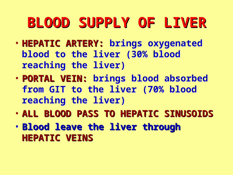
BLOOD SUPPLY OF LIVERBLOOD SUPPLY OF LIVER• HEPATIC ARTERY: HEPATIC ARTERY: brings oxygenated blood
to the liver (30% blood reaching the liver)
• PORTAL VEIN: PORTAL VEIN: brings blood absorbed from GIT to the liver (70% blood reaching the liver)
• ALL BLOOD PASS TO HEPATIC SINUSOIDSALL BLOOD PASS TO HEPATIC SINUSOIDS
• Blood leave the liver throughBlood leave the liver through HEPATIC HEPATIC VEINSVEINS
LIVERLIVERLYMPHATIC DRAINAGE:LYMPHATIC DRAINAGE:• Mainly into hepatic lymph nodeshepatic lymph nodes in porta hepatis.
Efferent vessels pass to celiac lymph nodesceliac lymph nodesNERVE SUPPLY: NERVE SUPPLY: Hepatic plexus of autonomic fibersHepatic plexus of autonomic fibers
derived from celiac plexusceliac plexusSUPPORT:SUPPORT:1. Hepatic veins (main support):(main support): suspend liver from
IVC & have no extrahepatic course2. Peritoneal folds.3. Surrounding organs4. Tone of anterior abdominal wall
BILIARY SYSTEMBILIARY SYSTEM
1.1. BILIARY DUCTS BILIARY DUCTS
2.2. GALL BLADDERGALL BLADDER

BILIARY SYSTEMBILIARY SYSTEM
COMMON BILE DUCTCOMMON BILE DUCT
BILIARY DUCTSBILIARY DUCTSRIGHT & LEFT HEPATIC DUCTS:RIGHT & LEFT HEPATIC DUCTS:• Emerge from porta hepatis• Drain bile from right & left lobes of liver• Unite to form common hepatic duct
COMMON HEPATIC DUCT:COMMON HEPATIC DUCT:• One and half inches in length• Formed by the union of the 2 hepatic ducts• Unite with the cystic duct to form the
common bile duct

BILIARY DUCTSBILIARY DUCTS
CYSTIC DUCT:CYSTIC DUCT:• One and half inches in length
• Connects the neck of gall bladder to common hepatic duct
• The mucous membrane of the duct as well as that of neck of gall bladder is raised forming the spiral valvethe spiral valve. The valve keeps the lumen open
BILIARY DUCTSBILIARY DUCTS
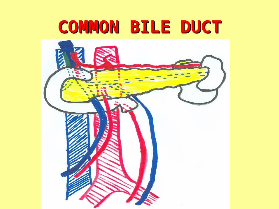
COMMON BILE DUCT:COMMON BILE DUCT:• Formation:Formation: Formed by union of cystic & common
hepatic duct• Length:Length: Three inches• Course & relations:Course & relations: It descends:1. In the free margin of lesser omentum (to the right
side of hepatic artery, anterior to portal vein)2. Behind 1st part of duodenum (to the right side of
gastroduodenal artery, anterior to portal vein)3. Behind the head of pancreas, anterior to IVC• Termination:Termination: usually join the main pancreatic duct
forming the ampulla of Vater that opens into the lumen of 22ndnd part of duodenum part of duodenum through the major duodenal papilla

GALL BLADDERGALL BLADDER
SHAPE: SHAPE: Pear-shaped sac
SITE: SITE: Lies in gall bladder fossa of liver
LENGTH: LENGTH: 3 – 4 inches
CAPACITY: CAPACITY: 30 – 50 ml
GALL BLADDERGALL BLADDER
PARTS:PARTS: Formed of 3 parts:1.1. Fundus:Fundus: projects below inferior margin
of liver, lies opposite the tip of right 9th costal cartilage, related anteriorlyanteriorly to anterior abdominal wall & posteriorlyposteriorly to transverse colon
• Body:Body: related anteriorlyanteriorly to liver & posteriorlyposteriorly to 1st part of duodenum
• Neck:Neck: continuous with cystic duct has same relation as body
GALL BLADDERGALL BLADDER
PERITONEAL COVERING:PERITONEAL COVERING:
• Fundus:Fundus: completely covered by peritoneum
• Body & neck:Body & neck: only covered posteriorly
FUNCTION: FUNCTION: Stores & concentrates bile
SUPPLY OF BILIARY SYSTEMSUPPLY OF BILIARY SYSTEM
• ARTERIES: ARTERIES: Cystic branch of right hepatic artery
• VEINS: VEINS: Cystic vein that drains into the right branch of portal vein
• LYMPH DRAINAGE: LYMPH DRAINAGE: Cystic lymph nodes, then to hepatic & finally to celiac lymph nodes
• NERVES: NERVES: autonomic fibers from celiac lymph nodes