Embed Size (px)
Citation preview

STIMULATION OF CONNECTIVE TISSUETYPE MAST CELLPROLIFERATION BY CROSSLINKING OF CELL-BOUND IgE
BY MINEO TAKAGI,' TATSUTOSHI NAKAHATA,* KENICHI KOIKE,*TOSHIMI KOBAYASHI," KOHICHIRO TSUJI,' SOMEI KOJIMA,ITAKAO HIRANO,§ ATSUSHI MIYAJIMA,II KEN-ICHI ARAI,II
AND TARO AKABANE*
From the *Department of Pediatrics, Shinshu University School ofMedicine, Matsumoto 390;the $Department of Parasitology, Chiba University School of Medicine, Chiba 280;
and the §Department of Internal Medicine and Rheumatology,Juntendo University School ofMedicine, Tokyo 113, ,Japan; and the
IIDNAX Research Institute of Molecular and Cellular Biology, Palo Alto, California 94304
There are two phenotypically distinct subpopulations of mast cells in rodents, i.e.,connective tissue-type mast cells (CTMC)' and mucosal mast cells (MMC). Thesepopulations differ in their location, cell size, staining characteristics, ultrastructure,mediator content, and T cell dependency (1-3) . Although proliferation of MMCwasknown to be Tcell dependent in vivo and thought to be IL-3 dependent in vitro,the factors on which CTMC proliferation depends remained elusive. We recentlyfound that mature CTMC separated from mouse peritoneal cells could proliferateextensively in methylcellulose culture and maintain the appearance and functionof CTMC (4). Clonal growth of CTMC could not be induced by IL-3 alone andrequired the presence of both IL-3 and IL-4/B cell stimulatory factor 1 (BSF1) (5).
Mast cells bear high affinity IgE receptors, and crosslinking of cell-bound IgEby multivalent antigen or anti-IgE antibody triggers the release ofa variety of chem-ical mediators (6).
In B cell proliferation, crosslinking of surface IgM on B cells by anti-IgM anti-bodies causes entry of resting B cells into the Gi phase of the cell cycle (7). BSF1also plays a role in B cell proliferation as an activating factor (8).These findings led us to examine whether crosslinking of IgE molecules bound
to IgE receptors on CTMC plays some role in the proliferation of these cells . Wereport here that IgE-sensitized CTMC can proliferate in vitro after the stimulationwith the specific antigen or anti-IgE antibody in the presence of IL-3 . Our dataindicate the possibility that antigen stimulation not only triggers the release of chemicalmediators from CTMC but participates in the mechanisms of their proliferation.
This work was supported by grants from the Ministry of Education, Science and Culture, Japan. Ad-dress correspondence to Tatsutoshi Nakahata, Department of Pediatrics, Shinshu University Schoolof Medicine, 3-1-1 Asahi, Matsumoto, 390 Japan.
1 Abbreviations used in this paper. BSF1, B cell stimulatory factor 1 ; CTMC, connective tissue-type mastcells ; MMC, mucosal mast cells ; Sj, Schistosomajaponicum antigen ; 48/80, compound 48/80
J. Exp. MED. C The Rockefeller University Press - 0022-1007/89/07/0233/12 $2.00
233Volume 170 July 1989 233-244
on June 9, 2017D
ownloaded from
Published July 1, 1989
234
ANTIGEN-INDUCED CLONAL GROWTH OF MAST CELLS
Materials and MethodsCell Preparation.
20-25-wk-old male BDFj mice were obtained from the Shizuoka Ex-perimental Animal Center (Shizuoka, Japan) . Peritoneal cells were collected as describedpreviously (9). Mature CTMC (398% purity) were separated from peritoneal cells by theremoval of phagocytes followed by Percoll (Sigma Chemical Co., St . Louis, MO) density-gradient centrifugation as described in detail (10) .IgE Antibodies, Antigens, and Other Secretagogues.
Both mouse monoclonal anti-DNP-BSAIgE antibody (B53) (11) and mouse monoclonal anti-Schislosoma japonicum (Sj) IgE antibody(12) were used as diluted ascitic fluid . Anti-DNP IgE at a dilution of 1/1,000 and anti-Sj IgEat a dilution of 1/1,000 corresponded to 80 ng/ml and 100 ng/ml IgE, respectively. Purifiedrat anti-mouse IgE mAb (6HD5) was prepared as described previously (13) . DNP-BSA con-tained 6.8 DNP groups per BSA molecule . Sj antigen was extracted from adult worms of
Si as described previously (12) . Compound 48/80 (48/80) was purchased from Sigma .IL-3, IL-4, and Anti-IL-4 Antibody.
Both mouse rIL-3 (14) and rIL-4 (15) were producedin yeast cells . Their activities were determined by using P cell clone IC2 (16), and a colori-metric assay (17) . Rat anti-IL-4 mAb was purified from 11B11-containing ascitic fluid (18) .
Clonal Cell Cultures .
Methylcellulose cultures were carried out by using a modification oftechnique described previously (9, 19) . Culture mixture (1 ml) containing 10 3 purifiedCTMC, a-medium (Flow Laboratories, Inc ., Rockville, MD), 30% FCS (HyClone Labora-tories, Inc ., Logan, UT), 1% deionized BSA (Sigma Chemical, Co.), 10 -4 M 2-ME (EastmanOrganic Chemicals, Rochester, NY), and 100 U IL-3 were preincubated in the presence orabsence of anti-DNP IgE (or anti-Sj IgE) at 37°C in 5% C02/95% air for passive sensiti-zation . After 12 h, varying concentrations ofspecific antigen (or anti-IgE antibody) and methyl-cellulose (final concentration, 0.8%) (Shinetsu Chemical Co., Tokyo, Japan) were added tothe preincubation mixture . This mixture was then incubated in a culture dish at 37°C ina humidified atmosphere flushed with 5% C02 in air. Mast cell colonies containing 320 cellswere counted on day 16 .Serum free culture was carried out by using a modification of the technique described pre-
viously (20, 21) . Culture mixture contained 1% crystallized globulin-free deionized BSA(Calbiochem-Behring Corp ., LaJolla, CA), 300 hg/ml fully saturated human transferrin (SigmaChemical Co.), 160 ug/ml soybean lecithin (Sigma Chemical Co.), 96 p.g/ml cholesterol (NakaraiChemicals Ltd ., Kyoto, Japan) and 10' M sodium selenite (Sigma Chemical Co.), insteadof FCS.
Staining of Cultured Cells .
Individual colonies were lifted from the methylcellulose cultureusing a 3 ul Eppendorf pipette under direct microscopic visualization and were collected inEppendorf microcentrifuge tubes containing 0.5 ml PBS. After washing, the samples wereimmediately spun in a cytocentrifuge (Cytospin ; Shandon Southern Instruments, Sewickley,PA) at 800 rpm for 5 min and stained with alcian blue-safranin (22) or berberine sulfate(23) . Specimens stained with berberine sulfate were examined with an Olympus epifluores-cence microscope . Alcian blue(-)- safranin(+) mast cells are thought to be mature CTMC(22) . Berberine sulfate staining was reported to be highly specific for heparin-containinggranules of CTMC (23) . We confirmed this by demonstrating that the staining was blockedby treating the cells with heparinase from Flaoobacterium heparinum (0.5 IU/ml, pH 7.0 ; Seika-gaku Kogyo Co., Tokyo, Japan) for 30 min but not chondroitinase ABC from Proteus vulgaris(5 .0 IU/ml, pH 8.0 ; Seikagaku Kogyo Co.) for 4 h (9) .
Histamine Release.
Histamine release experiments were carried out as described previously(24) . Aliquots of cells sensitized with anti-DNP IgE (1/1,000) containing N5 x 104 mast cellswere incubated for 20 min with varying concentrations of DNP or anti-IgE antibody at 37°C .Histamine release from mast cells was measured by HPLC with fluorometry (25) .
Results
Clonal Growth of CTMC Sensitized with Anti-DNP IgE by Stimulation with DNPin thePresence ofIL-3. When normal mouse CTMC sensitized with monoclonal anti-DNPIgE (B53) were stimulated by DNP and then cultured in methylcellulose medium
on June 9, 2017D
ownloaded from
Published July 1, 1989

containing IL-3, pure mast cell colonies developed, depending on the concentrationof DNP (Fig . 1 a) . Proliferation of mast cells also depended on the concentrationof anti-DNP IgE (Fig . 1 b) . The maximal colony formation was seen with 1/1,000dilution of anti-DNP IgE and 1 ug/ml DNP About one-third of mature CTMCseparated from mouse peritoneal cells produced colonies in culture containing op-timal concentrations ofanti-DNP IgE, DNP, and IL-3 . Neither DNP nor IL-3 alonestimulated clonal growth of sensitized CTMC. IL-3 and/or DNP had no effects onproliferation of nonsensitized CTMC. When sensitized cells were washed with ct-me-dium after stimulation with DNP and then cultured in methylcellulose medium con-taining DNP with IL-3, anti-DNP IgE with IL-3, or IL-3 alone, no mast cell colo-nies developed. In contrast, many mast cell colonies developed when the same washedcells were incubated with DNP, anti-DNP IgE, and IL-3 (data not shown) . Theseresults suggest that antigen stimulation only in early stage of culture is not sufficientto induce subsequent mast cell colony formation in the presence of IL-3, and thatcontinuous presence of anti-DNP IgE, DNP, and IL-3 in culture is required forextensive proliferation of CTMC .
Cytocentrifuged preparations ofmast cell colonies were individually stained withalcian blue-safranin and berberine sulfate to determine the characteristics of these
U
U
X
NaNQ1
.COOU
NU
NtCEOOZ
0 10'610-5 10 -~IU10(Si) (gml-t)
(anti-Sj IgE)0
2
TAKAGI ET AL .
b
0 10 -6 10' 510' 4 10 -3 10' 2(anti-DNP IgE)
d
0
10-9 Id- I 4-1
(anti-IgE) (gml ')
(anti-DNP IgE)
235
FIGURE 1 .
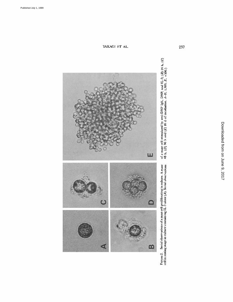
Dependence ofcol-ony growth on various stimula-tion . (Left) Mast cell colonygrowth from mature CTMC atvarying concentrations of (a)DNP-BSA, (c) Schistosomajaponi-cum (Sj) antigen, and (e) anti-mouse IgE antibody (Open circles)Cells sensitized with (a, e) anti-DNP IgE (1/1,000) or (c) anti-SjIgE (1/1,000) ; (filled circles) cellswithout sensitization . (Right)Mast cell colony growth fromcells sensitized with varyingconcentrations of (b,f) anti-DNPIgE and (d) anti-Sj IgE . Opencircles, cells stimulated by (b) 1tug/ml DNP, (d) 1 /~g/ml Sj, or(f) 0 .1 wg/ml anti-IgE antibody ;(filled circles) cells without stimu-lation . In different sets of sensi-tized cells, the rate ofhistaminerelease was determined . Spon-taneous release was 8% . Thesevalues were subtracted from theexperimental values. Data in thefigure are the average of tripri-cated cultures. Errors are smallerthan the symbols .
on June 9, 2017D
ownloaded from
Published July 1, 1989
236
ANTIGEN-INDUCED CLONAL GROWTH OF MAST CELLS
colonies . Most cells composing individual colonies were stained with both safranin(99.4 t 1.7%) and berberine sulphate (69 .2 t 8.7%) . Treatment with heparinasecompletely abolished the fluorescence of granules of proliferating mast cells in ourculture, whereas chondroitinase ABC did not. These results suggest that most de-veloping mast cells in culture actually contain heparin proteoglycan in their granules,and that the cells retain the characteristics in common with CTMC (4, 26). Safra-nin(+)-berberine sulfate(+) granules in the cells, however, were somewhat scarceras compared with CTMC separated from peritoneal cavity. In addition, althoughall mast cell colonies developed in culture were considered to be derived from safra-nin(+)-berberine sulfate(+) mature CTMC (4), about two-thirds of the coloniescontained a few alcian blue(+)-safranin(-)-berberine sulfate(-) mast cells .
To completely exclude the effects of accessory cells on colony formation in ourculture system, single-cell culture wasperformedaccording to the method describedpreviously (27) . Purified CTMC from peritoneal cells were plated in methylcellu-lose medium. A single CTMC was identified morphologically, lifted from the mediumusing a fine Pasteur pipette attached to a Leitz micromanipulator, transferred tofresh culture medium, and incubated for 16 d. 14 of 48 transferred single CTMCproduced colonies in the culture containing optimal concentrations of anti-DNPIgE, DNP, and IL-3 . This result demonstrates that clonal growth of CTMC in ourculture condition does not require the presence of accessory cells .
Serum-free culture was also carried out to exclude the effects of some factors inFCS on CTMC proliferation using atechnique described in Materials andMethods.60 ( ± 5) mast cell colonies developed from 500 purified CTMC in the serum-freeculture containing optimal concentrations . of anti-DNP IgE, DNP, and IL-3 .
Effects ofSj and Anti-IgE Antibody on Colony Formation.
To examine whether mastcell colony formation induced by antigen stimulation depends on the kinds of an-tigens, mature CTMC sensitized with monoclonal anti-S. japonicum (Sj) IgE werestimulated by Sj antigen and then cultured in the presence of IL-3 . Mast cell colo-nies also developed within a relatively small range of concentrations of Sj antigen(Fig . 1 c) . Anti-Sj IgE stimulated proliferation of CTMC dose dependently in thepresence of 1 /Ag/ml Sj antigen (Fig . 1 d) .The effects of anti-mouse IgE mAb (6HD5) on mast cell colony formation from
mature CTMC sensitized with anti-DNP IgEin the presence of IL-3 were also tested .Mast cell colonies developed, depending on the concentration ofboth anti-IgE anti-body and anti-DNP IgE (Fig . 1, e, f) .
Histamine Release Experiments and Serial Observations ofDividing Cells.
Together withcolony formation, we examined histamine release from mast cells as an index tothe degree of degranulation. When the cells sensitized with anti-DNP IgE were stimu-lated by DNP, both colony formation and histamine release increased in a dose-dependent manner (Fig . 1 a) . Anti-IgE antibody also stimulated both colony for-mation and histamine release at an optimal concentration of 0.1 hg/ml anti-IgEantibody (Fig . 1 e). However, whether dividing mast cells are similarly degranulatedremains unanswered . To examine this, serial observations of mast cells proliferatingin culture were performed. It was found that most dividing cells in the culture con-taining anti-DNP IgE, DNP, and IL-3 had pericellular halos which indicated de-granulation of CTMC as reported previously (28) (Fig . 2, B-D), whereas restingcells in culture containing IL-3 alone had no halos around them (Fig . 2 A) .
on June 9, 2017D
ownloaded from
Published July 1, 1989
TAKAGI ET AL .
N0
E~^OX
MaW
-DoGM
zq
a.zC
O OTbA i0
y ~Dua
O d~
N4 V)
^, OrU
0!J
4.w Gr: Oc.0.MuaVNof "S
O Gw OCO 47
eC ,~
N U.a .GO yOA
0l7q
N ya qO .^W u
237
on June 9, 2017D
ownloaded from
Published July 1, 1989

23 8
ANTIGEN-INDUCED CLONAL GROWTH OF MAST CELLS
UHU
X
daam00U
mUYNNE06Z
TABLE I
Effects of 48/80 on Mast Cell Colony Formation
Data represent the number of mast cell colonies on day 16 (48/80 10 tag/ml, IL-3100 U/ml, anti-DNP IgE 1/1,000, DNP 1 kg/ml) . Data shown in the table arethe mean t SD of triplicate cultures .
Effects ofNon-IgE-mediated Stimulation on Colony Formation.
48/80 is a secretagogueknown to stimulate CTMC directly (29) . Whether 48/80 could induce mast cell colonyformation in the presence of IL-3 as well as stimulation via IgE receptors was tested .When CTMC were stimulated by 10 Fig/ml 48/80 for 20 min, 43% total cellularhistamine was released from the cells . However, in three separate experiments, nomast cell colonies developed when CTMC were cultured with 10 Wg/ml 48/80 and100 U/ml IL-3 (Table I) . Addition of 10 fig/ml 48/80 neither enhanced nor inhibitedproliferation ofCTMC supported by anti-DNP IgE, DNP, and IL-3 . These resultsindicate that some mediators released from mast cells can not induce their proliferation .
Effects ofa Combination ofIL-4 and Antigen Stimulation on Colony Formation.
Whenvarying concentrations ofIL-4 were added to CTMC sensitized with anti-DNP IgEin the presence of IL-3, mast cell colony formation increased in a dose-dependentmanner and reached a plateau at 100 U/ml IL-4 . However, the combination of IL-4and DNP increased the number ofcolonies exceeding by far the maximal level sup-ported by IL-4 (Fig . 3) . When cells were incubated with IL-4 and IL-3 without sen-sitization, the dose-response curve was about equal to that ofthe sensitized cells (data
(IL-4 ) (Umt-')
FIGURE 3 .
The effects of a combination of IL-4 andDNP on colony formation . 10 3 CTMC sensitized withanti-DNP IgE (1/1,000) were cultured at varying con-centrations ofIL-4 with (open circle) or without (closed circle)stimulation of DNP (1 pg/ml) in the presence of IL-3(100 U/ml) . Data shown in the figure are the mean ±SD of triplicated cultures .
StimuliNo . of mast cell coloniesper 5 x 10 2 CTMC
IL-3 048/80 0IL-3 + 48/80 0IgE + DNP + IL-3 148 ± 4IgE + DNP + IL-3 + 48/80 134 ± 1
on June 9, 2017D
ownloaded from
Published July 1, 1989

UHU
X
am0°udU
NEE`o
Z
100
00
2.5
50anti-IL-4 (111311) (10-'pt'')
TAKAGI ET AL .
FIGURE 4.
The effects of anti-IL-4 antibody on colony formation.5 x 10'CTMC were incubated with ; DNP + anti-DNP IgE + IL-3(circles), IL-4 + IL-3 (squares) or anti-DNP IgE + DNP + IL-4 +IL-3 (triangles) (anti-DNP IgE 1/1,000, DNP 1 tAg/ml, IL-3 100 U/ml,IL-4 100 U/ml). Varying concentrations of anti-IL-4 antibody wereadded to the cultures on day0. Data shown in the figure are the meant SD of triplicated cultures.
not shown) . When sensitized cells were incubated with both IL-4 and DNP in theabsence of IL-3, no mast cell colonies developed . It is of interest that the combina-tion of IL-4 and DNP in the presence of IL-3 not only increased the number ofcolonies but greatly enlarged both the colony size and the cell volume (data not shown) .Addition of anti-IL-4 antibody, 111311, to the culture containing IL-4 (100 U/ml) andIL-3 (100 U/ml) resulted in a dose-dependent decrease of clonal growth of CTMC(Fig . 4) . About 70o7o of the colonies were suppressed by the addition of 50 lug/mlanti-IL-4 antibody. On the other hand, in culture containing optimal concentra-tions of anti-DNP IgE, DNP, and IL-3, their growth was not affected by the addi-tion ofanti-IL-4 antibody at the same concentration. Anti-IL-4 antibody also neu-tralized effects of a combination ofIL-4 andDNP on proliferation ofsensitized CTMCand depressed numbers ofmast cell colonies to the level supported by anti-IgE DNP,DNP, and IL-3 .
Replating Experiments .
Whether both IL-4 and antigen stimulation could exertcolony-inducing effects on a single CTMC was also examined . Since it is impossible
TABLE II
Replating Studies of Mast Cell Colonies
Mast cell colonies supported by IL-4 with IL-3, or anti-DNP IgE andDNPwithIL-3 were individually lifted from the culture on day 16, suspended in a-mediumand gently pipetted . Aliquots ofthe cell suspension were added to the secondaryculture medium containing: anti-DNP IgE + DNP + IL-3, IL-4 + IL-3, orIL-3 (anti-DNP IgE 1/1,000, DNP 1 ug/ml, IL-3 100 U/ml, IL-4 100 U/ml).One quadrant of the sample was stained with alcian blue-safranin and berber-ine sulphate to determine the characteristics of the colony . Data represent thenumber of mast cell colonies on day 16 of the secondary culture .
239
Colonyno .
Colonysize Primary stimuli
Secondary stimuli
IgE + DNP IL-4+ IL-3 + IL-3 IL-3
1 170 IgE + DNP + IL-3 13 12 02 210 IgE + DNP + IL-3 17 24 03 260 IgE + DNP + IL-3 37 61 14 110 IL-4 + IL-3 6 8 05 470 IL-4 + IL-3 66 61 26 1,000 IL-4 + IL-3 99 237 2
on June 9, 2017D
ownloaded from
Published July 1, 1989

240
ANTIGEN-INDUCED CLONAL GROWTH OF MAST CELLS
to culture a CTMC with two different stimuli, individual colonies, which derivedfrom an ancestor cell, supported by IL-4 with IL-3 or anti-DNP IgE and DNP withIL-3 were divided and replated in secondary cultures containing different stimuli .All of the mast cell colonies supported by IL-4 with IL-3 produced many secondarymast cell colonies in culture containing anti-DNP IgE, DNP, and IL-3 . The reverserelation was also observed as shown in Table II . It should also be noted that CTMCcan not proliferate extensively without successive stimulation of anti-DNP IgE andDNP or IL-4 in the culture containing IL-3 .
DiscussionThe present experiments show that crosslinking ofcell-bound IgE on mature CTMC
separated from mouse peritoneal cells by multivalent antigen or anti-IgE antibody,which leads to degranulation, also induces clonal growth of CTMC in the presenceof IL-3 . When CTMC sensitized with anti-DNP IgE were stimulated by DNP inthe presence of IL-3, mast cell colonies developed in methylcellulose culture, de-pending on the concentration of both DNP and anti-DNP IgE. Our various prein-cubation studies revealed that antigen stimulation only in early stage of culture isnot sufficient to induce extensive proliferation of CTMC in the presence of IL-3,and that continuous presence ofIgE, antigen, and IL-3 was required for the prolifer-ation ofCTMC . These results suggest that IgE-antigen complexes remaining in cul-ture play some role in proliferation of CTMC. Evidences for a direct effect o£ anti-DNP IgE and DNP on proliferation of CTMC were provided by the experimentsof both single cell culture and serum free culture . Neither accessory cells nor somefactors in FCSwere not required for clonal growth ofCTMC in our culture condition .
Cytological analyses revealed that all mast cell colonies developed in our cultureconsisted of cells with CTMC-like features . However, safranin(+)-berberine sul-fate(+) granules in a cell were somewhat scarcer than those in the donor cells . Inaddition, about two-thirds ofthe colonies contained a few alcian blue(+)-safranin(-)-berberine sulfate(-) mast cells . It is of interest that proliferating CTMC in culturecontaining PWM-stimulated spleen cell-conditioned medium (30) or IL-3 and IL-4(5) similarly lose some of their staining with safranin and berberine sulfate. Thesefindings may suggest the possibility of transdifferentiation from mature CTMC toalcian blue(+)-safranin(-)-berberine(-) mast cells in methylcellulose culture in agree-ment with the in vivo observation by Sonoda et al . (31) .
Crosslinking of cell bound IgE by Sj antigen or anti-IgE antibody in the presenceof IL-3 also induced clonal growth of CTMC . However, no mast cell colonies devel-oped when CTMC were directly stimulated by 48/80. These results suggest thatstimulation ofCTMC via their IgE receptors causes proliferation of CTMC. It seemsunlikely that some mediators released from CTMC by crosslinking of cell-boundIgE induce clonal growth of CTMC.
Histamine release experiments showed that the concentrations ofantigen and anti-IgE antibody for optimal proliferation of CTMC approximately correspond to thosefor maximal degranulation . In addition, serial observations of cultures on an in-verted microscope revealed that most dividing cells stimulated by antigen had pericel-lular degranulation halos, suggesting that crosslinking of IgE molecules bound to
on June 9, 2017D
ownloaded from
Published July 1, 1989

TAKAGI ET AL.
24 1
high affinity IgE receptors on CTMC can stimulate both degranulation and prolifer-ation of these cells in methylcellulose culture. Recently, however, a second class ofreceptors with a low affinity for the Fc portion of IgE (FceR2) has been identifiedon macrophages, platelets, eosinophils, B lymphocytes, and T lymphocytes (32, 33,34). Antibody to CD23 antigen on Bcells, which is known to be equivalent to FceR2(35), promote DNA synthesis in B cells activated with 12-0-tetradecanoylphorbol13-acetate (36) . Although presence of FceR2 on mast cells has not yet been reported,our data do not preclude a possibility that crosslinking of FceR2 on CTMC partici-pates in the mechanisms of their proliferation . Further studies will be needed to in-vestigate the possible role of FceR2 in the proliferation of CTMC.
Replating experiments suggested that both IL-4 and antigen stimulation can exertcolony-inducing effects on a single CTMC . It seems unlikely that there are two sub-populations in CTMC, i.e., one can proliferate by stimulation with IL-4 and an-other with IgE-antigen complexes . Replating experiments also indicated that IL-3can not support successive cell division without IL-4 or IgE-antigen complexes inculture .A recent study by Brown et al . (37) has shown that IL-4 mRNA is expressed by
IL-3-dependent nontransformed mast cell lines. A question remains whether IL-4is involved in the mechanisms of proliferation of CTMC stimulated via their IgEreceptors. The present experiments show that the combination of IL-4 and antigenstimulation increased the numbers of mast cell colonies, exceeding by far the max-imal level supported by IL-4 . In addition, clonal growth of CTMC supported byantigen stimulation was not neutralized by the addition of anti-IL-4 antibody. Theseresults maysuggest that IL-4 and antigen stimulation support proliferation ofCTMCby different mechanisms. However, another possibility: that crosslinking ofcell-boundIgE generates IL-4 by CTMC in an autocrine manner and IL-4 in turn supportstheir proliferation cannot be ruled out. Although our preliminary Northern blothybridization analysis could not demonstrate IL-4 mRNA in antigen-stimulated mouseperitoneal CTMC, further investigations, using more sensitive techniques and largernumbers of cells, are required to answer the question .
In any case, the present experiments show a dual effect of antigen stimulationon IgE-sensitized mouse mature CTMC, i.e ., stimulating both degranulation andproliferation. Intestinal nematode infection is associated with both potentiation ofIgE production (38) and hyperplasia of immature mucosal mast cells with activesecretory functions (39) . A recent study by Czarnetzki et al . (40) has shown thatthe number of CTMC in biopsied skin of sensitized rats increased after multipleinjection of Ascaris antigen into the skin . Although they speculated that mast cellprecursors immigrate into tissue sites in response to chemotactic factors to differen-tiate there into mature cells under the influence of specific growth factors, our resultsprovide another possibility : that increased CTMC in the skin results from prolifera-tion of CTMC by antigen stimulation. IL-4 secreted by T cells also enhances IgEproduction by B cells (38, 41), and locally produced IgE binds to the IgE receptorson CTMC. Crosslinking ofcell-bound IgE by multivalent antigen mayinduce prolifer-ation of CTMC in combination with IL-3, while triggering the release of variouschemical mediators and cytolytic factors (42) . Our findings should allow the eluci-dation of many pathophysiological mechanisms involving the CTMC.
on June 9, 2017D
ownloaded from
Published July 1, 1989

242
ANTIGEN-INDUCED CLONAL GROWTH OF MAST CELLS
SummaryCrosslinking of cell-bound IgE on mouse connective tissue-type mast cells (CTMC)
by multivalent antigen or anti-IgE antibody induced clonal growth of CTMC inmethylcellulose culture containing IL-3 . Continuous presence of antigen, IgE anti-body, and IL-3 in culture was required for extensive proliferation of CTMC. Op-timal concentrations of antigen and anti-IgE antibody for proliferation of sensitizedCTMC approximately corresponded to those for maximal histamine release from thecells, and it was observed that most dividing cells stimulated by antigen had pericel-lular degranulation halos in culture . Experiments of both single cell culture and serumfree culture provided evidence for a direct effect of antigen stimulation on prolifera-tion of CTMC. Neither accessory cells nor some factors in FCS were required forthe clonal growth of CTMC in our culture condition . Compound 48/80, a directstimulator of CTMC, also triggered histamine release from CTMC but failed tosupport their proliferation . These results suggest that stimulation of CTMC via IgEreceptors not only triggers the release of chemical mediators from the cells but in-duces clonal growth of CTMC in the presence of IL-3 . Our data indicate the possi-bility that antigen stimulation may play another role in the proliferation ofCTMC .
The authors thank Dr. Allan Waitz (DNAX Research Institute of Molecular and CellularBiology, Palo Alto, CA) for a constructive review of the manuscript and Drs . Shigeru Ikedaand Hidetada Komatsu (Central Research Laboratories ofKissei Pharmacological Co., Mat-sumoto, Japan) for their assistance in measurements of histamine .
Received for publication 24 January 1989 and in revisedform 4 April 1989 .
References
1 . Enerback, L . 1981 . The gut mucosal mast cell. Monogr Allergy. 17 :222 .2 . Bienenstock, J ., A. D. Befus, F. Pearce, J . Denburg, and R . Goodacre . 1982 . Mast cell
heterogenity : derivation and function, with emphasis on the intestine .J. Allergy Clin. Im-munol. 70:407 .
3 . Galli, S . J ., A . M. Dvorak, and H . F. Dvorak . 1984 . Basophils and mast cells : morpho-logic insights into their biology, secretory patterns, and function . Pro,. Allergy. 34 :1 .
4 . Nakahata, T., T. Kobayashi, A . Ishiguro, K . Tsuji, K . Naganuma, O. Ando, Y. Yagi,K . Tadokoro, and T. Akabane . 1986 . Extensive proliferation of mature connective-tissuetype mast cells in vitro . Nature (Loud.). 324:65 .
5 . Hamaguchi, Y., Y. Kanakura, J . Fujita, S. Takeda, T. Nakano, S . Tarui, T. Honjo, andY. Kitamura. 1987 . Interleukin 4 as an essential factor for in vitro clonal growth of mu-rine connective tissue-type mast cells . J Exp . Med. 165 :268 .
6 . White, J . R ., D. H . Pluznik, K . Ishizaka, and T. Ishizaka . 1985 . Antigen-induced in-crease in protein kinase C activity in plasma membrane of mast cells . Proc. Natl. AcadSci. USA . 82 :8193 .
7 . Defranco, A. L., E . S. Raveche, R. Asofsky, and W. E . Paul . 1982 . Frequency of B lym-phocytes responsive to anti-immunoglobulin . J Exp . Med. 155 :1523 .
8 . Rabin, E . M.,J . Ohara, and W. E . Paul . 1985 . B-cell stimulatory factor 1 activates restingB cells . Proc. Natl. Acad. Sci. USA . 82:2935 .
9 . Kobayashi, T., T. Nakano, T. Nakahata, H . Asai, Y. Yagi, K. Tsuji, A . Komiyama, T.Akabane, S . Kcjima, and Y. Kitamura . 1986 . Formation of mast cell colonies in methyl-cellulose by mouse peritoneal cells and differentiation of these cloned cells in both theskin and the gastric mucosa of W/W" mice : evidence that a common precursor can give
on June 9, 2017D
ownloaded from
Published July 1, 1989

TAKAGI ET AL.
243
rise to both connective tissue-type and mucosal mast cells . J Immunol. 136:1378 .10 . Sonoda, T., Y. Kanayama, H. Hara, C . Hayashi, M. Tadokoro, T. Yonezawa, and Y.
Kitamura . 1984 . Proliferation of peritoneal mast cells in the skin ofWIWI mice that ge-netically lack mast cells . J Exp. Med. 160:138.
11 . B6ttcher, I ., M. Ulrich, N . Hirayama, and Z. Ovary. 1980 . Production of monoclonalmouse IgE antibodies with DNP specificity by hybrid cell lines . Int. Arch . Allergy Appl.Immunol. 61:248 .
12 . Kojirna, S ., M. Niimura, and T. Kanazawa. 1987 . Production and properties of a mousemonoclonal IgE antibody to Schistosoma japonicum . J Immunol. 139:2044 .
13 . Hirano, T, H. Miyajima, H . Kitagawa, N . Watanabe, M. Azuma, O. Taniguchi, H .Hashimoto, S. Hirose, H. Yagita, S . Furusawa, Z . Ovary, and K. Okumura . 1988 . Studieson murine IgE with monoclonal antibodies . Int. Arch. Allergy Appl. Immunol. 85:47 .
14 . Yokota, T., F. Lee, D . Rennick, C . Hall, N. Arai, T. Mosmann, G. Nabel, H . Cantor,and K . Arai . 1984 . Isolation and characterization of a mouse cDNA clone that expressesmast-cell growth-factor activity in monkey cells . Proc. Nad. Acad Sci. USA . 81:1070 .
15 . Lee, F., T Yokota, T. Otsuka, P. Meyerson, D. Villaret, R . Coffman, T Mosmann, D.Rennick, N. Roehm, C . Smith, A . Zlotnik, and K. Arai . 1986 . Isolation and character-ization ofa mouse interleukin cDNA clone that expresses B-cell stimulatory factor 1 ac-tivities and T-cell- and mast-cell-stimulating activities . Proc. Natl. Acad. Sci. USA . 83:2061 .
16 . Koyasu, S ., H . Nakauchi, K. Kitamura, S . Yonehara, K . Okumura, T Tada, and I .Yahara. 1985 . Production of interleukin 3 and 'Y-interferon by an antigen specific mousesuppressor T cell clone. J. Immunol 134:3130 .
17 . Mosmann, T. 1983 . Rapid colorimetric assay for cellular growth and survival : applica-tion to proliferation and cytotoxicity assay. J Immunol. Methods. 65:55 .
18 . Ohara, J ., and W. E . Paul. 1987 . Recepto r for B-cell stimulatory factor-1 expressed oncells of haernatopoietic lineage . Nature (Land.) . 325 :537 .
19 . Nakahata, T., S . S. Spicer, J . R . Cantey, and M. Ogawa . 1982 . Clonal assay of mousemast cell colonies in methylcellulose culture . Blood. 60:352 .
20 . Iscove, N . N ., L . J . Guilbert, and C . Weyman . 1980 . Complete replacement of serumin primary cultures oferythropoietin-dependent red cell precursors (CFU-E) by albumin,transferrin, iron, unsaturated fatty acid, lecithin and cholesterol . Exp . Cell Res. 126:121 .
21 . Koike, K., T. Nakahata, M . Takagi, T Kobayashi, A . Ishiguro, K . Tsuji, K . Naganuma,A . Okano, Y. Akiyama, and T. Akabane . 1988 . Synergism of BSF2/interleukin 6 andinterleukin 3 on multi-potential hemopoietic progenitors in serum-free culture . J Exp .Med. 168:879 .
22 . Pretlow, T G., and I . M . Cassady. 1970 . Separation of mast cells in successive stagesof differentiation using programmed gradient sedimentation . Am. J. Pathol. 61 :323 .
23 . Enerback, L . 1974 . Berberine sulphate binding to mast cell polyanions : a cytofluoro-metric method for quantitation of heparin . Histochemistry. 42:301 .
24 . Ishizaka, T, F. Hirata, K . Ishizaka, and J . Axelrod . 1980 . Stimulation of phospholipidmethylation, Cat' influx, and histamine release by bridging of IgE receptors on rat mastcells . Proc. Natl. Acad. Sci. USA . 77:1903 .
25 . Yamatodani, A., K . Maeyama, T. Watanabe, H. Wada, and Y. Kitamura . 1982 . Tissuedistribution of histamine in a mutant mouse deficient in mast cells . Biochem . Pharmacol.31:305 .
26 . Nakano, T., T. Sonoda, C . Hayashi, A . Yamatodani, Y. Kanayama, T. Yamamura, H.Asai, T. Yonezawa, Y. Kitamura, and S . J . Galli . 1985 . Fate of bone marrow-derivedcultured mast cells after intracutaneous, intraperitoneal and intravenous transfer intogenetically mast cell deficient W/W° mice. Evidence that cultured mast cells can giverise to both connective tissue-type and mucosal mast cells . J. Exp. Med. 162:1025 .
27 . Nakahata, T., K . Tsuji, A . Ishiguro, O. Ando, N . Norose, K . Koike, and T. Akabane .
on June 9, 2017D
ownloaded from
Published July 1, 1989
244
ANTIGEN-INDUCED CLONAL GROWTH OF MAST CELLS
1985 . Single-cell origin of human mixed hemopoietic colonies expressing various combi-nations of cell lineages . Blood. 65:1010 .
28 . Fernadez, J . M., E . Neher, and B. D . Gomperts . 1984 . Capacitance measurements re-veal stepwise fusion events in degranulating mast cells . Nature (Land.). 312 :453 .
29 . Johnson, A. R., and N. C . Moran. 1969. Selective release of histamine from rat mastcells by compound 48/80 and antigen . Am . J Physiol. 216:453 .
30 . Kobayashi, T., and T. Nakahata . 1989 . Stochastic model for mast cell proliferation inculture of murine peritoneal cells . J Cell. Physiol . 138:24 .
31 . Sonoda, S., T. Sonoda, T. Nakano, Y. Kanayama, Y. Kanakura, H . Asai, T. Yonezawa,and Y. Kitamura . 1986 . Development of mucosal mast cells after injection of a singleconnective tissue-type mast cells in the stomach mucosa of genetically mast cell defficientW/W° mice. J Immunol. 137:1319 .
32 . Capron, M ., T. Jouault, L . Prin, M. Joseph, J.-C . Ameisen, A . E . Butterworth, J.-P.Papin, J .-P. Kusnierz, and A . Capron . 1986 . Functional study ofa monoclonal antibodyto IgE Fc receptor (Fcc-R2) of eosinophils, platelets, and macrophages .J Exp. Med. 164:72 .
33 . Spiegelberg, H . L . 1984 . Structure and function of Fc receptor for IgE on lymphocytes,monocytes, and macrophage . Adv. Immunol. 35:61 .
34 . Yodoi, J ., and K. Ishizaka. 1979 . Lymphocytes bearing Fc receptor for IgE . 1 . Presenceof human and rat T lymphocyte with Fce-receptor. J Immunol. 122:2577 .
35 . Yukawa, K., H . Kikutani, H . Owaki, K . Yamasaki, A . Yokota, H . Nakamura, E . L .Barsumian, R. R . Hardy, M. Suemura, and T. Kishimoto. 1987 . A B cell-specific differen-tiation antigen, CD23, is a receptor for IgE (FcER) on lymphocytes .J Immunol. 138:2576 .
36 . Gordon, J., A . J . Webb, L . Walker, G . R . Guy, and M . Rowe . 1986 . Evidence for anassociation between CD23 and the receptor for a low molecular weight B cell growthfactor. Eur. J. Immunol. 16:1627 .
37 . Brown, M . A ., J . H . Pierce, C . J . Watson, J . Falco, J . N. Ihle, and W. E . Paul . 1987 .B cell stimulatory factor-1/interleukin-4 mRNA is expressed by normal and transformedmast cells . Cell. 50:809 .
38 . Azuma, M., T. Hirano, H . Miyajima, N. Watanabe, H. Yagita, S. Enomoto, S . Furusawa,Z . Ovary, T Kinashi, T Honjo, and K. Okumura. 1987 . Regulation ofmurine IgE produc-tion in SJA/9 and nude mice : potentiation of IgE production by recombinant IL-4 . JImmunol. 139:2538 .
39 . Woodbury, R. G., H . R . P. Miller, J . E Huntley, G . F. J . Newlands, A . C . Palliser, andD. Wakelin . 1984 . Mucosal mast cells are functionally active during spontaneous expul-sion of intestinal nematode infections in rat . Nature (Land.). 312 :450 .
40 . Czarnetzki, B . M., and G . Mecklenburg . 1987 . In vivo increase of cutaneous mast cellsin response to specific mediators and ascaris antigen . Int . Arch. AllergvAppl. Immunol. 82 :259 .
41 . Coffman, R . L ., J . Ohara, M. W. Bond, J . Carty, A . Zlotnik, and W. E . Paul . 1986 .B cell stimulatory factor-1 enhances the IgE response of lipopolysaccharide-activated Bcells . J. Immunol . 136:4538 .
42 . Young, J . D . E ., C . C . Liu, G . Butler, Z . A . Cohn, and S. J . Galli . 1987 . Identification,purification, and characterization of a mast cell-accociated cytolytic factor related to tumornecrosis factor. Proc . Natl. Acad. Sci. USA . 84:9175 .
on June 9, 2017D
ownloaded from
Published July 1, 1989


![EMBRYONIC CONNECTIVE TISSUE CONNECTIVE TISSUE · PDF fileConnective tissue is composed of two elements: ØCells ØExtracelullar matrix [ECM] üFibers -collagen fibers, elastic fibers,](https://img.dokumen.tips/doc/110x75/5aa81cc77f8b9aa7258b710d/embryonic-connective-tissue-connective-tissue-tissue-is-composed-of-two-elements.jpg)


