Embed Size (px)
Citation preview

Title: Red Marrow Absorbed Dose for non-Hodgkin’s Lymphoma Patients treated with the novel
anti-CD37 antibody radionuclide conjugate 177Lu-lilotomab satetraxetan
Authors: Johan Blakkisrud1, Ayca Løndalen2, Jostein Dahle3, Simon Turner3, Harald Holte4,
Arne Kolstad4 & Caroline Stokke1,5*
1The Intervention Centre, Oslo University Hospital, Oslo, NORWAY
2Department of Radiology and Nuclear Medicine, Oslo University Hospital, Oslo, NORWAY
3Nordic Nanovector ASA, Oslo, NORWAY
4Department of Oncology, The Norwegian Radium Hospital, Oslo University Hospital, Oslo,
NORWAY
5Department of Life Science and Health, Oslo and Akershus University College of Applied
Sciences, Oslo, NORWAY
Disclaimer: The study was sponsored by Nordic Nanovector ASA. Johan Blakkisrud was in part
supported by grants from Nordic Nanovector ASA. Harald Holte and Arne Kolstad were both in
part supported by grants from the Norwegian Cancer Society.
*Corresponding author: Caroline Stokke, The Intervention Centre, Oslo University Hospital,
P.box 4950 Nydalen,0424 Oslo, NORWAY, Telephone: (0047)23071737, Fax: (0047)23070110,
Email: [email protected]
Journal of Nuclear Medicine, published on September 1, 2016 as doi:10.2967/jnumed.116.180471by NTNU University Library on September 5, 2016. For personal use only. jnm.snmjournals.org Downloaded from

First author: Johan Blakkisrud, The Intervention Centre, Oslo University Hospital, P.box 4950
Nydalen,0424 Oslo, NORWAY, Telephone: (0047)48146588, Fax: (0047)23070110, Email:
Word count: 4922
Running title: RM Dose for 177Lu-lilotomab satetraxetan
by NTNU University Library on September 5, 2016. For personal use only. jnm.snmjournals.org Downloaded from

1
ABSTRACT
Red bone marrow (RM) is often the primary organ at risk in radioimmunotherapy; irradiation of
marrow may induce short and long term hematological toxicity. 177Lu-lilotomab satetraxetan is a
novel anti-CD37 antibody-radionuclide-conjugate (ARC) currently in phase 1/2a. Two pre-
dosing regimens have been investigated, one with 40 mg unlabeled lilotomab antibody (arm 1)
and one without (arm 2). The aim of this work was to compare RM absorbed doses for the two
arms and to correlate absorbed doses with hematological toxicity. Methods: Eight patients with
relapsed CD37+ indolent B-cell non-Hodgkin’s lymphoma were included for RM dosimetry.
Hybrid Single Photon Emission Computed Tomography (SPECT) and Computed Tomography
(CT) images were used to estimate activity concentration in the RM of lumbar L2-L4.
Pharmacokinetic parameters were calculated after measurement of 177Lu-lilotomab satetraxetan
concentration in blood samples. Adverse events were graded according to Common Terminology
Criteria for Adverse Events (CTCAE) version 4.0. Results: The mean absorbed doses to RM
were 0.94 mGy/MBq for arm 1 (lilotomab+) and 1.53 mGy/MBq for arm 2 (lilotomab-). There
was a statistically significant difference between arm 1 and 2 (student t-test, p = 0.02). Total RM
absorbed doses ranged from 67 to 127 cGy in arm 1 and from 158 to 207 cGy in arm 2. For
blood, the area under the curve (AUCblood) was higher with lilotomab pre-dosing compared to
without pre-dosing (p = 0.001), while the volume of distribution and the clearance of 177Lu-
lilotomab satetraxetan was significantly lower (p = 0.01 and p = 0.03, respectively). Patients
with Grade 3/4 thrombocytopenia had received significantly higher radiation doses to RM than
patients with Grade 1/2 thrombocytopenia (p = 0.02). A surrogate, non-imaging based, method
underestimated the RM dose and did not show any correlation with toxicity. Conclusion: Pre-
dosing with lilotomab reduces the RM absorbed dose for 177Lu-lilotomab satetraxetan patients.
by NTNU University Library on September 5, 2016. For personal use only. jnm.snmjournals.org Downloaded from
2
The decrease in RM dose could be explained by the lower volume of distribution. Hematological
toxicity was more severe for patients receiving higher absorbed radiation doses, indicating that
adverse events possibly can be predicted by the calculation of absorbed dose to RM from
SPECT/CT images.
Key words: Red marrow absorbed dose, antibody-radionuclide-conjugate, non-Hodgkin’s lymphoma, 177Lu-lilotomab satetraxetan
by NTNU University Library on September 5, 2016. For personal use only. jnm.snmjournals.org Downloaded from
3
INTRODUCTION
Radioimmunotherapy, or ARC-therapy, utilizes targeting antibodies linked to a radionuclide, and
ARC therapy based on CD20 specific antibodies has been routinely used for treatment of non-
Hodgkin’s lymphoma. 177Lu-lilotomab satetraxetan (previously referred to as 177Lu-DOTA-HH1,
trade name Betalutin®) is a novel ARC which targets the CD37 antigen expressed on malignant
B-cells (1). Myelosuppression is the main adverse effect for the CD20 based ARC-therapies
131Iodine-tositumomab (Bexxar) and 90Yttrium-ibritumomab-tiuxetan (Zevalin), and is widely
regarded to be a consequence of marrow irradiation (2,3). ARCs composed of a CD37 antibody
labeled with Iodine-131 and a CD20 antibody labeled with Lutetium-177 also demonstrated
hematological toxicity (4,5). Estimating the absorbed dose to the RM is therefore an imperative
when a new ARC is studied. Preclinical studies and preliminary phase 1/2a clinical results
indicate that myelosuppression is dosage limiting also for 177Lu-lilotomab satetraxetan (6,7).
The distributed nature of the marrow, intricate micro-structure and dependence on sex and
age result in dosimetric challenges. Extensive work has resulted in S-values of the skeleton,
making dosimetry in accordance with the Medical Internal Radiation Dose scheme possible (8).
A requirement is then to estimate the activity concentration both in the RM itself and in the
surrounding tissues contributing to cross fire dose (9). An indirect measuring procedure for RM
itself has traditionally been the method of choice, with blood doses being used as a surrogate.
There is a growing consensus that this surrogate is sub-optimal for ARC therapy dose estimation,
mainly given the often observed lack of correlation with toxicity and deviations found when
compared to direct imaging methods (10-12). By imaging the uptake in marrow itself,
correlations between absorbed dose and toxic effects have been found (13). Correlations have
by NTNU University Library on September 5, 2016. For personal use only. jnm.snmjournals.org Downloaded from
4
been found to improve with the use of three-dimensional modalities, i.e. SPECT or Positron
Emission Tomography, compared to planar imaging (14).
Pre-dosing with unlabeled antibody the same day as the radioactive antibody has been
demonstrated effective as a way of blocking accessible non-cancerous B-cells for treatment with
131I-tositumomab (15,16). In the present phase 1/2a trial only patients in arm 1 received pre-
dosing with lilotomab. In addition, all patients were pre-treated with a larger amount of the anti-
CD20 antibody rituximab before 177Lu-lilotomab satetraxetan injection.
The aim of this work was to calculate RM doses using SPECT/CT images of patients
receiving treatment with 177Lu-lilotomab satetraxetan and investigate whether pre-dosing with
unlabeled lilotomab affects the RM dose. We have also investigated the correlation between
absorbed doses to RM and hematological toxicity measured by reduction in thrombocytes and
neutrophils.
by NTNU University Library on September 5, 2016. For personal use only. jnm.snmjournals.org Downloaded from

5
MATERIALSANDMETHODS
PatientPopulation
8 patients with relapsed indolent non-Hodgkin’s lymphoma treated in the phase 1
LYMRIT-37-01 trial were included for RM dosimetry. All patents had received prior
chemotherapy treatments (Table 1). Patients with prior external beam radiation therapy to L2-L4
were excluded. The study was approved by the regional ethical committee and all patients had
signed informed consent. The participants received a single injection of 177Lu-lilotomab
satetraxetan. Patients were pre-treated with 375 mg/m2 of the anti-CD20 antibody rituximab 4
weeks and 3 weeks before injection. In arm 1 the patients received pre-dosing with 40 mg
lilotomab before administration of 177Lu-lilotomab satetraxetan, and in arm 2 they did not.
HematologicalAnalyses
Blood samples were collected prior to administration of 177Lu-lilotomab satetraxetan and
several times day 0 as well as day 1, 2, 3, 4, 7, and then every week until week 4 and later every 6
months. The blood samples from the first month were decay corrected to yield blood activity
concentration curves. AUCblood, clearance and volume of distribution were found analytically
after mono-exponential curve fitting.
The decrease in thrombocytes and neutrophils at nadir relative to baseline were
calculated. Hematological adverse events, thrombocytopenia and neutropenia, were graded by the
CTCAE version 4.0 (17).
ImageAcquisitionandProbeMeasurements
The SPECT/CT imaging protocol has been described in part 1 (1). In brief, attenuation
and scatter corrected SPECT/CT images were acquired approximately 96 and 168 hours post
injection (p.i.) of 177Lu-lilotomab satetraxetan. Patients 13, 14 and 15 had an additional scan 24
by NTNU University Library on September 5, 2016. For personal use only. jnm.snmjournals.org Downloaded from

6
hours p.i. Whole body activity half-lives were determined by anterior and posterior probe
measurements at a fixed distance of the patients, at the height of the sternum. The first
measurement was performed pre-void, within 5 minutes p.i. Additional measurements were
performed 10 minutes p.i., and 4, 24, 96 and 196 hours p.i.
QuantificationandDosimetry
Absorbed dose to RM was found both by a surrogate method, using blood and whole
body (WB) measurements, and a method based on SPECT/CT images. The surrogate method was
primarily performed for comparison purposes. Both methods include both a contribution from the
marrow itself (self-dose) and cross-dose from the “remainder of body” (RB). The time-integrated
activity coefficient for RB is:
(1)
To find , time-activity curves were calculated by the geometrical mean of the probe
measurements, fitted to mono-exponential curves and integrated analytically. When not available,
a mean was used.
SPECT/CTMethod
The lumbar vertebrae L2-L4 were used for activity quantification performed using
SPECT/CT images as previously described for tumors in part 1(1). The mass of the marrow in
L2-L4 was estimated for each patient by drawing a volume of interest defining the interior space
of the vertebrae corpus. This volume, , mainly consists of RM, yellow marrow and
trabecular bone. Activity in trabecular bone was assumed zero, and a multiplicative correction
factor (1 - fTB) was applied to the interior volume. The factor fTB was assumed 0.135 for male and
by NTNU University Library on September 5, 2016. For personal use only. jnm.snmjournals.org Downloaded from
7
0.148 for female patients (18). The rest of was assumed RM. The RM activity
concentration in L2-L4 is then
1
(2)
with being the activity in L2-L4.
Activity concentration points were fitted by mono-exponential curves and integrated analytically,
resulting in RM cumulative activity in L2-L4, . RM time-integrated activity
coefficient L2-L4 and L2-L4 RM mass, and , can then be written
as
(3)
and
∗ 1 (4)
with being administered activity.
It was assumed equal cumulative concentrations throughout the marrow, and that L2-L4 account
for 6.7 % of total RM, therefore and were both scaled by (1/0.067)
(19). These parameters, together with , were used as input to OLINDA/EXM,
resulting in the image derived RM dose (20).
SurrogateMethod
by NTNU University Library on September 5, 2016. For personal use only. jnm.snmjournals.org Downloaded from

8
The surrogate RM dosimetry is based on the assumption that cumulative concentration in
RM is proportional to that of blood (21). Cumulative concentration in blood is derived from
AUCblood. Assuming a proportionality constant of unity and expressing the whole RM mass with
reference RM, reference WB mass and patient WB mass as
, (5)
, the time-integrated activity coefficient of RM can be expressed as
(6)
Reference values for WB and RM were taken from OLINDA/EXM for male and female
phantoms. , and were used as input in OLINDA/EXM,
resulting in dose to RM;
Statistics
Mean RM dose for arm 1 and 2 was compared using a two-sided student-t-test. AUCblood,
volume of distribution and clearance from the blood measurements were also compared for arm 1
and 2 using the same test. The difference in RM dose for the two groups CTCAE grade 1/2
versus 3/4 was investigated by a two-sided student-t-test. RM doses were individually tested by a
Pearson-test for correlation with thrombocyte and neutrophil values at nadir. and
were compared with a paired student-t-test. For all statistical tests, a significance
level of 0.05 was used.
by NTNU University Library on September 5, 2016. For personal use only. jnm.snmjournals.org Downloaded from
9
RESULTS
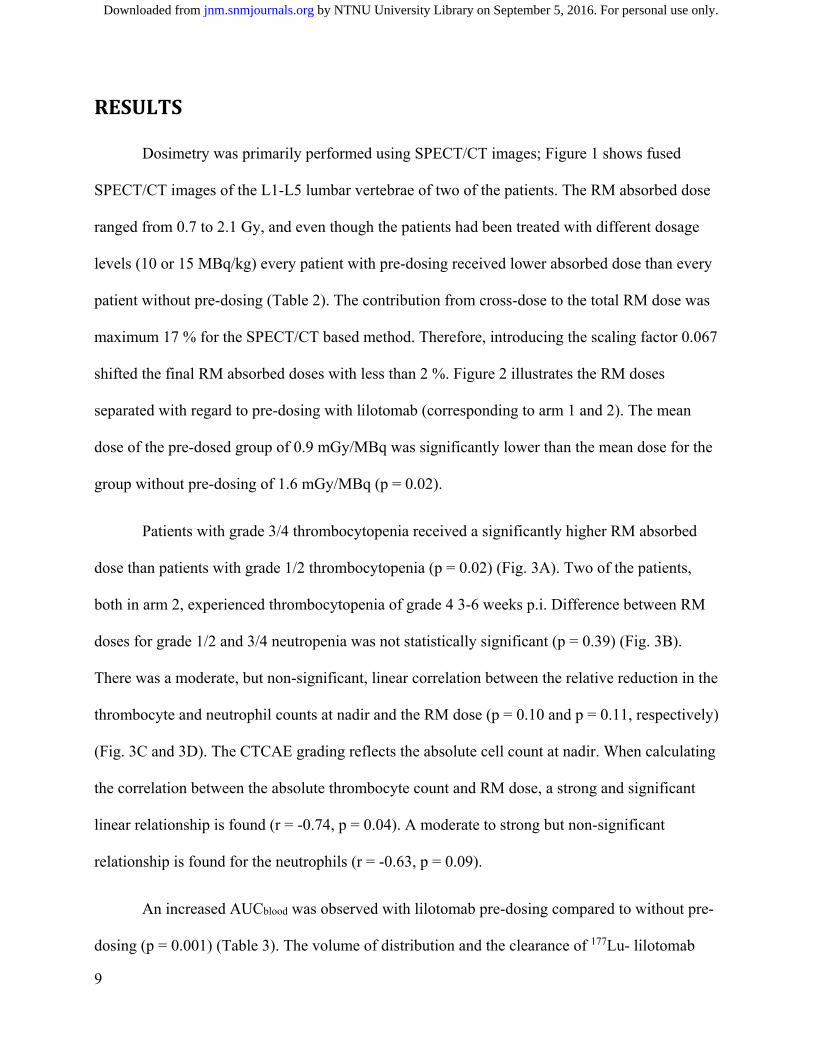
Dosimetry was primarily performed using SPECT/CT images; Figure 1 shows fused
SPECT/CT images of the L1-L5 lumbar vertebrae of two of the patients. The RM absorbed dose
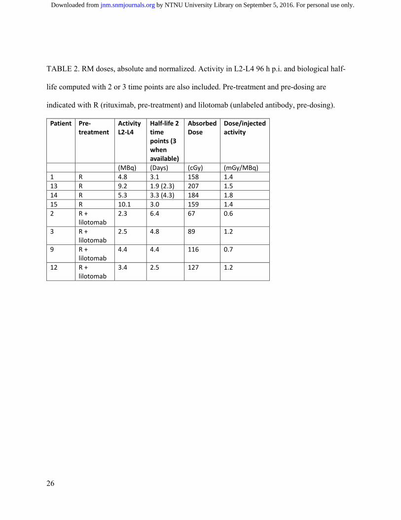
ranged from 0.7 to 2.1 Gy, and even though the patients had been treated with different dosage
levels (10 or 15 MBq/kg) every patient with pre-dosing received lower absorbed dose than every
patient without pre-dosing (Table 2). The contribution from cross-dose to the total RM dose was
maximum 17 % for the SPECT/CT based method. Therefore, introducing the scaling factor 0.067
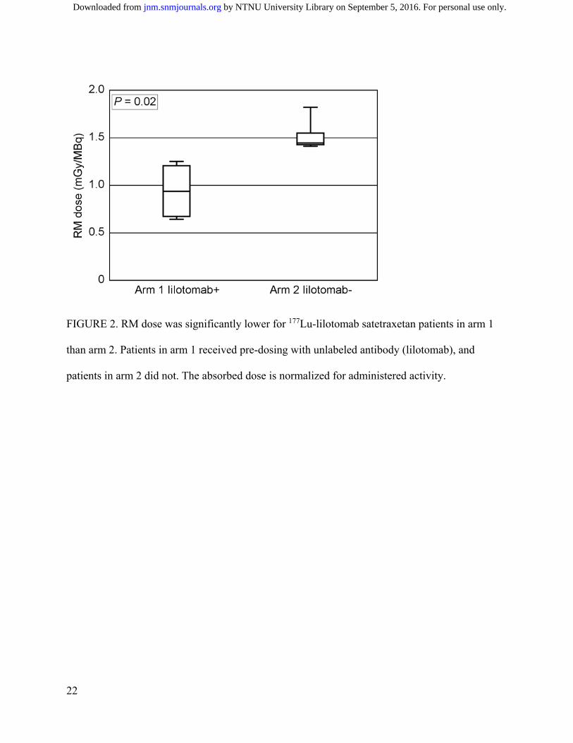
shifted the final RM absorbed doses with less than 2 %. Figure 2 illustrates the RM doses
separated with regard to pre-dosing with lilotomab (corresponding to arm 1 and 2). The mean
dose of the pre-dosed group of 0.9 mGy/MBq was significantly lower than the mean dose for the
group without pre-dosing of 1.6 mGy/MBq (p = 0.02).
Patients with grade 3/4 thrombocytopenia received a significantly higher RM absorbed
dose than patients with grade 1/2 thrombocytopenia (p = 0.02) (Fig. 3A). Two of the patients,
both in arm 2, experienced thrombocytopenia of grade 4 3-6 weeks p.i. Difference between RM
doses for grade 1/2 and 3/4 neutropenia was not statistically significant (p = 0.39) (Fig. 3B).
There was a moderate, but non-significant, linear correlation between the relative reduction in the
thrombocyte and neutrophil counts at nadir and the RM dose (p = 0.10 and p = 0.11, respectively)
(Fig. 3C and 3D). The CTCAE grading reflects the absolute cell count at nadir. When calculating
the correlation between the absolute thrombocyte count and RM dose, a strong and significant
linear relationship is found (r = -0.74, p = 0.04). A moderate to strong but non-significant
relationship is found for the neutrophils (r = -0.63, p = 0.09).
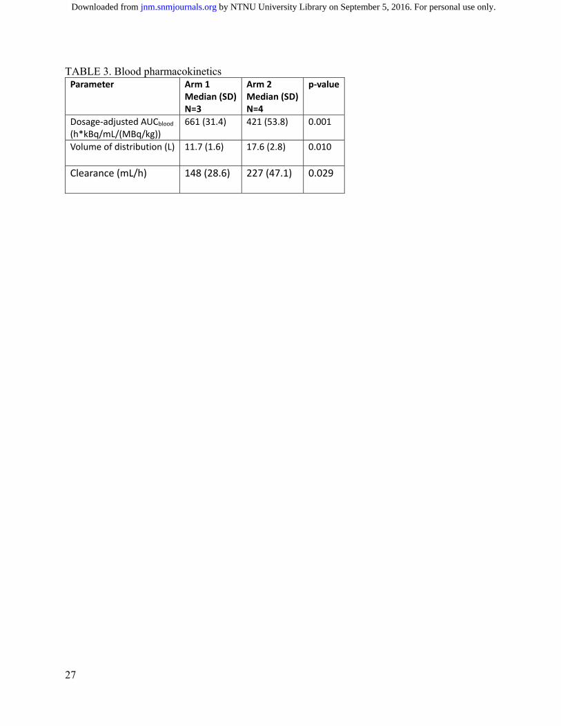
An increased AUCblood was observed with lilotomab pre-dosing compared to without pre-
dosing (p = 0.001) (Table 3). The volume of distribution and the clearance of 177Lu- lilotomab
by NTNU University Library on September 5, 2016. For personal use only. jnm.snmjournals.org Downloaded from

10
satetraxetan was significantly lower for patients given lilotomab compared to those not given
lilotomab (p = 0.01 and p = 0.03, respectively).
The surrogate method resulted in a significant underestimation of RM dose compared to
SPECT/CT-derived dose (p = 0.002). The relative difference ranged from 80 to 638 %. RM dose
calculated by the surrogate method did not show any correlation with hematological toxicity (Fig.
4).
by NTNU University Library on September 5, 2016. For personal use only. jnm.snmjournals.org Downloaded from

11
DISCUSSION
RM is one of the most radiation sensitive organs in the body. In this work we have
calculated the RM doses and correlated them to hematological adverse events for 8 patients
treated with 177Lu-lilotomab satetraxetan.
The RM doses were significantly higher for arm 2 (lilotomab-) than for arm 1
(lilotomab+). This difference indicates that pre-dosing with lilotomab will have a protective
effect for RM, most likely because the unlabeled antibody blocks binding to CD37 in the RM.
The activity in blood, AUCblood, was higher, and the volume of distribution and the clearance
were lower for arm 1 than for arm 2. This is likely due to binding of unlabeled lilotomab to CD37
expressed on cells in the highly perfused compartment including peripheral blood and RM. This
binding by lilotomab to the readily accessible CD37 target antigens then prevents 177Lu-lilotomab
satetraxetan binding to the cells in this compartment, increasing the concentration in the blood,
reducing the available volume of distribution and eventually resulting in reduced amounts of
radioactivity in RM. There is a risk that pre-dosing with cold antibody could block the CD37
antigen on tumor tissues as well, but there was no difference in the tumor absorbed dose for arm
1 and 2 (1). The reduced distribution volume and clearance in arm1 might explain this finding
because it implies that the concentration of 177Lu-lilotomab satetraxetan was higher for arm 1
patients than for arm 2 patients and the increased concentration will counteract an eventual
blocking of CD37 on tumor tissue. The combined findings of these works recommend to use pre-
dosing with lilotomab before treatment with 177Lu-lilotomab satetraxetan. The optimal amount of
unlabeled antibody has yet to be investigated.
Although the numbers of patients are limited, a clear tendency of increasing RM dose
with patient dosage (10 MBq/kg vs 15 MBq/kg, Table 2) can be seen for each arm. This is in
by NTNU University Library on September 5, 2016. For personal use only. jnm.snmjournals.org Downloaded from
12
accordance with the findings for tumors in part 1, where the absorbed dose significantly increased
with patient dosage level (1).
The overall RM dose range was 0.64 to 1.82 mGy/MBq. These doses are of the same
order of magnitude as the RM doses listed in the package inserts of 90Y-ibritumomab-tiuxetan
and 131I-tositumomab (22,23). Somewhat varying 90Y-ibritumomab-tiuxetan RM doses have been
reported, possibly since substitute radioligands have been used for planar imaging and dosimetry
(24,25). For 131I-tositumomab, the dosage is adjusted to produce a 0.75 Gy whole body dose,
shown to correspond with SPECT/CT-derived RM doses no higher than 1.9 Gy and a median of
1.56 Gy with typical dosage (26). Here, RM absorbed doses for 177Lu-lilotomab satetraxetan are
demonstrated in accordance with typical dose ranges reported for other ARC treatments.
Clear relationships between RM doses and hematological toxicity for ARC-therapies have
traditionally been difficult to establish, and possible explanations include heterogeneous patient
groups and dosimetric methodology. In our study, patients developing thrombocytopenia grade
3/4 had received significant higher RM doses than the grade 1/2 group. For the group level
neutropenia analysis, the difference was not significant, and a larger patient material should be
investigated. This is also demonstrated by the absolute neutrophil count and RM dose statistical
analyses (p = 0.09). Prior chemotherapy, limiting the RM reserve, can also alter the relationship
between RM dose and hematological toxicity (27). All patients in our study had undergone prior
chemotherapy, with the number of previous treatments ranging considerably (Table 1). While the
limited number of patients prevents quantitative analyses regarding the influence of prior
treatments, our findings suggest that the dose-toxicity relationship also depends on the extent of
prior chemotherapy. E.g. patient 2 had received the most extensive prior treatment, possibly
leading to a reduced marrow reserve and explaining the unexpected neutropenia grade 3 after an
RM dose of only 67 cGy. On the contrary patient 15, whom received an RM dose of 159 cGy,
by NTNU University Library on September 5, 2016. For personal use only. jnm.snmjournals.org Downloaded from

13
suffered only minor hematological toxicity. This well tolerated RM radiation could potentially be
explained by the relatively limited prior treatment of only one chemotherapy regime.
Our RM dose calculation relies on L2-L4 being representative for the whole marrow. This
part of the skeleton has frequently been used, and the resulting doses have shown correlation with
hematological toxicity (11,12,19). Ideally; analyses of all skeletal sites containing RM would
strengthen the dosimetry, visual inspection of the SPECT/CT-images did however suggest similar
uptake in other skeletal sites, e.g. costae, the sacrum, the sternum and ilium. A two-point
dosimetry model was used to avoid introducing systematic errors. An additional time point was
available for 2 patients (patient 13 and 14) and calculations for these three-point curves
demonstrated a low relative difference in RM dose (0 and 8 %, data not shown). For other
radionuclide treatments it has been suggested to calculate RM doses based on the radioactivity
concentration in blood, and this method is sometimes also erroneous used to estimate doses for
ARCs with specific RM binding. Assuming a conservative estimate of equal activity
concentration in blood and RM we found doses between 80 and 638 % lower than the SPECT/CT
derived RM doses and no correlation with toxicity (Fig. 4). This clearly shows that this surrogate
method should not be used to calculate RM doses for patients treated with 177Lu-lilotomab
satetraxetan. However; a seemingly inverse proportionality between AUCblood itself and RM dose
suggests that alternative models for linking AUCblood and RM dose can possibly be developed.
The reduction in cell count relative to baseline is commonly used to evaluate RM dose
against toxicity (11,13,28,29). When extrapolating the regression curve in Figure 3B, a 100 %
reduction of both thrombocytes and neutrophils are found at 2 – 3 Gy. While our data suggest
linear regression, sigmoidal fits have been demonstrated in other works, and this value should be
considered an estimate (29). This is further supported by the range in prior chemotherapies for
the patient population; this variation can preclude trends for different patient groups. The two
by NTNU University Library on September 5, 2016. For personal use only. jnm.snmjournals.org Downloaded from

14
patients in our study (patient 13 and 14) that experienced grade 4 thrombocytopenia had received
the highest RM doses and were also the only patients receiving an RM dose above 1.8 Gy. The
widely used 2 Gy dose limit for RM was initially determined for treatment of differentiated
thyroid cancer using 131 I in the early 60s (30). Later, the potential differences in biological and
physical factors (e.g. dose rate and electron energy) have been suggested to demonstrate a need
for empiric determination of dose limits for other/novel therapies. Our results support an RM
absorbed dose limit of approximately 2 Gy for patients treated with 177Lu-lilotomab satetraxetan.
by NTNU University Library on September 5, 2016. For personal use only. jnm.snmjournals.org Downloaded from

15
CONCLUSION
While pre-dosing with 40 mg unlabeled lilotomab significantly reduces the RM absorbed
dose for patients treated with 177Lu-lilotomab satetraxetan, the tumor absorbed dose is not
affected by this amount of unlabeled antibody. These findings support to use pre-dosing with
lilotomab for patients to be treated with 177Lu-lilotomab satetraxetan, and encourage
investigations regarding the optimal amount of pre-dosing, which currently is ongoing.
Hematological toxicity was more severe for patients receiving higher absorbed radiation doses,
and our results indicate an RM absorbed dose limit of about 2 Gy for 177Lu-lilotomab satetraxetan
therapy. Given the extent of prior chemotherapy for the population, a somewhat higher dose limit
can be expected for patients without such treatment. A surrogate method based on blood
sampling instead of imaging demonstrated severe shortcomings for 177Lu-lilotomab satetraxetan
patients. The calculation of RM absorbed dose, based on SPECT/CT imaging approximately day
4 and 7, can possibly predict adverse events weeks before occurring. In our experience, such
calculations can be performed by trained and prepared personnel within 2 days of the imaging.
by NTNU University Library on September 5, 2016. For personal use only. jnm.snmjournals.org Downloaded from
16
DISCLOSURE
This study was sponsored by Nordic Nanovector ASA. Johan Blakkisrud was in part
supported by grants from Nordic Nanovector ASA. Arne Kolstad is a member of the Nordic
Nanovector Scientific ASA Advisory board. Harald Holte and Arne Kolstad were both in part
supported by grants from the Norwegian Cancer Society.
by NTNU University Library on September 5, 2016. For personal use only. jnm.snmjournals.org Downloaded from

17
ACKNOWLEDGMENTS
We thank the personnel at the Nuclear Medicine section at Oslo University Hospital for
technical assistance with the acquisitions. Stine Nygaard, study nurse at the Department of
Oncology, is also greatly acknowledged.
by NTNU University Library on September 5, 2016. For personal use only. jnm.snmjournals.org Downloaded from

18
REFERENCES
1. Blakkisrud J, Løndalen A, Martinsen ACT, et al. Tumor absorbed dose for non-Hodgkin’s lymphoma patients treated with the novel anti-CD37 antibody radionuclide conjugate 177Lu-lilotomab satetraxetan. J Nucl Med. 4. August 2016 [Epub ahead of print].
2. Zelenetz AD. A clinical and scientific overview of tositumomab and iodine I 131 tositumomab. Semin Oncol. 2003;30:22-30.
3. Jacene HA, Filice R, Kasecamp W, Wahl RL. Comparison of 90Y-ibritumomab tiuxetan and 131I-tositumomab in clinical practice. J Nucl Med. 2007;48:1767-1776.
4. Kaminski MS, Fig LM, Zasadny KR, et al. Imaging, dosimetry, and radioimmunotherapy with iodine 131-labeled anti-CD37 antibody in B-cell lymphoma. J Clin Oncol. 1992;10:1696-1711.
5. Forrer F, Oechslin-Oberholzer C, Campana B, et al. Radioimmunotherapy with 177Lu-DOTA-Rituximab: final results of a phase I/II Study in 31 patients with relapsing follicular, mantle cell, and other indolent B-cell lymphomas. J Nucl Med. 2013;54:1045-1052.
6. Repetto-Llamazares AHV, Larsen RH, Mollatt C, Lassmann M, Dahle J. Biodistribution and dosimetry of (177)Lu-tetulomab, a new radioimmunoconjugate for treatment of non-hodgkin lymphoma. Curr Radiopharm. 2013;6:20-27.
7. Kolstad A, Madsbu U, Beasley M, et al. Efficacy and safety results of Betalutin® (177-Lu-DOTA-HH1) in a phase 1/2 study of patients with non-Hodgkin B-Cell lymphoma (NHL). AACR Annual Meeting [Poster]. New Orleans, Louisiana, USA; 2016.
8. Stabin MG, Eckerman KF, Bolch WE, Bouchet LG, Patton PW. Evolution and status of bone and marrow dose models. Cancer Biother Radiopharm. 2002;17:427-433.
9. Hindorf C, Glatting G, Chiesa C, Linden O, Flux G. EANM dosimetry committee guidelines for bone marrow and whole-body dosimetry. Eur J Nucl Med Mol Imaging. 2010;37:1238-1250.
10. Boucek JA, Turner JH. Personalized dosimetry of 131I-rituximab radioimmunotherapy of non-hodgkin lymphoma defined by pharmacokinetics in bone marrow and blood. Cancer Biother Radiopharm. 2014;29:18-25.
by NTNU University Library on September 5, 2016. For personal use only. jnm.snmjournals.org Downloaded from

19
11. Shen S, Meredith RF, Duan J, et al. Improved prediction of myelotoxicity using a patient-specific imaging dose estimate for non-marrow-targeting (90)Y-antibody therapy. J Nucl Med. 2002;43:1245-1253.
12. Ferrer L, Kraeber-Bodéré F, Bodet-Milin C, et al. Three methods assessing red marrow dosimetry in lymphoma patients treated with radioimmunotherapy. Cancer. 2010;116:1093-1100.
13. Pauwels S, Barone R, Walrand S, et al. Practical dosimetry of peptide receptor radionuclide therapy with (90)Y-labeled somatostatin analogs. J Nucl Med. 2005;46 Suppl 1:92-98.
14. Woliner-van der Weg W, Schoffelen R, Hobbs RF, et al. Tumor and red bone marrow dosimetry: comparison of methods for prospective treatment planning in pretargeted radioimmunotherapy. EJNMMI Phys. 2015;2:5.
15. Wahl RL. Tositumomab and 131I therapy in non-hodgkin’s lymphoma. J Nucl Med. 2005;46:128-140.
16. Kaminski MS, Zasadny KR, Francis IR, et al. Radioimmunotherapy of B-cell lymphoma with [131I]anti-B1 (anti-CD20) antibody. N Engl J Med. 1993;329:459-465.
17. Common terminology criteria for adverse events v4.0. National Cancer Institute.
NIH#09-7473. 4.0 ed; 2009.
18. Schwartz J, Humm JL, Divgi CR, Larson SM, O'Donoghue JA. Bone marrow dosimetry using 124I-PET. J Nucl Med. 2012;53:615-621.
19. Herrmann K, Lapa C, Wester H-J, et al. Biodistribution and radiation dosimetry for the chemokine receptor CXCR4-targeting probe 68Ga-pentixafor. J Nucl Med. 2015;56:410-416.
20. Stabin MG, Sparks RB, Crowe E. OLINDA/EXM: the second-generation personal computer software for internal dose assessment in nuclear medicine. J Nucl Med. 2005;46:1023-1027.
21. Sgouros G. Bone marrow dosimetry for radioimmunotherapy: theoretical considerations. J Nucl Med. 1993;34:689-694.
22. Zevalin prescribing information [package insert]. Amsterdam, Netherlands: Spectrum Pharmaceuticals, Inc. http://www.zevalin.com/downloads/Zevalin_Package_Insert.pdf. Accessed 22/6/2016.
by NTNU University Library on September 5, 2016. For personal use only. jnm.snmjournals.org Downloaded from

20
23. Bexxar prescribing information [package insert]. London, England: GlaxoSmithKline plc. https://www.gsksource.com/pharma/content/dam/GlaxoSmithKline/US/en/Prescribing_Information/Bexxar/pdf/BEXXAR.PDF. Accessed 24/6/2016.
24. Fisher DR, Shen S, Meredith RF. MIRD dose estimate report no. 20: radiation absorbed-dose estimates for 111In- and 90Y-ibritumomab tiuxetan. J Nucl Med. 2009;50:644-652.
25. Wiseman GA, Kornmehl E, Leigh B, et al. Radiation dosimetry results and safety correlations from 90Y-ibritumomab tiuxetan radioimmunotherapy for relapsed or refractory non-Hodgkin's lymphoma: combined data from 4 clinical trials. J Nucl Med. 2003;44:465-474.
26. Boucek JA, Turner JH. Validation of prospective whole-body bone marrow dosimetry by SPECT/CT multimodality imaging in (131)I-anti-CD20 rituximab radioimmunotherapy of non-Hodgkin's lymphoma. Eur J Nucl Med Mol Imaging. 2005;32:458-469.
27. Siegel JA. Update: establishing a clinically meaningful predictive model of hematologic toxicity in nonmyeloablative targeted radiotherapy: practical aspects and limitations of red marrow dosimetry. Cancer Biother Radiopharm. 2005;20:126-140.
28. Chiesa C, Botta F, Coliva A, et al. Absorbed dose and biologically effective dose in patients with high-risk non-Hodgkin's lymphoma treated with high-activity myeloablative 90Y-ibritumomab tiuxetan (Zevalin). Eur J Nucl Med Mol Imaging. 2009;36:1745-1757.
29. Walrand S, Barone R, Pauwels S, Jamar F. Experimental facts supporting a red marrow uptake due to radiometal transchelation in 90Y-DOTATOC therapy and relationship to the decrease of platelet counts. Eur J Nucl Med Mol Imaging. 2011;38:1270-1280.
30. Benua RS, Cicale NR, Sonenberg M, Rawson RW. The relation of radioiodine dosimetry to results and complications in the treatment of metastatic thyroid cancer. Am J Roentgenol Radium Ther Nucl Med. 1962;87:171-182.
by NTNU University Library on September 5, 2016. For personal use only. jnm.snmjournals.org Downloaded from
21
FIGURELEGENDS
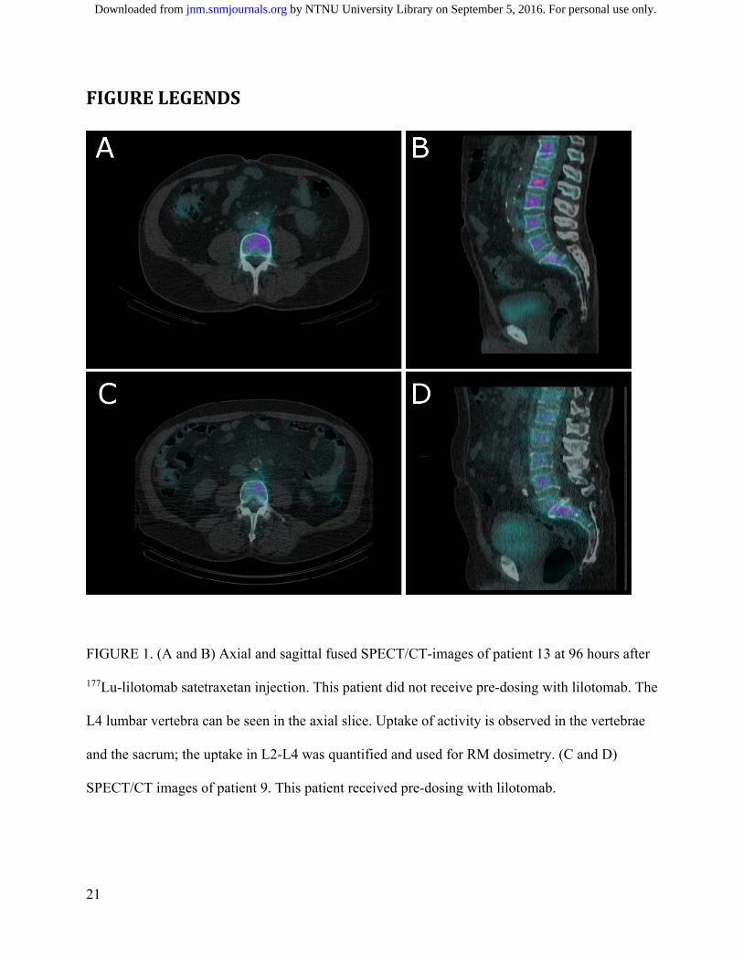
FIGURE 1. (A and B) Axial and sagittal fused SPECT/CT-images of patient 13 at 96 hours after
177Lu-lilotomab satetraxetan injection. This patient did not receive pre-dosing with lilotomab. The
L4 lumbar vertebra can be seen in the axial slice. Uptake of activity is observed in the vertebrae
and the sacrum; the uptake in L2-L4 was quantified and used for RM dosimetry. (C and D)
SPECT/CT images of patient 9. This patient received pre-dosing with lilotomab.
by NTNU University Library on September 5, 2016. For personal use only. jnm.snmjournals.org Downloaded from
22
FIGURE 2. RM dose was significantly lower for 177Lu-lilotomab satetraxetan patients in arm 1
than arm 2. Patients in arm 1 received pre-dosing with unlabeled antibody (lilotomab), and
patients in arm 2 did not. The absorbed dose is normalized for administered activity.
by NTNU University Library on September 5, 2016. For personal use only. jnm.snmjournals.org Downloaded from

23
FIGURE 3. Hematological toxicity versus RM absorbed dose for patients receiving 177Lu-
lilotomab satetraxetan treatment. (A) CTCAE grading of thrombocytopenia plotted against dose.
The dose was significantly higher for patients with grade 3/4 thrombocytopenia than grade 1/2
thrombocytopenia (p = 0.02). (B) CTCAE grading of neutropenia plotted against dose. Higher
doses were found for patients with grade 3/4 than grade 1/2 neutropenia but the difference was
not statistically significant (p = 0.39). (C and D) The relative reduction in blood-cells at nadir
with respect to RM dose. Thrombocytes are shown in C and neutrophils in D. Filled symbols
represent patients without pre-dosing (arm 2) and open symbols patients with pre-dosing (arm 1).
by NTNU University Library on September 5, 2016. For personal use only. jnm.snmjournals.org Downloaded from

24
FIGURE 4. The lack of correlation between RM dose derived by surrogate method and the
reduction in thrombocyte and neutrophil counts demonstrates that this non-imaging based method
is unfit to predict marrow toxicity for 177Lu-lilotomab satetraxetan therapy. Thrombocytes are
shown in A and neutrophils in B. Filled symbols represent patients without pre-dosing (arm 2)
and open symbols patients with pre-dosing (arm 1).
by NTNU University Library on September 5, 2016. For personal use only. jnm.snmjournals.org Downloaded from
25
TABLES
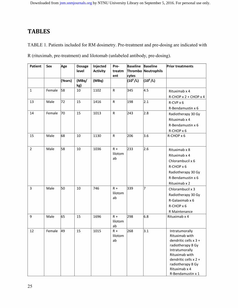
TABLE 1. Patients included for RM dosimetry. Pre-treatment and pre-dosing are indicated with
R (rituximab, pre-treatment) and lilotomab (unlabeled antibody, pre-dosing).
Patient Sex Age Dosage level
Injected Activity
Pre‐treatment
Baseline Thrombocytes
Baseline Neutrophils
Prior treatments
(Years) (MBq/kg)
(MBq) (109 /L) (109 /L)
1 Female 58 10 1102 R 345 4.5 Rituximab x 4
R‐CHOP x 2 + CHOP x 4
13 Male 72 15 1416 R 198 2.1 R‐CVP x 6
R‐Bendamustin x 6
14 Female 70 15 1013 R 243 2.8 Radiotherapy 30 Gy
Rituximab x 4
R‐Bendamustin x 6
R‐CHOP x 6
15 Male 68 10 1130 R 206 3.6 R‐CHOP x 6
2 Male 58 10 1036 R + lilotomab
233 2.6 Rituximab x 8
Rituximab x 4
Chlorambucil x 6
R‐CHOP x 6
Radiotherapy 30 Gy
R‐Bendamustin x 6
Rituximab x 2
3 Male 50 10 746 R + lilotomab
339 7 Chlorambucil x 3
Radiotherapy 30 Gy
R‐Galaximab x 6
R‐CHOP x 6
R Maintenance
9 Male 65 15 1696 R + lilotomab
298 6.8 Rituximab x 4
12 Female 49 15 1015 R + lilotomab
268 3.1 Intratumorally Rituximab with dendritic cells x 3 + radiotherapy 8 Gy Intratumorally Rituximab with dendritic cells x 2 + radiotherapy 8 Gy Rituximab x 4 R‐Bendamustin x 1
by NTNU University Library on September 5, 2016. For personal use only. jnm.snmjournals.org Downloaded from
26
TABLE 2. RM doses, absolute and normalized. Activity in L2-L4 96 h p.i. and biological half-
life computed with 2 or 3 time points are also included. Pre-treatment and pre-dosing are
indicated with R (rituximab, pre-treatment) and lilotomab (unlabeled antibody, pre-dosing).
Patient Pre‐treatment
Activity L2‐L4
Half‐life 2 time points (3 when available)
Absorbed Dose
Dose/injected activity
(MBq) (Days) (cGy) (mGy/MBq)
1 R 4.8 3.1 158 1.4
13 R 9.2 1.9 (2.3) 207 1.5
14 R 5.3 3.3 (4.3) 184 1.8
15 R 10.1 3.0 159 1.4
2 R + lilotomab
2.3 6.4 67 0.6
3 R + lilotomab
2.5 4.8 89 1.2
9 R + lilotomab
4.4 4.4 116 0.7
12 R + lilotomab
3.4 2.5 127 1.2
by NTNU University Library on September 5, 2016. For personal use only. jnm.snmjournals.org Downloaded from
27
TABLE 3. Blood pharmacokinetics Parameter Arm 1
Median (SD) N=3
Arm 2 Median (SD) N=4
p‐value
Dosage‐adjusted AUCblood (h*kBq/mL/(MBq/kg))
661 (31.4) 421 (53.8) 0.001
Volume of distribution (L) 11.7 (1.6)
17.6 (2.8) 0.010
Clearance (mL/h) 148 (28.6)
227 (47.1) 0.029
by NTNU University Library on September 5, 2016. For personal use only. jnm.snmjournals.org Downloaded from

Doi: 10.2967/jnumed.116.180471Published online: September 1, 2016.J Nucl Med. Johan Blakkisrud, Ayca Løndalen, Jostein Dahle, Simon Turner, Harald Holte, Arne Kolstad and Caroline Stokke
Lu-lilotomab satetraxetan177novel anti-CD37 antibody radionuclide conjugate Red Marrow Absorbed Dose for non-Hodgkin's Lymphoma Patients treated with the
http://jnm.snmjournals.org/content/early/2016/08/31/jnumed.116.180471This article and updated information are available at:
http://jnm.snmjournals.org/site/subscriptions/online.xhtml
Information about subscriptions to JNM can be found at:
http://jnm.snmjournals.org/site/misc/permission.xhtmlInformation about reproducing figures, tables, or other portions of this article can be found online at:
and the final, published version.proofreading, and author review. This process may lead to differences between the accepted version of the manuscript
ahead of print area, they will be prepared for print and online publication, which includes copyediting, typesetting,JNMcopyedited, nor have they appeared in a print or online issue of the journal. Once the accepted manuscripts appear in the
. They have not beenJNM ahead of print articles have been peer reviewed and accepted for publication in JNM
(Print ISSN: 0161-5505, Online ISSN: 2159-662X)1850 Samuel Morse Drive, Reston, VA 20190.SNMMI | Society of Nuclear Medicine and Molecular Imaging
is published monthly.The Journal of Nuclear Medicine
© Copyright 2016 SNMMI; all rights reserved.
by NTNU University Library on September 5, 2016. For personal use only. jnm.snmjournals.org Downloaded from


![STN 125011 Tositumomab Therapeutic Regimen (TTR) [tositumomab plus I-131 tositumomab] Oncologic Drugs Advisory Committee December 17, 2002](https://img.dokumen.tips/doc/110x75/56649dc45503460f94ab6f7b/stn-125011-tositumomab-therapeutic-regimen-ttr-tositumomab-plus-i-131-tositumomab.jpg)


![STN 125011 Tositumomab Therapeutic Regimen (TTR) [tositumomab plus I-131 tositumomab]](https://img.dokumen.tips/doc/110x75/568145aa550346895db2a1c5/stn-125011-tositumomab-therapeutic-regimen-ttr-tositumomab-plus-i-131-tositumomab-56958b79cf2de.jpg)

