Embed Size (px)
Citation preview

EU Scientific Seminar 2013 Radiation Induced Long-term Health Effects after Medical Exposures
Luxembourg, 19 November 2013
Dosimetry in Radiodiagnostic Procedures, Risk Issues and Research Needs
Renato Padovani
Medical Physics Dpt, S. Maria della Misericordia University Hospital, Udine, Italy

Objectives
• To discuss present status of dosimetry in diagnostic and interventional radiology and radiation risk assessment and to identify possible research and action needs
2

Content
• Examination frequencies and population exposures in Europe
• Dosimetry status and needs
• Radiation risk assessment and research needs
• Optimisation of exposure status and action needs
3

DoseDataMed2 EC project (2012): frequency X-ray procedures
4
> 1.5 proc/year
<0.7 proc/year
Large variability for high dose precedures (CT, IR)

DoseDataMed2 EC project (2012): collective effective dose for
X-ray procedures (mSv/person.year)
5
> 1.5 mSv/year
<0.5 mSv/year

DDM2: European figure
• Europe has lowest mean dose level if compared with US (~ 3mSv/y) or Japan (~2 mSv/y)
• Large differences with regards to justification and optimisation levels
6

Question rising
• Which has been the impact of 15 years of research, actions, ME Directive and EC guidelines in Europe? – Several actions and research projects:
• Development of DRLs concepts, referral criteria, optimisation tools, equipment with dose information, etc
• Several guidelines (education and training, audit, etc.)
– Development of safety culture in radiology area • Implementation of QA programmes, patient dose monitoring, DRLs
assessment and use
• Development of a category of medical physicists experts in diagnostic imaging
• IEC and DICOM developments
• But, in Europe we have still large differences in the justification and optimisation of radiological procedures
7

Patient dosimetry status and needs
8
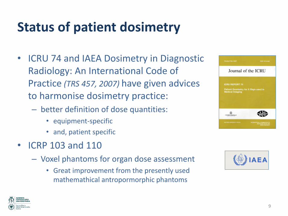
Status of patient dosimetry
• ICRU 74 and IAEA Dosimetry in Diagnostic Radiology: An International Code of Practice (TRS 457, 2007) have given advices to harmonise dosimetry practice:
– better definition of dose quantities:
• equipment-specific
• and, patient specific
• ICRP 103 and 110
– Voxel phantoms for organ dose assessment
• Great improvement from the presently used mathemathical antropormorphic phantoms
9

Equipment specific dose quantities (names and symbols, ICRU 74)
• Incident air kerma, Ki
• Entrance surface air kerma Ke= Ki B unit: Gy
• X-ray tube output Y(d) = K(d)/Pit unit: GyC-1
• Air kerma-area product PKA unit: Gym2
• Air kerma-lenght product PKL unit: Gym
• Computed Tomography Kerma Index, C100 unit: Gy
IEC standards, MED requirements 10
A
KA dxdyyxKP ),(
L
airKL dzzKP )(
50
50
1100 )( dzzKC
nT

Patient specific dose quantity
• The absorbed dose D, is the energy absorbed per unit mass.
Organ dose is expressed as an mean absorbed dose to an organ/tissue
11

Organ dose
• Methods to assess mean organ dose: – To measure organ dose in a
physical antropomorphic phantom
– To simulate with the Monte Carlo method the irradiation of a phantom (mathematical or voxel) • Conversion coefficients normalised
to equipment specific dose quantities
12

Computational anatomical phantoms
• To evaluate the energy deposition in organs from internal and external radiation exposures.
• Mathematical phantoms: – Mathematical expressions describing the shape and position of
idealised body organs (Oak Ridge National Laboratory (Fisher and Snyder, 1967, 1968; Snyder et al., 1969, 1978; Cristy,
1980; Cristy and Eckerman, 1987) for the Medical Internal Radiation Dose (MIRD) Committee)
– From the original adult MIRD phantom, paediatric phantoms were derived to represent infants and children of various ages (Cristy, 1980).
– Hermaphrodite models, male and female adult mathematical models called Adam’ and ‘Eva’ were introduced (Kramer, 1982)
13

Computational anatomical phantoms
• ‘Tomographic’ or ‘voxel’ phantoms – Large number of volume elements (voxels) for a detailed
representation of human anatomy (Zankl et al., 1988; Zubal et al., 1994, 1996; Dimbylow, 1996; Caon et al., 1999; Xu et al., 2000; Zankl and Wittmann, 2001; Petoussi-Henss et al., 2002; Zaidi and Xu, 2007)
– Voxel phantoms can be used for a wide spectrum of applications.
ICRP 110: … phantoms will be used by ICRP in establishing radiation protection guidance, e.g. effective dose coefficients and other secondary dosimetric quantities.
14

Example: Absorbed dose per air kerma to Stomach from an Anterio-Posterior photon exposure for male and female ICRP 110 phantoms
- Different MC codes used - Comparison with previous conversion
coefficients (.) (ICRP 74)
Progresses mainly to improve accuracy of exposure and risk assessment to workers and population
Development for patient dose
assessment are possible: - to develop phantoms patient-
specific (e.g. different age, obese patients, pregnant women, etc)
- to improve accuracy in patient exposure assessment
15

Dosimetry needs: equipment-specific dose information
• Computed Tomography – Dose quantity
• CTDI is a dose quantity related to specific geometrical phantoms (CT dosimetry phantoms)
– For the wide beams used in some MSCT the quantity is inaccurate
• CTDI is not a real equipment specific and it is not a patient specific – A new CT dosimetry methodology should be probably necessary
– Information from the CT of the angular current modulation is not always available • IEC should require availability of angular current modulation and DICOM
should export the data
• Interventional radiology – Real time skin dose distribution not yet available to monitor in skin
dose in high dose IR procedures • Patient skin models and patient-to-equipment registration methods have to be
develop and implemented in IR equipment
16

Dosimetry needs: equipment-specific dose information
• ConeBeam CT
– A solid dosimetry is not available • KAP is used, but x-ray beam is not fully intercepted by the patient
body
• A better dosimetry method should be developed
17

Dosimetry needs: patient specific dosimetry
• To develop patient models aiming to compute organ doses in the different procedure – Patient models
• Age/Size/Gender specifics
e.g. paediatric, obese patient models, pregnant women, etc.
• Models for specific applications: – Interventional radiology: patient skin model to compute skin dose
distribution and peak skin dose
– Mammography : average glandular dose
• Models to take into account the different irradiation modalities: – Tomosynthesis
– CT
– ConebeamCT
18

Dosimetry needs: implementation of patient specific dosimetry
• To implement in radiological equipment patient specific dosimetry – CT
• organ dose patient-specific taking information from CT images
– IR • from the patient skin model, patient-to-equipment registration, to
compute and provide the real time skin dose map
– Mammography • Average glandular dose (AGD) is calculated for standard breast,
equipment software is not using the information on breast composition (glandularity) derived from the automatic exposure system
• A more solid dosimetry can be implemented
19

Dosimetry needs: risk assessment and communication
• Effective dose is a fortunate synthetic quantity to quantity workers and population exposures and compare with limits
• Today cumulative effective dose is frequently adopted to quantify population exposures, also for medical exposures, when age classes, gender, pathology can modify substantially risk factors!
Probably we need a similar synthetic quantity to properly express patient radiation exposure – should be age/gender/(pathology?) specific
Or do we need to develop a quantity to assess the risk in a form that can be understood by the patient and can be compared with other risks?
20

Optimisation of radiological procedures
21

Optimisation tools: DRLs
• Status of Diagnostic Reference Levels (DRLs) – Required by MED
– Present situation in Europe is given by DDM2 study: • Several countries have adopted the (old) EC DRLs
• Most Countries have never updated DRLs
• Very few Countries have DRLs for high dose/risk procedures (paediatric CT, interventional radiology)
• Few information on the effective use of DRLs in EU countries
• General thinking is that the compliance with DRLs certifies an optimised practice!
– No great impact of DRLs is seen in most of EU countries (see DDM2)
22

Optimisation tools: DRLs
• The need:
– To redefine DRLs: role, assessment and use
– To add a second quantity, like Achievable Reference Level (ARLs) ? • It can be easier to understand the purpose of DRLs to identify
unacceptable practices
– Regulatory needs • More stringent requirements from regulations
– periodic dose assessment and communication to regulatory bodies, ...
• Development of national /regional patient dose archives
• External audits
23

Optimisation tools: the clinical protocol
• To develop and implement in the radiological equipment intelligent tools supporting staff in planning a procedure:
– To develop models and objective procedures on how to set the pre-programmed dose levels in an optimal way.
– We should find the ‘task’ to be optimized • Example:
The task: high contrast visualisation of thin linear structure, moving on a cardiac background. The aim: to develop an algorithm to apply on the object (like a model observer or another relevant SNR), and access to raw data to calculate such a model observer
24

Optimisation in interventional procedures
1. Deterministic risks should not be a post procedure surprise. 2. A center doing interventional cardiac work and never observing any deterministic risks
in their patients is probably not trying hard enough. (S. Balter, US)
• The need:
– Real time skin dose maps: • from the patient skin model, patient-to-equipment registration, to
compute and provide the real time skin dose map
– Patient dose archives • To identify repeated procedures • To compute cumulative exposures, including skin dose maps, and to
transfer the information also to the to x-ray equipment • To identify patient for clinical follow-up
25

Optimisation in interventional procedures: staff exposure
• Status of staff exposure in IR (from ISEMIR, IAEA): – Not harmonised monitoring, – Low compliance with rules by the staff – The actual exposures are probably not known in several hospitals – Dated technology (passive dosimeters) applied to staff monitoring – New dose limit for eye lens of20 mSv/y in EU BSS
• The need: – To improve staff monitoring:
• Dosimetry: models to assess eye doses, computational dosimetry • Technologies:
– active dosimeters, electronic archives providing real time information, – integration of staff and patient exposures
• To improve dosimetry practices – Inspection/audit – Exposure monitoring: to integrate national dose archives with personal data
(e.g. clinical tasks & workload)
26

Optimisation tools: large patient dose archives
• Status of standards – DICOM has developed and is updating the RDSR (Radiation Dose
Structured Report) – IHE has developed the REM profile (Radiation Exposure Monitoring
Integration Profile, draft 2008) thinking to a patient dose tracking tool at the department/hospital level
– Some examples of dose archives implemented in small network of hospitals
• Present developments allows to develop dose archives at regional/country level to easily: – provide periodic information to radiological staff – provide cumulative doses to individual patient – compare practice/protocols between clinics/hospitals – assess and update DRLs/ARLs – support clinical audits
27

Level of knowledge of exposure and risk levels in specific group of
patients and applications
28

Repeated exposures: CT
• Repeated exposure to head and neck CT is significantly associated with increased risk of cataracts Mei-Kang Yuan et al., Taiwan (AJR, September 2013, Vol. 201:3pp. 626-630).
• Magnitude of repeated exposures in a hospital (Udine, 2013): – 2.4% of patients submitted to CT examination have received a
cumulative DLP > 6700 mGycm (100 mSv for a reference adult man) • A 71 y patient has performed 8 CTs with a cumulative DLP of 516000
mGycm (415 mSv) • Another patient 28 y old has performed 8 CTs with a cumulative DLP
of 917000 mGycm (209 mSv)
29

Risk Assessment in diagnostic radiology
30
• Very few information on risk are available for specific group of patients/pathologies
– Adult chronic disease patients with repeated procedures, sometimes for the whole life: • ESKD (end stage kidney disease),
• IBD (inflammatory bowel disease),
• CAD (coronary artery disease),
• HT (heart transplant)

Risk Assessment in diagnostic radiology
• For paediatric patients, only studies reporting cumulative effective dose!
– Paediatric patients with pathologies with positive outcome and long life expectation and repeated x-ray and nuclear medicine procedures: • Lymphoma
• Crown disease, cystic fibrosis, hydrocephalous,
• CHD (congenital heart disease),
• haemophilia , bleeding disorders
31

Needs for risk assessment in IR & CT
• Dose and dose-rate effectiveness factor (DDREF). ICRP combines the LNT model with a value of 2 for the DDREF and considers it a prudent basis for the practical purposes of radiological protection, ... should be applied to chronic exposures at dose rates less than 6 mGy/h averaged over the first few hours. ICRP refers (paragraph A62 of the 2007 recommendations) that: “When dose rates are lower than around 0.1 Gy/hour there is repair of cellular radiation injury during the irradiation”.
• In IR procedures, dose and dose rates can be higher. During a cine frame a skin dose of 1 mGy can be imparted in <10 ms dose rate of 360 Gy/h
• .. also in CT dose rate is of the same order of magnitude
radiation risk factors could be higher
32

Summary
– In the 80s-90s European outcomes from researches and regulations have been the basis for the implementation of actions, rules and safety culture in medical exposure • These developments have been taken as models at worldwide level
– It is necessary another great effort to fulfil the job to provide harmonised practice to all European patients • Developing optimisation methods and implement them in existing and
new coming practices • Improving knowledge on low dose radiation risks • Developing communication strategies
These will allow to continue to be ahead in this field bvgat worldwide level
33