Embed Size (px)
Citation preview

Dormancy cycling in seeds: mechanisms and regulation
Susanne M. C. Claessens

Thesis committee
Thesis supervisor
Prof. dr. L. H. W. van der Plas
Professor of Plant Physiology
Wageningen University
Thesis co-supervisors
Dr. H. W. M. Hilhorst
Associate professor, Laboratory of Plant Physiology
Wageningen University
Dr. P. E. Toorop
Senior scientist diagnostics, Seed Conservation Department
Millennium Seed Bank Project, Royal Botanic Gardens Kew, UK
Other members
Prof. dr. ir. M. Koornneef, Wageningen University/
Max Planck Institut, Köln, Germany
Dr. H. van As, Wageningen University
Prof. H.W. Pritchard, Botanic Gardens Kew, UK
Prof. dr. O. Leprince, Institut National de la Recherche Agronomique,
Angers,France
This research was conducted under the auspices of the Graduate School of Experimental Plant Sciences
Dormancy cycling in seeds: mechanisms and regulation
Susanne M. C.Claessens
Thesis
submitted in fulfilment of the requirements for the degree of doctor
at Wageningen University
by the authority of the Rector Magnificus
Prof. dr. M.J. Kropff,
in the presence of the
Thesis Committee appointed by the Academic Board
to be defended in public
on Monday 20 February 2012
at 4 p.m. in the Aula.

Susanne M. C. Claessens
Dormancy cycling in seeds: mechanisms and regulation
161 pages
Thesis Wageningen University, Wageningen, NL (2012)
With references with summaries in Dutch and English
ISBN 978-94-6173-190-6
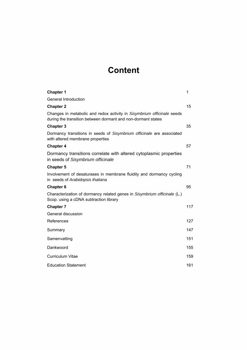
Content
Chapter 1
General Introduction
1
Chapter 2
Changes in metabolic and redox activity in Sisymbrium officinale seeds during the transition between dormant and non-dormant states
15
Chapter 3
Dormancy transitions in seeds of Sisymbrium officinale are associated with altered membrane properties
35
Chapter 4
Dormancy transitions correlate with altered cytoplasmic properties in seeds of Sisymbrium officinale
57
Chapter 5
Involvement of desaturases in membrane fluidity and dormancy cycling in seeds of Arabidopsis thaliana
71
Chapter 6
Characterization of dormancy related genes in Sisymbrium officinale (L.) Scop. using a cDNA subtraction library
95
Chapter 7
General discussion
117
References 127
Summary 147
Samenvatting 151
Dankwoord 155
Curriculum Vitae 159
Education Statement 161


1
Chapter 1
General Introduction
The current study was aimed at further delineating the regulatory principles of
dormancy. The main focus was on a biophysical approach to study cellular
properties that may reflect the seed’s dormancy status, including membrane
properties, metabolic activity and dormancy-associated gene expression. We have
employed Electron Paramagnetic Resonance (EPR) techniques and differential
screening for gene expression. In this chapter, after a general physiological
introduction to the dormancy phenomenon a short introduction will be given to EPR
based research methods, as well as to differential gene expression. Finally, the
objectives and scope of this thesis are described.
Dormancy and dormancy cycling
The life cycle of most plants starts, and often ends, at the seed stage. In most
species mature seeds are shed and dispersed. At this stage of its life cycle the
seed may be dormant and will, by definition, not germinate under favourable
conditions (Bewley, 1997). Nikolaeva (2004) formulated a dormancy classification
system reflecting the fact that dormancy is determined by morphological and
physiological properties, as well as ecological and geographical peculiarities.
Based on this system Baskin and Baskin (2004) have proposed a classification
system which includes 5 classes of seed dormancy: physiological, morphological,
morphophysiological, physical and combinational dormancy. Physiological
dormancy is divided in three levels: deep, intermediate and non-deep. Seeds with
deep physiological dormancy require a prolonged cold or warm incubation in the
imbibed state (stratification) before germination can take place. Seeds with non-

2
deep physiological dormancy cannot germinate without either excision of the
embryo, gibberellic acid, scarification, after-ripening, dry storage or a short warm or
cold stratification. Morphological dormancy is characterised by an underdeveloped,
but differentiated embryo. Embryos of morphologically dormant seeds need time
and suitable temperatures to grow before germination can take place.
Morphophysiological dormancy is characterised by an underdeveloped embryo that
also has a physiological component to its dormancy. Physical dormancy is usually
characterised by water-impermeable layers of palisade cells in the seed or fruit
coat that control water uptake. Mechanical or chemical scarification can break
physical dormancy. Combinational dormancy is characterised by both water
impermeability and a physiological dormancy component.
Physiological dormancy is one of the most common forms of seed
dormancy. In this thesis Arabidopsis thaliana and Sisymbrium officinale seeds
were chosen, as their germination and dormancy behaviour is well described.
Arabidopsis thaliana has well known advantages for molecular studies and in
Sisymbrium officinale dormancy can be fully separated from the germination event
(Hilhorst and Karssen, 1989).
Arabidopsis thaliana and Sisymbrium officinale seeds are classified as
possessing non-deep physiological dormancy. Different states of physiological
dormancy can be distinguished. Seeds may be primary dormant upon shedding, as
at this stage the seeds are not sensitive to germination-inducing factors such as
light or nitrate (Hilhorst, 1990 a, b). Primary dormancy is usually lost after
(prolonged) dry storage; this is called after-ripening. Once this block of primary
dormancy is removed, imbibed seeds are sub-dormant; they can start germination
by exposure to light, or become secondary dormant if environmental conditions
disfavour germination. Although secondary dormancy is similar in nature to primary
dormancy, in Sisymbrium officinale there are differences, such as the sensitivity to
dormancy breaking agents such as light, gibberellins and nitrate (Derkx and
Karssen, 1993a; see below). In addition, Cadman et al. (2006) have found that in
Arabidopsis thaliana, freshly imbibed primary dormant seeds showed a gene
3
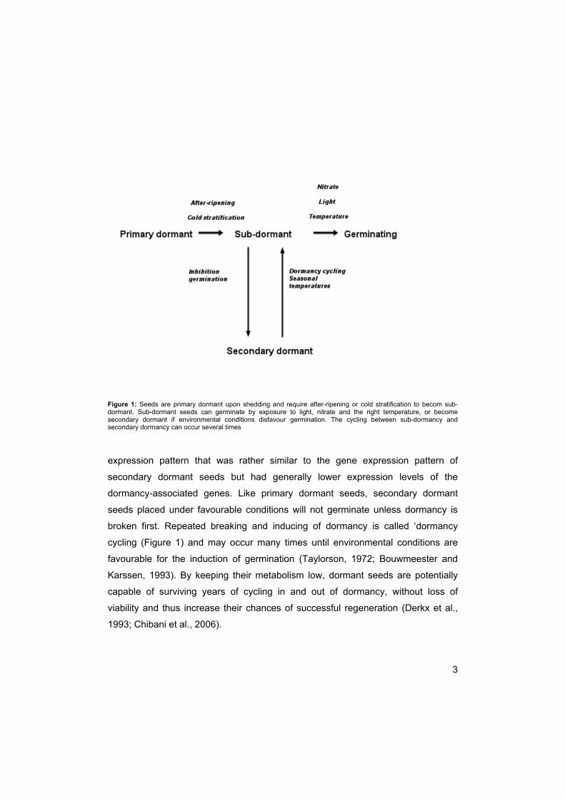
Figure 1: Seeds are primary dormant upon shedding and require after-ripening or cold stratification to becom sub-dormant. Sub-dormant seeds can germinate by exposure to light, nitrate and the right temperature, or become secondary dormant if environmental conditions disfavour germination. The cycling between sub-dormancy and secondary dormancy can occur several times
expression pattern that was rather similar to the gene expression pattern of
secondary dormant seeds but had generally lower expression levels of the
dormancy-associated genes. Like primary dormant seeds, secondary dormant
seeds placed under favourable conditions will not germinate unless dormancy is
broken first. Repeated breaking and inducing of dormancy is called ‘dormancy
cycling (Figure 1) and may occur many times until environmental conditions are
favourable for the induction of germination (Taylorson, 1972; Bouwmeester and
Karssen, 1993). By keeping their metabolism low, dormant seeds are potentially
capable of surviving years of cycling in and out of dormancy, without loss of
viability and thus increase their chances of successful regeneration (Derkx et al.,
1993; Chibani et al., 2006).
4
Temperature
Temperature is one of the main environmental factors controlling dormancy and
germination (Hilhorst, 1998). For example, germination in the field is restricted to a
limited period of time when the field temperature overlaps with the temperature
range in which germination can take place (Vleeshouwers et al., 1995), the
‘germination temperature window’. Sub-dormant seeds exposed to light exhibit a
broad window and they can germinate over a wide temperature range. Dormant
seeds (exposed to light) exhibit a narrow window and germination can only take
place within a small range of temperatures (Karssen, 1982; Bouwmeester and
Karssen 1992). The temperature required for breaking of dormancy may differ from
the temperature that is optimal for germination. While cold/heat shocks can break
dormancy, prolonged periods of time at sub-optimal temperatures and in darkness
can induce (secondary) dormancy (Kępczyński and Bihun, 2002). The induction of
dormancy by a sub-optimal temperature treatment is more rapid at higher
temperatures than at lower temperatures.
Light and nitrate
Sisymbrium officinale seeds are not only dependent on the right temperature for
germination, they are also dependent on the simultaneous presence of light
(through phytochrome) and nitrate (Hilhorst et al., 1986; Hilhorst, 1990a, b; Derkx
and Karssen, 1993). In species such as Arabidopsis thaliana there is no need for
the simultaneous presence of light and nitrate: its seeds can germinate in the
presence of light only but the presence of nitrate may reduce the requirement for
light (Batak, 2002). Dormancy cycling is dependent on the sensitivity to dormancy
breaking factors, such as light and nitrate. Thus, this sensitivity may change over
time (Hilhorst, 1990 a, b). The sensitivity/responsiveness to phytochrome and
nitrate is thought to be regulated by the changes in the number of available
phytochrome and nitrate receptors by variations in (seasonal) temperatures (Derkx
and Karssen, 1993).

5
Phytochrome
One of the most important environmental sensors in plants are the phytochromes.
Phytochromes are biliproteins that are synthesised in the inactive red light
absorbing (Pr) form (Casal and Sanchez, 1998) and red light converts them into
bioactive far-red absorbing (Pfr) absorbing isomers (Whitelam and Devlin, 1997).
Pfr converts back into Pr in the dark. In lower plants phytochromes are probably
rigid cytosolic, probably plasma membrane-associated. Hilhorst (1998)
hypothesized that in higher plants the phytochrome and nitrate receptors may also
be (temporarily) associated with membranes. But in higher plants there is a switch
from primarily cytosolic towards a more dominating nuclear function (Nagy and
Schäfer, 2000). Phytochromes are known to regulate GA synthesis, which
promotes germination (Hilhorst and Karssen 1998; Yamaguchi et al., 1998, 2004;
Ogawa et al., 2003). As temperature has been shown to influence endogenous GA
concentration in seeds (Derkx, Vermeer and Karssen, 1994; Yamaguchi et al.,
2004), and sensitivity to GA (Derkx and Karssen, 1993; Yamaguchi et al., 2004)
phytochrome could also have a temperature dependent effect. The effect
temperature has on phytochrome was further analysed by Donohue et al. (2007),
who showed that phytochromes mediate dormancy and germination responses to
seasonal cues that the seed experiences during maturation and after dispersal.
Heschel et al. (2007) showed that in Arabidopsis thaliana 5 different phytochromes
seem to be working at different temperatures, suggesting that phytochromes have
a potential role in regulating seasonal timing of germination.
Gibberellins (GAs)
The need for light and nitrate in seed germination can be circumvented by
application of gibberellins (GAs) to the seeds. It was hypothesized that nitrate can
act as a cofactor to light and that light may induce the biosynthesis of gibberellins.
Applying GAs can thus lead to completion of germination without application of
light and nitrate (Hilhorst et al., 1986). It has indeed been shown that light

6
stimulates GA-biosynthesis, through a direct effect of phytochrome on the GA 3-
oxidase gene (Yamauchi et al., 2004). GAs are suggested to stimulate germination
by 2 different actions directed at (1) the embryo, by promoting the growth potential,
and (2) the surrounding tissues, particularly the endosperm. Seed germination may
be prevented or delayed by the mechanical constraint of the seed coat that the
embryo has to overcome before it can take up water and nutrients (Chen and
Bradford, 2000; Leubner-Metzger, 2001; McIntyre, 1996). The strength of this
barrier can be reduced by stimulating endosperm degradation. In gibberellin-
deficient seeds only exogenous GA4+7 or endosperm plus testa removal can induce
germination, indicating that GA4+7 can induce endosperm weakening (Hilhorst and
Karssen, 1992; Debeaujon and Koornneef, 2000). GA4+7 in the embryo is believed
to migrate to the endosperm (Hilhorst and Karssen, 1992) where it induces
expression of genes encoding enzymes that hydrolyze the endosperm cell walls
(Debeaujon and Koornneef, 2000; Chen and Bradford 2000; Nonogaki et al., 2000;
Manz et al., 2005). After endosperm weakening, the embryo can take up more
water (Manz et al., 2005), metabolic activity of the embryo is promoted and an
additional degree of cell turgor required for the elongation of the radicle is acquired.
Membrane involvement in dormancy
For the past 3 decades, based on a wealth of circumstantial evidence, membranes
have been suggested to be involved in the regulation of dormancy. Hendricks and
Taylorson (1976, 1978 and 1979) have shown that dormancy induction by higher
temperatures is accompanied by an increased leakage of amino acids. The
increased leakage was suggested to be linked to the membrane transition
temperature and this transition was considered the main limiting factor in
germination over a wider temperature range. The membrane transition is
accompanied by a disordering of the membrane and changes in membrane order
may influence phytochrome activity, as the phytochrome receptor (or steps in its
signal transduction pathway) may be membrane-associated (Hendricks and
Taylorson, 1978; Nagy and Schäfer, 2000). These changes in membrane

7
Figure 2: Model based on Hilhorst (1998). Nitrate receptors are suggested to occur in membranes. Under the
influence of temperature and time the membrane fluidity changes, allowing the receptors to move to the membrane
surface, where nitrate and phytochrome can bind and subsequently germination can take place. Xi and Xa are inactive
and active (nitrate) receptors, respectively. Pr and Pfr are inactive and active forms of phytochrome, respectively. r is
red light and fr is far red light.
organisation are a possible explanation for changes in responsiveness to light and
nitrate. A hypothesis was suggested by Hilhorst (1998) (Figure 2), in which the
changes in responsiveness to light and nitrate were explained. Phytochrome and
nitrate receptors may be associated with membranes. Temperature has a profound
influence on membrane fluidity which, on its turn, may determine the magnitude of
movement of the receptors within the membranes. In one fluidity conformation the
receptors will be at the surface, available for nitrate and phytochrome to bind whilst
in the other conformation the receptors are within the membrane and therefore not
available for binding of nitrate and phytochrome. One aspect of temperature-
induced membrane changes is homeoviscous adaptation (Sinensky, 1974), the
mechanism by which unsaturated fatty acids aid in maintaining membranes in a
8
fluid state, necessary for biological functioning (Cyril et al., 2002). A temperature
induced increase or decrease in membrane fluidity can be counteracted by an
increase in synthesis of de novo fatty acids or by desaturation of existing fatty
acids (Sato and Murata, 1980), in order to maintain the fluidity.
Electron Paramagnetic Spectroscopy (EPR)
Spin label ESR is a non-destructive technique whereby a paramagnetic molecule
(i.e. spin label) is used to tag macromolecules in specific regions. Using the EPR
spectra, from the spin label, the type of environment in which the spin probe is
located can be determined.
EPR can be very useful in studying seed dormancy behaviour as it can thus give
detailed information about the structural and dynamic properties of the cytoplasm
or lipid fraction, including membranes, of the seed sample (Marsh,1981). The
rotational correlation time (τR) of the spin probe in its local environment can be
measured by EPR. The τR is defined as the time it takes for the spin probe to rotate
one radian (~60°) around its axis. In other words, the shorter the τR, the faster the
rotational motion of the spin probe. The choice of spin probe is important.
Depending on the polarity of the spin probe, it will partition into the a-polar oil
phase, the polar aqueous cytoplasm, and/or in the membrane, making it a useful
tool to study different cellular properties of dormancy and germination, e.g.
membrane fluidity and cytoplasmic viscosity. By measuring oxidation-reduction
rates of the probe, metabolic activity can be investigated with these spin probes. In
this thesis 3 different spin probes were used: 4-Oxo-2,2,6,6-tetramethyl-1-
piperidinyloxy (TEMPONE; Figure 3A), 3- carboxy-proxyl (Figure 3B) and a
methyl ester of 5-doxyl stearic acid (Figure 3C).
TEMPONE is a small (MW 168) molecule that is easily soluble in water but slightly
apolar, making it also soluble in the lipid environment. Thus, TEMPONE can be
found in both the aqueous cytoplasm and in lipid bodies. TEMPONE can give
information based on the partitioning of this spin probe between the aqueous

9
Figure 3A: Structure of TEMPONE spin probe
Figure 3B: Structure of 3-carboxy-proxyl spin probe
Figure 3C: Structure of methyl ester of 5-doxyl stearic acid spin probe
cytoplasm and oil bodies. TEMPONE can be used to study the cytoplasmic volume
and cytoplasmic viscosity (Golovina and Hoekstra, 2002), but also the metabolic
activity, as it can be reduced to non-paramagnetic species, depending on the
reducing power of cells. This reduction can be used to study cellular metabolic
rates (Jung et al., 1998).
3-Carboxy-proxyl (CP) is also a relatively small (MW 186) spin probe that is easily
soluble in water. CP is more polar than TEMPONE due to the presence of an OH
group. This group increases the probability of these molecules to form hydrogen
bonds, making it particular suitable to study cytoplasmic viscosity.
5-Doxyl stearic acid is often used to study membrane fluidity (Benatti et al., 2001;
Bianconi et al., 1988; Turchiello et al., 2000). The methyl ester of 5-doxyl stearic

10
acid is targeted to cellular membranes. This spin probe is weakly anchored in the
head group area due to the high hydrophobicity of the methyl ester. As a result the
methylated spin label is localized in a deeper position in the bilayer than its un-
methylated counterpart (Sanson et al., 1976).
Seed dormancy and gene expression
Several methods have been used to analyse the transcriptional differences
between primary dormant and long term dormant seeds. Micro-array analysis is a
much used technique and has been employed to study dormancy transitions in
Arabidopsis thaliana (Cadman et al., 2006; Finch-Savage et al., 2007). Here the
cDNA subtraction library of S. officinale was chosen over micro-array analysis.
With a cDNA subtraction library the cDNA of interest is tagged and the cDNA you
want to compare this with is subtracted from the tagged cDNA. The rationale for a
cDNA subtraction library over micro-array analysis was to prevent cross-species
hybridization difficulties. A major issue with cross-species hybridization is the effect
of sequence divergence on probe affinity, which is not only a function of
phylogenetic distance. Due to differences in sequence divergence rates, such
effects are not uniform across all genes. At present it is difficult to correct for such
effects during the analysis of micro-array data (Bar-Or et al., 2007). Subtraction
libraries have been used to identify key genes and pathways in plants and
seedlings (de los Reyes, 2003). cDNA subtraction libraries, however, pose a whole
new set of difficulties; a large number of clones needs to be sequenced in order to
obtain an overall impression of the transcriptome of a developmental state of the
seed. Gene expression of most genes needs to be verified, as some of the genes
picked up may appear in both the forward and reversed libraries. Primary dormant
seeds and long-term primary dormant seeds were used, as compared to the
primary dormant seeds and secondary dormant seeds used in other chapters.
Primary and secondary dormant seeds are imbibed in different media, making
comparison difficult. Long-term primary dormant seeds are seeds imbibed in water
in the dark for 10 days, making the comparison with primary dormant seeds easier.
11
Long-term primary dormant seeds do not differ from secondary dormant seeds in
gene expression patterns (Cadman et al., 2006).
Objectives
Over the past decades significant progress has been made in understanding seed
dormancy. Although apparently similar in nature, some genuine differences have
been found between primary and secondary dormancy, e.g. the sensitivity to
dormancy breaking factors (Derkx and Karssen, 1993) or differences in expression
intensity of dormancy related genes (Cadman et al., 2006). These differences may
reflect the differences in depth of dormancy. The general aim of this thesis was to
not only study the differences and similarities between primary and secondary
dormancy, but also sub-dormancy and germination in Sisymbrium officinale and
Arabidopsis thaliana in order to enlarge our understanding of this topic. More
specifically, the objectives of this thesis were:
To analyse membrane involvement in the regulation of dormancy;
To analyse changes in metabolic activity and in cytoplasmic viscosity in dormancy
cycling;
To identify differences in gene expression between different dormancy states.
Scope of the thesis
Chapter 1: General Introduction
A short introduction to dormancy, dormancy cycling, and the membrane
involvement in dormancy regulation is presented as well as the research
approaches in this thesis.

12
Chapter 2: Changes in metabolism in Sisymbrium officinale seeds
during the transition between dormant and non-dormant states, as
measured by electron paramagnetic resonance spectroscopy.
The changes in dormancy and germination were linked to general seed
metabolism in Sisymbrium officinale seeds. Low cytoplasmic volume and reduced
metabolism were linked to dormancy, while germinating seeds exhibited a high
cytoplasmic volume and metabolism. Sub-dormant seeds exhibited an intermediate
metabolism.
Chapter 3: Altered membrane properties are associated with
dormancy transitions in seeds of Sisymbrium officinale
The changes in dormancy and germination were linked to changes in membrane
properties in Sisymbrium officinale. At low and high temperatures membrane
fluidity could be linked to dormancy, particularly primary dormancy. However, these
changes did not seem to be caused by (changes in) fatty acid unsaturation.
Chapter 4: Altered cytoplasmic properties are associated with
dormancy transitions in seeds of Sisymbrium officinale
Cytoplasmic properties were studied using the spin probe 3-carboxyl-proxyl. The
observed changes in cytoplasmic viscosity may be linked to changes in
metabolism and the changes in vitrification temperature may be linked to changes
in the content of high-molecular weight compounds.
Chapter 5: Desaturases are associated with dormancy transitions in
Arabidopsis thaliana seeds
The effects of mutations in desaturases were assessed on the induction of
dormancy and membrane fluidity in Arabidopsis thaliana seeds. The conversion of

13
linoleic acid (18:2) into linolenic acid (18:3), appeared to be the most important
conversion associated with dormancy induction, especially in the fatty acid
desaturases 3 (fad3) mutant. However, desaturases activity did not show any
involvement in changes in membrane fluidity.
Chapter 6: Characterization of large scale differences in transcription
between short term and long term primary dormant seeds
Using a cDNA subtraction library differences in gene expression between primary
dormant and long term primary dormant seeds were studied in Sisymbrium
officinale and compared to Arabidopsis thaliana seeds. This yielded a set of
conserved dormancy related genes. Candidate genes involved in dormancy are
genes involved in maintaining the stability and integrity of cell compartments and
macromolecules.
Chapter 7: General Discussion
All the results are combined to obtain a general picture of the molecular and
biophysical changes taking place during transitions among dormancy states.

14

15
CHAPTER 2
Changes in metabolic and redox
activity in Sisymbrium officinale seeds
during the transition between dormant
and non-dormant states
Susanne MC Claessens1,2, Elena A Golovina1, Mieke van Zeijl3, Bert
van Duijn3, Folkert A Hoekstra1, Peter E Toorop2, Henk WM
HiIlhorst1
1Laboratory of Plant Physiology, Wageningen University, P.O. Box 658
6700 AR, Wageningen, The Netherlands; 2Seed Conservation Dept., Royal Botanic Gardens Kew, Wakehurst Place, Ardingly, W Sussex, RH17 6TN, U.K.; 3Fytagoras BV / Leiden University, Institute Biology Leiden, P.O. Box
546, 2300 AM Leiden, The Netherlands
Abstract
Physiological dormancy is reversible and this enables seeds to cycle in and out of
dormancy until the conditions are favourable for germination. In this way seeds can
survive in the soil for extended periods of time. It has been argued that dormancy
cycling must be an energy-efficient process to explain long-term survival. In
Sisymbrium officinale seeds, storage lipids are the main source of energy.

16
Mobilisation of these lipids is expected to occur if energy is required for dormancy
cycling. As yet, there is no evidence that changes in respiration rate, respiratory
pathways or their enzymes are essentially linked to the regulation of dormancy.
EPR results with a TEMPONE spin probe showed that high R-values (water over
lipid content ratio) were associated with the breaking of dormancy, indicating high
cytoplasmic volume. When germination was inhibited and secondary dormancy
induced, R-values and metabolic activity were reduced. Germinating seeds
displayed a quick and substantial chemical reduction of the spin probe, leading to a
decay of the EPR signal. In sub-dormant seeds the signal decay rate was not as
high as that of germinating seeds, but much higher than that of dormant seeds.
The oxygen consumption of primary dormant and secondary dormant seeds was
very low (less than 0.5% of total oxygen content in 63 hours), but slightly faster in
primary dormant than in secondary dormant seeds. In conclusion, germination was
linked to a higher cytoplasmic volume and higher metabolic activity or higher
capacity for reduction. When germination was inhibited and dormancy induced the
cytoplasmic volume and metabolic activity were reduced.
Introduction
Dormancy is the failure of an intact viable seed to germinate under favourable
conditions (Bewley, 1997). One of the most common forms of seed dormancy is
called ‘physiological dormancy’. It is distinguished from other dormancy types by its
reversible nature, allowing dormancy cycling (Taylorson, 1972; Bouwmeester and
Karssen, 1993). Physiological dormancy may occur as primary dormancy or as
secondary dormancy. Primary dormancy is acquired on the mother plant during
maturation, and is observed in seeds upon shedding. Primary dormancy can be
lost during dry storage, which is commonly referred to as dry after-ripening. Once
primary dormancy is lost, imbibed seeds are sub-dormant; they are capable to
complete germination provided that a final dormancy releasing factor, usually light,

17
is available, or become secondary dormant if environmental conditions disfavour
germination. Like primary dormant seeds, secondary dormant seeds placed under
favourable conditions will not complete germination unless dormancy is broken
first. Upon breaking of dormancy, the right environmental cues trigger germination.
Seeds can cycle in and out of dormancy repeatedly, if environmental conditions
allow dormancy to be broken but are not favourable for germination (Taylorson,
1972; Bouwmeester and Karssen, 1993). Although apparently similar in nature,
genuine differences in primary and secondary dormancy have been observed. S.
officinale seeds differ in their sensitivity to dormancy breaking factors such as
gibberellic acid and nitrate (Derkx and Karssen, 1993a), with the lowest sensitivity
in the seeds with secondary dormancy. This difference in sensitivity may reflect the
difference in depth of dormancy.
Mature dry seeds usually contain 5-10% water on a fresh weight basis (Hilhorst
and Karssen, 1992). Under these conditions metabolic activity is virtually reduced
to zero. Metabolic activity increases upon the uptake of water, which is a triphasic
process. Rapid initial uptake (I) is followed by a plateau phase (II) and a further
uptake (III) once germination is completed and radicle protrusion begins (Bewley,
1997). One of the first changes upon imbibition is the resumption of respiratory
activity. After a steep initial increase in oxygen consumption the rate declines until
the radicle penetrates the surrounding structures. At this time, a second burst of
respiratory activity occurs (Bewley, 1997). In dormant seeds it is likely that for
successful survival the metabolic activity of the seeds is reduced to avoid untimely
depletion of reserves. However, although reduction in O2 uptake has been
demonstrated after dormancy induction in Sisymbrium (Derkx et al., 1993) and in
lettuce (Powell et al., 1983) there was no correlation between O2 uptake and
dormancy cycling (Derkx et al., 1993). Proteomic analysis of A. thaliana has shown
that, upon imbibition, two different sets of enzymes controlling metabolism can
accumulate, one set is up-regulated in both dormant and non-dormant seeds, while
the other set is only up-regulated in non-dormant seeds (Chibani et al., 2006),
implying that this additional metabolic activity, is only observed in non-dormant

18
seeds. Proteins up-regulated only in the non-dormant seeds include the
neoglucogenesis enzymes 1,6-Fru bisphosphate aldolase and cytosolic isoforms of
GAPDH, providing energy from stored lipids required for seedling establishment,
and isocitrate lyase, an enzyme involved in storage lipid mobilization.
There is no evidence that changes in respiration rate, respiratory pathways, or their
enzymes are essentially linked to the regulation of dormancy (Bewley, 1997). Upon
release from secondary dormancy, there is an increase in respiration, but this is
slower than in seeds emerging from primary dormancy (Powell et al., 1984). These
observations raise the question as to how much or how little of the respiration
measured in primary dormant seeds is really essential for its maintenance, and
how much is excess, resulting in a high background that masks any subtle
metabolic changes taking place (Bewley, 1997; Powell et al., 1984).
In this paper we tested the hypothesis that the metabolic rate of primary and
secondary dormant seeds is lower than that of sub-dormant and germinating
seeds. We used S. officinale (hedge mustard) as it has a well-described
germination and dormancy behaviour, and the breaking of dormancy can be fully
separated from the germination event (Hilhorst and Karssen, 1989). Electron
paramagnetic resonance (EPR) was used to study changes in cytoplasmic volume
and metabolic activity, measured as reducing power of cells, in dormant and sub-
dormant seeds. The metabolic activity was further characterised by measuring
single seed oxygen consumption.
Materials and Methods
Germination
Seeds of Sisymbrium officinale (L.) Scop. were collected in a field in the vicinity of
Wageningen, The Netherlands in 2004. Seeds were cleaned, dried at 20ºC to 85
mg water/g dry seed, and stored at 5 ºC until use (2005-2008). Prior to
19
germination, seeds were surface sterilized in 1% sodium hypochlorite for 1 minute
and rinsed with demineralized water for 5 minutes. Triplicates of 30 seeds were
sown in 5-cm Petri dishes on two layers of filter paper (Schleicher & Schuell No
595), moistened with 1.5 ml of either water, 25 mM potassium nitrate (Fisher) or
100 µM GA4+7 (Sigma). Seeds were imbibed for 1 or 10d in the dark at 25°C after
which they were irradiated with a saturating red light (620-700nm, Philips) pulse for
10 minutes, or kept in the dark (Hilhorst and Karssen, 1988). After irradiation,
seeds were transferred back to the dark at 25ºC. Germination was scored every
day after irradiation, for 1 month, under safe green light.
Germination tests in 96 wells plates, for concomitant oxygen
measurements, were done in triplicate. To each well 2 filter papers were added
(Schleicher & Schuell No 595), cut to the size of the wells. Filter papers were
moistened with 20 µl H2O, 25 mM KNO3 or 100 μM GA4+7 before one seed per well
was added. After 1 or 10d of imbibition in the dark at 25°C, seeds were irradiated
for 10 minutes with a saturating red light (620-700 nm) pulse. Germination was
scored every hour after irradiation.
Water content
Upon surface sterilization, imbibition and dark-incubation for 1 or 10 days, seeds
were placed shortly in a 9-cm Petri dish filled with 2 filter papers and 3 ml H2O, to
ensure that an equal amount of water was attached to the seeds’ surface of each
replicate, making the standard error of each measurement comparable. Water
content was assessed of 15 replicates of 10 seeds, by weighing before and after
drying at 103ºC for 17 hours on a 7-decimal balance. Water content was expressed
on a dry weight basis, in g H2O/ g dry weight.
20
EPR
Seed coats of dry seeds were treated with sandpaper for 20 seconds resulting in a
superficial bruising of the seed coat, before surface sterilization and imbibition, in
order to facilitate entry of the spin probe into the seed. A parallel germination test
showed no effect of this treatment on the germination behaviour. Seeds were
imbibed for 1 or 10d in the dark. Seed samples were then incubated in
perdeuterated 4-oxo-2,2,6,6-tetramethyl-1-piperidinyloxy (TEMPONE) spin probe
and 120 mM potassium ferricyanide. Due to paramagnetic interactions, ferricyanide
causes broadening of the TEMPONE signal to apparent invisibility. Because
ferricyanide does not pass the plasma membrane, the non-broadened narrow lines
in EPR spectra originate from the intracellular location of TEMPONE molecules
(Golovina et al., 1997; Golovina et al., 2001). After 10 minutes of incubation the
seed samples were loaded into a 2-mm capillary for spectra recording.
Ferricyanide was not used in the experiments where the reduction rate of spin
probe was determined. All seeds were kept in the dark and measurements were
done under dimmed white light. EPR spectra were recorded using an X-band EPR
spectrometer (Bruker Elexis E500 CW_EPR , Rheinstetten, Germany). To prevent
overmodulation and saturation of the EPR signal, microwave power was limited to
5 mW, the modulation amplitude was 0.3 Gauss (G) and the scan range was 100
G.
Oxygen uptake
Following surface sterilization seeds were pre-incubated in the dark for 1 or 10d.
After pre-incubation, seeds were irradiated with red light (620-700 nm) for 10
minutes and kept in the dark for 5h, before transferring 64 seeds per treatment to a
96-well plate, one seed per well, containing 2 filter papers (Schleicher & Schuell No
595) moistened with 20 µl of solution. Oxygen uptake was measured using the Q2-
test (ASTEC Inc.; www.astec-global.com), which is a non-invasive method whereby
oxygen levels in closed wells containing a single seed are determined using

21
fluorescence life-time properties of an oxygen sensitive dye. An air-tight
transparent foil with dots of the fluorescent oxygen sensitive dye was placed on top
of the plates and sealed to close every well individually. Complete darkness could
not be guaranteed for all plates, and therefore all plates received a light pulse. As a
control, the seal on one of the empty wells was pierced; scans were taken every 30
min in the dark.
Up to 32 wells were left empty in the 96-well plate. The O2 measurements of these
wells were used for calibration of the data. After calibration, data were smoothed
using 9-point symmetrical smoothing analysis (with a start and finish ramp). The
differences in respiration rate can be analyzed by measuring differences in the
slopes of the oxygen uptake. The differences in the slopes were measured by
synchronization analysis, setting the steepest point in the oxygen consumption
(15% oxygen levels) for both treatments at 45h, followed by a subsequent t-test of
the whole process of oxygen uptake.
Results
In S.officinale seeds the different physiological states could be clearly distinguished
and were easy to manipulate. Seeds imbibed in H2O that failed to germinate, both
in the light and dark (Figure 1b, 1g), were considered primary dormant. Seeds
imbibed in KNO3 solution (‘sub-dormant’) only required a light pulse (620-700 nm)
of at least 10 minutes, to release (sub) dormancy and to complete germination
(Fig.1a, 1f). The light pulse was required within a certain time window; when
delivered after 10d of dark imbibition on KNO3, seeds had acquired secondary
dormancy and the light pulse was not sufficient to induce germination (Fig. 1c).
Seeds imbibed in 100 µm GA4+7 completed germination both in the light (Fig. 1d)
and in the dark (Fig.1e), which indicates that the light requirement was bypassed.
Seeds imbibed on GA4+7 in the light germinated faster than seeds imbibed on KNO3
in the light and than GA treated seeds in the dark. We studied aspects of metabolic

22
Figure 1: Germination vs time curves of Sisymbrium officinale seeds after the following pre-treatments:
a. 25mM KNO3 for 1d + light pulse (●; germinating);
b. H2O for 1d in darkness (□; primary dormant, dark);
c. 25 mM KNO3 for 10d in darkness + light pulse (♦; secondary dormant);
d. 100 μM GA4+7 for 1d + light pulse (▲; germinating GA light);
e. 100 μM GA4+7 for 1d in darkness (∆; germinating GA dark);
f. 25 mM KNO3 for 1d in darkness (◊; sub-dormant, non-germinating).
g. H2O for 1d + light pulse (■; primary dormant, light)
Where applicable, the light pulse was given after 8h of imbibition, after which seeds were placed back in the dark
activity in order to associate the different physiological states with different
metabolic rates. To achieve this we used EPR spectroscopy to measure
cytoplasmic and metabolic properties and the Q2-test to measure O2 uptake.

23
Figure 2. A. EPR spectra of 1 mM TEMPONE in seeds in different states of dormancy, in germinating Sisymbrium seeds and in seedlings. (Sub-dormant seeds were imbibed in 25mm KNO3; Germinating seeds were imbibed in 100 μM GA4+7). No radicle protrusion had occurred in any of the treatments, except of the seedlings. Seedlings were sampled after 7d of imbibition in GA4+7.
Cytoplasmic volume changes and lipid mobilization in seeds of
different dormancy states
The EPR spectrum of perdeuterated TEMPONE from seeds is the superposition of
spectra coming from the aqueous cytoplasm, oil bodies and the seed coat
(Golovina and Hoekstra, 2002). The relatively broad component of the spectrum
originates from the spin probe located in the seed coat and is not analyzed in this
study. The spectra of TEMPONE located in the aqueous cytoplasm and lipid
bodies each have 3 narrow lines due to fast rotation of the spin probe molecules
(Figure 2A). However, these narrow line spectra differ in the distance between the
lines. These distance is called the isotropic hyperfine splitting constant and
24
depends on the polarity of the environment. The spectra from TEMPONE in oil
bodies have a smaller distance between the lines in comparison with the spectra
originating from TEMPONE in aqueous cytoplasm. These two kinds of spectra are
resolved in the high field (right-hand) region of the spectrum because of the
combined effects of the changes in the g-value and the isotropic hyperfine-splitting
constant in the peak position of both components (Golovina et al., 1997; Golovina
and Hoekstra, 2002). The g-value is the most characteristic value that describes an
EPR spectrum, and is a unitless measurement of the intrinsic magnetic moment of
the electron. The g-value can give information about the paramagnetic center’s
electronic structure. The g-value is strongly affected by the environment of the
unpaired electron of the spin label. The g-value for a free electron, ge, is
2.0023193. The value of g can vary, and can be calculated. The interaction of the
unpaired electron with other electrons in the same atom is usually treated as a
coupling of the unpaired electron spin with its orbital momentum. Such coupling
produces a splitting of the signal into three separate transitions with characteristic
g-values. The high-field region of the spectra consists of two peaks of which the left
peak represents the lipid (L) component and the right peak (W) represents the
water component (Figure 2A). At a first approximation the heights of the peaks are
proportional to the number of TEMPONE molecules in each compartment, which,
at a given partition coefficient depends on the volumes of these compartments.
The changes in ratio (R) of W over L will indicate the changes in the cytoplasmic
volume, or in lipid content, or both. Seedlings and germinating seeds imbibed in
GA4+7 showed a high R-value (Figure 2B) in comparison with dormant and sub-
dormant seeds. This is indicative of cellular expansion or oil mobilization, or both.
Sub-dormant and primary dormant seeds showed comparable R values, 0.93 and
0.91, respectively, whereas secondary dormant seeds had a lower R value of 0.46.
A low R value could also be an indication of water loss. However, the water content
of the whole sub-dormant, primary dormant and secondary dormant seeds showed
no significant differences among treatments (Table 1; P values of 0.07, 0.7 and
0.06, respectively).

25
Treatment WC (g/g dry weight)
Standard deviation
Dry weight (g) Standard deviation
GA4+7 1d, germinating 2.28 0.14 0.00297 0.00032
KNO3 1d, sub-dormant 2.10 0.14 0.00284 0.00016
H2O 1d primary dormant 2.02 0.10 0.00297 0.00018
KNO3 10d, secondary dormant 2.12 0.029 0.00285 0.00016
Table 1: Water content (WC) and dry weights of primary dormant (H2O 1d), secondary dormant (KNO3 10d), sub-
dormant (KNO3, 1d) and germinating seeds (GA4+7, 1d). Number of observations per average is 10.
Reduction of the spin probe signal to measure rate of cellular
metabolism
Although spin probes are stable free radicals, they can be reduced to non-
paramagnetic species in living cells. Ferricyanide can rapidly reoxidize the reduced
forms of spin labels even if the ferricyanide is located in the apoplast, due to fast
exchange of spin probe molecules over the plasma membrane (Kaplan et al.,
1973). This allows the observation of the stable in time EPR signal from living
material. However, the reduction of spin probe moleucules can also be used as a
tool to study the rate of cellular metabolism. IN this case spin probes are used
without ferricyanide and the decay rate of the EPR signal is indicative of the
concentration of reducing agents inside the cells. Redox activity increases within
germinating seeds, as a response to the oxidative burst that occurs upon
resumption of metabolic activity.The oxidative burst may impose stress to the seed,
thus influencing germination (Wojtyla et al., 2006). To scavenge the reactive
oxygen species, antioxidants are produced. Antioxidants are necessary to re-
establish the reducing intra-cellular redox environment to prevent inhibition of
protein synthesis (Wojtyla et al., 2006). Antioxidants, especially glutathione and
ascorbic acid are very effective in reducing spin probes (Fuchs et al., 1997).
Reduction of the spin probe can be also caused by activation of the electron

26
Figure 3. Decrease in the amplitude h0 of the central spectral line of the ESR spectra of TEMPONE with time as an
estimation of the rate of cellular metabolism. Primary dormant (■), secondary dormant (♦), sub-dormant non-
germinating (●) and germinating (▼) Sisymbrium seeds were analyzed.
transport chain (Chapman et al., 1985) The EPR signal intensity decay can
therefore be used as a good measure of the redox activity and, hence, the
metabolic activity in the seed (Jung et al., 1998).
To study metabolic activity in S. officinale seeds in different physiological states,
we labelled these in a solution of perdeuterated TEMPONE, without ferricyanide,
and monitored the changes in the amplitude of the central spectral line in time. The
height of the central component of the EPR spectra (h0) in this case is a good
estimation of the total number of paramagnetic species in the sample.
Germinating seeds displayed a 38% reduction of the spin probe in 90
minutes (Figure 3). In sub-dormant seeds primary dormancy is broken but light is
still required to complete germination. The signal decay rate of these seeds was

27
not as high as that of germinating seeds, but much higher than that of primary
dormant seeds. The signal decay rate of seeds with secondary dormancy was not
as high as in sub-dormant seed, but much higher than that in seeds imbibed in H2O
for 1 day that possess primary dormancy.
Respiration
With differences in the rate of the spin probe reduction we also expected
differences in respiration rates because of the possible involvement of the electron
transport chain. Therefore, O2 uptake was measured using the Q2-test. This test is
a non-invasive method whereby fluorescence life-time properties of an oxygen
sensitive dye are recorded. In the Q2-machine, single seed measurements were
performed and a germination test under identical conditions was carried out
alongside the Q2-test. As complete darkness could not be guaranteed in the Q2-
machine, oxygen measurements were only done on primary dormant, secondary
dormant, KNO3-imbibed germinating and GA4+7-imbibed germinating seeds.
Figure 4A shows O2 uptake and germination percentages over time for all
the different conditions. Both seeds imbibed in GA4+7 and in KNO3 in the light,
reached almost 100% germination within 48 hours. The t50 values for germination
were 25 and 29h, respectively, showing that half maximal germination was reached
4h faster when seeds were imbibed in GA4+7. The t50 values for O2 uptake were 27h
for seeds in GA4+7 and 34h for seeds imbibed in KNO3, showing that half maximal
O2 uptake was reached 7h faster in seeds imbibed in GA4+7 as compared to seeds
imbibed in KNO3. Figure 4B shows that oxygen uptake started earlier (P= 1.6*10-6)
in GA4+7 imbibed seeds than in KNO3 imbibed seeds. Synchronization analysis,
setting the steepest point in the oxygen consumption (15% oxygen levels for both
treatments) for both treatments at 45 h, and subsequent t-tests of the whole
process of oxygen uptake (Figure 5A) showed that the process of oxygen uptake
for these 2 treatments was significantly different, within the 1% significance level,
with seeds imbibed in KNO3 displaying slower O2 uptake. Primary and secondary
28
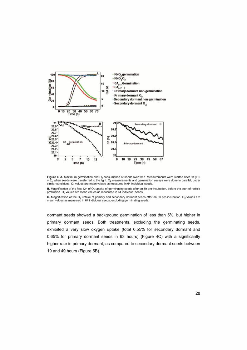
Figure 4. A. Maximum germination and O2 consumption of seeds over time. Measurements were started after 8h (T 0 = 8), when seeds were transferred to the light. O2 measurements and germination assays were done in parallel, under similar conditions. O2 values are mean values as measured in 64 individual seeds.
B. Magnification of the first 12h of O2 uptake of germinating seeds after an 8h pre-incubation, before the start of radicle protrusion. O2 values are mean values as measured in 64 individual seeds.
C. Magnification of the O2 uptake of primary and secondary dormant seeds after an 8h pre-incubation. O2 values are mean values as measured in 64 individual seeds, excluding germinating seeds.
dormant seeds showed a background germination of less than 5%, but higher in
primary dormant seeds. Both treatments, excluding the germinating seeds,
exhibited a very slow oxygen uptake (total 0.55% for secondary dormant and
0.65% for primary dormant seeds in 63 hours) (Figure 4C) with a significantly
higher rate in primary dormant, as compared to secondary dormant seeds between
19 and 49 hours (Figure 5B).

29
Figure 5. A. P-values of synchronized O2 consumption measurements of germinating seeds imbibed in KNO3 and in
GA4+7. Synchronizing the oxygen consumption at its steepest point allowed analyzing differences in the process of
oxygen uptake. As a consequence of the synchronization at 45 h the P-value is approximately 1.
B. P-values of O2 consumption measurements of primary and secondary dormant seeds imbibed in H2O for 1 day and
KNO3 for 10 days. Oxygen consumption measurements were not synchronized, as there is not a steepest point.
In panels A and B, P-values below 0.01 are significantly different.
Discussion
Seeds imbibed in water for 1d (primary dormant) and seeds imbibed in KNO3 for
10d (secondary dormant) did not complete germination in the light or dark, thus
displaying a similar absence of the light response leading to visible germination
(Figure 1). However, in the light, the sensitivity to the dormancy breaking factor
nitrate differed, since primary dormant seeds responded to nitrate but secondary
dormant seeds did not (Hilhorst, 1990b). Thus, secondary dormancy induced by
prolonged darkness at the optimal germination temperature was different from
primary dormancy in S. officinale. Dormancy induction was faster than in

30
Arabidopsis thaliana in which it took up to 80 days of dark incubation for seeds to
lose their sensitivity to light and nitrate (Cadman et al., 2006).
Primary dormant and secondary dormant seeds displayed differences in
the ratio between water and lipid components of the EPR spectra. Water content
measurements (Table 1) showed that in both dormancy states amounts of water
were not significantly different. Lipid mobilization is necessary for the transition
from dormancy to germination and in seedling establishment in Arabidopsis
thaliana, as was proven with the comatose mutants. However, lipid breakdown
itself appeared not to be an important prerequisite for germination but rather
functioned as a signal (Footitt et al., 2002, 2006). Here the seed dry weight of the
dry, primary dormant and secondary dormant seeds did not differ significantly
(Table 1), indicating that no significant lipid mobilization, degradation and utilization
in energy metabolism had occurred. However, the dry weight measurements may
not have been precise enough to determine small differences. Also the low R-
values for the dormant states suggest an absence of lipid mobilization and/or cell
expansion (Figure 2). The differences in R-value could also be due to a change in
lipid/water content locally. These small changes will be averaged out in the water
content measurements as water content is measured of the whole seed.
For successful survival of seeds in the soil one would expect the metabolic
activity of dormant states to be low, to prevent fast depletion of reserves. Metabolic
activity, as measured by reducing capacity, of primary dormant seeds was hardly
detectable (Figure 3), whereas metabolic activity but not oxygen consumption
(Figure 4A, C) in the used measurement set-up, of the secondary dormant seeds
was considerably higher. The primary dormant seeds had been imbibed in water
whereas the secondary dormant seeds had been imbibed in nitrate. This suggests
that nitrate had a stimulating effect on reducing activity without increasing
respiration, even when the seeds were dormant. The dormant seed is known to
have a restricted availability of metabolites and energy (Garciarrubio et al., 1997),
due to presence of ABA. It is possible that nitrate treatment led to metabolic
changes enabling the seed to overcome this inhibition (Matakiadis et al., 2009).

31
Although both types of seed were dormant the different imbibition medium could
thus be responsible for changes in metabolism. The difference between primary
and secondary dormancy in these seeds was very clear. However, as they had
been imbibed in different media, comparisons are difficult to make. Comparing
secondary dormant and sub-dormant nitrate-imbibed seeds is less complicated;
both were imbibed in nitrate in the dark and differed only in incubation time.
Secondary dormant seeds have passed through a phase of light-responsiveness
that applies to the seeds in which dormancy is released by nitrate (Derkx &
Karssen, 1993b). Metabolic activity (Figure 3) was considerably higher in sub-
dormant non-germinating seeds than in secondary dormant seeds, which shows
that the initially high metabolic activity slows down when germination is not initiated
and secondary dormancy is induced. This is in contrast with earlier findings,
reporting that dormant and sub-dormant seeds do not differ appreciably in their
metabolic activity (reviewed by Bewley, 1997). Reduction in metabolic activity
observed in these secondary dormant seeds may conserve energy and
presumably serves to prevent depletion of seed reserves and reduced viability
during dormancy cycling in the soil (Derkx and Karssen, 1993a). We hypothesize
that the metabolic rate slows down even further after longer incubation, to a similar
level as in primary dormant seeds that were not exposed to nitrate.
Energy metabolism and oxygen consumption are expected to be related,
thus, increased oxygen consumption was anticipated for secondary dormant
seeds, as compared to primary dormant seeds. The oxygen consumption of
primary dormant and secondary dormant seeds was very low (less than 0.5% in 63
hours), but slightly slower in secondary dormant than in primary dormant seeds.
This difference was significant between 16 and 50h of imbibition, and thus
suggests that reducing capacity and oxygen consumption are not coupled.
Dormant seeds have been shown to consume oxygen at very low rates (Derkx et
al., 1993; Powell et al., 1984). In the study by Derkx et al (1993) on seeds of
S.officinale it was shown that it took approximately 20d of induction of secondary
dormancy at 24 °C to attain the minimum very low levels of oxygen consumption.
32
Thus, an alternative explanation for this discrepancy between oxygen consumption
and metabolic activity is that secondary dormancy was not fully attained after 10d,
despite the fact that germination did not occur.
Even though seeds imbibed both in nitrate and GA4+7 germinated to a high
percentage in the light, seeds imbibed in GA4+7 circumvented the light requirement.
It has been proposed that nitrate can act as a cofactor to light-induced biosynthesis
of GAs and to subsequent completion of germination (Hilhorst et al., 1986). In
Arabidopsis, light activates phytochrome via PHYB, an inducer of GA-3-oxidases
(GA3OX1 and GA3OX2), which catalyse the final step in the synthesis of bioactive
GAs, even if germination does not occur afterwards (Yamaguchi, et al., 1998; Oh
et al., 2006). Thus, completion of germination can take place after imbibition in
GA4+7 without light, but not on nitrate without light (Hilhorst et al., 1986). Light,
however, may sensitize the seed to GA4+7, facilitating the germination process and
resulting in faster germination (Figure 1; Hilhorst et al., 1986; Derkx and Karssen
1993b).
Upon imbibition, seeds start taking up water which is sufficient for
metabolic activity and oxygen consumption to resume. However, after a steep
initial increase the oxygen uptake rate declines until the radicle penetrates the
surrounding structures (Bewley, 1997). Indeed, when seeds were non-dormant but
had not received a light pulse (‘sub-dormant’), their water content did not appear to
be different from that of their dormant counterparts (Table 1). When seeds were
induced to germinate in GA4+7 the R-value was very high (Figure 2B), which could
indicate an increase in cytoplasmic volume due to cell enlargement and/or oil body
degradation, prior to radicle protrusion, however, the water content measurement
of germinating seeds did not show this (Table 1). The difference in R-value could
be due to a change in lipid/water content locally, which is averaged out in the
whole seed water content measurements.
Nitrate and GA4+7 can both stimulate the growth potential of the embryo
(McIntyre, 1996; Debeaujon and Koornneef, 2000; Alboresi, 2005). A possible
explanation for the differences in the cytoplasmic volume could be that the seed

33
coat acts as a mechanical constraint that the embryo has to overcome before it can
take up further water and nutrients (Chen & Bradford, 2000; Leubner-Metzger,
2001; McIntyre, 1996). This barrier can be reduced in time by stimulating seed coat
and endosperm degradation. For example, in gibberellin-deficient seeds of tomato
or Arabidopsis only exogenous GA4+7 or endosperm and testa removal could
induce germination (Hilhorst and Karssen, 1992; Debeaujon and Koornneef, 2000),
indicating that GA4+7 can induce endosperm weakening. GA4+7 in the embryo is
believed to migrate to the endosperm (Hilhorst and Karssen, 1992) where it
induces expression of genes encoding for enzymes that hydrolyze the endosperm
cell walls (Debeaujon and Koornneef, 2000; Chen and Bradford 2000; Nonogaki et
al., 2000; Manz et al., 2005). Nitrate in combination with light can stimulate GA
production which may result in seed coat degradation. However, when light is not
supplied it cannot (Hilhorst et al., 1986). After endosperm weakening, the embryo
can take up more water (Manz et al., 2005), metabolic activity of the embryo is
promoted and an additional higher degree of cell turgor required for the elongation
of the radicle is acquired. EPR results showed that seeds were metabolically active
when imbibed in nitrate or GA4+7 (Figure 3), but the germinating seeds imbibed in
GA4+7 were metabolically more active than the sub-dormant seeds imbibed in
nitrate. Q2-tests were performed after a light pulse was given; therefore both KNO3-
and GA4+7-imbibed seeds readily completed germination. O2 uptake was faster and
started earlier in GA4+7 imbibed seeds, but this extra amount of O2 uptake did not
seem necessary for germination, as both germinated to a similar high percentage.
However, it did result in faster germination.
In conclusion, germination was linked to a higher cytoplasmic volume
locally and higher metabolic activity or higher capacity for reduction. When
germination was prevented, dormancy was induced and the cytoplasmic volume
and metabolic activity did not increase. The use of non-destructive spin-label EPR
spectroscopy to measure changes in cytoplasmic volume and lipid mobilization has
proven to be a unique tool to monitor germination related phenomena in time in
living seeds

34

35
Chapter 3
Dormancy transitions in seeds of
Sisymbrium officinale are associated
with altered membrane properties
Susanne MC Claessens1, 2, Elena A Golovina1, Magdalena Witek3, Thomas
Roach2, Ilse Kranner2, Folkert A Hoekstra1, Peter E Toorop2, Henk WM HiIlhorst1
1Laboratory of Plant Physiology, Wageningen University, P.O. Box 658, 6700 AR Wageningen, The Netherlands; 2Seed Conservation Dept., Royal Botanic Gardens Kew, Wakehurst Place, Ardingly, W Sussex, RH17 6TN, U.K. 3Wageningen NMR
Centre.
Abstract
Temperature is the main environmental factor involved in the regulation of seed
dormancy. As membranes are often considered the primary target for temperature
perception they have been implicated in the regulation of dormancy. Membrane
properties may be altered to adapt to the temperature. One way of achieving this is
by homeoviscous adaptation, the mechanism by which unsaturated fatty acids aid
in maintaining the membranes in a fluid state. Here we tested the hypothesis that
changes in dormancy and germination concur with changes in some membrane
properties. At low and high temperatures the membrane fluidity could indeed be
linked to dormancy, especially primary dormancy. Breaking of dormancy induced
the membranes to become more fluid, whereas membrane rigidity was partially
restored in secondary dormant seeds. The changes in fluidity were not related with
changes in glutathione levels. The changes in membrane fluidity did not appear to
36
be caused by changes in unsaturation. At temperatures that are physiologically
relevant for germination the fluidity of membranes did not differ between different
dormancy states.
Introduction
Temperature is the principal environmental factor involved in the (seasonal) control
of dormancy and plays a decisive role in the regulation of both dormancy and
germination (Hilhorst 1998). Seasonal changes in temperature determine the
responsiveness of seeds in the soil seed bank to factors that further break
dormancy and induce germination, including light and nitrate. In addition, the
prevailing field temperature has to overlap with a permissive range of germination
temperatures (determined by the dormancy status of the seeds) before germination
can take place, which illustrates the dual role of temperature in seasonal flushes of
seedling emergence (Karssen, 1982; Bouwmeester and Karssen, 1992).
Membranes have often been implicated in the regulation of dormancy in
seeds, as these are considered the primary target of temperature perception at the
cellular level (Minorsky, 1989; Murata and Los, 1997, Penfield 2008). However,
the principles of temperature perception, as well as how thermal history is
remembered are not yet understood. Membranes are capable of altering their
properties in order to adapt to changes in environmental temperature. One aspect
of these changes is homeoviscous adaptation (Sinensky, 1974), the mechanism by
which unsaturated fatty acids aid in maintaining membranes in a fluid state
necessary for biological functioning (Sato and Murata, 1980; de Vivrille et al.,
2002). When the temperature increases, membranes become more fluid because
of an increased rotational and lateral movement of membrane lipids. To counteract
this increase in fluidity, the amount of unsaturated fatty acids is decreased by
suppression of desaturation of fatty acids and acceleration of de novo synthesis of
saturated fatty acids. When the temperature decreases, synthesis of saturated fatty

37
acids ceases and existing fatty acids are desaturated to counteract the decrease in
membrane fluidity (Sato and Murata, 1980). Membrane fluidity influences general
membrane properties, such as membrane permeability, for amino acids, and the
movement or orientation of molecules associated with or incorporated in the
membrane (Hilhorst, 1998).
Based on a wealth of circumstantial evidence Hilhorst (1998) has proposed
a model in which temperature alters membrane fluidity, which in turn would result
in altered conformation of membrane proteins (e.g. receptors) and/or membrane
permeability, and ultimately in a change in dormancy status (cf. chapter 1). The
level of unsaturation of the membrane phospholipids would function as
‘temperature memory’. Changes in dormancy coincide with changes in sensitivity
and responsiveness to naturally occurring factors such as light and nitrate
(Hilhorst, 1990a, b). The model suggests that the availability of the receptor sites
for light and nitrate depends on their synthesis and accessibility. The accessibility
of the receptor may be a function of membrane fluidity. With the increase of
membrane fluidity the receptor protein moves to the membrane surface and
becomes available for perception of phytochrome and nitrate signals.
Here we test the hypothesis that changes in dormancy and germination
concur with changes in membrane properties. Membrane fluidity was studied with
electron paramagnetic resonance (EPR) spectroscopy. This technique allows the
study of the physical properties of an (membrane) environment where a spin probe
is embedded. The motion characteristics of the probe derived from the shape of
EPR spectra are used as a measure of the fluidity of the membrane.
Spin probe approach can also be used to determine the redox status of the
environment. Nitroxide radicals can be reduced to non-paramagnetic
hydroxylamines by reducing agents. Reduced forms of spin labels can be
reoxidized to paramagnetic forms (Swartz, 1987). The observed intensity of the
EPR spectrum relates to the equilibrium state between these reactions. Any shift in
equilibrium between reduction of the spin probe molecules and re-oxidation of their
reduced forms will change the spectral intensity. Redox status of the seed has
38
been suggested to play a role in dormancy and germination: not only does it
change in response to biotic and abiotic stress but it can also interplay with
hormonal signalling (Bailly et al., 2008). Glutathione is an essential component of
the redox status, and is capable of reducing spin probes (Bobko et al., 2007). Here
changes in the intensity of EPR spectra due to reduction of the spin probe were
further analysed by measuring oxidised and reduced glutathione content of the
seed. Desaturase involvement in membrane fluidity changes was characterised by
NMR. Sisymbrium officinale (Hedge mustard) seeds were used, as this species
has a well-described germination and dormancy behaviour, and the breaking of
dormancy can be fully separated from the actual germination event (Hilhorst and
Karssen, 1989).
Materials and Methods
Germination
Seeds of Sisymbrium officinale (L.) Scop. were collected in a field in the vicinity of
Wageningen, The Netherlands in 2004. Seeds were cleaned, dried at 20ºC to 85
mg water/g dry seed, and hermetically stored at 5ºC until use (2005-2008). Prior to
germination, seeds were surface sterilized in 1% sodium hypochlorite for 1 minute
and rinsed with demineralized water for 5 minutes. Triplicates of 30 seeds were
sown in 5-cm Petri dishes on two layers of filter paper (Schleicher & Schuell No
595), moistened with 1.5 ml of either demineralised water or 25 mM potassium
nitrate (Fisher). Seeds were imbibed for 1 or 10 days in the dark at 25°C after
which they were irradiated with a saturating red light (620-700nm, Phillips) pulse
for 10 minutes or kept in the dark (Hilhorst and Karssen, 1989). After irradiation,
seeds were transferred back to the dark at 25ºC. Germination was scored daily
after irradiation, for 1 month under safe green light.

39
EPR
The methyl ester of 5 doxyl stearic acid (5-mDS(A)) was used as a spin probe. This
spin probe is only weakly anchored in the phospholipid head group area due to the
high hydrophobicity of the methyl ester. As a result, the methylated spin label is
localized in a deeper position in the membrane bilayer than its unmethylated
counterpart (5-DS(A)) (Sanson et al., 1976). 5-mDS(A) has often been used to
study membrane fluidity (Benatti et al., 2001; Bianconi et al., 1988; Turchiello et al.,
2000).
Seeds were imbibed in water or 25 mM potassium nitrate for 8 hours or
10d in the dark. Medium attached to the surface of the seed was removed with
filter paper before seed coats were removed using tweezers, after which seeds
were dried at room temperature. Dry seeds were placed in a 1mM solution of
membrane spin probe in hexane. After 1d the spin probe solution was removed,
seeds were washed twice with hexane and placed at 3% RH for 3-4 days to
remove the remaining hexane from the seeds. Seeds were re-hydrated by
humidification for 3 hours at 100% RH. EPR spectra were recorded with an X-band
EPR spectrometer (Bruker model 300E Analytik, Rheinstetten, Germany). To
prevent over-modulation and saturation of the EPR signal, microwave power was
limited to 5 mW and modulation amplitude of 3G for solid-state and 1G for fluid
type spectra was used. In the case of 2-component spectra the lowest modulation
amplitude of 1G was used. Field scan widths of 100G were used.
Nuclear Magnetic Resonance Spectroscopy (NMR)
The NMR spectra were recorded on an Avance II spectrometer (Bruker,
Rheinstetten, Germany), operating at 300.13 MHz for protons and at 75.47 MHz for
carbons, and equipped with a solid-state magic angle spinning (MAS) probe. Dry
Sisymbrium officinale samples with seed coats and without additional treatments
were packed into a 7 mm Zirconia rotor and spun under a magic angle at a
spinning speed of 5 kHz. 13C MAS single-pulse excitation spectra were obtained

40
with 20-30 K scans, using a recycle delay of 2s, 200 kHz spectral width, 8k data
points and under low-power decoupling. The 13C 90°C pulse width was 5 μs. 13C
NMR chemical shifts were assigned according to the literature (Gunstone, 1993;
Fan, 1996; Jie and Mustafa, 1997).
Single-pulse 13C NMR MAS (magic angle spinning) was used for analyses of the
fatty acid (FA) composition of lipids in seeds of different physiological states. By
spinning the sample under the magic angle θm (ca. 54.74°, where cos2θm=1/3) with
respect to the direction of the magnetic field, the normally broad lines become
narrower, increasing the resolution for better identification and analysis of the
spectrum. Since magic angle sample spinning eliminates line broadening arising
from differences in magnetic susceptibility, it significantly improves NMR
spectroscopy of liquids that are found in an inhomogeneous environment. The use
of this technique facilitates nondestructive measurements of oil composition in
viable plant seeds (Rutar, 1989). The cellular components, which are in a solid
state in dry seeds (proteins and carbohydrates) were present as very broad lines in
the 13C spectra in our experiments and were treated as a base line.
The area between 127-132 ppm is derived from olefin carbon atoms of fatty acids
(http://lipidlibrary.aocs.org/nmr/nmr.html; Table 1).
The fatty acids with one, two and three double bonds can be identified in 13C NMR
MAS spectra of all Sisymbrium samples (Figure 1). We attributed the shifts in the
spectra to fatty acids commonly present in seeds: oleic acid (18:1), linoleic acid
(18:2) and α-linolenic acid (18:3) (http://lipidlibrary.aocs.org/nmr/nmr.html; Table 1).
The peak at ≈14 ppm is characteristic of the terminal methyl (CH3) group in all
types of fatty acids

41
Fatty acid Double bonds Chemical shift (ppm)
Oleic (18:1) -C9=C10- ≈130 (9, 10)
linoleic acid (18:2) -C9=C10-C-C12=C13- ≈130 (9, 13); ≈128(10, 12)
α-linolenic acid (18:3) C9=C10-C-C12=C13-C-C15=C16- ≈130 (9); ≈128 (10, 12, 13); ≈127(15); ≈132(16)
All fatty acids -C18H3 ≈14(18)
Table 1:. NMR spectroscopy of fatty acids and their derivates. Results are shown for primary dormant, sub-dormant and germinating seeds of Sisymbrium officinale. For attributing shifts to specific fatty acids see table 1 and http://lipidlibrary.aocs.org/nmr/nmr.html
Figure 1. NMR spectroscopy of fatty acids and their derivates. Results are shown for primary dormant, sub-dormant and germinating seeds of Sisymbrium officinale.
The integrated area under the peak is proportional to the number of
carbons participating in double bonds. Taking into account the data presented in
Table 1, the fatty acid composition of seed lipids was calculated as follows:
42
I is the integrated area under a specific peak
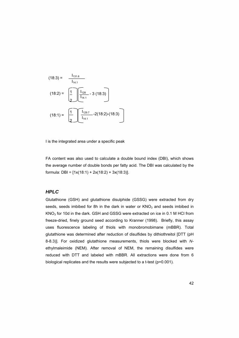
FA content was also used to calculate a double bound index (DBI), which shows
the average number of double bonds per fatty acid. The DBI was calculated by the
formula: DBI = [1x(18:1) + 2x(18:2) + 3x(18:3)].
HPLC
Glutathione (GSH) and glutathione disulphide (GSSG) were extracted from dry
seeds, seeds imbibed for 8h in the dark in water or KNO3 and seeds imbibed in
KNO3 for 10d in the dark. GSH and GSSG were extracted on ice in 0.1 M HCl from
freeze-dried, finely ground seed according to Kranner (1998). Briefly, this assay
uses fluorescence labeling of thiols with monobromobimane (mBBR). Total
glutathione was determined after reduction of disulfides by dithiothreitol [DTT (pH
8-8.3)]. For oxidized glutathione measurements, thiols were blocked with N-
ethylmaleimide (NEM). After removal of NEM, the remaining disulfides were
reduced with DTT and labeled with mBBR. All extractions were done from 6
biological replicates and the results were subjected to a t-test (p=0.001).
(18:3) = I131.6
I14.1
(18:2) = 1
2
I128 I14.1
- 3 (18:3)
(18:1) = I14.1
I129.7 -2(18:2)-(18:3) 1
2

43
Results
Germination
In S.officinale the different dormancy states could be clearly distinguished and
were easy to manipulate (see Chapter 2, Figure 1). Seeds that failed to germinate
in H2O were considered primary dormant. Seeds required a combination of KNO3,
to alleviate dormancy, and a light pulse (620-700 nm) of at least 10 minutes to
complete germination. The light pulse was required within a certain time window;
when delivered after 10d of imbibition on KNO3, seeds had become secondary
dormant and the light pulse was not sufficient to induce germination, and other
methods were needed to break dormancy. The primary dormant, secondary
dormant and sub-dormant states were used to study membrane fluidity by using
the EPR spin probe technique.
Membrane fluidity
A methyl ester of 5 doxyl stearic acid (5-mDS(A)) was used as a spin probe. The
location of the spin probe is relatively deep in the phospholipid bilayer, so that the
spectra of 5-mDS(A) give information about the average fluidity of the membranes
(Benatti et al., 2001; Bianconi et al.,1988; Turchiello et al., 2000). At 220K the 5-
mDS(A) spectrum in hydrated Sisymbrium seed membranes is of the ‘powder’ type
(Figure 2). A powder type spectrum originates from randomly orientated completely
immobilized spin label molecules (Marsh, 1981). Increasing the temperature up to
260 K gradually increases the motional freedom of the spin probe without changing
the anisotropic character of the spectra. This increase in motional freedom of spin
probe within the ordered spectra is characterized by a decrease of the distance
between the outermost extremes, 2Amax, of the spectra, and narrowing of the
spectral lines (figure 2). 2Amax can be used to characterize the degree of
membrane ordering at low temperatures. An increase in this parameter

44
Figure 2. EPR spectra of 5-mDS(A) in primary dormant Sisymbrium officinale seeds. Spectra were recorded at 220K (top) and 260K (bottom). The distance between the outermost extremes, 2Amax, is indicated by the arrow. 2Amax
decreases with increasing temperature. corresponds to an increased ordering of spin label in the membrane environment
(Golovina and Hoekstra, 2003). In Sisymbrium seeds 2Amax decreased with
increasing temperature due to temperature induced membrane disordering (Figure
3). The decrease in 2Amax started later in primary dormant seeds than in
secondary dormant and sub-dormant seeds. This indicates that spin probe
molecules are more immobilized in membranes of primary dormant seeds.
The changes in the spectral shape at temperatures above 270K indicate the
appearance of a mobile component along with the immobile component (Golovina
and Hoekstra 2002, 2003; Figure 4A). 2Amax for the immobile component could not
be determined precisely anymore. There was not much difference in temperature
of appearance of the mobile component between the different dormancy states,
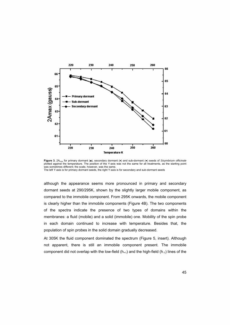
45
Figure 3. 2Amax for primary dormant (■), secondary dormant (♦) and sub-dormant (●) seeds of Sisymbrium officinale plotted against the temperature. The position of the Y-axis was not the same for all treatments, as the starting point was sometimes different; the scale, however, was the same. The left Y-axis is for primary dormant seeds, the right Y-axis is for secondary and sub-dormant seeds
although the appearance seems more pronounced in primary and secondary
dormant seeds at 290/295K, shown by the slightly larger mobile component, as
compared to the immobile component. From 295K onwards, the mobile component
is clearly higher than the immobile components (Figure 4B). The two components
of the spectra indicate the presence of two types of domains within the
membranes: a fluid (mobile) and a solid (immobile) one. Mobility of the spin probe
in each domain continued to increase with temperature. Besides that, the
population of spin probes in the solid domain gradually decreased.
At 305K the fluid component dominated the spectrum (Figure 5, insert). Although
not apparent, there is still an immobile component present. The immobile
component did not overlap with the low-field (h+1) and the high-field (h-1) lines of the

46
Figure 4 The changes of the spectral shape of the 5-mDS(A) spin probe with an increase in temperature. A. The transition from a one-component spectrum to a two-component spectrum occurred within the range of temperatures from 270K to 295 K. B. Increasing the temperature above 295 K reduced the immobile component whereas the mobile component increased in the range between 300K and 330K.
Results are shown for primary dormant, sub-dormant and secondary dormant seeds of Sisymbrium officinale.
mobile component, and the broadening of these lines, originating from rotational
motion of the spin probe, is different for the low-field (h+1) and the high-field (h-1)
lines. The high-field and low-field lines can be used to calculate the viscosity in the
fluid domains in the membranes (Golovina and Hoekstra, 2002). For that we used
the ratio between the low-field and high-field lines as the empirical parameter,
which is proportional to viscosity (Kuznetzov et al., 1971). The fluid domains in

47
Figure 5: Changes in the ratio of the low-field (h+1) and high-field (h-1) signals (see insert, spectrum at 305K) of the
mobile component of 5MESL spectra in seeds with temperature. This ratio is an indicator of the viscosity of the spin
probe environment in membranes. Results are shown for primary dormant, sub-dormant and secondary dormant
seeds of Sisymbrium officinale.
primary dormant seed membranes had the highest viscosity, while in sub-dormant
seed membranes the viscosity was the lowest (Figure 5). The viscosity of the
membranes of secondary dormant seeds was the same as that of primary dormant
seeds at 310 K, however it decreased quicker with temperature but not to the same
degree as that of sub-dormant seeds.
The spectra recorded from labelled seeds are the superposition of all spectra from
different membranes of different cells within the seeds. If only a small amount of all
the cellular membranes is involved in seed dormancy, the dynamic parameters
obtained from EPR spectra will not show this because of averaging of the signal.
The indirect approach to study the changes taking place in parts of the membrane
is to analyze changes in the integrated intensity of the EPR spectrum. This will
show the total number of paramagnetic centres responsible for the spectrum.

48
Figure 6: The integrated intensity of the EPR spectra plotted against the temperature. Note that the y-axes of all treatments are on a different scale for easy comparison of the curves.
Results are shown for primary dormant, sub-dormant and secondary dormant seeds of Sisymbrium officinale.
Integrated intensity of the spectrum is a cumulative parameter, which is sensitive to
changes even in small amounts of cellular membranes (Golovina, et al., 2010).
The intensity of the EPR spectrum is determined by the number of nitroxide
radicals in the sample. The intensity of the spectrum can decrease or increase with
temperature. The decrease of the signal is caused by chemical reduction of spin
label and the increase of the signal is caused by re-oxidation of the reduced forms
of spin labelled molecules. At lower temperatures the oxidation is the dominating
reaction and at higher temperature the reduction is dominating (E. Golovina,
personal communication). The oxidation of the spin label is possibly related to the
diffusion of oxygen (Chen and Swartz, 1988). The reduction of the spin label
depends on the presence of reducing molecules and on the molecular mobility in
the system, which allows molecular diffusion. The integrated intensity of the EPR

49
Figure 7: The integrated intensity of EPR spectra plotted against the temperature. Both samples represent primary
dormant seeds but dormant 1 are partially hydrated seeds and dormant 2 are excessively hydrated seeds, showing
that when bulk water is present there is a sharp decrease in the integrated intensity at 273 K
spectra was plotted against the temperature (Figure 6) and showed that the
temperature at which domination of the oxidation of the spin label switched to
domination of the reduction of the spin label was at 245K in sub-dormant and 250K
in secondary dormant seeds. However, in primary dormant seeds the switch
occurred at a higher temperature (260K). Due to this shift the level of the spectral
intensity at higher temperatures was higher for primary dormant seeds than for
secondary dormant and sub-dormant seeds.
When bulk water is present a sharp decrease in integrated intensity can be
observed at the temperature of water melting at around 273K (Figure 7), which is
caused by the absorption of energy by free water present in the cavities and not by
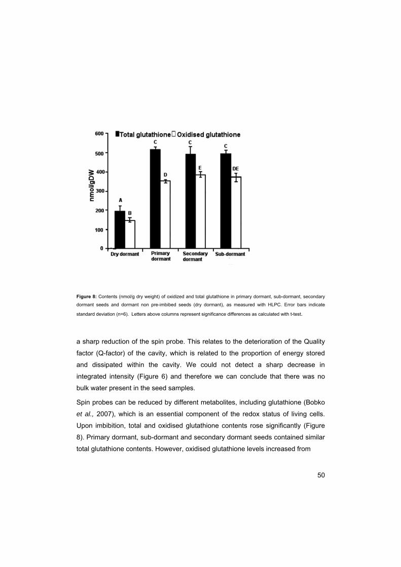
50
Figure 8: Contents (nmol/g dry weight) of oxidized and total glutathione in primary dormant, sub-dormant, secondary
dormant seeds and dormant non pre-imbibed seeds (dry dormant), as measured with HLPC. Error bars indicate
standard deviation (n=6). Letters above columns represent significance differences as calculated with t-test.
a sharp reduction of the spin probe. This relates to the deterioration of the Quality
factor (Q-factor) of the cavity, which is related to the proportion of energy stored
and dissipated within the cavity. We could not detect a sharp decrease in
integrated intensity (Figure 6) and therefore we can conclude that there was no
bulk water present in the seed samples.
Spin probes can be reduced by different metabolites, including glutathione (Bobko
et al., 2007), which is an essential component of the redox status of living cells.
Upon imbibition, total and oxidised glutathione contents rose significantly (Figure
8). Primary dormant, sub-dormant and secondary dormant seeds contained similar
total glutathione contents. However, oxidised glutathione levels increased from

51
Table 2: Relative content (percentage of total) of unsaturated fatty acid and double bond index (DBI) for Sisymbrium officinale seeds in different physiological states, as calculated from 13C NMR MAS spectra
primary dormant (350 nmol/g dry weight (DW)) to sub-dormant (370 nmol/g DW) to
secondary dormant (384 nmol/g DW) seeds, with a significant difference between
primary dormant and secondary dormant seeds (p= 0.001).
Fatty acid composition
Membrane fluidity can be adjusted by introducing or reducing the number of double
bonds in the fatty-acyl chains of the membrane lipids, making the membrane more
or less fluid (Los and Murata, 2004; Sato and Murata, 1980). The unsaturated fatty
acid percentage and the double bond index of total fatty acids of primary dormant,
sub-dormant and germinating seeds were measured by NMR and calculated. The
percentage of 18:1 appeared slightly reduced in primary dormant seeds, while the
percentage of 18:2 was slightly elevated when compared with sub-dormant and
germinating seeds (Table 2). Percentages 18:3 and DBI did not differ among
dormancy states. Because these data are obtained for bulk lipids, i.e. including oil,
care has to be taken when membrane fluidity is discussed. Membranes might have
fatty acid composition different from oil (Sheffer et al., 1986; Millar et al., 2000;
Voelker, 2001). Membrane fluidity can be also modified by sterols and other
compounds or membrane-editing by corresponding enzymes (Millar et al., 2000).
Sisymbrium 18:1 18:2 18:3 DBI
Primary dormant 32.0 28.3 39.7 2.03
Sub-dormant 34.6 24.5 40.9 2.01
Germinating 34.0 25.2 40.8 2.05

52
Discussion
Membranes have been suggested to be the primary target for temperature
perception (Murata and Los, 1997) and membranes and membrane fluidity have
often been implicated in the regulation of dormancy and germination (Hilhorst,
1998). Temperature induced changes in membrane fluidity have been proposed to
be the primary temperature sensing event (Murata and Los, 1997). However, the
non-specific effects of membrane-altering agents used in experiments testing this,
coupled with a lack of any evidence for defects in temperature-regulated gene
expression in Arabidopsis desaturase mutants, have lead to skepticism that this
could be a possible temperature sensing system in plants (Penfield, 2008;
Somerville and Browse, 1996).
We determined changes in membrane fluidity in the different physiological
states of S.officinale seeds. Seeds of this species require a combination of light
and nitrate for germination to take place. If the light pulse is not given within a
certain time window, seeds become secondary dormant, and will not germinate.
The light and nitrate pulses alone cannot break dormancy and induce germination;
the seed will need these triggers within a certain temperature window (Karssen,
1982). The present EPR measurements clearly show that primary dormant seeds
contained less-fluid membranes than secondary dormant and sub-dormant seeds
at low temperatures (Figure 3). When the temperature was increased to 270 K a
relatively mobile component appeared in the EPR spectrum, together with the
already present immobile component (Figure 4A). In this temperature range
membrane fluidity can be estimated by the proportion of mobile and immobile
components. The characteristics of the mobile component cannot be properly
calculated due to overlapping of the spectra. This proportion seems to be similar
for all dormancy types, indicating that at physiological temperatures that are
suitable for germination, the membranes for all dormancy types are equally fluid. At
temperatures above 300K (Figure 4B) the mobile component becomes the main
component in the ESR spectrum. Although these high temperatures are not

53
physiologically relevant for the seed they might show underlying differences
between the dormancy states. At T>300K membrane fluidity can be calculated
from the shape of the narrow-line spectrum. (Figure 5, inset). The ratio between
the low-field peak and the high-field peak was calculated as a parameter, which
relates to the viscosity of the spin label environment. The lesser values of this ratio
in the whole range of temperatures above room temperature in sub-dormant seeds
suggests that during the transition from the primary dormant to the sub-dormant
state, viscosity decreased and, hence, membrane fluidity increased notably. When
secondary dormancy was induced membranes became more rigid again at high
temperatures. At 310K primary dormant and secondary dormant seeds showed a
similar fluidity but at higher temperatures the membranes of secondary dormant
seeds remained more fluid than those of the primary dormant seeds. Primary
dormant seeds had been imbibed on water for 1d in the dark, while sub-dormant
seeds had been imbibed in nitrate for 1d in the dark. The difference in membrane
fluidity could thus be an effect of the imbibition medium rather than the different
dormancy states. However, secondary dormant seeds appeared to have a less
fluid membrane than sub-dormant seeds, while both had been imbibed on nitrate.
Secondary dormant seeds have been sub-dormant before becoming secondary
dormant due to absence of a light pulse within the required time (Derkx & Karssen,
1993a). This suggests that secondary dormant seeds had a more fluid membrane
after 1d when seeds were sub-dormant, but with induction of secondary dormancy
the membranes became more rigid again. These results support the membrane
hypothesis, which suggests that the changing responsiveness to naturally
occurring factors like light and nitrate (Hilhorst, 1990a,b) may indeed be a function
of membrane fluidity (Hilhorst, 1998).
The changes in integrated intensity with temperature (Figure 6) give more
information about the changes of the spin label within the seeds with temperature
that are not visible in the shape of the EPR spectra. The spin label reduction in
primary dormant seeds started at a higher temperature than in sub-dormant and
secondary dormant seeds. Although the temperatures at which the changes take
54
place are not physiologically relevant for dormancy and germination they do show
the possible differences between the dormancy states.
The reducing reaction depends on the presence and mobility of reducing
molecules. Glutathione is assumed to be the main redox compound for most living
cells (Gilbert, 1995). A central role for oxidized glutathione in overcoming seed
dormancy stages was suggested (Kranner and Grill 1996), as oxidized glutathione
accumulation could serve in protecting thiol groups from desiccation induced
oxidative injury; desiccation is one of the later developmental stages and may be
associated with dormancy. However, the role for glutathione in dormancy
alleviation has not been confirmed (Bahin et al., 2011).
Differences in oxidized glutathione levels were small but significant between
primary dormant and secondary dormant seeds and could be a sign of a local
effect. There were no differences between primary dormant and sub-dormant
seeds, indicating that glutathione was not the (only) cause of the reducing reaction
within the EPR spectra. This is in contrast with results of Wang and Faust (1994)
and Fontain et al. (1995) who found that the breaking of dormancy in barley seeds
and apple buds was closely related to an increase in reduced glutathione levels;
other people, however, were also not able to confirm a role for glutathione in
dormancy alleviation (Bahin et al., 2011). Glutathione levels were all measured at
room temperature, which is above the temperature where the switch from oxidation
to reduction of spin label takes place in primary dormant, as well as secondary
dormant and sub-dormant seeds. The location of glutathione (cytoplasm)
compared to the location of the 5-mDS(A) spin probe (membrane core) makes
glutathione a very unlikely candidate for the reducing reaction. In addition to that, in
a dry system with high molecular immobilization the reaction between the two is
hardly possible, and there is little to no metabolism at this temperature.
Glutathione, or other reducing factors, could still be present in the same amounts in
primary dormant, secondary dormant and sub-dormant seeds but in a more
immobile state and thus less accessible at a higher temperature in dormant seeds.
55
These data coincide with the lesser mobility of the spin probe molecules in
membranes in dormant seeds at higher temperatures (Figure 5).
Desaturases have often been implicated in membrane fluidity (Sato and Murata,
1980; Cyril et al., 2002) Based on 13C NMR spectra, we found slight differences in
18:1 and 18:2 fatty acids content of lipids between dormant and sub-
dormant/germinating seeds. It is possible that the total signal masks the
differences in fatty acid composition and double bond index of membrane lipids.
The desaturation of fatty acids counteracts the decrease in membrane fluidity (Sato
and Murata, 1980). According to the spin-label data, sub-dormant seeds have
more fluid membranes in comparison with primary dormant seeds, and therefore
an increased DBI of membrane lipids should be expected for these seeds.
However, the small differences found in 13C NMR spectra do not support this
expectation. Two reasons of such discrepancy can be mentioned. First, the FA
composition of (some) seed membranes might be different from that determined for
bulk lipids from 13C NMR spectra (Sheffer et al., 1986). Second, other than DBI
factors might modify membrane fluidity, such as sterols, flavanoids and other
amphiphiles.
In conclusion, particularly primary dormancy was linked to more rigid membranes
at low and high temperatures. Membranes became more fluid upon breaking of
dormancy. When secondary dormancy was induced, membranes became more
rigid at high temperatures but the membrane rigidity of primary dormant seeds was
not restored completely. At temperatures relevant for germination the fluidity of
membranes does not differ between different physiological states. Desaturation of
fatty acids of membrane lipids could be involved in membrane fluidity but changes
in desaturation of bulk lipids were so small that they cannot prove this mechanism.
The increased fluidity of membranes of sub-dormant seeds may allow the
receptors for nitrate and light to become available for binding and allow
germination to take place (Hilhorst, 1998), however, at physiological optimal
temperatures for germination no major differences in membrane fluidity were

56
found. As some changes in membrane fluidity are seen at temperatures not
relevant for germination the membrane hypothesis described in chapter 1 cannot
be completely discarded, however, it is not likely to be the main temperature
sensing event that leads to germination. A suggestion how temperature sensing
works and eventually leads to germination could be the circadian clock (Penfield,
2008). The circadian clock consists of at least three interlocking transcriptional
feedback loops. One of the key features of the circadian clock is their ability to
perform robustly across a wide range of temperature regimes. Several genes
involved in the circadian clock have also been shown to play a role in promotion of
seed germination (Penfield and Hall, 2009).

57
Chapter 4
Dormancy transitions correlate with
altered cytoplasmic properties in
seeds of Sisymbrium officinale
Susanne MC Claessens 1,2, Elena A Golovina1, Folkert A Hoekstra 1, Peter E
Toorop2, Henk WM HiIlhorst1
1Laboratory of Plant Physiology, Wageningen University, P.O. Box 658, 6700 AR Wageningen, The Netherlands; 2Seed Conservation Dept., Royal Botanic Gardens
Kew, Wakehurst Place, Ardingly, W Sussex, RH17 6TN, U.K.
Abstract
The regulation of seed dormancy is tightly linked with abiotic stress factors from the
environment. Seeds may anticipate a period of adverse conditions by shutting
down their metabolism and engaging a stress response in order to protect cellular
constituents from irreversible damage. The regulation of dormancy and survival of
seeds under conditions that mimic storage in the soil are largely dependent on the
composition of the cytoplasm. In this study the colligative properties of cytoplasmic
water were attempted to be linked to physiological dormancy states of Sisymbrium
officinale seeds. Cytoplasmic properties were studied using the spin probe 3-
carboxyl-proxyl. Tests on PEG, an osmoticum, showed that primary and secondary
dormant embryos did not grow as well as sub-dormant and germinating embryos
on higher concentrations of PEG and were, thus, less tolerant to osmotic stress.

58
Therefore, we conclude that dormancy of S. officinale seeds is also, at least
partially, located in the embryo.
The ‘ease’ at which vitrified water melts and the viscosity of the cytoplasm show
that dormant and sub-dormant seeds possessed a higher viscosity than the
germinating seeds. The viscosity of secondary seeds appeared to be intermediate,
but the ‘ease’ at which the vitrified water melted was similar to that of primary
dormant seeds.The higher viscosity of the dormant and non-dormant seed may be
a way of the seed to survive prolonged periods in the hydrated state. As a result of
the changes in viscosity the temperature of vitrified water melting was lower (265K)
for germinating seeds than for primary, sub- and secondary dormant seeds (280K).
In conclusion, we have shown, for the first time, that cytoplasmic viscosity may
increase upon the induction of dormancy. The observed changes in cytoplasmic
viscosity may be linked to changes in metabolism and the changes in vitrification
temperature may be linked to changes in the content of high-molecular weight
compounds.
Introduction
For a seed to germinate under a range of conditions, or withstand extended
periods of drought, excessive heat and frost, it needs to be able to adapt quickly to
the environment. Seeds that have already initiated germination cannot stop this
process anymore. Therefore, these seeds need to be able to continue germination
during changes in temperature, water availability, salinity, etc. For example, a seed
can only cope with changes in osmotic potential of the environment if it maintains
its ability to take up water and nutrients from the environment. In order to take up
water from an environment with low osmotic potential, the water potential of seed
cells and tissues has to be even lower (more negative).
For a dormant seed the main environmental changes that take place when buried
in the soil are changes in temperature and changes in its water content

59
(hydration/dehydration cycles). Changes in temperature have a profound influence
on the lipid and cytoplasmic properties of the (dry) seed. In general, higher
temperature will increase molecular mobility and, therefore, also the 2 or 3
dimensional diffusion of compounds in both membranes and the cytoplasm, at
least of the dry seed (Buitink et al., 2000a/b; Chapter 3).
By seasonal sensing, seeds are known to anticipate a stressful period by shutting
down their metabolism and engaging a stress response. One of the possible stress
responses in seeds is the loss of water from dormant tissue (Pnueli et al., 2002). At
reduced water content, many reactions that may have had the potential to damage
cells and cell constituents under such adverse physical conditions are suppressed.
The stress response, e.g. dehydration, may induce the synthesis of compounds
that protect the cells against dehydration damage, such as late embryogenesis-
abundant (LEA) proteins (Ingram and Bartels, 1996) and anti-oxidants. The LEA
proteins are implicated in stabilizing and protecting cellular structures (Shinozaki et
al., 1999) and may also protect cells during the course of de/rehydration (Pneuli et
al., 2002). The mechanism of dehydration and concomitant synthesis of protective
compounds to cope with adverse conditions has been described for buds (Rohde
and Bhalerao, 2007), but has been studied mainly in micro-organisms, such as
bacteria (Potts, 1994) and yeast (Hohmann, 2002), as well as invertebrates, such
as tardigrades and rotifers (Danks, 2000). The link between dormancy and stress
has hardly been studied in seeds, although Cadman et al. (2006) have shown that
the dormancy and stress responses in seeds of Arabidopsis thaliana are intimately
associated at the level of gene expression.
Although intermediate water content may reduce seed longevity (Roberts and Ellis,
1989) another possible survival strategy under frost conditions could be not the
loss of water, but vitrification, as opposed to crystallisation of water components
(Langis and Steponkus 1990). Vitrification of living tissue, or intracellular glass
formation, is generally induced by rapid cooling of cells. However, in seeds
intracellular glasses can be formed simply by drying to 10% moisture content, or
less (Leopold et al., 1994). It is not clear if we can compare these two forms of

60
vitrification, as one is a ‘water’-glass and the other is a ‘non-water’-glass. The
complex chemical matrix of seeds prevents crystallization. Raffinose
oligosaccharides and late embryogenesis abundant (LEA) proteins appear to be
decisive for the occurrence of glass formation rather than crystallization and play a
role in glass stabilization (Buitink et al., 2000b). A high viscosity glass can inhibit
protein unfolding and denaturation and provide protection against heat stress by
counteracting the effect of elevated temperature on diffusion in the cytoplasm and
protein unfolding (Klimov and Thirumalai; 1997). Vitrification may be necessary but
is not sufficient to protect the biological system during desiccation as the glass
transition behaviour appears to be identical in desiccation tolerant and desiccation
sensitive seeds and pollen and it is assumed that other factors are involved (Sun et
al., 1994; Buitink et al., 1996; Sun and Leopold 1997). So far, it is not clear if
vitrification is important for the regulation of dormancy.
In this study we have attempted to monitor the physical properties of the cytoplasm
in hydrated seeds to link these to the physiological dormancy states. Sisymbrium
officinale seeds were used, as their dormancy states are clearly distinguishable
and easy to manipulate (Hilhorst and Karssen, 1989). The cytoplasmic viscosity
and vitrification temperatures of the different dormancy states of the seeds were
analysed using Electron Paramagnetic Resonance (EPR). 3-Carboxyl-proxyl (CP)
was used as spin probe. CP is a polar molecule which resides mainly in the
aqueous environment.
Materials and Methods
Germination
Seeds from Sisymbrium officinale were collected in a field in the vicinity of
Wageningen, The Netherlands in 2004. Seeds were cleaned, dried at 20ºC to 85
mg water/g dry seed, and stored at 5ºC. Prior to germination, seeds were surface

61
sterilized in 1% sodium hypochlorite for 1 minute and rinsed with demineralized
water for 5 minutes. Triplicates of 30 seeds were sown in 5-cm Petri dishes on two
layers of filter paper (Schleicher & Schuell No 595), moistened with 1.5 ml of either
water, 25 mM KNO3 ,or 100 µM GA4+7 (Sigma-Aldrich, Zwijndrecht, The
Netherlands). Seeds were imbibed for 1 or 10d in the dark at 25°C after which they
were irradiated with a saturating red light (620-700nm, Phillips) pulse for 10
minutes or kept in the dark (Hilhorst and Karssen, 1988). After irradiation, seeds
were transferred back to the dark at 25ºC. Germination was scored every day after
irradiation, for 1 month under green ‘safe’ light.
Germination on PEG
Seeds were sown on Petri dishes on two layers of filter paper (Schleicher &
Schuell No 595), moistened with 1.5 ml of either water, 25 mM KNO3, or 100 µM
GA4+7 (Sigma-Aldrich, Zwijndrecht, The Netherlands). Seeds were imbibed for 1 or
10 days in the dark at 25°C. After this pre-incubation the seed coat and endosperm
were removed with tweezers and intact embryos were imbibed in different
concentrations of polyethylene glycol (PEG) in triplicate, at 25°C with an 8/16h
light/dark regime. The growth potential was assessed by measuring the length of
isolated embryos daily for 10d.
EPR
Seeds were imbibed in water, 25 mM KNO3, or 100 µM GA4+7 (Sigma-Aldrich,
Zwijndrecht, The Netherlands), for 1 or 10 d in the dark. Seed coats were removed
and seeds/embryos were then dried at 25°C. Embryo samples were then incubated
in 1 mM perdeuterated 3-carboxy-proxyl (CP) spin probe, with ferricyanide unless
measuring changes in signal intensity, for 10 minutes, hydrating the seeds
completely. After 10 minutes of incubation the embryo samples were removed from
the solution, blotted with filter paper and loaded into a 2-mm capillary for spectra
recording. EPR spectra were recorded using an X-band EPR spectrometer (Bruker
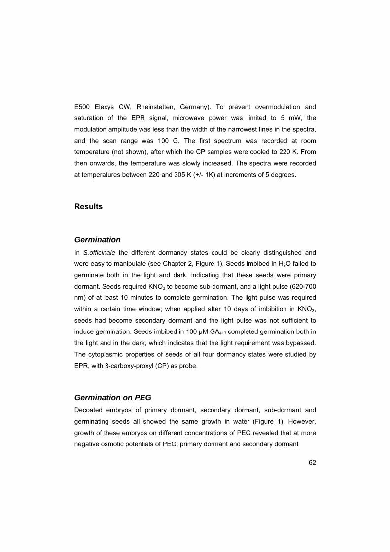
62
E500 Elexys CW, Rheinstetten, Germany). To prevent overmodulation and
saturation of the EPR signal, microwave power was limited to 5 mW, the
modulation amplitude was less than the width of the narrowest lines in the spectra,
and the scan range was 100 G. The first spectrum was recorded at room
temperature (not shown), after which the CP samples were cooled to 220 K. From
then onwards, the temperature was slowly increased. The spectra were recorded
at temperatures between 220 and 305 K (+/- 1K) at increments of 5 degrees.
Results
Germination
In S.officinale the different dormancy states could be clearly distinguished and
were easy to manipulate (see Chapter 2, Figure 1). Seeds imbibed in H2O failed to
germinate both in the light and dark, indicating that these seeds were primary
dormant. Seeds required KNO3 to become sub-dormant, and a light pulse (620-700
nm) of at least 10 minutes to complete germination. The light pulse was required
within a certain time window; when applied after 10 days of imbibition in KNO3,
seeds had become secondary dormant and the light pulse was not sufficient to
induce germination. Seeds imbibed in 100 µM GA4+7 completed germination both in
the light and in the dark, which indicates that the light requirement was bypassed.
The cytoplasmic properties of seeds of all four dormancy states were studied by
EPR, with 3-carboxy-proxyl (CP) as probe.
Germination on PEG
Decoated embryos of primary dormant, secondary dormant, sub-dormant and
germinating seeds all showed the same growth in water (Figure 1). However,
growth of these embryos on different concentrations of PEG revealed that at more
negative osmotic potentials of PEG, primary dormant and secondary dormant

63
Figure 1: Length (mm) of embryos after incubation for 144h in PEG solutions of different osmotic strength. Seeds were
pre-incubated in the dark on 25mM KNO3 for 1 day (●)(sub-dormant), H2O for 1 day (■)(primary dormant), 25 mM KNO3 for 10 days (♦)(secondary dormant) and GA4+7 for 1 day (▲)(germinating), before seed coats were removed and seeds were transferred to the PEG medium. Error bars represent standard deviation.
seeds were less resistant to the inhibitory action of PEG than germinating and sub-
dormant seeds (Figure 1).
EPR Measurements
3-Carboxy-proxyl spectra
For characterization of the water fraction in hydrated seeds in different
physiological states, seeds were labelled with perdeuterated 3-carboxyl-proxyl
(CP). CP is a relatively small (MW 186) molecule that is easily soluble in water. It is
a polar molecule, due to its OH group. This group increases the probability to form
hydrogen bonds and, thus, CP resides mainly in the aqueous cytoplasm. All seed

64
Figure 2: CP Spectra of Sisymbrium officinale seeds in different dormancy states. Seed samples are slowly heated
from 220 K to 300K. The melting of vitrified water occurred at 280 K in dormant, seco. ndary dormant and sub-dormant
seeds. Vitrified water melted at 265 K in germinating seeds
samples were cooled to 200 K and then slowly heated. The spectra were recorded
from 220 K to 305 K with 5-degree steps.
At low temperatures all the spectra showed vitrified water. Above 260 K
(germinating seeds) or 275 K (dormant and sub-dormant seeds) part of the water
appears in a liquid state as can be seen by the appearance of narrow-line spectra
(Figure 2). Complete melting of vitrified water occurred at about 280 K, in dormant,
secondary dormant and sub-dormant seeds; in germinating seeds melting of
vitrified water occurred at around 265 K. The narrow component in the spectra of
germinating seeds below 265 K originates from CP in oil (Figure 2). This can be
concluded from the distance between narrow lines, which are typical for
hydrophobic environment.
65
Figure 3: Changes of the distance between the outermost extremes, 2Amax, of the immobile (solid-like) component of CP spectra in Sisymbrium officinale seeds in different dormancy states, with temperature. Breaking points of curves
are indicated by the dotted lines.
Motional freedom, within the solid-like spectra, as measured by the distance
between the outermost extremes (2Amax) can be used as a tool to measure the
‘ease’ at which vitrified water melts (Kumler and Boyer, 1976) (2Amax is determined
only for solid-like spectra; see chapter 3). Plotting 2Amax against temperature will
show a linear temperature dependency of 2Amax due to increase of the motional
freedom. However, at a certain temperature, there is a break in the temperature
dependency, as from this break the rate of changes of the motional freedom with
temperature increases (Kumler and Boyer, 1976; Buitink et al, 1998; Buitink et al,
2000). The break occurred at 270K for primary and secondary dormant seeds, at
255Kfor sub-dormant seeds and at 250K for germinating seeds. Figure 3 shows
that vitrified water melted much easier in germinating seeds than in any other
seeds.

66
Figure 4: Rotational correlation time of CP in the cytoplasm of Sisymbrium officinale seeds in different dormancy states at different temperatures
The narrow line spectra of CP in aqueous cytoplasm can be used to calculate the
rotational correlation time for CP molecules according to the formula:

67
ΤR = 6.5 x 10⁻¹⁰ ∆W0 ((h0/h-1) 1/2-1)
in which h0 and h-1 are the heights of the central and high-field lines (Figure 4A)
and ∆W0 is the peak-to-peak width of the central line. The rotational correlation
time is proportional to the viscosity (Keith and Snipes, 1974). In Figure 4 the
rotational correlation time of CP in the cytoplasm at temperatures above the
melting temperature for all 4 seed samples is shown. The τR and, thus, cytoplasmic
viscosity were high in primary dormant and sub-dormant seeds, and low in
germinating seeds. In the secondary dormant seeds the viscosity had intermediate
values.
Discussion
3-Carboxy-proxyl (CP) was used in an EPR study to investigate the colligative
properties of cytoplasm water in seeds in different dormancy states, as it can give
information about how seeds adapt their cytoplasmic properties to periods of
dormancy. Sisymbrium officinale seeds were used for this, as their dormancy
states are clearly distinguished and easy to manipulate (Hilhorst and Karssen,
1989). Dormancy can be broken by supplying the seeds with nitrate and a light
pulse within a certain time frame. When the light pulse is suspended, seeds
become secondary dormant and will not germinate. When the seed coat (and
endosperm) is removed, embryos will grow on water, even when pre-incubated to
be dormant. This could be an indication that Sisymbrium officinale seeds have a
seed coat/endosperm imposed dormancy (Bewley, 1997). However, tests on PEG,
an osmoticum, showed that primary and secondary dormant embryos did not grow
as well as sub-dormant and germinating embryos on higher concentrations of PEG
(Figure 1) and were, thus, less tolerant to osmotic stress. Therefore, we conclude
that dormancy of S. officinale seeds is also, at least partially, located in the
embryo. This is the result of differences in the base water potential for germination,
which is higher (less negative) in dormant seeds, as compared to non-dormant

68
seeds (Bradford, 2005). This implies that the seed coat/endosperm imposes a
restriction on embryo growth, and the dormant embryo itself cannot gain sufficient
force to overcome this restriction. To enable the seed to germinate and grow in an
environment with a low osmotic potential (more negative) it needs to enhance the
water potential of lots of cells to be able to take up water. It was suggested by
Nabors and Lang (1971) that germination stimulating factors cause an increase of
the osmotic potential of the embryonic axis, and therefore increase the water-
absorbing capacity of the seed. The osmotic potential of the embryonic axis could
decrease by an increase in the concentration of low-molecular weight substances.
The low molecular weight substances will decrease the viscosity of the cytoplasm
and the temperature of water vitrification. Here we studied the viscosity of the
cytoplasm and the vitrification temperature of the seed, at different states of
physiological dormancy.
The ‘ease’ at which vitrified water melts (Figure 3) and the viscosity of the
cytoplasm (Figure 4) show that dormant and sub-dormant seeds possesed a
higher viscosity than the germinating seeds. The viscosity of secondary seeds
appeared to be intermediate, but the ‘ease’ at which the vitrified water melted was
similar to that of primary dormant seeds. This difference in cytoplasmic viscosity
could be caused by a change in presence of high-molecular weight substances.
Cytoplasmic viscosity could be of importance in the dormancy of the seed. At high
viscosity the seed is protected against protein unfolding (Klimov and Thirumalai,
1997) and heat stress due to reduced diffusion within the cytoplasm. The higher
viscosity of the dormant and non-dormant seed may be a way of the seed to
survive prolonged periods in the hydrated state. The high viscosity in the dormant
state was also found by Dijksterhuis et al. (2007), who studied fungal spores.
However, they came to the conclusion that the higher viscosity was not mainly
caused by a high concentration of solutes but by the high proportion of ordered
spin probe molecules in the vicinity of macro-structures. Sun (2000) linked reduced
metabolism in red oak seeds to an increased viscosity. In Chapter 2 we found a

69
reduction in metabolic activity for dormant and non-dormant seeds, as compared to
germinating seeds, which may be linked to the increased viscosity found here.
As a result of the changes in viscosity the temperature of vitrified water
melting was lower (265K) for germinating seeds than for primary, sub- and
secondary dormant seeds (280K) (Figure 2). The lower vitrification and melting
temperatures for germinating seeds could be due to the presence of higher
concentrations of low-molecular weight substances, as these are known to lower
the vitrification temperature (Wolfe and Bryant 1999). The low-molecular weight
substances could originate from the mobilisation of storage metabolites. While in
non-dormant and dormant seeds the higher molecular weight substances (e.g.
raffinose family of oligosacharides, sucrose, trehalose etc.) are more dominant
(Kou et al, 1988; Downie and Bewley, 2000) in germinating seeds high molecular
weight substances are hydrolysed into more low-molecular weight substances (e.g.
glucose, fructose) (Downie and Bewley, 2000). These low-molecular weight
substances increase the osmotic potential (make it more negative) and, therefore,
the water absorbing capacity of the seed (Nabors and Lang, 1971).
In conclusion, we have shown, for the first time, that cytoplasmic viscosity may
increase upon the induction of dormancy. The better water absorbing capacity of
germinating seeds may be caused by hydrolysis of storage metabolites into low
molecular weight compounds which will then cause a decrease in vitrification
temperature. Even though it is not clear if this is the main cause for a change in
viscosity of the cytoplasm, the increased cytoplasmic viscosity in the dormant state
may contribute to a long-term maintenance of seed viability. However, it is not clear
yet if the increase in cytoplasmic viscosity is the result or the cause of reduced
metabolic activity within the seed.

70

71
Chapter 5
Involvement of desaturases in
membrane fluidity and dormancy
cycling in seeds of Arabidopsis
thaliana
Susanne MC Claessens1, 2, Raquel Iglesias1, Magdalena Witek1, Elena A
Golovina1, Ronny Joosen1, Wilco Ligterink1, Folkert A Hoekstra1, Peter E Toorop2,
Henk WM HiIhorst1
1Laboratory of Plant Physiology, Wageningen University, P.O. Box 658, 6700 AR Wageningen, The Netherlands; 2Seed Conservation Dept., Royal Botanic Gardens Kew, Wakehurst Place, Ardingly, W Sussex, RH17 6TN, U.K. (email:
Abstract
Dormancy cycling in seeds depends on changes in sensitivity or responsiveness to
naturally occurring factors that are known to break dormancy or stimulate
germination. Receptors for these factors are likely to occur in membranes and
based on that, a model was proposed in which regulation of dormancy depends on
accessibility of membrane-bound receptors. Accessibility of membrane-bound
receptors can change by changing the membrane fluidity. Membrane fluidity is
largely determined by composition of unsaturated fatty acids in the membrane
phospholipids, which is a function of temperature and activity of desaturases. Here
we tested the hypothesis that unsaturated fatty acids and desaturase gene activity

72
are involved in the induction of secondary dormancy by changing membrane
fluidity. To test this hypothesis Arabidopsis thaliana desaturase mutants fad3, fad7,
fad8, ads1 and ads2 and wild type seeds were used. All mutants were
characterised in their dormancy phenotype. Membrane fluidity and fatty acid
composition were analysed when enough seeds where present, whilst the
expression of desaturase genes was analysed in wild type seeds.
Whilst ADS2 seems to have an influence on membrane fluidity and is expressed in
non-dormant seeds, it does not show any changes in dormancy phenotype; this
may be due to functional redundancy. FAD3 activity shows changes in relation to
dormancy and germination but it is not involved in changes in membrane fluidity.
Although desaturase activity may change the membrane fluidity or influence the
germination/dormancy phenotype, the two are not linked, unless the effects of
these enzymes are very localised within the seeds.
Introduction
Dormancy cycling in seeds of Arabidopsis thaliana and Sisymbrium officinale
depends on changes in sensitivity or responsiveness to naturally occurring factors
that are known to break dormancy or stimulate germination, such as light and
nitrate (Hilhorst, 1990 a/b). Sisymbrium officinale seeds have an absolute
dependency on a combination of light and nitrate to terminate dormancy and
initiate germination (Hilhorst et al., 1986, Hilhorst, 1990a, b). In Arabidopsis
thaliana light, but not nitrate, is an absolute requirement for germination (Derkx and
Karssen, 1994). There are several possible mechanisms that explain how
responsiveness to light and nitrate can change; two possible explanations suggest
that (1) either the amount of active receptors or (2) the accessibility of their ligands
changes with the induction and relief of dormancy (Raison et al., 1980; Di Nola and
Mayer, 1986; Hilhorst, 1998). Receptors for light (through phytochrome) and nitrate
are likely to occur in membranes and with that in mind Hilhorst (1998) proposed a
73
model in which regulation of dormancy cycling depends on accessibility of
membrane-bound receptors (chapter 1). In this model the magnitude of movement
of receptor proteins within the membrane depends on membrane fluidity. A higher
fluidity may cause the receptor to move to the membrane surface, where it then
becomes accessible for nitrate and phytochrome. Membrane fluidity is largely
determined by the content of unsaturated fatty acids in the membrane
phospholipids, which on its turn is a function of temperature and activity of
desaturases. Thus, the breaking and inducing of dormancy may be achieved by
changes in temperature that induce an increase or decrease of fatty acid
unsaturation.
A downward shift in temperature results in a decrease in membrane
fluidity, whereas an upward shift may cause fluidization of the membrane (Los and
Murata, 2004; Vigh, 1998). Decreased/increased membrane fluidity can cause
extensive damage to the membranes; therefore membranes are able to adjust to
the new conditions to maintain their fluidity. This is called homeoviscous adaptation
(Sinensky, 1974). Membrane fluidity can be adjusted at low temperature by
introducing double bonds into the fatty-acyl chains of membrane lipids, making the
fatty acids more unsaturated and, hence, the membrane more fluid (Los and
Muratta, 2004). The introduction of double bonds in fatty acids is mediated by
desaturases. Following an upward shift in temperature, the degree of unsaturation
of fatty acids of membrane phospholipids may again decrease, by suppression of
the desaturation of fatty acids and acceleration of de novo synthesis of saturated
fatty acids, thereby diluting the unsaturated fatty acids (Sato and Murata, 1980).
There are three types of desaturases: Acyl-CoA desaturases, Acyl-ACP
desaturases and Acyl-lipid desaturases (Murata and Wada, 1995). Acyl-CoA
desaturases introduce double bonds into fatty acids that are bound to Coenzyme
A, and are only found in animal, fungal and yeast cells. Acyl-ACP desaturases can
introduce the first double bond into plant fatty acids that are bound to an Acyl-
carrier protein (ACP), a reaction which occurs in the plastids of plant cells (Murata
and Wada, 1995). All double bonds can be introduced by Acyl-lipid-desaturases

74
into fatty acids that have been esterified to glycerolipids, bound to the
endoplasmatic reticulum or chloroplast membrane (Murata and Wada, 1995).
Since the Acyl-lipid desaturases introduce double bonds directly into the fatty acids
of membrane lipids they are most efficient in regulating unsaturation in response to
temperature changes (Murata and Wada, 1995). Acyl-lipid desaturases introduce
double bonds at specific sites within the carbon chains. The order in which these
desaturases operate is very strictly determined: the first double bond is introduced
by the ∆9 desaturase (an Acyl-ACP- or Acyl-lipid-desaturase), introducing a double
bond in the 9th position of a C18, fatty acid converting stearic acid (18:0) into oleic
acid (18:1) (Murata and Wada, 1995). The second double bond is introduced by a
∆12 or ∆6 desaturase (Acyl-lipid-desaturases), introducing a double bond in the
12th position of oleic acid (18:1) converting it into linoleic acid (18:2). This double
bond is only introduced into fatty acids that have a double bond at the ∆9 position.
The third and last double bond is introduced by an ω3 or ∆15 desaturase (Acyl-
lipid-desaturases), converting linoleic acid (18:2) into linolenic acid (18:3) (Matos et
al., 2007). This desaturase only introduces double bonds into fatty acids that have
a double bond at the ∆12 position (Hagashi and Murata, 1993).
The aim of the present work was to assess the effects of mutations in
desaturase genes on the induction of secondary dormancy. To test the hypothesis
that membrane desaturases are involved in the induction of secondary dormancy,
we used Arabidopsis thaliana fatty acid desaturase (FAD) mutants and wild type
seeds as control. Here we used the fad3, fad7, fad8, ads1 and ads2 mutants. The
fad3, fad7 and fad8 mutants are mutated in the ω3 desaturase genes (Murata and
Wada, 1995), whereas ads1 and ads2 are mutated in a ∆9 desaturase gene
(Murata and Wada, 1995). The fad3, fad7, fad8, ads1 and ads2 mutants were
characterised in their dormancy phenotype whilst the expression of desaturase
genes was analysed in wild type seeds.
75
Materials and Methods
Seed germination and plant growth to obtain higher quantity of
seeds
Seeds were obtained from the Arabidopsis Biological Resource Centre (ABRC).
Wild-type and fad3, fad7, fad8, ads1, ads2 mutants were all from the Columbia
genetic background. Of all seeds 2 different mutant lines in the same gene were
ordered, if available. Seeds were sterilized for 1h using a gas sterilizer containing
bleach and 1.2% HCl. Seeds were sown on 0.5 MS medium containing 2.3 g /l
vitamins, 0.8 % purified agar (Duchefa) pH = 6.0, 1.95 g/l MES hydrate (Sigma)
and 10 mg/l kanamycin. Petri dishes containing medium with seeds were kept at
4°C for 3 days. After 3 days dishes were transferred to 24°C under a 16h/8h
light/dark regime. After seeds had germinated and plants were big enough they
were transferred to the ARASYSTEM (www.arasystem.com) and kept at 24°C
(16h/8h, light/dark).
Seed germination
Freshly harvested wild type and mutant seeds were stratified at 4°C for 3 days to
break any dormancy present. After stratification seeds were irradiated with red light
for two hours and transferred back to the dark, at 5, 15, 20 and 25°C. Secondary
dormancy was induced after stratification at 4°C by incubating the seeds in the
dark for 10 days at a range of temperatures, before the light pulse was given and
seeds were germinated at the same temperature.
Isolation of genomic DNA
100-200 mg young leaves were collected in an Eppendorf tube and frozen in liquid
nitrogen. Leaf material was ground to a fine powder. DNA extraction buffer (0.3 M
76
NaCl, 50 mM Tris (pH 7.5), 20 mM EDTA, 2% Sarkosyl, 0.5% SDS, 5 M Urea and
5% (v/v) Phenol pH 7.5) was added and samples were refrozen and ground again.
A phenol/chloroform extraction was performed; samples were mixed gently and
spun at 12.000 rpm for 5 minutes. DNA was precipitated with iso-propanol and kept
at room temperature for 5 minutes, then washed with 70% ethanol and dried
briefly. DNA pellets were dissolved in TE buffer containing 10 μg/ml RNAse and
stored at 4°C.
RNA isolation from mutant seed material and gene expression
analysis for detection of homozygous plants
30 mg of seed material was collected per sample and RNA was isolated using the
SV Total RNA isolation system (Promega) according to the manufacturer’s
instructions. cDNA was synthesized, using iScripttm (Biorad), from 1 µg total RNA,
in a mixture containing RNase H+ iScript reverse transcriptase, RNase inhibitor,
oligo (dT) and random hexamers (used as random primers). The reaction protocol
was 5 min at 25ºC, 30 min at 42ºC followed by 5 min at 85ºC and hold at 4ºC. For
detection of homozygous mutant plants and gene expression analysis of mutant
plants quantitative real time PCR (QRT-PCR) was performed using the Bio-Rad
system (iCycler iQ Real Time PCR detection device). The iQ Sybr Green Supermix
(BioRad) was used, containing SYBR green I dye, hot-start iTaq DNA polymerase,
buffer and dNTPs qualified for quantitative PCR. The Supermix also contained
fluorescein used for the correction of the readings and 5 μl of cDNA. Primers used
are listed in Table 1 (for detection of homozygous plants) and Table 2 (for
expression analysis). The standard thermal profile used was 3 min at 95ºC,
followed by 45 cycles of 15s at 95ºC, 1s at 60ºC and 2 min at 72ºC.
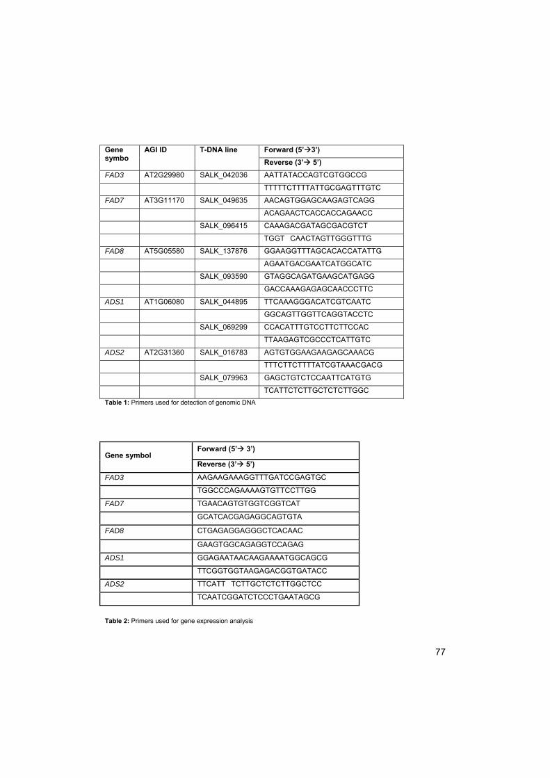
77
Gene symbo
AGI ID T-DNA line Forward (5’3’)
Reverse (3’ 5’)
FAD3 AT2G29980 SALK_042036 AATTATACCAGTCGTGGCCG
TTTTTCTTTTATTGCGAGTTTGTC
FAD7 AT3G11170 SALK_049635 AACAGTGGAGCAAGAGTCAGG
ACAGAACTCACCACCAGAACC
SALK_096415 CAAAGACGATAGCGACGTCT
TGGT CAACTAGTTGGGTTTG
FAD8 AT5G05580 SALK_137876 GGAAGGTTTAGCACACCATATTG
AGAATGACGAATCATGGCATC
SALK_093590 GTAGGCAGATGAAGCATGAGG
GACCAAAGAGAGCAACCCTTC
ADS1 AT1G06080 SALK_044895 TTCAAAGGGACATCGTCAATC
GGCAGTTGGTTCAGGTACCTC
SALK_069299 CCACATTTGTCCTTCTTCCAC
TTAAGAGTCGCCCTCATTGTC
ADS2 AT2G31360 SALK_016783 AGTGTGGAAGAAGAGCAAACG
TTTCTTCTTTTATCGTAAACGACG
SALK_079963 GAGCTGTCTCCAATTCATGTG
TCATTCTCTTGCTCTCTTGGC
Table 1: Primers used for detection of genomic DNA
Table 2: Primers used for gene expression analysis
Gene symbol Forward (5’ 3’)
Reverse (3’ 5’)
FAD3 AAGAAGAAAGGTTTGATCCGAGTGC
TGGCCCAGAAAAGTGTTCCTTGG
FAD7 TGAACAGTGTGGTCGGTCAT
GCATCACGAGAGGCAGTGTA
FAD8 CTGAGAGGAGGGCTCACAAC
GAAGTGGCAGAGGTCCAGAG
ADS1 GGAGAATAACAAGAAAATGGCAGCG
TTCGGTGGTAAGAGACGGTGATACC
ADS2 TTCATT TCTTGCTCTCTTGGCTCC
TCAATCGGATCTCCCTGAATAGCG

78
RNA isolation from wild type seeds for analysis of fatty acid
desaturase gene expression
Following treatment, seeds were stored in 2-ml tubes at -80°C. Total RNA was
extracted using a hot borate method (modified from Wan and Wilkins, 1994).
Seeds were ground with mortar and pestle, submerging the mortar and pestle in
liquid nitrogen before grinding. Ground seeds were kept submerged in liquid
nitrogen prior to RNA extraction. Ground seeds were re-suspended in 700 µl
extraction buffer [(0.2M sodium tetraborate decahydrate, 30 mM EGTA, 1% w/v
SDS, 1% w/v sodium deoxycholate, pH 9.0) containing 14 mg polyvinylpyrolidone
and 1.1 mg dithiothreitol], which had been heated to 80°C. The seed suspension
was added to 0.35 mg proteinase K in a 2 ml round-bottomed tube and incubated
at 42°C for 90 min. 56µl 2M KCL was then added and samples incubated on ice for
1h. Samples were centrifuged at 16000 g at 4°C, after which the supernatant was
transferred to a new tube and its volume determined. A one-third volume of 8M
LiCl (to give a final 2M LiCl concentration) was added and samples were incubated
overnight at 4°C. Samples were centrifuged, and pellets washed by vortexing in 2M
LiCl until the supernatant remained colourless. Pellets were re-suspended in 300 µl
diethylpolycarbonate (DEPC) treated water, and RNA precipitated by the addition
of 30 µl 2M potassium acetate and 990 µl ice-cold 100% ethanol, and incubation
overnight at -80°C. RNA was pelleted by centrifugation at 16000 g for 30 min at
4°C, followed by a wash with 70% ethanol. Partially dried pellets were re-
suspended in 50µl DEPC-treated water. RNA concentration and quality were
determined using a WPA lightwave s2000 UV/Vis spectrophotometer.
QRT-PCR of wild type seeds
Total RNA from 3 biological replicates per treatment were used. cDNA was
synthesized using a transcriptor first strand cDNA synthesis kit (Roche), according
to the manufacturer’s instructions. Primers were used as described in Table 2.
PCR reactions were performed using LightCycler FastStart DNA master SYBR

79
green I (Roche), in a mixture containing 5 µl cDNA in a volume of 25 µl. The
thermal profile used was 10 min at 95ºC, followed by 45 cycles of 1 min at 95ºC, 2
min at 60ºC and 1 min at 72ºC. Comparative Ct values were calculated and
ANOVA tests were used to statistically analyse the data.
Nuclear Magnetic Resonance Spectroscopy (NMR)
The NMR spectra were recorded on an Avance II spectrometer (Bruker,
Rheinstetten, Germany), as described in chapter 3. Dry Arabidopsis thaliana seeds
with seed coats and without additional treatments were packed into a 7 mm
Zirconia rotor and spun under a magic angle at spinning speed of 5 kHz. 1H MAS
spectra were recorded at room temperature with a spectral width of 6 kHz applying
single-pulse sequences with a 10 μs rf pulse and 5 s repetition delay between
scans. Time domain size of the spectra was 8 k with a number of accumulations of
32. The narrow range of proton chemical shifts makes it difficult to identify
individual fatty acids.
13C MAS single-pulse excitation spectra were obtained and analysed as described
in chapter 3, and was used for analyses of the fatty acid (FA) composition of the
mutants and wild type.
Electron Paramagnetic Resonance
The methyl ester of 5 doxyl stearic acid (5-mDS(A)) was used as a spin probe. This
spin probe is weakly anchored in the phospholipid head group area due to the high
hydrophobicity of the methyl ester. As a result, methylated spin label is localized in
a deeper position in the membrane bilayer than its un-methylated counterpart (5-
DS(A)) (Sanson et al., 1976). 5-mDS(A) is often used to study membrane fluidity
(Benatti et al., 2001; Bianconi et al., 1988; Turchiello et al., 2000; chapter 3).
Arabidopsis seeds were imbibed in water for 8h in the dark. Medium attached to
the surface of the seed was removed with filter paper before seed coats were

80
removed using tweezers, after which seeds were dried at room temperature. Dry
seeds were placed in a 1mM solution of membrane spin probe in hexane. After 1d
the spin probe solution was removed, seeds were washed twice with hexane and
placed at 30% RH for 24 hours to remove the remaining hexane from the seeds.
Seeds were re-hydrated by humidification for 3h at 100% RH.
EPR spectra were recorded with an X-band EPR spectrometer (Bruker E500
Elexys CW, Rheinstetten, Germany). To prevent over-modulation and saturation of
the EPR signal, microwave power was limited to 5 mW, the modulation amplitude
3G for solid-state and 1G for fluid type spectra. In the case of 2-component spectra
the lowest modulation amplitude of 1G was used. Field scan widths of 100G were
used.
Results
Identification of homozygous T-DNA insertion mutants
Nine SALK T-DNA lines (Alonso et al., 2003) representing T-DNA insertions in 5
desaturase genes were obtained from the ABRC (Table 1). Seeds from the
homozygous mutant plants were tested for expression of the desaturase genes
using qRT-PCR, and none showed significant expression (data not shown).
Phenotypic analysis
Phenotypic analysis did not show appreciable differences between wild type and
most mutant plants. However, the fatty acid desaturase 7 mutants (fad7;
Salk_096415) showed slow growth. Plants were lagging 2 weeks behind in
development and flowering. No differences in seed and seed coat structure could
be observed.
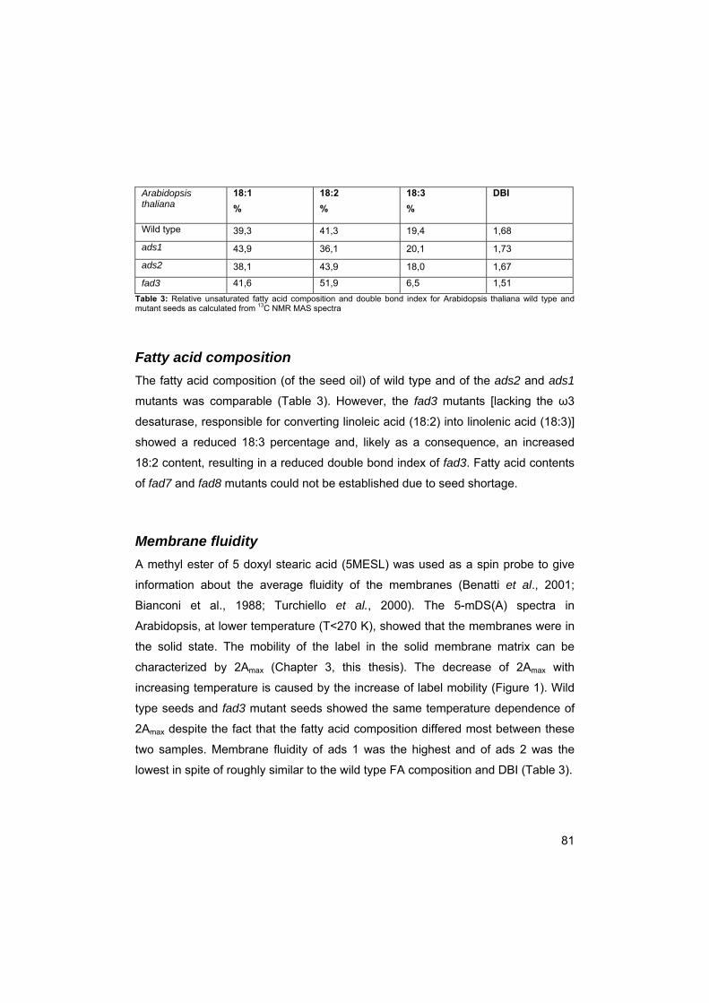
81
Arabidopsis thaliana
18:1
%
18:2
%
18:3
%
DBI
Wild type 39,3 41,3 19,4 1,68
ads1 43,9 36,1 20,1 1,73
ads2 38,1 43,9 18,0 1,67
fad3 41,6 51,9 6,5 1,51
Table 3: Relative unsaturated fatty acid composition and double bond index for Arabidopsis thaliana wild type and mutant seeds as calculated from 13C NMR MAS spectra
Fatty acid composition
The fatty acid composition (of the seed oil) of wild type and of the ads2 and ads1
mutants was comparable (Table 3). However, the fad3 mutants [lacking the ω3
desaturase, responsible for converting linoleic acid (18:2) into linolenic acid (18:3)]
showed a reduced 18:3 percentage and, likely as a consequence, an increased
18:2 content, resulting in a reduced double bond index of fad3. Fatty acid contents
of fad7 and fad8 mutants could not be established due to seed shortage.
Membrane fluidity
A methyl ester of 5 doxyl stearic acid (5MESL) was used as a spin probe to give
information about the average fluidity of the membranes (Benatti et al., 2001;
Bianconi et al., 1988; Turchiello et al., 2000). The 5-mDS(A) spectra in
Arabidopsis, at lower temperature (T<270 K), showed that the membranes were in
the solid state. The mobility of the label in the solid membrane matrix can be
characterized by 2Amax (Chapter 3, this thesis). The decrease of 2Amax with
increasing temperature is caused by the increase of label mobility (Figure 1). Wild
type seeds and fad3 mutant seeds showed the same temperature dependence of
2Amax despite the fact that the fatty acid composition differed most between these
two samples. Membrane fluidity of ads 1 was the highest and of ads 2 was the
lowest in spite of roughly similar to the wild type FA composition and DBI (Table 3).
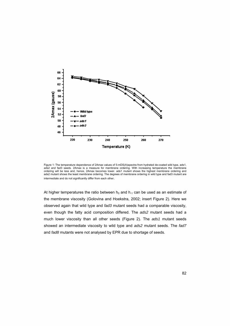
82
Figure 1: The temperature dependence of 2Amax values of 5-mDS(A)spectra from hydrated de-coated wild type, ads1, ads2 and fad3 seeds. 2Amax is a measure for membrane ordering. With increasing temperature the membrane ordering will be less and, hence, 2Amax becomes lower. ads1 mutant shows the highest membrane ordering and ads2 mutant shows the least membrane ordering. The degrees of membrane ordering in wild type and fad3 mutant are
intermediate and do not significantly differ from each other.
At higher temperatures the ratio between h0 and h-1 can be used as an estimate of
the membrane viscosity (Golovina and Hoekstra, 2002; insert Figure 2). Here we
observed again that wild type and fad3 mutant seeds had a comparable viscosity,
even though the fatty acid composition differed. The ads2 mutant seeds had a
much lower viscosity than all other seeds (Figure 2). The ads1 mutant seeds
showed an intermediate viscosity to wild type and ads2 mutant seeds. The fad7
and fad8 mutants were not analysed by EPR due to shortage of seeds.

83
Figure 2: The temperature induced changes in membrane fluidity of hydrated wild type and mutants ads1, ads2 and fad3. Membrane fluidity is expressed as the ratio of the central line height (h0) to the low-field (h-1) line height of 5-mDS(A) spectra (insert). The fad3 and wild type seeds show a comparable viscosity, ads2 mutants show a much lower viscosity, while ads1 mutant seeds show an intermediate viscosity
Germination
Freshly harvested wild type Columbia seeds were pre-incubated at 4°C for 3 days
to break any dormancy present. After pre-incubation seeds were irradiated with red
light for two hours and transferred back to the dark, at 5, 15, 20 and 25°C. Wild
type seeds germinated up to 100% (Figure 3A) at all temperatures. From the t50
values (Figure 3B) it can be seen that seeds reached half maximum germination
significantly earlier, as confirmed by t-test, at 20 and 25°C than at 5 and 15 °C (p-
value: 0.0000008 and 0.009), implying that 20 and 25°C are the more optimal
temperatures for germination than 5 and 15°C for this Arabidopsis accession. At
5°C maximum germination was achieved only after 24d, at the other temperatures

84
Figure 3: A. Total germination and B. time to half-maximal germination (t50) of wild type, ads1 and ads2 seeds. A and B in mutant names indicate different sites of the mutation in the same gene. A and B in wild type indicate replicates coming from different seed batches. Seeds were imbibed in water at 4°C for 3d before being transferred to the light (left) or kept in the dark for 10 d (right) before being transferred to the light. 3 replicates were used. Vertical bars represent standard deviation
maximum germination was achieved within 4d. All mutants reached similar levels
of germination (Figures 3 and 4), apart from the ads1A mutants. The ads1A mutant
germinated only for 55% at 5°C; however, the ads1B mutant germinated for 100%
at 5°C. The difference between A and B is caused by a different site of the
mutation in the same gene.
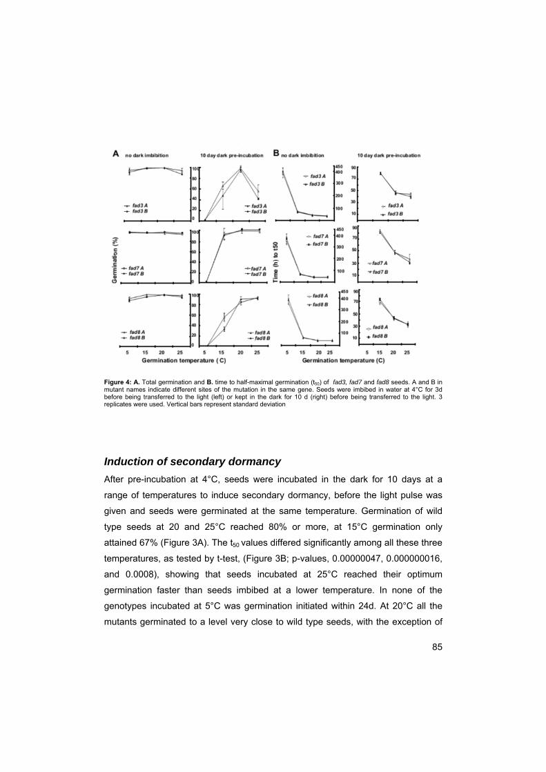
85
Figure 4: A. Total germination and B. time to half-maximal germination (t50) of fad3, fad7 and fad8 seeds. A and B in mutant names indicate different sites of the mutation in the same gene. Seeds were imbibed in water at 4°C for 3d before being transferred to the light (left) or kept in the dark for 10 d (right) before being transferred to the light. 3 replicates were used. Vertical bars represent standard deviation
Induction of secondary dormancy
After pre-incubation at 4°C, seeds were incubated in the dark for 10 days at a
range of temperatures to induce secondary dormancy, before the light pulse was
given and seeds were germinated at the same temperature. Germination of wild
type seeds at 20 and 25°C reached 80% or more, at 15°C germination only
attained 67% (Figure 3A). The t50 values differed significantly among all these three
temperatures, as tested by t-test, (Figure 3B; p-values, 0.00000047, 0.000000016,
and 0.0008), showing that seeds incubated at 25°C reached their optimum
germination faster than seeds imbibed at a lower temperature. In none of the
genotypes incubated at 5°C was germination initiated within 24d. At 20°C all the
mutants germinated to a level very close to wild type seeds, with the exception of

86
the ads1A mutant which germinated for only 60%. The greatest variation in
germination was observed at 15°C. Only the ads1B mutant germinated to the same
percentage as the wild type (65%). Of the other genotypes fad7A, fad7B
germinated at 15 C to a higher level (90%) than the wild type, whereas the rest
showed germination below that of the wild type of which the ads1A mutant showed
only 10% germination. The fad3 mutants showed wild type germination levels at
20°C, but was the only genotype that displayed lower germination (50%) at 25°C.
The t50 for all mutants was comparable with the wild type, with only the fad7
mutants showing a somewhat lower t50 (70 vs 80h at 15°C).
Fatty acid desaturase gene expression in wild type Arabidopsis
thaliana seeds
Desaturases were further analysed by studying their transcript abundance in wild
type seeds that were imbibed for 1 or 10 days at 15 or 25°C, to characterize
optimal and suboptimal conditions for germination. Although germination reached
comparable levels at both temperatures there is a difference in the t50 between the
two temperatures (figure 3).Gene expression analysis was done on seeds that
were pre-incubated for 1 day at 4 °C and were subsequently incubated in the dark
or in the light; seeds imbibed for 10 days were incubated for 10 days in the dark
before transferring them to either the light or keeping them in the dark for 8 hours.
Seeds imbibed in the dark do not germinate at all, seeds imbibed for 10 days in the
dark before transferring them to the light, do germinate (Figure 3), however, to a
lesser value than seeds not imbibed in the dark for 10 days. Gene expression was
analysed by the comparative Ct method and further analysed by t-test (p=0.05).
FAD3 gene expression (Figure 5) was significantly higher in the least dormant
seeds at 15 and 25°C. When seeds were light irradiated during imbibition for 1
day, gene expression was higher than when kept in the dark for 10 days at optimal
germination temperatures. There was no effect of the temperature on transcript
abundance which corresponds with comparable germination at 15 and 25°C, albeit

87
Figure 5: Relative transcript abundance of FAD3, FAD7, FAD8, ADS1 and ADS2 genes in wild type seeds. Seeds were imbibed at 15 or 25°C, for 1 (1d) or 10 (10d) days, after which they were transferred to the light (L) or kept in the dark (D). 5 replicates were used per sample. Gene expression was analysed with the comparative Ct method and further analysed with ANOVA, standard deviations are given. Germination status of the seeds is given, - stands for no germination; + for some germination; ++ for up to 100% germination.
with different t50. Although without prolonged dark imbibition fad3 mutants showed
100 % germination, they showed the most dormant phenotype after 10 days dark
pre-incubation at suboptimal temperatures (Figure 4A) suggesting that FAD3 gene
expression may be associated with optimal conditions for breaking dormancy and
88
inducing germination. FAD7 gene expression (Figure 5) was significantly higher in
seeds imbibed in the light for 1 day, at 15 and 25°C than in all other treatments, so
FAD7 seems to be associated with induction of germination too. FAD8 gene
expression (Figure 5) was significantly higher for 1 day imbibed seeds at 25°C as
compared with all other treatments. Both these 1-d imbibed seeds and 1-d imbibed
seeds at 15°C had their dormancy broken, however only 1-d imbibed seeds at
25°C showed higher FAD8 gene expression. ADS1 and ADS2 gene expression
(Figure 5) did not show a pattern that could be clearly related to dormancy,
although the abundance of ADS1 and ADS2 transcripts was significantly different
between the different treatments (e.g. ADS2 gene expression being significantly
higher for 1-d imbibed seeds in the light than for 10-d imbibed seeds
Discussion
The aim of this work was to determine the relation between desaturase activity and
seed dormancy. Arabidopsis thaliana Columbia wild type plants and fad3, fad7,
fad8, ads1 and ads2 mutant plants in a Columbia back ground were used. The
ads1 and ads2 mutants are mutated in a ∆9 desaturase. The ∆9 desaturases
introduce a double bond in the 9th position of the C18, converting stearic acid to oleic
acid (Murata and Wada, 1995).The fad3, fad7 and fad8 mutants are mutated in ω3
desaturase and therefore are not capable of forming double bonds at the ∆15
position of the C18 acids (Murata and Wada, 1995), converting linoleic acid (18:2) to
linolenic acid (18:3) (Matos et al., 2007).
NMR data showed that the FA composition of ads1 and ads2 mutants did not differ
much from wild type seeds.
ads1 and ads2 Mutant and wild type seeds both are able to germinate up
to 100% in the light at temperatures ranging from 5 to 25 °C after a short cold
imbibition at 4 °C (Figure 3), showing that both wild type and mutant seeds
exhibited similar vigour. A shift from 4°C to higher temperatures, such as 25°C
89
generally increases membrane fluidity. As ads1 and ads2 mutants did not show
any differences in FA composition, no differences in germination pattern were
expected. Imbibition of WT seeds for 10 days in the dark before they are
transferred to the light should induce secondary dormancy as in e.g. Sisymbrium
officinale seeds (Derkx et al., 1993; Hilhorst, 1990a/b; Chapter 2 of this thesis).
However, in Arabidopsis seeds induction of secondary dormancy has been shown
to take longer in the Cvi ecotype (Cadman et al., 2006). In Figure 3A we see that
after 10 day pre-incubation wild type seeds did not longer germinate at 5°C, but
still germinated up to a high percentage at 20-25°C, at 15 C germination
percentage was somewhat less than at 20 – 25 C (Figure 3A). After 10 days of
dark incubation, seeds completed germination faster when imbibed at 25°C (Figure
3B), making this the more optimal temperature for germination. Apparently, the 10
day pre-incubation had made the germination temperature window narrower, and
germination was reduced outside this window. Thus, Arabidopsis seeds acquire
secondary dormancy at low temperatures, which underlines the winter annual
properties of the Columbia accession (Cadman et al., 2006).
The germination pattern was similar for ads1B and ads2A and B mutants
but the ads1A mutant showed significantly reduced germination at 5 °C (without
dark imbibition) and at 15°C (after 10 days of dark pre-incubation ) as compared
with wild type (Figure 3A). The abundance of ADS1 and ADS2 transcripts was
significantly different between the different treatments. ADS2 gene expression was
significantly higher for 1-d imbibed seeds in the light than for 10-d imbibed seeds.
The fatty acid composition of ads1 and ads2 mutants was comparable to wild type
seeds but the membranes of ads2 mutants were more fluid and of lower viscosity
than that of ads1 and wild type seeds (Figs. 2, 3). Thus, although ads1A mutants
show a different germination pattern than wild type seeds, the germination pattern
results of the ads1B combined with the membrane fluidity, FA composition and
gene expression levels lead to the conclusion that ADS1 genes are not involved in
dormancy and germination. ADS2 gene activity did not seem to show a clear
relation with germination and dormancy (Figure 5; compare the results for the ads2

90
mutants), nor with the fatty acid composition, but it had the most profoundly
changed membrane fluidity and viscosity. ADS1 and ADS2 are both
endoplasmatic reticulum based desaturases that catalyze the formation of the first
double bond in the FA, and if this double bond is not formed no other double bonds
are formed (Murata and Wada, 1995). Thus, it is very likely that these mutants
show functional redundancy since membranes cannot exist if only saturated fatty
acids are present (Stubbs and Smith, 1984; Los and Murata, 1998). ADS2,
however, has been shown to be able to change membrane fluidity whereas ADS1
is not (Fukuchi-Muzutani et al., 1998). This is in agreement with our results. So
while some functions of ADS1 and ADS2 might be interchangeable, not all
functions are. The fatty acid content obtained here is the content for bulk lipids, i.e.
oil and membranes. Membranes might have fatty acid composition different from
oil (Sheffer et al., 1986; Millar et al., 2000; Voelker, 2001). Therefore changes
found in membrane fluidity but not in fatty acid content might not mean that fatty
acids and desaturases are not the cause of the change in fluidity. However,
membrane fluidity can be also modified by sterols, flavonoids and other
compounds. Testing of ads1/ads2 double mutants would be a next step in
analyzing the functions; however, ADS1, ADS2 and ADS3 are known to be able to
switch regiospecificity (Heillman et al., 2004) making analysis more difficult.
NMR data confirmed that the fad3 mutant is impaired in the conversion of
linoleic acid to linolenic acid, although still one third of the linolenic acid is present
(Table 3). Mutant and wild type seeds both germinated up to 100% at temperatures
ranging from 5 to 25 °C after a short cold imbibition (Figure 3A, 4A), showing that
both wild type and mutant seeds exhibited similar vigour. A shift from 4°C to higher
temperatures, such as 25°C generally increases membrane fluidity. The fluidity is
maintained by decreasing the degree of unsaturation of fatty acids of membrane
lipids, which is achieved by acceleration of de novo synthesis of saturated fatty
acids, as this dilutes the unsaturated fatty acids (Sato and Murata, 1980). As this
process does not involve the action of desaturases itself, mutants were not
expected to exhibit a germination pattern different from wild type seeds.
91
The fad3 mutants showed increased dormancy after 10 day pre-incubation
at 25°C, as compared to wild type seeds, with clearly narrowed germination
temperature window. However, at 20°C, the germination percentages of fad3
mutants and wild type seeds are high and both up to 100 %. At 25°C wild type
seeds showed a high percentage of germination but this was not seen in the fad3
mutant seeds where germination was reduced to 50%. At 15°C the germination of
fad3 mutants seems slightly reduced after 10 day pre-incubation compared to wild
type seeds, however, this is not significant.
Even though desaturases are known to introduce double bonds at low
temperatures and not at high temperatures (Los and Murata, 2004), FAD3 was
expressed to similar levels at both 25 and 15°C (Figure 5). In fad3 seeds 18:3
production is reduced (Table 3; Nishida and Murata, 1996; Horiguchi et al., 2000;
Matos et al., 2007), thus an increase in 18:3 level, mediated by FAD3, could be
necessary to keep the membranes fluid enough for dormancy breaking (Hilhorst,
1998). Nevertheless, EPR measurements did not show appreciable differences in
membrane fluidity between wild type and fad3 mutants (Figure 1 and 2). However,
measurements were done after 1d, when there is also no difference in germination
between the two treatments. The expectation would be that after a 10-d pre-
incubation, when there is a clear difference in dormancy and germination pattern
between genotypes, there would be a difference in membrane fluidity;
unfortunately this could not be measured here, due to a shortage of seeds.
Previously, we have found that secondary dormancy induction is accompanied by
membranes becoming less fluid (This thesis, Chapter 3). In fad3 mutant seeds
membranes can only become slightly more fluid and at higher temperatures (10d at
25°C) a much higher fluidity might be needed to keep seeds non-dormant. The
lower fluidity of fad3 mutants compared to wild type seeds at higher temperatures
might be the reason for partial induction of secondary dormancy at 25°C which did
not occur in wild type seeds. The FAD3 gene expression pattern found here is
similar to that found by Finch-Savage et al. (2007) in the Cvi accession of A.
thaliana. The present results combined with those of Finch-Savage et al. (2007)

92
show that expression of FAD3 is low in dormant seeds and higher when dormant
seeds are exposed to cold or light. It is interesting to see that in wild type seeds
that germinated after a 10-d pre-incubation, there was no FAD3 expression. FAD3
is post-transcriptionally controlled by temperature (Collados et al., 2006) but
transcriptionally by light (Collados et al., 2006). The light induced transcription
combined with the reduced sensitivity to light (Hilhorst 1990a/b) after prolonged
incubation might explain why there is no FAD3 expression after 10 days.
The fad8 mutant seeds showed a similar germination pattern as wild type
seeds. However, at 15°C, germination only reached a maximum of 55% after a 10
day pre-incubation (Figure 3, 4), suggesting that the germination window had
narrowed. Gibson et al (1994) reported that FAD8 transcript levels may increase at
low temperature, making the membrane more fluid. This could be the cause of
changes in germination induction at low temperature as seen here. However,
FAD8 gene expression increases when dormancy is broken and/or germination is
induced, but only at 25°C (Figure 5). This is in disagreement with results in the eFP
browser that showed higher FAD8 expression levels when dormancy is broken and
germination is induced and where no differences in expression between cold
imbibition or imbibition at 22°C were found. The reason for the discrepancy
between Gibson et al (1994) and eFP browser is unknown.
FAD7 is a chloroplast based light-induced desaturase (Los and Murata,
1998). The gene expression results (Figure 5) show a light dependency for 1-d
imbibed seeds. After 10d of pre-incubation, however, the FAD7 gene was not up-
regulated by light anymore. After secondary dormancy is induced seeds become
less responsive to light (Hilhorst, 1990a/b) and this coincides with FAD7 not being
up-regulated. Although the expression pattern suggests FAD7 to be involved in
germination, the mutant germination pattern did not confirm this. This could be due
to functional redundancy, as there are more ω3 desaturases and fad7 mutants only
show the mutant phenotype under certain conditions, as other ω3 desaturases
may take over its role (Gibson et al., 1994). As FAD7 protein is mainly present in
the thylakoid membrane of chloroplasts (Poghosyan et al., 1999) gene expression

93
will not lead to functional proteins within the seed and therefore FAD7 is unlikely to
be involved in germination itself.
Some but not all desaturases are shown here to change in relation to
dormancy and germination. FAD3, FAD7 and FAD8 all catalyze the conversion of
linoleic acid (18:2) to linolenic acid (18:3) (Matos et al., 2007). From the present
results we can conclude that this is an important conversion and is likely to be
involved in dormancy and germination. Although all three FADs catalyze the same
conversion, their functions in dormancy and germination appear to be different.
Especially FAD3 is shown to have a relation with dormancy and germination.
However, FAD3 involvement was not supported by measurements of membrane
fluidity. This could indicate that desaturases may influence the
germination/dormancy phenotype of Arabidopsis seeds but, possibly not via
changes in membrane fluidity unless effects of these enzymes are very localized
within the seed, e.g. in the radicle.
Therefore the question remains how in seed germination responsiveness to light
and nitrate can change; this probably cannot be explained by changes of
membrane fluidity due to desaturase activity leading to changes in accessibility of
ligands.

94

95
Chapter 6
Characterization of dormancy related
genes in Sisymbrium officinale (L.)
Scop. using a cDNA subtraction
library
S. M. C. Claessens1,2, P. E. Toorop2, H. W. M. Hilhorst1
1Laboratory of Plant Physiology, Wageningen University, P.O. Box 658, 6700 AR Wageningen, The Netherlands; 2Seed Conservation Dept., Royal Botanic
Gardens Kew, Wakehurst Place, Ardingly, W Sussex, RH17 6TN, U.K.
Abstract
Dormancy depth of imbibed seeds changes continuously in response to the
environment. These changes in dormancy depth might be the result of differences
in sensitivity to dormancy breaking factors. Sisymbrium officinale seeds show
reduced sensitivity to nitrate after long imbibition in the dark. Transcriptional
differences between primary and long-term primary dormant seeds (seeds imbibed
for 1 and 10 days in water, respectively, before application of light) of S. officinale
were analysed using two cDNA subtraction libraries and compared to Arabidopsis
thaliana. Several genes were differentially expressed between dormancy states, in
S. officinale as well as A. thaliana, however, a large amount of genes found in S.
officinale were differently expressed in A. thaliana in similar dormancy states. The

96
genes found in the subtraction libraries were classified using FunCat and TAGGIT
classification. A large set of genes found were stress related genes which might be
the seeds strategy to cope with the adverse conditions met during dormancy.
However, the exact role of these genes in the regulation of primary and long term
primary dormancy can only be verified by functional analysis, e.g. by reverse
genetics.
Introduction
Seeds are equipped to survive extended periods of unfavourable conditions before
germination and plant establishment take place under more favourable conditions.
The absence of germination under favourable conditions for germination is called
dormancy. One of the most widely occurring types of seed dormancy is
physiological dormancy. Physiological dormancy acquired during maturation on the
mother plant, is called primary dormancy and is observed in seeds upon shedding.
Primary dormancy in Arabidopsis can be removed during dry storage (after-
ripening) and/or cold stratification (pre-chilling). After removal of these blocks to
germination seeds are capable of completing germination if environmental
conditions are favourable; if environmental conditions are not favourable for
germination, seeds may become secondary dormant. Seeds can cycle in and out
of dormancy repeatedly; this is called dormancy cycling (Taylorson, 1972;
Bouwmeester and Karssen, 1993).
The depth of the dormancy of imbibed seeds changes continuously in
response to the environment (Finch-Savage et al., 2007). After-ripened or pre-
chilled seeds may respond to dormancy breaking factors such as light and nitrate
and start germination. However, when primary dormant seeds are imbibed for a
long time (long term primary dormant) or when seeds are secondary dormant, they
become less responsive to these dormancy breaking factors (Derkx and Karssen,
1993a), especially nitrate. The difference in sensitivity may reflect the difference in

97
depth of dormancy. Several methods have been used to analyse the transcriptional
differences between primary dormant and long-term primary dormant seeds. Micro-
array analysis of dormancy states in Arabidopsis thaliana has shown that genes
expressed in long-term primary dormant seeds group away from genes expressed
in primary dormant seeds in a principal component analysis (Cadman et al., 2006).
It was confirmed that long term primary dormancy is a deeper dormancy than
primary dormancy. To analyse transcriptional differences between the primary
dormant and long term primary dormant seeds of Sisymbrium officinale, cDNA
subtraction libraries were generated. Although cDNA subtraction library analysis is
very labour intensive it was chosen over micro-array analysis to prevent cross-
species hybridization difficulties. Genes found in the libraries were compared to
gene expression in Arabidopsis thaliana using the eFP browser and classified
using FunCat and TAGGIT classification systems. A stress response is thought to
be the seeds way of coping with adverse conditions met during dormancy;
dormancy and stress responses are strongly linked and largely overlapping
(Cadman et al., 2006; Hilhorst, 1995), therefore the genes found in the subtraction
libraries were compared to known stress related genes.
Material and Methods
Plant material, germination conditions
Seeds of Sisymbrium officinale (L.) Scop. were collected in a field in the vicinity of
Wageningen, The Netherlands in 2004. Seeds were cleaned, dried at 20ºC to 85
mg water/g dry seed, and stored at 5ºC until use (2005-2008). Prior to germination,
seeds were surface sterilized in 1% sodium hypochlorite for 1 minute and rinsed
with demineralized water for 5 minutes. Triplicates of 30 seeds were sown in 5-cm
Petri dishes on two layers of filter paper (Schleicher & Schuell No 595), moistened
with 1.5 ml of either demineralised water or 25 mM potassium nitrate (Fisher).
Seeds were imbibed for 1 (primary dormant, water; non-dormant, potassium
98
nitrate) or 10 (long term primary dormant, water; secondary dormant, potassium
nitrate)) days in the dark at 25°C after which they were irradiated with a saturating
red light (620-700nm, Phillips) pulse for 10 minutes or kept in the dark (Hilhorst and
Karssen, 1989). After irradiation, seeds were transferred back to the dark at 25ºC.
Germination was scored every day after irradiation, for 1 month under safe green
light.
Total RNA isolation from seeds
Following treatment, seeds were frozen in liquid nitrogen and stored in 2-ml tubes
at -80°C. Total RNA was extracted using a hot borate method (modified from Wan
and Wilkins, 1994). Seeds were ground with mortar and pestle, submerging the
mortar and pestle in liquid nitrogen before grinding. Ground seeds were kept
submerged in liquid nitrogen prior to RNA extraction. Ground seeds were re-
suspended in 700 µl extraction buffer [(0.2 M sodium tetraborate decahydrate, 30
mM EDTA, 1% w/v SDS, 1% w/v sodium deoxycholate, pH 9.0) containing 14 mg
polyvinylpyrrolidone and 1.1 mg dithiothreitol], which had been heated to 80°C. The
seed suspension was added to 0.35 mg proteinase K in a 2 ml round-bottomed
tube and incubated at 42°C for 90 minutes. 56µl 2M KCL was then added and
samples incubated on ice for 1h. Samples were centrifuged at 16000 g at 4°C,
after which the supernatant was moved to a new tube and its volume determined.
A one-third volume of 8M LiCl (to give a final 2M LiCl concentration) was added
and samples were incubated at 4°C overnight. Samples were centrifuged, and
pellets washed by vortexing in 2M LiCl until the supernatant remained colourless.
Pellets were re-suspended in 300 µl diethylpolycarbonate (DEPC) treated water to
inactivate RNAses, and RNA precipitated by the addition of 30 µl 2M potassium
acetate and 990 µl ice-cold 100% ethanol, and incubation at -80°C overnight. RNA
was pelleted by centrifugation at 16000 g for 30 min at 4°C, followed by a wash
with 70% ethanol. Dried pellets were re-suspended in 50-µl DEPC-treated water.

99
Gene Forward (5’--> 3)
Reverse (3’--> 5)
XERO1 AGAAGAAGGGAATTACGGAGAAAA
CCGGGCAGGTACGCAAACAAGT
LEA CCTGGTGGGATCGCCGGTTCA
CTT CCCGTCGCACCCGTCAAAAC
AVP1 TGGCAAAGAAGGGAGCGAAGA
ACTGGTGGTGCCTGGGACAAC
ANACO60 GCGGCCGAGGTTTCTAGGGATACA
GGGGATACTTCTTACCACGAGCAC
FAD2 TTGATAATCGGAAAAGGCGTGGTG
CGCTCAATCCCTCGCTCTTTCC
RIN2 AGGTGCTGGTGGAAGGA
GAGAGCAATGAACAGAAGC
Table 1: Primers used for expression analysis of Dehydrin XERO1 (XERO1), Late embryogenesis abundant domain containing protein (LEA), Vacuolar-type H+-pumping pyrophosphatase (AVP1), NAC domain containing protein 60 (ANACO60), Delta-12-fatty acid dehydrogenasse (FAD2), and RPM1 interacting protein 2 (RIN2) genes
RNA concentration was determined using a WPA light wave s2000 UV/Vis
spectrophotometer.
cDNA subtraction libraries
The cDNA subtraction libraries were constructed using the PCR-select cDNA
subtraction kit (CLONTECH). cDNAs of 1-day H2O imbibed seeds (primary
dormant) and of 10-day H2O imbibed seeds (long-term primary dormant) were
used as tester and driver. Fragments of the forward and reverse subtraction
libraries were cloned into E. coli competent cells (Promega) and plated on LB agar
plates supplemented with ampicillin (0.1 mg/ml) and grown overnight at 37°C.
Plasmids were isolated using the High Pure Plasmid Isolation Kit (Roche). A

100
minimum insert size of 200bp was set as criterion for further investigation, to avoid
difficulties with finding homologue genes in the NCBI database. Sequencing was
performed at the Wolfson Institute for Biomedical Research (UCL) using AB
sequencing technology.
qRT PCR validation of subtraction library data
The expression of selected genes was quantified by qRT-PCR. Total RNA from 3
biological replicates per treatment was used. cDNA was synthesized using a
transcriptor first strand cDNA synthesis kit (Roche), according to the
manufacturer’s instructions. Primers for real-time PCR were generated using
DNAstar software PrimerSelect 5.01 with the sequences obtained in the
subtraction library. Gene specific primer sequences are given in Table 1. Genes
chosen for validation were normalized with 18S rRNA. PCR reactions were
performed using LightCycler FastStart DNA master SYBR green I (Roche).
Reactions were performed in triplicate for each biological replicate, in a mixture
containing 5μl of first-strand cDNA in a volume of 20μl. The standard thermal
profile used was 10 min at 95 °C followed by 45 cycles of 10 sec at 95C°, 20 sec at
60°C and 10 sec at 72 °C. Comparative Ct values were calculated and ANOVA
tests were used to statistically analyse the data.
Results and Discussion
Germination
In S. officinale different dormancy levels can be distinguished which are easy to
manipulate. After-ripened seeds imbibed in H2O failed to germinate in the dark and

101
Figure 1: Germination of Sisymbrium officinale seeds after pre-treatment in 25mM KNO3 for 1 day, germinating (light)
(■), H2O for 1 day, primary dormant (♦), H2O for 10 days, long term primary dormant (●), 25 mM KNO3 without a subsequent light-pulse, sub-dormant (dark) (▲). A light pulse was given after 8 hours of imbibition, except for treatments labeled ‘dark’, and seeds were placed back in the dark.
in the light and were, thus, considered primary dormant (Figure 1). Seeds required
both KNO3 and light to break dormancy and initiate germination. When H2O
imbibed seeds are not given KNO3 and light within a certain time frame (10 days)
seeds will not respond to KNO3 and light anymore because they have become
long- term primary dormant. Other factors e.g. chilling or nitrate/light at higher
concentrations/intensities may be required to relieve dormancy, making long-term
primary dormancy a deeper dormancy than primary dormancy. To find possible
differences between primary dormant and long-term primary dormant seeds, a
cDNA subtraction library of primary dormant vs. long-term primary dormant seeds
was constructed. In previous chapters of this thesis differences between primary
and secondary dormancy were sought after. However, the primary and secondary
102
dormancy phenotypes not only differed in time of imbibition, but also in imbibition
medium (water versus nitrate). If these were used for the subtraction library, not
only differences in dept of dormancy would be found but also differences in
response to water and nitrate. Therefore primary dormant and long-term primary
dormant seeds were used here, as these only differed in imbibition time (1d vs.
10d).
cDNA subtraction library construction and sequence selection
Two reciprocal cDNA subtraction libraries were constructed, one enriched for rare
messages in primary dormant seeds; and the other one enriched for rare
messages in long term primary dormant seeds. One tenth of the subtraction library
was cloned into E. coli. Fifty percent of the colonies contained plasmid with a single
insert of sufficient size (above 200bp) for sequencing and analysis. Of each library,
50 genes were sequenced. We found at least 12 sequences per library appearing
more than once in, mainly, identical fragments. All sequences showed Arabidopsis
thaliana orthologs with BLASTN, only e-values of 2E-09 or better were accepted. In
Table 2 the gene lists are given for all the genes that were differentially expressed
in the subtraction library enriched for primary dormant (Table 2a) and long term
primary dormant (Table 2b) seeds. The Arabidopsis thaliana orthologs, functional
description, cDNA length, query coverage, e-value and FunCat MIPS classification,
expression levels as found on the eFP browser and the ratio between the
expression levels are given. Sequences that were found more than once are only
mentioned once.
Table 2: List of genes found in the primary dormant (2A) and long term primary dormant (2B) libraries. The primary dormant library is the subtraction library enriched for primary dormant genes while the long term primary dormant library is enriched for long term primary dormant genes. The Arabidopsis thaliana orthologs, functional description, cDNA length, query coverage, e-value and FunCat MIPS classification, expression values, as found with the eFP browser, and the ratio between the expression values are given. Sequences that were found more than once are only mentioned once. Comparison with the eFP browser was done by using 24 hour water imbibed Arabidopsis thaliana seeds as a comparison for primary dormant seeds in Sisymbrium officinale (PD24h) and 30 day water imbibed Arabidopsis thaliana seeds were used as a comparison for long term primary dormant seeds in Sisymbrium officinale (PD30d).

103
Arabidopsis thaliana orthologue
Gene description
cDNA length (bp)
% query coverage
e- value MIPS classification Class
MIPS subclass
PD24h PD30d Ratio
AT1G01930 Zinc finger protein-related, mRNA 749 64 0.0E+00 99
860.02 196.65 4.4
AT1G07920 Elongation factor 1-alpha/EF-1-alpha 776 76 1.0E-67 12, 16, 70
16.01 70.16 4328.92 2525.2 1.7
AT1G13690 ATPase E1; nucleic acid binding (ATE1) 235 83 1.0E-113 16
16.03 317.37 522.61 0.6
AT1G30880 Unknown protein, mRNA 855 42 4.0E-142 70
70.16 1221.25 2372.9 0.5
AT1G35190 Oxidoreductase, 2OG-Fe(II) oxygenase family protein mRNA, complete cds 628 58 3.0E-80 1
1.20
604.95 725.49 0.8 AT1G51200 Zinc finger (AN1-like)
family protien 587 78 3.0E-112 16 16.03 16.17 1844.52 3978.8 0.5
AT1G68790 LINC3 (Little nuclei13)(LINC3) mRNA 673 57 0.0E+00 99
300.37 314.96 1
AT1G79920 Heat shock protein 70, putative/HSP70 558 94 8.0E-69 16
16.19 251.8 468.97 0.5
AT2G16590 FIP15.3 mFRNA
214 81 6.0E-137 Not found Not
present Not present -
AT2G36460 Fructose-biphosphate aldolase 275 94 4.0E-63 1, 2, 70
1.05 70.16 1419.65 1419.4 1
AT2G38530 LIPID TRANSFER PROTEIN 2 (LTP2); lipid binding 380 85 2.0E-129 16, 20 42
16.09
6279.95 3188.1 2 AT3G04120 Glyceraldehyde-3-
phosphate dehydrogenase C subunit (GAPC) 366 91 2.0E-78 1, 2, 32, 34, 70
1.05, 32.01 70.03, 70.16 477.22 195.95 2.4
AT3G12120 Delta-12-fatty acid dehydrogenase (FAD2) mRNA 241 94 3.0E-144 1, 70
1.06 70.07
291.6 370.04 0.8 AT3G22490 Late embryogenesis
abundant protein, putative LEA protein, putative mRNA 636 71 2.0E-121 41
2725 2648.6 1 AT4G22640 Cupin family protein
mRNA 617 83 6.0E-149 4
7250.82 641.32 11.3 AT3G41768 Cytosolic small ribosomal
subunit 335 89 0.0E+00 12, 16, 70 70.03
Not present
Not present -
AT3G44110 DnaJ homologue 3 (ATJ3)
541 79 1.0E-129 14, 16 16.01 1376.67 1788.9 0.8
AT3G44290 NAC domain containing protein 60; transcription factor (ANACO60) mRNA 549 70 0.0E+00 11, 41
Not present
Not present -
AT3G54860 Vacuolar protein sorting 33; protein transporter (ATVPS33) mRNA 612 95 8.0E-65 14, 20, 42
209.3 289.56 0.7 AT4G00810 60S acidic ribosomal
protein P1 (RPP1B) 300 75 2.0E-100 12, 70 70.03
481.37 199.22 2.4 AT4G13200 Unknown protein mRNA 411 78 9.0E-50 99 484.37 135.34 3.6 AT4G16150 Calmodulin-binding protein
mRNA 718 24 1.0E-179 11, 16 16.01
Not present
Not present -
AT4G25230 RPM1 interacting protein 2; proteinbinding/ zinc ion binding (RIN2) mRNA 534 93 2.0E-126 14, 16, 40, 70
16.01,16.17 16.19,70.02
161.57 141.22 1.1 AT4G28520 Cruciferin 3; nutrient
reservoir (CRU3) 470 95 9.0E-82 4, 41
7119.2 6988.3 1 AT4G34660 AME3; kinase (AME3)
mRNA 247 86 2.0E-57 14, 70 70.16
49.3 44.3 1.1 AT4G40030
Histone H3.2 mRNA 195 80 4.0E-116 16, 42, 70
16.03,70.10 70.26 2487 3888.7 0.6
AT5G22290 NAC domain containing protein 89; transcription factor (ANACO89) mRNA 549 75 1.0E-139 11, 41
283.37 335.57 0.8 AT5G24350 Unknown protein mRNA 374 92 99 185.12 214.37 0.9
104
Arabidopsis thaliana orthologue
Gene description cDNA length (bp)
% query coverage
e- value MIPS classification
class
MIPS
Sub-class
PD24h PD30d ratio
AT1G08060 Maintenance of Methylation (MOM)
262 77 3.0E-18 1, 10, 11, 42, 70
70.10 142.7 232.7 0.6
AT1G08420 Kelch repeat-containing protein/ serine/threonine phosphoesterase family protein
352 87 1.0E-106 14 118.2 152.6 0.8
AT1G15280 Glycine-rich protein 214 87 4.0E-28 99 175.9 302.3 0.6
AT1G15690 AVP1 (vacuolar-type H+- pumping pyrophospahtase
535 82 0.0E+00 1, 14, 20, 32, 34, 70
1.04,32.01, 70.02,70.16, 70.22
1491 1469.7 1
AT1G23290 Ribosomal protein L27A 529 38 2.0E-76 12, 14, 70 70.03 1662.3 657.7 2.5
AT1G67430 60S ribosomal protein L17 684 38 0.0E+00 12, 14, 70 70.03 2027.2 709.1 2.9
AT1G75830 Low-molecular-weight cysteine-rich 67 (LCR67/PDF1.1) mRNA
371 72 1.0E-57 32, 70 32.05,70.011 5035.3 4517.1 1.1
AT1G79260 Unknown protein mRNA 183 87 8.0E-37 99 30.8 20.2 1.5
AT2G04270 Glycoside hydrolase starch-binding domain containing protein mRNA
359 80 1.0E-121 1, 11, 70 1.03, 1.05, 70.03,70.16,70.26
54.9 76.7 0.7
AT2G23110 Unknown protein mRNA 125 88 2.0E-09 99 975.7 1032.3 0.9
AT3G01650 RING domain ligase1 protein binding, zinc ion binding (RGLG1) mRNA
214 64 2.0E-65 14 209.5 374.4 0.6
AT3G11830 Chaperonin, putative mRNA 397 81 1.0E-12 14, 16 16.01,16.19 380.8 1104.2 0.3
AT3G13330 Binding mRNA 289 49 1.0E-100 99 289.7 281.8 1
AT3G43520 Unknown protein 372 86 3.0E-109 70 70.26 945 787.4 1.2
AT3G50980 Dehydrin XERO1 mRNA 410 91 2.0E-75 32, 34 32.01 4399.6 3708.7 1.2
AT3G55010 Phosphoribosylformylglycinamidinecyc-loligase (ATPURM/PUR5) mRNA
397 72 4.0E-95 1, 70 1.03,70.03, 70.26
123.3 131.3 0.9
AT3G56340 40S ribosomal protein S26 559 90 8.0E-24 12, 16, 70 70.03 469.8 162.8 2.9
AT3G56350 Superoxide dismutase (Mn), putative/manganese superoxide dismutase, putative mRNA
450 37 5.0E-165 32, 70 70.16 2273.1 1697.4 1.3
AT4G21020 Late embryogenesis abundant domain containing protein/ LEA domain-containing protein mRNA
615 90 4.0E-54 41, 70 70.16 1454.9 2019.1 0.7
AT4G24690 Ubiquitin-associated (UBA)/TS-N domain-containing protein/octicosapeptide/Phox/Bemp1 mRNA
374 52 1.0E-95 16, 70 16.17, 70.03 13.4 36.4 0.4
AT5G11710 (EPSIN1); binding mRNA 475 87 3.0E-161 14, 16, 20, 70 16, 70.04, 70.08
102.5 58.6 1.8
AT5G35180 Unknown protein mRNA 576 89 0.0E+00 99 142.7 232.7 0.6
AT5G35430 Binding mRNA 211 92 6.0E-45 99 118.2 152.6 0.8
AT5G35440 MOK9_2 mRNA 211 88 7.0E-45 70 175.9 302.3 0.6
AT5G44430 PDF1.2c 291 84 5.0E-73 32 1491 1469.7 1
AT5G60980 Nuclear transport factor 2 (NTF2) (RRM)-containing protein
306 84 5.0E-99 16, 20 16.03 1662.3 657.7 2.5
AT5G64840 General control non-repressible 5 (ATGCN5)
310 87 2.0E-117 20, 34, 70 70.26 2027.2 709.1 2.9
AT5G66120 3-Dehydroquinate synthase, putative
241 89 2.0E-70 1, 70 1.01, 1.04 1.05, 1.20 70.16,70.26
5035.3 4517.1 1.1
105
eFP browser
Both the primary dormant and the long term primary dormant libraries were
compared with (microarray) expression data from Arabidopsis, using the eFP
browser (www.bar.utoronto.ca; Geisler-Lee et al., 2007; Winter et al., 2007) (Table
2). The PD24h and PD30d treatments were used for comparison; PD24h are
Arabidopsis seeds imbibed for 24 hours in water in the dark, these seeds require
light for germination. PD30d are Arabidopsis seeds imbibed for 30 days in water in
the dark. These seeds also require light for germination but have been shown to
possess a deeper dormancy (Cadman et al., 2006), and this is the best
comparable treatment to long term primary dormant Sisymbrium seeds.
Normalized expression levels are given in Table 2. The ratios between PD24h and
PD30d are expected to be above 1 in the primary dormant Sisymbrium library and
below 1 in the long-term primary dormant library. Of the primary dormant library
(Table 2a) 43% showed the expected gene expression pattern of up-regulated
genes of PD24h as compared to PD30d. 46% showed a gene expression pattern
completely opposite to what we expected and for 11% the PD24h and PD30d
treatments did not differ in gene expression. Of the long term primary dormant
library (Table 2b) 46% showed a gene expression pattern with up-regulated
expression for PD30d as compared to PD24h, as expected; 37% showed a
completely opposite expression pattern from what was expected and for 17%
expression of both PD24h and PD30d did not differ. The 4 genes that did not
appear in the eFP browser are disregarded here. The subtraction library was
generated from Sisymbrium officinale seeds whereas the Cadman (2006) and
Finch-Savage (2007) gene sets and the eFP browser gene sets were all generated
from Arabidopsis thaliana. The genes that were expressed in a similar fashion in
seeds from both species are the more conserved genes. In Arabidopsis genes
related to metabolism and energy are more highly expressed during early
imbibition of primary dormant seeds, as compared with the long-term dormant
states (Cadman et al., 2006). In contrast, the long-term dormancy states contain a
greater representation in the transcription, cell cycle and DNA processing

106
functional groups (Cadman et al., 2006). The gene expression differences between
the Sisymbrium subtraction library and the analysed Arabidopsis gene sets could
suggest that the subtraction library was not 100% accurate. However, since two
different species are compared, the genes found differentially expressed between
subtraction library and Arabidopsis gene set could also be species-specific,
dormancy-associated genes.
Functional Classification (FunCat)
Arabidopsis thaliana orthologs were classified according to the Functional
Catalogue (FunCat) of the Munich Information Centre for Protein Sequences
(MIPS) (Figure 2, Table 2). FunCat enables the assignment of multiple categories
to a single gene (Ruepp et al., 2004). Figure 2 shows a chart with the functional
classification of genes differentially expressed in the primary dormant (A) and long-
term primary dormant (B) subtraction libraries. Classification categories for both
dormancy types were very different, most notable differences were in protein with
binding function or co-factor requirement (16), that had a larger representation in
the long term primary dormant category (19% vs. 9%), while the cell rescue,
defence and virulence category (32) displayed a greater representation of genes in
primary dormant (8.7%) than long term primary dormant library (1.9%). Also the
sub-cellular localization functional category (70) displayed a greater representation
of genes in the primary dormant (26%) than in the long term primary dormant
library (17%). These classes were further sub-classified to display the differences
between the two libraries in more detail (Figure 2 A, B). In class 16 5 genes were
found in the primary dormant library compared to 10 in the long-term primary
dormant library (Table 2A/2B). 3 genes of the long term primary dormant library
classified in more than 1 sub-class. One gene of the long term primary dormant
library was classified in the lipid binding (16.09) sub-class, compared to 0 in the
primary dormant library. The cell rescue, defence and virulence (32) class showed
5 genes in the primary dormant library compared to 1 in the long term primary
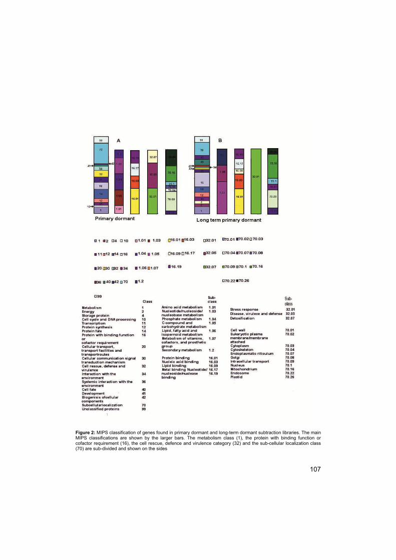
107
Figure 2: MIPS classification of genes found in primary dormant and long-term dormant subtraction libraries. The main MIPS classifications are shown by the larger bars. The metabolism class (1), the protein with binding function or cofactor requirement (16), the cell rescue, defence and virulence category (32) and the sub-cellular localization class (70) are sub-divided and shown on the sides

108
dormant library (Table 2A/2B). For the subcellular localization class (70) 16 genes
were found in the primary dormant library while 10 were found in the long term
primary dormant library (Table 2A/2B). Sub-dividing this class shows that genes
present in both libraries classify for more than one sub-class. The primary dormant
library had a greater representation in cytoplasm (70.03) (6 versus 3) and plastids
(70.26) (5 versus 1).
Cadman et al. (2006) have shown that the metabolism class (1) exhibited
major differences between primary dormant and long term primary dormant seeds,
which is in contrast to what was found here. The metabolism class was further
subdivided (Figure 2) to display possible differences between the two dormancy
types. Even though both libraries show almost the same number of genes in the
metabolism class (i.e. 5 and 4, see Table 2A and 2B), subdividing this class shows
that genes present in the primary dormant library classify in more than one sub-
class, while genes present in the long term primary dormant library are only
present in one subdivision each (Figure 2). Primary dormant seeds show more
genes in nucleotide/nucleoside/nucleobase metabolism (2 versus 0; cat. 1.03) and
phosphate metabolism (2 versus 0; cat. 1.04) (Figure 2). The long-term primary
dormant library shows one gene present in the sub-class of lipid/fatty
acid/isoprenoid metabolism (cat. 1.06) where no genes of the primary dormant
library are present. Only one tenth of the subtraction library was analysed, this
should be an appropriate amount to give a rough estimate of the genes
represented in each class and subclass; however, analysing more genes will give a
much more representable classification.
TAGGIT classification
MIPS classification can be unsatisfactory because the classification system does
not reflect specific processes previously shown to be important for seed biology
(Carrera et al., 2007). To distinguish genes encoding proteins related to seed

109
dormancy, development and germination, we analysed the Arabidopsis thaliana
orthologs of genes differentially expressed in the subtraction libraries with the
TAGGIT workflow. The TAGGIT workflow re-annotates gene lists according to
defined functions and provides more useful information related to seed biology
(Carrera et al., 2007), giving an indication of the developmental status of the seed.
However, only 4 genes of each subtraction library could be assigned to a category
in the TAGGIT classification. This could be due to the still small number of
genespresent in the TAGGIT workflow, the small number of genes present in the
subtraction libraries or to the fact that the genes present in the subtraction libraries
may not be seed specific. The newly assigned genes were found in the dormancy
category (AT3G50980, a dehydrin, in the primary dormant library), the translation
category (AT1G23290 and AT1G67430, both encoding for ribosomal proteins, in
the primary dormant library and AT1G07920 and AT4G00810, an elongation factor
and a ribosomal protein, respectively, in the long term primary dormant library) and
in the seed storage proteins/late embryogenesis abundant category (AT4G21020,
a LEA protein in the primary dormant and AT3G22490 and AT4G28520, a LEA
protein and cruciferin, respectively, in the long term primary dormant library
Stress related gene expression
In many organisms the regulation of dormancy is associated with abiotic stress
responses. The combination of metabolic/developmental arrest and stress
resistance/tolerance is a powerful strategy for the (long-term) survival of organisms
under adverse conditions (Lubzens et al., 2010). At the level of gene expression,
dormancy and stress responses are strongly linked and largely overlapping
(Cadman et al., 2006; Hilhorst, 1995). In general, competent organisms anticipate
a stressful period by shutting down their metabolism and engaging in a stress
response. One key method to resist stress is the expelling of water from dormant
tissue (Pnueli et al., 2002), as water has a potential to damage cells under adverse
physical conditions, e.g. freezing. Thus, the stress response may include
110
dehydration, but also the generation of protective compounds such as late
embryogenesis-abundant (LEA) proteins and anti-oxidants (Ingram and Bartels,
1996; Raynal, 1999; Singh, 2007). Other stress responses are engaged to harden
the seed against cold, salinity stress or pathogen attacks. In the subtraction library
a large group of stress related genes was found (Table 2A,B); in the primary
dormant library (Table 2A) one LEA gene was found (ortholog to AT4G21020), a
water and, consequently, freezing related stress gene Dehydrin XERO1 (ortholog
to AT3G50980), a salinity stress related gene, AVP1 (ortholog to AT1G15690) and
two pathogen related genes (orthologs to AT1G75830, LCR67/PDF1.1 and
AT5G44430, PDF1.2c). In the long term primary dormant library (Table 2B) one
LEA (ortholog to AT3G22490), one cold stress related gene FAD2 (ortholog to
AT3G12120), 2 heat-shock related genes (orthologs to AT1G07920, an elongation
factor, and AT3G44110, a DnaJ homologue 3/heat shock protein) and one
pathogen attack related RIN2 (ortholog to AT4G25230) were found.
Expression of randomly chosen stress related genes was analysed by qRT-PCR
under different dormancy conditions. Gene expression was analysed for primary
dormant seeds in the dark and in the light, long term primary dormant seeds in the
dark, secondary dormant seeds in the dark, sub-dormant seeds in the dark and in
the light. Secondary dormancy was included here, to analyse differences between
two varieties of deep dormancy; both are induced by a long imbibition time, but for
secondary dormant seeds nitrate was the imbibition medium. This would disclose
the effect that nitrate may have on gene expression associated with germination
and dormancy. Dormancy conditions were obtained in the same seed batch as in
which the subtraction library was made, apart from primary dormant 1dH(D) and
long term-primary dormant 10dH(D).
The LEA proteins are implicated in stabilizing and protecting cellular structures
(Shinozaki et al., 1999) and may also protect cells during the course of
de/rehydration (Pnueli et al., 2002). LEA are found in the primary dormant and
long-term primary dormant library and therefore are an interesting gene to
analyse.The LEA (ortholog to AT4G21020) found in the primary dormant
111
subtraction library and expressed higher in PD30d seeds of Arabidopsis,
(http://bar.utoronto.ca) with a ratio of PD24h vs. PD30d of 0.7 (Table 2A),
appeared to be constitutively expressed at low levels in Sisymbrium and did not
show significant expression differences between treatments (Figure 3). The water
and freezing stress tolerance related gene dehydrin XERO1 (Roberton and
Chandler, 1992) (ortholog to AT3G50980) has been proposed to contribute to
freezing tolerance by its protective effect on membranes (Puhakainen et al., 2004).
Dehydration related genes are also expressed during late embryogenesis when
plants naturally lose water, and ABA is thought to be involved in the expression of
deydrins (Roberton and Chandler, 1992). XERO1 was found in the primary
dormant library and qRT-PCR analysis showed that seeds imbibed in water for 1
day, light or dark (1dH (L) and 1dH(D)), exhibited higher transcript abundance than
the long term primary dormant and non-dormant seeds (10A, 10dN(D), 10dH(D)
and 1dN(L), 1dN(D)) (Figure 3). This could indicate that XERO1 expression levels
drop with increasing depth of dormancy or disappearance of dormancy. However,
the transcript abundance of 1A is not significantly different from 10A, as standard
errors are too big, showing there is no clear link between dormancy depth and
XERO1 expression levels. In Arabidopsis the ratio between PD24h and PD30d
was 1.2.
The salinity stress related gene AVP1 (ortholog to AT1G15690) is a vacuolar
proton pump that increases salt and drought tolerance of higher plants, by
increasing cation uptake and retaining water (Gaxiola et al., 2001). Salt and
drought tolerance are important for non-dormant and germinating seeds when
seedlings have to establish themselves under stress conditions. Expression of this
gene was reported to increase sharply in 10-d old seedlings of Arabidopsis when
exposed to various kinds of stress, including cold, osmotic, salt and heat, as well
as to ABA (Zeller et al., 2009). Thus, it is not surprising that, in anticipation of
transition to the seedling stage, the increased expression of AVP1 in primary
dormant seeds increased even further in non-dormant/germinating seeds, and

112
Figure 3: Expression of 3 genes selected from the primary-dormant subtraction library: Dehydrin XERO1 (XERO1) and Late embryogenesis abundant domain containing protein (LEA) and Vacuolar-type H+-pumping pyrophosphatase (AVP1), Eight treatments are shown: 1A: primary dormant seeds used in library, 10A: long-term dormant seeds used in library, 1d N(L): seeds imbibed for 1 day in KNO3 in the light (germinate),1dN (D): seeds imbibed for 1 day in KNO3 in the dark (do not germinate; need light), 1dH (L): seeds imbibed in water for 1 day in the light (do not germinate; need nitrate), 1dH (D): seeds imbibed in water for 1 day in the dark (do not germinate; need light and nitrate), 10dN (D): seeds imbibed in KNO3 for 10 days in the dark (do not germinate), 10dH (D): seeds imbibed in water for 10 days in the dark (do not germinate). Error bars represent standard deviations. a, b, c and d represent significant differences

113
Figure 4: Gene expression of 3 genes selected from the long-term primary dormant subtraction library: NAC domain containing protein 60 (ANACO60), Delta-12-fatty acid dehydrogenasse (FAD2), and RPM1 interacting protein 2 (RIN2). Treatments as in Fig. 3. Error bars represent standard deviations. a, b, c, and d represent the significant differences.
appeared to be under the control of light (Fig. 3). The ratio between 1A and 10A
was 2.95 and significant. Cadman et al. (2006) found a ratio of 1 for Arabidopsis.
Gene expression of some of the stress–associated genes in the long-term primary
dormant library was also analysed. Fatty acid desaturases aid in adjusting
membrane fluidity in response to temperature changes (Los and Murata, 1998;
Chapter 5). The ortholog to the FAD2 (AT3G12120) gene is involved in cold stress
114
tolerance in plants (Matos et al., 2007). FAD2 is a fatty acid desaturase 2 involved
the conversion of 18:1 to 18:2 fatty acids, allowing changes in temperature.
Although the Arabidopsis gene sets of Cadman et al., (2006) and Finch-Savage et
al., (2007) showed that PD30d seeds exhibitied a higher expression of FAD2 than
PD24h seeds (Table 2b) with a ratio of 0.8, qRT-PCR analysis showed no
differences in the FAD2 expression pattern between short and long term incubation
(Fig. 4) with a non-significant ratio between 1A and 10A of 0.57, however, it is a
similar type of response. Apart from 1A, expression seemed to be somewhat
higher in the 1d treatments but this was not significant. Interestingly 1A -1dH(D)
and 10A -10dH(D) have received a similar treatment, however expression of FAD2
is not comparable. Dormancy states have been obtained using different seed
batches for these treatments, and this might explain the difference in expression
levels. Standard deviation for 1dH(D) is very large and makes the results difficult to
interpret.
RPM1 interacting protein RIN2 (ortholog to AT4G25230) is involved in the
hypersensitive response to pathogens e.g. Pseudomonas syringae. RIN2 is
predominantly located in the plasma membrane where it contributes to the RPM1
hypersensitive response (Kawasaki et al., 2005). RIN2 was found in the long-term
primary dormant library. The analysed gene sets did not show differences in
expression of RIN2 between short and long term dormancy states (Figure 4). The
ratio between 1A and 10A was 0.67, and was not significant. The ratio found for
Arabidopsis was 1.1 (http://bar.utoronto.ca; table 2B). In the long-term primary
dormant library AT3G44290, a NAC domain containing protein 60, ANACO60, was
found. ANACO60 encodes for a plant-specific transcriptional regulator, expressed
in developmental stages (Ooka et al., 2003). qRT-PCR analysis (Figure 4) showed
that ANACO60 was not significantly differentially expressed between primary
dormant and long term primary dormant seeds, displaying a ratio of 0.4 (not
significant). This gene showed high expression in 4-d old seedlings of 5 out of 34
Arabidopsis accessions and may be related to their specific stress phenotypes
(Lempe et al., 2005). The qRT-PCR results showed that although stress responses
115
are involved in dormancy control, the genes selected from the subtraction library
were often not differentially expressed between treatments.
Conclusions
The cDNA subtraction library was chosen, over micro-array analysis, as the
preferred method to find transcriptional differences between primary dormant and
long term primary dormant seeds of Sisymbrium officinale. The rationale for this
choice was the prevention of cross-species hybridization difficulties. A major issue
with cross-species microarray hybridization is the effect of sequence divergence on
probe affinity, which is not only a function of phylogenetic distance. Due to
differences in sequence divergence rates, such effects are not uniform across all
genes. At present it is difficult to correct for such effects during the analysis of
microarray data (Bar-Or et al., 2007).
A cDNA subtraction library, however, poses a whole new set of difficulties; a large
number of clones needs to be sequenced in order to obtain an overall impression
of the transcriptome of a certain developmental state of the seed. Gene expression
of most genes needs to be verified, as some of the genes picked up may appear in
both the forward and reversed libraries. Not all genes picked up in one library were
found to be expressed differentially by qRT-PCR. This could be due to the
sensitivity of the method, or the genes may have been selected erroneously.
Despite these methodological problems, several genes (in the primary dormant
subtraction library orthologs AT1G75830, AT1G79260, AT3G43520, AT3G60980,
AT3G56340, AT3G56350, AT5G11710, AT5G60980, AT5G64840 and
AT5G66120, in the long term primary dormant subtraction library orthologs
AT1G13690, AT1G30880, AT1G35190, AT1G51200, AT1G79920, AT3G12120,
AT3G44290, AT3G54860, AT4G40030, AT5G22290 and AT5G24350) were
identified that are similarly expressed in Sisymbrium and Arabidopsis, although not
verified by PCR. The exact role of these genes in the regulation of primary and

116
long term primary dormancy can only be verified by functional analysis, e.g. by
reverse genetics.

117
Chapter 7
Summarizing Discussion
Dormancy, diapause, rest and quiescence are all terms that are in use to indicate a
state of (temporary) lowered metabolic activity (‘hypometabolic state’) in living
organisms. Dormancy occurs in many organisms from all kingdoms, ranging from
bears to mice and frogs, from fish to buds and seeds, and from crustaceans to
yeast and bacteria (Lubzens et al., 2010). Dormancy and stress responses are
closely associated. Many of the above organisms become dormant before they are
exposed to stress conditions. The timing of this may depend on developmental
phase (e.g. sexual versus asexual), seasonal rhythms, environmental cues and
food supply, among others. The common denominators appear to be controlled
reduction of metabolic activity, arrest of the cell cycle and the initiation of protective
mechanisms.
However, whether the underlying mechanisms are similar between different
organisms is a question yet to be answered. The first comparisons indicate
remarkable similarities (Hilhorst, 2008). Some of the similarities are metabolic
control mediated by AMP-activated protein kinase and the induction of protective
late embryogenesis abundant (LEA) and heat-shock (HSP) proteins (Hilhorst,
2008).
Seasonal dormancy cycling is a characteristic, so far, only found in seeds. Being
able to cycle in and out of dormancy allows the seed to survive decades or even
centuries in the soil, allowing germination to be spread over time but only when
optimal conditions are available, not only for germination but also for seedling
establishment. The ecological significance of dormancy cycling has long been
established (Karssen, 1982; Bouwmeester, 1990) but the physiological and
molecular mechanisms behind it have only been studied in recent years. In this

118
thesis we have attempted to further elucidate the mechanisms behind dormancy,
germination and dormancy cycling in seeds.
Germination stimulatory factors: light, nitrate and gibberellic
acid (GA)
Sisymbrium officinale seeds do not complete germination after imibibition in water
or nitrate when no light pulse has been given. The seeds need nitrate to become
non-dormant and light to start germination (Chapters 2, 3, 5, 6). Nitrate acts in part
by reducing the abscisic acid levels (Matakiadis et al., 2009), which at high levels
promote dormancy. If the light pulse is not given within a certain time frame, seeds
will become secondary dormant and the seed is not sensitive to light and nitrate
anymore. Other methods are then needed to break the dormancy (Derkx and
Karssen, 1993).
GAs can be used to circumvent the light and nitrate requirement for
germination (Chapter 6). GA biosynthesis has been suggested to be mediated by
light and nitrate as a cofactor (Hilhorst et al., 1986). Light both induces
Phytochrome B (PHYB) expression and activates the resulting PhyB protein. The
active PHYB protein reduces phytochrome-interacting factor 3-like 5 (PIL5) protein
levels. PIL5 suppresses the AtGA3ox1 and AtGA3ox2 and activates AtGA2ox2.
Reduction of PIL5 allows up-regulation of AtGA3ox1 and AtGA3ox2 and down-
regulation of AtGA2ox2 (Yamaguchi, 2008), although PIL5 does not bind
AtGA3ox1, AtGA3ox2 or AtGA2ox2 genes (Yamaguchi, 2008), suggesting that
additional components are required. ATGA3OX1 and ATGA3OX2 catalyse the
final step in the production of bioactive GAs, even without germination taking place
afterwards (Yamaguchi et al., 1998; Oh et al., 2006). Sisymbrium officinale seeds
are thought to possess seed coat imposed dormancy but we found that embryos of
dormant seeds lack growth force to overcome these restrictions (Chapter 3). It was
therefore concluded that dormancy of this species is also located in the embryo.
119
GAs are capable of inducing enzymes that hydrolyze the endosperm cell walls
(Debeaujon and Koornneef, 2000; Chen and Bradford, 2000; Nonogaki et al.,
2000; Manz et al., 2005) and in this way GAs could be involved in overcoming
restrictions imposed by the seed coat. On the other hand, GAs may also increase
the embryo growth potential.
Metabolism and respiratory activity during changes in
dormancy
For successful survival of the dormant seed, metabolic activity is reduced (see
above), to avoid rapid depletion of reserves. The metabolic state of the seed was
measured using electron paramagnetic resonance (EPR), with TEMPONE as a
spin probe, and the respiratory activity was measured with the Q2-test (Chapter 2).
Primary dormant seeds of Sisymbrium officinale showed hardly any metabolic or
respiratory activity, but this was increased considerably when dormancy was
broken by nitrate. However, when the light pulse was not given and the seeds had
become secondary dormant the metabolic activity slowed down. The reduction of
metabolism and respiratory activity in secondary dormant seeds did not reach the
same low activity of metabolism of primary dormant seeds. However, we propose
that after longer induction of secondary dormancy the metabolic activity and
respiratory activity will slow down even further, eventually likely to a similar level as
in primary dormant seeds.
Cytoplasmic changes during dormancy cycling
Dormancy has often been linked to the survival of the seed during periods of
abiotic stress. In order to survive the stress periods seeds may adapt their
cytoplasmic properties to the changing environment (Chapter 4). We tested this by
EPR, using carboxyl-proxyl (CP) spin probe. Vitrification temperatures for
cytoplasmic water were higher for dormant than for germinating seeds. The
120
vitrification temperature depends on the composition of the cytoplasm. In
germinating seeds the storage reserve mobilization has started, allowing more low-
molecular weight compounds in the cytoplasm. This causes a lower vitrification
temperature. Also, the germinating seeds had a less viscous cytoplasm than the
dormant seeds. The dormant seeds possess higher quantities of high-molecular
weight compounds in their cytoplasm than germinating seeds in which these have
been metabolized. The high molecular weight compounds have a greater influence
on the viscosity than the low-molecular weight compounds. However, the
differences in quantity of high and low-molecular weight compounds might not be
the main cause for differences in viscosity. Dijksterhuis (et al., 2007) also found
changes in cytoplasmic viscosity, in their research on fungal spores but they came
to the conclusion that the higher viscosity was not mainly caused by a high
concentration of solutes but by the high proportion of ordered PDT molecules in the
vicinity of macro-structures. Sun (2000) linked increased viscosity to reduced
metabolism of red oak seeds. We also found reduced metabolism in dormant
seeds (Chapter 2). A more viscous cytoplasm could protect dormant seed, making
them capable of surviving longer periods under abiotic stress, e.g. by protecting
cellular structures (Shinozaki et al, 1999) and preventing de/rehydration (Pneuli et
al, 2002).
The membrane hypothesis analyzed
Membranes have been implicated in dormancy over the past decades, as
membranes are the primary target for temperature perception (Hilhorst, 1998). A
hypothesis was put forward in which changes in responsiveness to naturally
occurring dormancy breaking factors like light and nitrate was a function of
membrane fluidity (Hilhorst, 1998). In Chapter 3 we showed that dormancy is
indeed a function of membrane fluidity. Primary dormant seeds of Sisymbrium
officinale appeared to have very rigid membranes, whereas breaking dormancy
increased membrane fluidity considerably. However, when non-dormant seeds
121
became secondary dormant membrane fluidity decreased again, but not to the
rigidity seen in primary dormant seeds. A more viscous membrane could affect the
functions of membrane proteins and could either be a response to stress, e.g.
because of the lipid-/protein-mediated activation of a signal pathway, or play a role
in cell injury, e.g. because some proteins such as respiratory and transport proteins
only function in a fluid membrane (Beney and Gervais, 2001).
Membranes are known to be able to adapt to changes in temperature by
homeoviscous adaptation (Sinesky, 1974), the mechanism whereby unsaturated
fatty acids aid in maintaining the membranes in a fluid state necessary for
biological functioning (Cyril et al., 2002). The introduction of double bonds into the
fatty acids is catalyzed by desaturases (Los and Murata, 1998). Desaturase
involvement in changes in membrane fluidity due to changes in dormancy was
tested in Chapter 3 (using Sisymbrium officinale) and Chapter 5 (using Arabidopsis
thaliana). Although FAD3, involved in the conversion of linoleic acid to linolenic
acid (Matos et al., 2005) could be related to dormancy and germination (Chapter
5), it was not found to be related to membrane fluidity in Arabidopsis thaliana
(Chapter 5). The ADS2 mutant, normally involved in the conversion of stearic acid
into oleic acid (Murata and Wada, 1995), on the other hand, did not show changes
in fatty acid composition, or a different germination phenotype, but it did show a
gene expression pattern with high expression in non-dormant seeds, enhanced by
light, and increased membrane fluidity (Chapter 5). In Sisymbrium officinale there
was no difference in fatty acid composition between different dormancy types,
indicating no involvement of desaturases in changes in membrane fluidity.
Although desaturase mutants seem to change germination behaviour and/or
membrane fluidity, desaturases are not the cause of these changes in wild type
seeds. Likely candidates for changing membrane fluidity are changes in the acyl
chain length (Ohlrogge and Browse, 1995) or lipid-protein interactions (Yeagle,
1989). The transition from the dormant to the non-dormant state is marked by
changes in the composition of membrane-associated proteins, mainly in the
plasma membrane enriched fractions (DiNola et al., 1989).

122
‘Old’ model, new perspective
In the Membrane hypothesis, as formulated by Hilhorst (1998), phytochrome
receptors may be (temporarily) associated with membranes. Temperature has a
profound influence on membrane fluidity which, on its turn, may determine the
magnitude of movement of receptors within the membranes. In one conformation
the receptors will be at the surface, available for nitrate and phytochrome to bind
whilst in the other conformation the receptors are within the membrane and
therefore not available for binding of nitrate and phytochrome. Homeoviscous
adaptation might be responsible for the changes in membrane fluidity (Chapter 1).
However, continuing research on various aspects of this hypothesis has altered
this view. Below the new findings will be discussed and a new hypothesis will be
put forward.
The phytochromes that are thought to be involved in germination are PhyA,
PhyB and PhyE (Nagy, 2000). PhyB is active over a range of temperatures and is
induced by red/far red light (Nagy, 2000), which makes it the most likely candidate
in our model. However, there is no evidence of membrane association for
phytochrome. Studies have shown that on photoconversion of Pr to Pfr the
phytochromes are translocated from the cytoplasm into the nucleus where they
form intranuclear bodies of various sizes in light quality dependent and light
quantity dependent fashion (Yamaguchi, 1999; Kircher, 1999/2002). However, a
significant portion of the phytochrome remains cytosolic (Nagy, 2000). The active
phytochrome protein reduces phytochrome-interacting factor 3-like 5 (PIL5) protein
levels. Reduction of PIL5 allows up-regulation of GIBBERELLIN 3-OXIDASE 1,
AtGA3ox1, and GIBBERELLIN 2-OXIDASE 2, AtGA3ox2 and down-regulation of
AtGA2ox2 (Yamaguchi, 2008). ATGA3OX1 and ATGA3OX2 catalyse the final step
in the production of bioactive GAs, even without germination taking place
afterwards, whereas AtGA2ox2 is a key gene in the degradation of GAs
(Yamaguchi et al., 1998; Oh et al., 2006).
In Arabidopsis high affinity nitrate transporter 2.7 (NRT2.7) has been
found to be the primary nitrate transporter (Chopin et al., 2007) and the dual affinity
123
nitrate transporter CHL1 has been reported to act as a sensor for nitrate (Ho et al.,
2009). Within the seed nitrate enhances the degradation of ABA via the
cytocrhome P450 8'-hydroxylase for ABA (CYP707A2: Matakiadis et al, 2009). The
nitrate and GA pathways are loosely linked by ABA. ABA and GA metabolism are
linked during seed germination, as seen by the increased GA biosynthesis in the
aba2-2 mutant and the suppression of GA biosynthesis by ABA during seed
germination (Seo et al., 2006). The reduction of ABA by nitrate allows the
biosynthesis of GA.
In Chapter 3 we found that membrane fluidity is indeed linked to dormancy and
germination, however, not via the saturation and un-saturation of fatty acids.
These data together lead to the conclusion that the membrane hypothesis, as it
exists, needs to be revised. Although membrane fluidity is involved in the
dormancy/germination process, phytochrome light receptors are not located within
the membrane. The nitrate transporter is located in the membrane, and changes in
membrane fluidity could thus alter the accessibility of the nitrate transporter.
However, as nitrate is not only a signal in the breaking of dormancy but also a
nutrient for plant growth and development (Crawford, 1995) this is an unlikely
option. So nitrate and light probably affect germination without a direct involvement
of membranes.
Other factors related (changes in) cell membranes might be involved that lead to
changes in dormancy and germination. Another gene involved in dormancy
regulation and containing transmembranes domain is DELAY OF GERMINATION
(DOG1) (Bentsink 2006). DOG1 accounts for genetic variation in natural
populations and has been characterized at the molecular level. DOG1, encodes a
novel plant-specific protein of unknown mode of action.DOG1 is functionally related
to primary dormancy in Arabidopsis Cvi. Its expression decreased with after-
ripening and during imbibition, which led to the conclusion that the gene is related
to dormancy induction during seed development (Bentsink, 2006). However, DOG1
expression may increase again upon induction of secondary dormancy, as was
shown in Arabidopsis Cvi (Finch-Savage et al., 2007). Because of the presence of

124
Figure 1: Model for the induction of germination. Under the influence of temperature (and with changes in dormancy) the membrane fluidity changes. The changes in membrane fluidity allow the (proteolytical) activation of a membrane anchored dormancy related transcription factor. The transcription factor (Tf) is transported to the nucleus where it activates the germination process. The germination process starts with an increase in metabolism, respiration and the mobilization of storage reserves. The effects of nitrate and light on dormancy and germination take place without membrane involvement and therefore are not included in this model
a transmembrane domain. DOG1 might be the link between the changes in
dormancy and membrane fluidity we found in this thesis. DOG1 could be
proteolytically activated by remodeling the membrane fluidity.
Seo et al. (2010) proposed a model whereby the membrane bound transcription
factor NTL6 is proteolytically activated by remodeling of the membrane fluidity.
NTL6 plays a role in cold-induced pathogen resistance. They found that the
regulation by NTL6 changes with changes in membrane fluidity and their
suggestion is that a cold-regulated membrane-associated metalloprotease may
serve as a primary regulator for NTL6-mediated cold signaling. The

125
transcriptionally active NTL6 form is translocated from the plasma membrane to the
nucleus where it triggers a pathogen resistance response.
Our suggestion is that a similar process may be present here (Figure 1). A
membrane anchored dormancy related protein/transcription factor, e. g. DOG1, is
activated by increasing the membrane fluidity. The activation of the transcription
factor can be proteolytcally or by releasing the whole transcription factor from the
membrane. In our findings the membrane fluidity was increased at extreme
temperatures (chapter 3) which makes proteolytical activation less likely than
activation by releasing the whole transcription factor. The activated form is
transported to the nucleus where it activates the germination process. The
germination process includes the increase in metabolism (Chapter 2) and
respiration (Chapter 2) and the mobilization of storage reserves (Chapter 2).
Mobilization of storage reserves leads to a decrease in cytoplasmic viscosity as
found here in Chapter 4.

126

127
References
Alboresi A., Gestin C., Leydecker M.-T., Bedu M., Meyer C., Truong H.-N.
2005. Nitrate, a signal relieving seed dormancy in Arabidopsis. Plant, Cell and
Environment 28: 500-512.
Alonso J. M., Stepanova A. N., Leisse T. J., Kim C. J., Chen H., Shinn P.,
Stevenson D. K., Zimmerman J., Barajas P., Cheuk R., Gadrinab C., Heller C.,
Jeske A., Koesema E., Meyers C. C., Parker H., Prednis L., Ansari Y., Choy N.,
Deen H., Geralt M., Hazari N., Hom E., Karnes M., Mulholland C., Ndubaku R.,
Schmidt I., Guzman P., Aguilar-Henonin L., Schmid M., Weigel D., Carter D.
E., Marchand T., Risseeuw E., Brogden D., Zeko A., Crosby W. L., Berry C. C.,
Ecker J. R. 2003 Genome-wide insertional mutagenesis of Arabidopsis thaliana.
Science 301: 653-657.
Bahin E., Bailly C., Sotta B., Kranner I., Corbineau F., Leymarie J. 2011.
Crosstalk between reactive oxygen species and hormonal signalling pathways
regulates grain dormancy in barley. Plant Cell & Environment 34: 980-933.
Bailly C., El-Maarouf-Bouteau H., Corbineau F. 2008. From intracellular
signaling networks to cell-death: the dual role of reactive oxygen species in seed
physiology. Comptes Rendus Biologies 331: 806-814.
Bar-Or C., Czosnek H. and Koltai H. 2007. Cross species microarray
hybridizations: developing a tool for studying species diversity. Trends in Genetics
23, 200-207.
Baskin J. M., Baskin C. C. 2004. A classification system for seed dormancy. Seed
Science Research 14: 1-16.
Batak I., Devic M., Gibal Z., Grubišić D., Poff K. L., Konjević R. 2002. The
effects of potassium nitrate and NO-donors on phytochrome A- and phytochrome
B-specific induced germination of Arabidopsis thaliana seeds. Seed Science
Research 12: 253-259.

128
Beney L., Gervais P. 2001. Influence of the fluidity of the membrane on the
response of microorganisms to environmental stresses. Applied microbiology
biotechnology 57: 34-42.
Bentsink L., Jowett J., Hanhart C. J., Koornneef M. 2006. Cloning of DOG1, a
quantatitative trait locus controlling seed dormancy in Arabidopsis. Proceedings of
the National Academy of Sciences, USA 103: 1742-1747.
Benatti C. R, Feitosa E., Fernandez R. M., Lamy-Freund M., T. 2001. Structural
and thermal characterization of dioctadecyldimethylammonium bromide
dispersions by spin labels. Chemistry and Physics of Lipids 111: 93-104.
Bewley J. D. 1997. Seed germination and dormancy. The Plant Cell 9: 1055-1066.
Bianconi M. L., do Amaral A. T., Schreier S. 1988. Use of membrane spin label
spectra to monitor rates of reaction of partitioning compounds: hydrolysis of a local
anesthetic analog. Biochemical and Biophysical Research Communications 152:
344-350.
Bobko A. A., Kirilyuk I. A., Grigor’ev I. A., Zweier J. L., Khramtsov V. V. 2007.
Reversible reduction of nitroxides to hydroxylamines: Roles for ascorbate and
glutathione. Free Radical Biology and Medicine 42: 404-412.
Bouwmeester H. J. 1990. The effect of environmental conditions on the seasonal
dormancy pattern and germination of weed seeds. PhD thesis, Wageningen
University, The Netherlands: Agricultural University
Bouwmeester, H. J. and Karssen, C. M. 1992. The dual role of temperature in
the regulation of the seasonal changes in dormancy and germination of seeds of
Polygonum persicaria L. Oecologia 90: 88-94
Bouwmeester H. J., Karssen C. M. 1993. Annual changes in dormancy and
germination in seeds of Sisymbrium officinale (L.) Scop. New Phytologist 124: 179-
191.
Bradford, K.J. 2005. Threshold models applied to seed germination ecology. New
Phytologist 165: 338-341.
129
Buitink J., Claessens M.M.A.E., Hemminga M. A., Hoekstra F. A. 1998.
Influence of water content and temperature on molecular mobility and intracellular
glasses in seeds and pollen. Plant Physiology 118: 531-541
Buitink, J., Hemminga, M.A., Hoekstra, F.A. 2000. Is there a role for
oligosaccharides in seed longevity? An assessment of the intracellular glass
stability. Plant Physiology 122: 1217-1224.
Buitink J., Leprince O., Hemminga M. A., Hoekstra F. A. 2000b. Molecular
mobility of the cytoplasm: an approach to describe and predict the lifespan of dry
germplasm. Proceedings of the National Academy of Sciences of the United States
of America. 97: 2385-2390.
Buitink J., Van den Dries I. J., Hoekstra F. A., Alberda M., Hemminga M. A.
2000a. High critical temperature above Tg may contribute to the stability of
biological systems. Biophysical Journal 79: 1119-1128
Buitink J., Walters-Vertucci C, Hoekstra F. A., Leprince O. 1996. Calorimetric
properties of dehydrating pollen: analysis of a desiccation-tolerant and intolerant
species. Plant physiology 111: 235-242.
Buitink J., Walters C., Hoekstra F. A., Crane J. 1998. Storage behavior of Typha
latifolia pollen at lower water contents: interpretation on the basis of water activity
and glass concepts. Physiologia Plantarum 103: 145-153.
Cadman S. C., Toorop P. E., Hilhorst H. W. M., Finch-Savage W. E. 2006. Gene
expression profiles of Arabidopsis Cvi Seeds during dormancy cycling indicate a
common underlying dormancy control mechanism. The Plant Journal 46: 805-822
Carrera E., Holman T., Medhurst A., Peer W., Schmuths H., Footitt S.,
Theodoulou F. L., Holdsworth M. J. 2007. Gene expression profiling reveals
defined functions of the ATP-binding cassette transporter COMATOSE late in
phase II of germination. Plant Physiology 143: 1669-1679.
Casal J. J., Sanchez R. A. 1998. Phytochromes and seed germination. Seed
Science Research 8: 317-329.

130
Chapman D. A., Killian G. J., Gelerinter E., Jarrett M. T. 1985. Reduction of the
spin-label TEMPONE by ubiquinol in the electron transport chain of intact rabbit
spermatozoa. Biology of Reproduction 32: 884-894
Chen F., Bradford K. J. 2000. Expression of an expansin is associated with
endosperm weakening during tomato seed germination. Plant Physiology 124:
1265-1274.
Chen K., Swartz H. M. 1988. Oxidation of hydroxylamines to nitroxide spin labels
in living cells. Biochimica et Biophysica Acta 970: 270-277.
Chibani K., Ali-Rachedi S., Job C., Job D., Jullien M., Grappin P. 2006.
Proteomic analysis of seed dormancy in Arabidopsis. Plant Physiology 142: 1493-
1510
Chopin F., Orsel M., Dorbe M.F., Chardon F., Truong H-N., Miller A. J., Krapp
A., Daniel-Vedele F. 2007. The Arabidopsis ATNRT2.7 nitrate transporter controls
nitrate content in seeds. The Plant Cell 19: 1590-1602.
Collados R., Andreu V., Picorel R., Alfonso M. 2006. A light-sensitive
mechanism differently regulates transcription and transcript stability of ω3 fatty-
acid desaturases (FAD3, FAD7 and FAD8) in soybean photosynthetic cell
suspensions. FEBS letters 20: 4934-4940.
Crawford, N. M. 1995. Nitrate: nutrient and signal for plant growth. The Plant Cell
7: 859-868.
Cyril, J., Powell, G. L., Duncan, R. R., Baird, W. V. 2002. Changes in membrane
polar lipid fatty acids of seashore paspalum in response to low temperature
exposure. Crop science 42: 2031-2037.
Danks H. V. 200. Dehydration in dormant insects. Journal of Insect Physiology 46:
837-852.
Debeaujon, I., Koornneef, M. 2000. Gibberellin requirement for Arabidopsis seed
germination is determined both by testa characteristics and embryonic abscisic
acid. Plant physiology 122: 415-424
131
De los Reyes B. G., Gibbons M. M., Varma T. S. N., McGrath J. M., Halgren R.,
Redus M. 2003. A snapshot of the low temperature stress transcriptome of
developing rice seedlings (Oryza sativa L.) via ESTs from subtracted cDNA library.
Theoretical and Applied Genetics 107: 1071-1082
Derkx M. P. M., Karssen C. M. 1993a. Changing sensitivity to light and nitrate but
not to gibberellins regulates seasonal dormancy patterns in Sisymbrium officinale
seeds. Plant, Cell and Environment 16: 469-479.
Derkx, M. P. M. and Karssen, C. M. 1994. Are seasonal dormancy patterns in
Arabidopsis thaliana regulated by changes in seed sensitivity to light, nitrate and
gibberellin? Annals of Botany 73: 129-136
Derkx M. P. M., Karssen C. M. 1993b. Effects of light and temperature on seed
dormancy and gibberellin-stimulated germination in Arabidopsis thaliana: studies
with gibberellin-deficient and -insensitive mutants. Physiologia Plantarum 89: 360-
368.
Derkx M. P. M., Smidt W. J. van der Plas L. H. W., Karssen C. M. 1993.
Changes in dormancy in Sisymbrium officinale do not depend on changes in
resperatory activity. Physiologia Plantarum 89: 707-718.
Derkx M. P. M., Vermeer E., Karssen C. M. 1994. Gibberellins in seeds of
Arabidopsis thaliana: biological activities, identification and effects of light and
chilling on endogenous levels. Plant Growth Regulation 15: 223-234.
Dijksterhuis J., Nijsse J., Hoekstra F. A., Golovina E. A. 2007. High viscosity
and anisotropy characterize the cytoplasm of fungal dormant stress-resistant
spores. Eukaryotic cell 6: 157-170
Di Nola, L. and Mayer, A. M. 1986. The effect of temperature of imbibition and
germination on the lipid and fatty acid content and compositionin phospholipids of
embryonic axes from pea seeds. Phytochemistry 25: 2725-2731.
DiNola L., Mischke C. F., Taylorson R. B. (1989). Changes in the composition
and synthesis of proteins in cellular membranes of Echinochloa crus-galli (L.)

132
Beauv. Seeds during the transition from dormancy to germination. Plant physiology
92: 427-433.
Downie B., Bewley J. D. 2000. Soluble sugar content of white spruce (Picea
glauca) seeds during and after germination. Physiologia plantarum 110: 1-12.
Donohue K., Heschel M. S., Chiang G. C. K., Butler C. M., Barua D. 2007.
Phytochrome mediates germination responses to multiple seasonal cues. Plant,
Cell and Environment 30: 202-212.
De Vivrille J. D., Cantrell C., Bousquet A.-L., Hoffelt M., Tenreiro A.-M., Vaz
Pinto V., Arrabaça J. D., Caiveau O., Moreau F., Zachowiski A. 2002.
Homeoviscous and functional adaptations of mitochondrial membranes to growth
temperature in soybean seedlings. Plant, Cell & Environment 25: 1289-1297.
Fan T. W. M. 1996. Recent advances in profiling plant metabolites by multi-nuclear
and multi-dimensional NMR. Current topics in plant physiology. Nuclear resonance
in plant biology. Y. Shachar-Hill and P. E. Pfeffer (Eds.). American Society of Plant
Physiologists 16: 181-254.
Finch-Savage W. E., Cadman C. S., C., Toorop P. E., Lynn J. R., Hilhorst H.
W.H. 2007. Seed dormancy release in Arabidopsis Cvi by dry after-ripening, low
temperature, nitrate and light shows common quantitative patterns of gene
expression directed by environmentally specific sensing. The Plant Journal 51: 60-
78
Fontain O., Billard J. P., Huault C. 1995. Effect of glutathione on dormancy
breakage in barley seeds. Plant Growth Regulation 16: 55-58.
Footitt S., Slocombe S. P., Larner V., Kurup S., Yaosheng W., Larson T.,
Graham I., Baker A., Holdsworth M. 2002. Control of germination and lipid
mobilization by COMATOSE, the Arabidopsis homologue of human ALDP. EMBO
Journal 21: 2912-2922.
Footitt S., Marquez J., Schmuths H., Baker A., Theodoulou F. L., Holdsworth
M. 2006. Analysis of the role of COMATOSE and peroxisomal beta-oxidation in the

133
determination of germination potential in Arabidopsis. Journal of Experimental
Botany 57: 2805-2814.
Fuchs J., Groth N., Herrling T., Zimmer G. 1997. Electron paramagnetic
resonance studies on nitroxide radical 2,2,5,5-tetramethyl-4-piperidin-1-oxyl
(TEMPO) redox reactions in human skin. Free Radical Biology & Medicine 22: 967-
976.
Fukuchi-Mizutani M., Tasaka Y., Tanaka Y., Ashikari T., Kusumi T., Murata N.
1998. Characterization of ∆9 acyl-lipid desaturase homologues from Arabidopsis
thaliana. Plant Cell Physiology 39: 247-253.
Garciarrubio A., Legaria J. P., Covarrubias A. A. 1997. Abscisic acid inhibits
germination of mature Arabidopsis seeds by limiting the availability of energy and
nutrients. Planta 203: 182-187
Gaxiola R. A., Li J., Undurraga S., Dang L. M., Allen G. J., Alper S. L., Fink G.
R. 2001. Drought- and salt-tolerant plants result from overexpression of the
AVP1H+-pump. Proceedings of the National Academy of Sciences of the United
States of America 98: 11444-11449
Geisler-Lee J., O’Toole N., Ammar R., Provart N. J., Harvey Millar A., Geisler
A. 2007. A predicted interactome for Arabidopsis. Plant Physiology 145: 317-329.
Gibson S., Arondel V., Iba K., Somerville C. 1994. Cloning of a temperature-
regulated gene encoding a chloroplast ω-3 desaturase from Arabidopsis thaliana.
Plant Physiology 106: 1615-1621.
Gilbert H. F., 1995. Thiol/disulfide exchange equilibria and disulfide bond stability.
Biothiols Part A Monothiols and Dithiols, Protein Thiols, and Thiol Radicals.
Methods in Enzymology 251: 8-28.
Golovina E. A., Hoekstra F. A. 2002. Membrane behavior as influenced by
partitioning of amphiphiles during drying: a comparative study of anhydrobiotic
plant systems. Comparative Biochemistry and Physiology Part A 131: 545-558.

134
Golovina E. A., Hoekstra F. A. 2003. Acquisition of desiccation tolerance in
developing wheat embryos correlates with appearance of a fluid phase in
membranes. Plant, Cell and Environment 26: 1815-1826
Golovina E. A., Hoekstra F. A., Van Aelst A. C. 2001. The competence to
acquire cellular desiccation tolerance is independent of seed morphological
development. Journal of Experimental Botany 52: 1015-1027.
Golovina E. A., Tikhonov A. N., Hoekstra F. A. 1997. An electron paramagnetic
resonance spin-probe study of membrane-permeability changes with seed aging.
Plant Physiology 114: 383-389.
Golovina E. A., Van As H., Hoekstra F. A. 2010. Membrane chemical stability
and seed longevity. European Biophisics Journal 39: 657-668.
Gunstone F. D. 1993. High-resolution of 13C-NMR spectroscopy of lipids.
Advances in lipid methodology 2, ed. W. W. Christie. Dundee: Oily Press 1-68.
Hendricks S. B., Taylorson R. B. 1976. Variation in germination and amino acid
leakage of seeds with temperature related to membrane phase change. Plant
Physiology 58: 7-11
Hendricks S. B., Taylorson R. B. 1978. Dependence of phytochrome action in
seeds on membrane organization. Plant Physiology 61: 17-19.
Hendricks S. B., Taylorson R. B. 1979. Dependence of thermal responses of
seeds on membrane transitions. Proceedings of the National Academy of Sciences
of the United States of America 76: 778-781.
Heschel M. S., Selby J., Butler C., Whitelam G. C., Sharrock R. A., Donohue K.
2007. A new role for phytochromes in temperature dependent germination. New
Phytologist 174: 735-741.
Hilhorst, H.W.M. 1995. A critical update on seed dormancy. I. Primary dormancy.
Seed Science Research 5: 61-73.

135
Hilhorst H. W. M. 1990a. Dose-response analysis of factors involved in
germination and secondary dormancy of seeds of Sisymbrium officinale. I.
Phytochrome. Plant Physiology 94: 1090-1095.
Hilhorst H. W. M. 1990b. Dose-response analysis of factors involved in
germination and secondary dormancy of seeds of Sisymbrium officinale. II. Nitrate.
Plant Physiology 94: 1096-1102.
Hilhorst H. W. M. 1998. The regulation of secondary dormancy. The membrane
hypothesis revisited. Seed Science Research 8: 77-90.
Hilhorst H. W. M 2008. Sleeping beauties, dormancy and resistance in harsh
environments: molecular, proteomic and metabolomic aspects – Berlin, Germany,
18-20 may 2008. Seed Science Research 18: 185-187.
Hilhorst H. W. M., Karssen C. M. 1988. Dual effect of light on the gibberellin- and
nitrate-stimulated seed germination of Sisymbrium officinale and Arabidopsis
thaliana. Plant Physiology 86: 591-597.
Hilhorst H. W. M., Karssen C. M. 1989. Nitrate reductase independent stimulation
of seed germination in Sisymbrium officinale L. (Hedge mustard) by light and
nitrate. Annals of Botany 63: 131-138.
Hilhorst H. W. M., Karssen C. M. 1992. Seed dormancy and germination: the role
of abscisic acid and gibberellins and the importance of hormone mutants. Plant
Growth Regulation 11: 225-238
Hilhorst H. W. M., Smitt A. I., Karssen C. M. 1986. Gibberellin-biosynthesis and –
sensitivity mediated stimulation of seed germination of Sisymbrium officinale by red
light and nitrate. Physiologia Plantarum 67: 285-290.
Ho C-H., Lin S-H., Hu H. C., Tsay Y. F. 2009. CHL1 functions as a nitrate sensor
in plants. Cell 6: 1184-1194
Hohmann S. 2002. Osmotic stress signalling and osmoadaption in yeasts.
Microbiology and Molecular Biology Reviews 66: 300-372.

136
Horiguchi G., Fuse T., Kawakami N., Kodama H., Iba K. 2000. Temperature-
dependent translational regulation of ER omega-3 fatty acid desaturase gene in
wheat root tips. Plant Journal 24: 805-813.
Ingram J. Bartels D. 1996. The molecular basis of dehydration tolerance in plants.
Annual Review of Plant Physiology and Plant Molecular Biology 47: 377-403.
Jie M., Mustafa J. 1997. High resolution nuclear magnetic resonance
spectroscopy- applications to fatty acids and triacylglycerols. Lipids 32: 1019-1034.
Jung K., Ristori S., Gallori E., Martini G. 1998. Stability of water-soluble and
lipid-soluble paramagnetic probes in Bacillus subtilis. Biochimica et Biophysica
Acta 1425: 387-397.
Kaplan J., Canonico P. G., Caspary W. J. 1973. Electron-spin resonance studies
of spin-labeled mammalian cells by detection of surface-membrane signals.
Proceedings of the National Academy of Sciences of the United States of America
70: 66-70.
Karssen C. M. 1982. Seasonal patterns of dormancy in weed seeds. Pp 243-270.
in Khan, (ed.) The physiology and biochemistry of seed development, dormancy
and germination. Amsterdam, Elsevier Biomedical Press
Kawasaki T., Nam J., Boyes D. C., Holt B. F., III, Hubert D. A., Wiig A., Dangl J.
L. 2005. A duplicated pair of Arabidopsis RING-finger E3 ligases contribute to the
RPM1- and RPS2-mediated hypersensitive response. The Plant Journal 44: 258-
270.
Keith A. D., Snipes W. 1974. Viscosity of the cellular protoplasm. Science 183:
666-668.
Kępczyński J., Bihun M., 2002. Induction of secondary dormancy in Amaranthus
caudatus seeds. Plant Growth Regulation 38: 135-140
Kircher S., Kozma-Bognar L, Kim L., Adam E., Harter K., Schafer E., Nagy E.
1999 Light quality-dependent nuclear import of the plant photoreceptors
phytochrome A and B, Plant Cell 11 1445–1456.
137
Kircher S., Gil P., Kozma-Bognar L., Fejes E., Speth V.,Husselstein-Muller T.,
Bauer D., Adam E., Schafer E., Nagy F. 2002 Nucleocytoplasmic partitioning of
the plant photoreceptors phytochrome A, B, C, D, and E is regulated differentially
by light and exhibits a diurnal rhythm, Plant Cell 14. 1541–1555
Klimov D. K., Thirumalia D. 1997. Viscosity dependence of the folding rates of
proteins. Physical review letters 79: 317-320.
Kou T. M., VanMiddlesworth J. F., Wolf W. J. 1988. Content of raffinose
oligosacharides and sucrose in various plants. Journal of agricultural and food
chemistry. 36: 32-36.
Kranner I. 1998. Determination of glutathione, glutathione disulfide, and two
related enzymes, glutathione reductase and glucose-6-phosphate dehydrogenase,
in fungal and plant cells. In Varma A. (ed.) Mycorrhiza Manual, Springer Verlag,
227-241.
Kranner I., Grill D. 1996. Significance of thiol-disulfide exchange in resting stages
of plant development. Botanica Acta 109: 8-14.
Kumler P. L., Boyer R. F. 1976. ESR studies of polymer transitions.
Macromolecules 9: 903-910.
Kuznetsov A. N., Wasserman A. M., Volkov A. J. J., Korst N. N. 1971.
Determination of rotational correlation time of nitric oxyderadicals in a viscous
medium. Chemical Physics Letters 12: 103-106
Langis R., Steponkus P. L. 1990. Cryopreservation of Rye protoplast by
vitrification. Plant physiology 92: 666-671.
Leopold A. C., Sun W. Q., Bernal-Lugo I. 1994. The glassy state in seeds:
analysis and function. Seed Science Research 4: 267-274.
Leubner-Metzger G. 2001. Brassinosteroids and gibberellins promote tobacco
seed germination by distinct pathways. Planta 213: 758-763.
Los D. A., Murata N. 1998. Structure and expression of fatty acid desaturases.
Biochimica et biophysica acta 1394: 3-15.

138
Lubzens, E., Cerdà, J. and Clark, M. (Eds.) 2010. Dormancy and resistance in
harsh environments. Topics in Current Genetics 21. Springer-Verlag, Berlin
Heidelberg
Manz B., Müller K., Kucera B., Volke F., Leubner-Metzger G. 2005. Water
uptake and distribution in germinating Tobacco seeds investigated in vivo by
nuclear magnetic resonance imaging. Plant Physiology 138: 1538-1551.
Marsh D. 1981. Electron spin resonance: spin labels. In: Grell, E (Ed.), Membrane
spectroscopy. Molecular biology, biochemistry and biophysics, Vol. 31. Springer
Verlag, Berlin, 51-142.
Matakiadis T., Alboresie A., Jikumaru Y., Tatematsu K., Pichon O., Renou J-
P., Kamiya Y., Nambara E., Truong H-N. 2009. The Arabidopsis abscisic acid
catabolic gene CYP707A2 plays a key role in nitrate control of seed dormancy.
Plant Physiology 149: 949-960.
Matos, A. R., Hourton-Cabassa, C., Ciçek, D., Rezé, N., Arrrabaça, J. D.,
Zachowski, A., Moreau, F. 2007. Alternative oxidase involvement in cold stress
response of Arabidopsis thaliana fad2 and FAD3+ cell suspensions altered ion
membrane lipid composition. Plant & Cell Physiology 48: 856-865.
McIntyre G. I., Cessna A. J., Hsiao A. I. 1996. Seed dormancy in Avena Fatua:
Interacting effects of nitrate, water and seed coat injury. Physiologia Plantarum 97:
291-302.
Millar A. A., Smith M. A., Kunst L. 2000. All fatty acids are not equal:
discrimination in plant membrane lipids. Trends in plant science 5: 95-101.
Minorsky, P. V. 1989. Temperature sensing by plants: a review and hypothesis.
Plant, Cell and Environment 12: 119-135.
Murata N., Los D. A. 1997. Membrane fluidity and temperature perception. Plant
Physiology 115: 875-879.

139
Murata N., Wada H. 1995. Acyl-lipid desaturases and their importance in the
tolerance and acclimatization to cold of cyanobacteria. Biochemical Journal 308: 1-
8.
Nabors M. W., Lang A. 1971. The growth physics and water relations of red-light
induced germination in lettuce seeds. I. embryos germinating in osmoticum. Planta
101: 1-25.
Nagy F., Kircher S., Schafer E. 2000. Nucleo-cytoplasmic partitioning of the plant
photoreceptors phytochromes, Seminars in Cell & Developmental Biology 11: 505–
510
Nagy, F. and Schäfer, E. 2000. Nuclear and cytosolic events of light-induced,
phytochrome-regulated signaling in higher plants. EMBO Journal 19: 157-163.
Nikolaeva, M. G. 2004. On criteria to use in studies of seed evolution. Seed
Science Research 14: 315-320.
Nishida I., Murata N. 1996. Chilling sensitivity in plants and cyanobacteria: the
crucial contribution of membrane lipids. Annual Review of Plant Physiology and
Plant Molecular Biology 47: 541-568.
Nonogaki, H., Gee, O. H., Bradford, K. J. 2000. A germination-specific endo-β -
mannanase gene is expressed in the micropylar endosperm cap of tomato seeds.
Plant physiology 123: 1235-1245.
Ogawa M., Hanada A., Yamauchi Y., Kuwahara A., Kamiya Y., amaguchi S.
2003. Gibberellin biosynthesis and response during Arabidopsis seed germination.
Plant Cell 15: 1591-1604.
Oh E., Yamaguchi S., Kamiya Y., Bae G., Chung W-II., Choi G. 2006. Light
activates the degradation of PIL5 protein to promote seed germination through
gibberellin in Arabidopsis. Plant Journal 47: 124-139.
Ohlrogge J., Browse J. 1995. Lipid biosynthesis. The Plant Cell 7: 957-970.
Ooka H., Satoh K., Doi K., Nagata T., Otoma Y., Murakami K., Matsubara K.,
Osato N., Kawai J., Carninci P., Hayashizaki Y., Suzuki K., Kojima K.,
140
Takahara Y., Yamamoto K., Kikuchi S. 2003. Comprehensive analysis of NAC
family genes in Oryza sativa and Arabidopsis thaliana. DNA Research 10: 239-247
Penfield S. 2008. Temperature perception and signal transduction in plants. New
Phytologist 179: 615-628.
Penfield S., Hall A. 2009. A role for multiple circadian clock genes in the response
to signals that break seed dormancy in Arabidopsis. The Plant Cell 21: 1722-1732.
Pnueli L., Hallak-Herr E., Rozenberg M., Cohen M., Goloubinoff P., Kaplan A.,
Mittler R. 2002. Molecular and biochemical mechanisms associated with dormancy
and drought tolerance in the desert legume Retama raetem. The Plant Journal 31:
319-330.
Poghyosyan Z. P., Haralampidis K., Martsinkovskaya A. I., Murphy D. J.,
Hatzopoulos P. 1999. Developmental regulation and spatial expression of a
plastidial fatty acid desaturase from Olea europaea. Plant Physiology and
Biochemistry 37: 109-119.
Powell A.D., Dulson J., Bewley J.D. 1984. Changes in germination and
respiratory potential of embryos of dormant Grand rapids lettuce seeds during
long-term imbibed storage, and related changes in the endosperm. Planta 162: 40-
45.
Powell A. D., Leung D. W. M., Bewley J. D. 1983. Long-term storage of dormant
Grand Rapis lettuce seeds in the imbibed state: physiological and metabolic
changes. Planta 159: 182-188.
Potts M. 1994. Desiccation tolerance of prokaryotes. Microbiology and Molecular
Biology Reviews. 58: 755-805.
Puhakainen T., Hess M. W., Mäkelä P., Svensson J., Heino P., Palva E. T.
2004. Overexpression of multiple dehydrin genes enhances tolerance to freezing
stress in Arabidopsis. Plant Molecular Biology 54: 743-753.
Raison J. K., Berry J. A., Armond P. A., Pike C. S. 1980. Membrane properties
in relation to the adaptation of plants to temperature stress. In: Turner N.C.,

141
Kramer P.J. (Eds.) Adaptation of plants to water and high temperature stress. New
York, Wiley, pp. 261-273.
Roberton M., Chandler P. M. 1992. Pea dehydrins: identification, characterisation
and expression. Plant Molecular Biology 19: 1031-1044.
Roberts E. H., Ellis R. H. 1989. Water and seed survival. Annals of Botany 63: 39-
52.
Rohde A., Bhalerao R. P. 2007. Plant dormancy in the perennial context. Trends
in Plant Science 12: 217-223.
Ruepp A., Zollner A., Maier D., Albermann K., Hani J., Mokrejs M., Tetko I.,
Güldener U., Mannhaupt G., Münsterkötter M., Mewes H. W. 2004. The FunCat,
a functional annotation scheme for systematic classification of proteins from whole
genomes. Nucleic Acid Research 32: 5539-5543.
Rutar V. 1989. Magic angle sample spinning NMR spectroscopy of liquids as a
non-destructive method for studies of plant seeds. Journal of Agricultural and Food
Chemistry 37: 67-70.
Ryanal M., Guilleminot J., Gueguen C., Cooke R., Delseny M., Gruber V. 1999.
Structure, organization and expression of two closely related novel Lea (late
embryogenesis-abundant) genes in Arabidopsis thaliana. Plant Molecular Biology
40: 153-165.
Sanson A., Ptak M., Rigaud J. L., Gary-Bobo C. M. 1976. An ESR study of the
anchoring of spin labeled stearic acid in lecithin multilayers. Chemistry and Physics
of Lipids 17: 435-444.
Sato N., Murata N. 1980. Temperature shift-induced responses in lipids in the
blue-green alga, Anabaena variabilis. The central role of
diacylmonogalactosylglycerol in thermo-adaptation. Biochimica et Biophysica Acta
619: 354-366
Seo M., Hanada A., Kuwahara A., Endo A., Okamoto M., Yamauchi Y., North
H., Marion-Poll A., Sun T-P., Koshiba T., Kamiya Y., Yamaguchi S., Nambara

142
E. 2006. Regulation of hormone metabolism in Arabidopsis seeds: phytochrome
regulation of abscisic acid metabolism and abscisic acid regulation of gibberellins
metabolism. The Plant Journal 48: 354-366.
Seo P. J. Kim M. J., Song J-S., Kim Y-S., Kim H-J., Park C-M. 2010. Proteolytic
processing of an Arabidopsis membrane bound NAC transcription factor is
triggered by cold-induced changes in membrane fluidity. Biochemical journal 427:
359-367.
Sinesky M. 1974. Homeoviscous adaptation - A homeostatic process that
regulates viscosity of membrane lipids in Escherichia coli. Proceedings of the
National Academy of Sciences of the United States of America 71: 522-525
Sheffer M., Fried A., Gottlieb H. E., Tietz A., Avron M. 1986. Lipid composition of
the plasma-membrane of the halotolerant alga, Dunaliella salina. Biochemica et
Biophysica Acta 857: 165-172
Shinozaki K., Yamaguchi-Shinozaki K., Liu Q., Kasuga M., Ichimura K.,
Mizoguchi T., Urao T., Miyata S., Nakashima K., Shinwari Z. K., Abo H.,
Sakuma Y., Ito T., Seki M. 1999. In: Smallwood, M. F., Calvert, C. M., Bowles, D.
J. (Eds.) Plant responses to environmental stress, Oxford, BIOS Scientific
Publishers.
Singh S., Cornilescu C. C., Tyler R. C., Cornilescu G., Tonelli M., Lee M. S.,
Markley J. L. 2007. Solution structure of a late embryogenesis abundant protein
(LEA14) from Arabidopsis thaliana, a cellular stress-related protein. Protein
Science 14: 2601-2609
Somerville C., Browse J. 1996. Dissecting desaturation: plants prove
advantageous. Trends in Cell Biology 6: 148-153.
Stubbs C. D., Smith A. D. 1984. The modification of mammalian membrane
polyunsaturated fatty acid composition in relation to membrane fluidity and
function. Biochimica et Biophysica Acta 779: 89-137
143
Sun W. Q. 2000. Dielectric relaxation of water and water-plasticized biomolecules
in relation to cellular water organization, cytoplasmic viscosity, and desiccation
tolerance in recalcitrant seed tissues. Plant physiology 124: 1203-1216.
Sun W. Q., Irving T. C., Leopold T. C. 1994. The role of sugar, vitrification and
membrane phase transition in desiccation tolerance. Physiologia Plantarum 90:
621-628.
Sun W. Q., Leopold A. C. 1997. Cytoplasmic vitrification and survival of
anhydrobiotic organisms. Comparative Biochemistry and physiology 117A: 327-
333.
Swartz H. M. 1987. Use of nitroxides to measure redox metabolism in cells and
tissues. Journal of the Chemical Society, Faraday Transactions 1: Physical
Chemistry in Condensed phases 83: 191-202.
Taylorson R. B. 1972. Phytochrome controlled changes in dormancy and
germination of buried weed seeds. Weed Science 20: 417-422.
Turchiello R. F., Juliano L., Ito A. S., Lamy-Freundl M. T. 2000. How bradykinin
alters the lipid membrane structure: a spin label comparative study with bradykinin
fragments and other cations. Biopolymers 54: 211-221.
Vigh L., Maresca B., Harwood L. J. 1998. Does the membrane’s physical state
control the expression of heat shock and other genes? Trends in Biochemical
Sciences 23: 369-374
Vleeshouwers L. M., Bouwmeester H. J., Karssen C. M. 1995. Redefining seed
dormancy: An attempt to integrate physiology and ecology. Journal of Ecology 83:
1031-1037
Voelker T, Kinney A. J. 2001. Variations in the synthesis of seed-storage lipids.
Annual Review of Plant Physiology and Plant Molecular Biology 52: 335-361.
Wan C.-Y., Wilkins T.A. 1994. A modified hot borate method significantly
enhances the yield of high-quality RNA from cotton (Gossypium hirsutum L.).
Analytical Biochemistry 223: 7–12.

144
Wang S. Y., Faust M. 1994. Changes in the antioxidant system associated with
budbreak in ‘Anna’ apple (Malus domestica Borkh.) buds. Journal of the American
Society for Horticultural Science 119: 735-741.
Whitelam G. C., Devlin P. F. 1997. Roles of different phytochromes in Arabidopsis
photomorphogenesis. Plant, Cell & environment 20: 752-758.
Winter D., Vinegar B., Nahal H., Ammar R., Wilson G. V., Provart N. J. 2007. An
“electronic fluorescent pictograph” browser for exploring and analyzing large-scale
biological data sets. PloS ONE 8: 1-12.
Wojtyla L., Garnczarska M., Zalewski T., Bednarski W., Ratjczak, L., Jurga S.
2006. A comparative study of water distribution, free radical production and
activation of antioxidative metabolism in germinating pea seeds. Journal of Plant
Physiology 163: 1207-1220.
Wolfe J., Bryant G. 1999. Freezing, drying, and/or vitrification of membrane
solute-water systems. Cryobiology 39: 103-129.
Yamaguchi S. 2008. Gibberellin metabolism and its regulation. Annual Review of
Plant Biology 59: 225-251.
Yamaguchi S., Smith M. W., Brown R. G. S., Kamiya Y., Sun T. P. 1998.
Phytochrome regulation and differential expression of gibberellin 3beta-
hydroxylase genes in germinating Arabidopsis seeds. Plant Cell 10: 2115-2126.
Yamauchi, Y., Ogawa, M., Kuwahara A., Hanada A., Kamiya Y., and
Yamaguchi S. 2004. Activation of gibberellin biosynthesis and response pathways
by low temperature during imbibition of Arabidopsis thaliana seeds. The Plant Cell
16: 367-378.
Yeagle P. L. 1989. Regulation of membrane-function through composoition,
structure, and dynamics. Annals of the New York academy of sciences 568: 29-34.
Zeller, G.,Henz, S.R., Widmer, C.K., Sachsenberg, T., Rätsch, G., Weigel, D.
and Laubinger, S. 2009. Stress-induced changes in the Arabidopsis thaliana

145
transcriptome analyzed using whole-genome tiling arrays. The Plant Journal 58:
1068–1082.

146
147
Summary
The life cycle of most plants starts, and ends, at the seed stage. In most species
mature seeds are shed and dispersed on the ground. At this stage of its life cycle
the seed may be dormant and will, by definition, not germinate under favourable
conditions (Bewley, 1997).
Seasonal dormancy cycling is a characteristic found in plant seeds. Being able to
cycle in and out of dormancy allows the seed to survive decades or even centuries,
allowing germination to be spread over time, but only when optimal conditions are
available, not only for germination but especially for seedling establishment. In this
thesis we have attempted to further elucidate the mechanisms behind dormancy,
germination and dormancy cycling.
Sisymbrium officinale seeds need nitrate and light to start germination (Chapter 2,
3, 4, 6). Nitrate acts in part by reducing the abscisic acid (ABA) levels (a plant
hormone that elevates dormancy levels). The action of light and nitrate can also be
reached by applying gibberellins (GAs) to the seeds (Chapter 2, 3, 4, 6). GAs are
capable of inducing enzymes that hydrolyze the ensdosperm walls (Debeaujon and
Koornneef, 2000; Chen and Bradford, 2000; Nonogaki et al., 2000; Manz et al.,
2005) In this way GAs could be involved in lowering the physical restrictions
imposed by the resistance of the seed coat and the endosperm. On the other hand,
GAs may also increase the embryo growth potential.
For successful survival of the dormant seed, metabolic activity is reduced to avoid
rapid depletion of reserves. The metabolic activity of the seed was measured using
electron paramagnetic resonance (EPR), with TEMPONE as a spin probe, and the
respiratory activity was measured with the Q2-test (Chapter 2).We showed that
primary dormancy was accompanied by hardly any metabolic or respiratory activity,
and this increased considerably when dormancy was broken by nitrate. However,
148
when the light pulse was not given and the seeds had become secondary dormant
the metabolic activity slowed down.
Regulation of dormancy is tightly linked with abiotic stress factors from the
environment. The regulation and survival of the seed under stress conditions is
largely dependent on the composition of the cytoplasm. We tested this by EPR,
using carboxyl-proxyl (CP) spin probe (Chapter 4). The primary dormant and sub-
dormant seeds possessed a higher viscosity than the germinating seeds. The
viscosity of secondary dormant seeds appeared intermediate; however, the ease at
which the vitrified water melted was similar to that of primary dormant seeds. As a
result of the differences in viscosity, the temperature of vitrified water melting
differed between the different dormancy states. The changes in cytoplasmic
viscosity and vitrified water melting may be linked to changes in metabolism and
the content of high molecular weight compounds.
As membranes are the primary target for temperature perception, they are often
implicated in regulating dormancy. Therefore, Hilhorst (1998) put forward a
hypothesis in which changes in responsiveness to dormancy breaking factors like
nitrate and light was a function of cellular membrane fluidity. In Chapter 3 we
indeed showed that dormancy is a function of membrane fluidity. Primary dormant
seeds of Sisymbrium officinale appeared to have very rigid membranes, whereas
breaking dormancy increased membrane fluidity considerably. However, when
sub-dormant seeds became secondary dormant membrane fluidity decreased
again, but not to the rigidity seen in primary dormant seeds. One of the most
common ways in which cells control membrane fluidity is by homeoviscous
adaptation with the help of desaturases. Desaturase involvement in changes in
membrane fluidity due to changes in dormancy was tested in Chapter 3 (using
Sisymbrium officinale) and Chapter 5 (using Arabidopsis thaliana). Here we found
that although desaturase activity may change the membrane fluidity or influence
the germination/dormancy phenotype, the two are not linked, unless the effects of
these enzymes are very local within the seed. Finally, in Chapter 7, we presented a
new model in which a membrane anchored dormancy related protein/transcription

149
factor is activated by changes in membrane fluidity. The activated form is
transported to the nucleus, where it starts the germination process, which includes
changes in metabolism and mobilization of storage reserves.

150

151
Samenvatting
De levenscyclus van de meeste planten begint en eindigt als zaad. Meestal vallen
de rijpe zaden van de moederplant en komen dan verspreid op en in de grond
terecht. In dit stadium van de levens-cyclus kan het zaad dormant zijn en zal dan -
ook onder gunstige omstandigheden - niet kiemen(Bewley, 1997).
De seizoengebonden dormantie-cyclus is eigen aan planten. Het in en uit kiemrust
kunnen komen zorgt er voor dat het zaad decennia- of zelfs eeuwenlang kan
overleven, waardoor kieming zich over de tijd kan spreiden en alleen dan optreden
als de optimale condities - voor kieming maar vooral ook voor overleving van de
zaailing - zich voordoen. In dit proefschrift hebben we het dormantiemechanisme
en de dormantie-cyclus verder onderzocht.
Sisymbrium officinale zaden hebben nitraat en licht nodig om te kiemen (Hoofdstuk
2, 3, 4, 6). Nitraat zorgt, gedeeltelijk, voor een vermindering van het gehalte aan
abscisinezuur (ABA), een plantenhormoon dat de kiemrust bevordert. Het effect
van licht en nitraat kan ook worden bewerkstelligd door aan zaden gibberellines
(GAs) toe te voegen (Hoofdstuk 2, 3, 4,6). GAs kunnen enzymen induceren die de
celwanden van het endosperm hydrolyseren (Debeaujon and Koornneef, 2000;
Chen and Bradford, 2000; Nonogaki et al., 2000; Manz et al., 2005). Op deze
manier zouden GAs betrokken kunnen zijn bij het verminderen van de fysieke
beperkingen die worden veroorzaakt door de weerstand van zaadhuid en
endosperm. Daarnaast kunnen GAs ook de groeipotentiaal van het embryo
verhogen.
Voor succesvol overleven van het dormante zaad moet metabolische activiteit
worden gereduceerd om ervoor te zorgen dat reserves niet worden uitgeput.
Metabolische activiteit van het zaad is gemeten met de elektron paramagnetisch
resonantie techniek (EPR), met TEMPONE als spin probe. De
ademhalingsactiviteit is gemeten met de Q2 –test (Hoofdstuk 2). We hebben laten

152
zien dat primair dormante zaden bijna geen metabolische- en ademhalings-
activiteit vertonen. De metabolische- en ademhalings-activiteit nam aanzienlijk toe
als dormantie werd gebroken met nitraat. Zonder een lichtpuls werden de zaden
echter secundair dormant en nam de metabolische activiteit af.
De regulatie van kiemrust is nauw verbonden met stressfactoren uit de omgeving.
Regulatie en overleving van het zaad onder stress condities hangt in belangrijke
mate af van de compositie van het cytoplasma. We hebben dit getest met behulp
van EPR, met carboxyl-proxyl (CP) als spin probe (Hoofdstuk 4). De primair
dormante en sub-dormante zaden vertonen een hogere viscositeit dan de
kiemende zaden. De viscositeit van secundair dormante zaden leek intermediair.
Het gevitrificeerde water smolt echter op ongeveer dezelfde wijze als bij primair
dormante zaden. Als gevolg van het verschil in viscositeit smolt het gevitrificeerde
water in de verschillende kiemruststadia bij verschillende temperaturen. De
veranderingen in cytoplasmatische viscositeit en de verschillende temperaturen
waarbij gevitrificeerd water smolt kon worden gekoppeld aan veranderingen in
metabolisme en aan het gehalte van verbindingen met een hoog moleculair
gewicht.
Aangezien membranen de eerste barrière zijn voor het “waarnemen” van
temperatuur, wordt verwacht dat ze betrokken zijn bij de regulatie van kiemrust. In
dat kader heeft Hilhorst (1998) een hypothese opgesteld waarin de veranderingen
in reactie op kiemrustbrekende factoren, zoals nitraat en licht, afhangen van de
vloeibaarheid van cellulaire membranen. In Hoofdstuk 3 hebben we laten zien dat
dormantie inderdaad afhangt van de vloeibaarheid van het membraan. Primair
dormante zaden van Sisymbrium officinale hadden erg rigide membranen, terwijl
het breken van dormantie de membraanvloeibaarheid aanzienlijk verhoogde.
Echter, als sub-dormante zaden secundair dormant werden, nam de vloeibaarheid
weer af, maar niet tot dezelfde stijfheid als bij primair dormante zaden. Een van de
meest voorkomende manieren om membraanvloeibaarheid te veranderen is door
homeovisceuze adaptatie met behulp van desaturases. De rol van desaturases in
veranderingen van membraanvloeibaarheid hebben we getest in Hoofstuk 3

153
(Sisymbrium officinale) en Hoofdstuk 5 (Arabidopsis thaliana). Hier hebben we
gevonden dat, alhoewel desaturase activiteit membraanvloeibaarheid kan
veranderen of betrokken kan zijn bij veranderingen in het kiemings/dormantie
fenotype, de twee niet aan elkaar gekoppeld zijn, behalve als het enzym een erg
lokaal effect zou hebben in het zaad.
In hoofdstuk 7 presenteren we een nieuw model, waarin een - in het membraan
verankerde dormantie gerelateerd eiwit/transcriptie factor - wordt geactiveerd door
veranderingen in membraanvloeibaarheid. De geactiveerde vorm wordt naar de
kern getransporteerd en start daar het kiemproces, en daarmee de veranderingen
in het metabolisme en de mobilisatie van opslagreserves.

154
155
Dankwoord In 2004 verwachtte ik niet dat mijn promotie zoveel tijd – en vooral doorlooptijd – zou vergen. Nu is het dan eindelijk zo ver; het proefschrift is af. Terugkijkend was het een boeiende en leerzame tijd. Sommige zaken duurden wat langer dan gepland en dan bedoel ik niet alleen de schrijfperiode maar bijvoorbeeld ook het werken aan de subtraction library en de anderhalf jaar die ik in Wakehurst Place heb gewoond.
Er zijn veel mensen belangrijk geweest bij het tot stand komen van dit proefschrift. Hieronder een opsomming van een aantal van hen, maar het overzicht is ongetwijfeld niet compleet. Voor degenen die ik vergeten ben, bij deze mijn excuses en bedankt!
Als eerste wil ik mijn begeleiders Henk Hilhorst en Peter Toorop bedanken. Jullie hebben me de mogelijkheid geboden om promotieonderzoek te doen in zowel Nederland als Engeland. Soms was dat wat moeilijk, maar toch bovenal uitdagend. Werken in Nederland en Engeland was voor mij persoonlijk een fijne balans.
Peter, je was als dagelijkse begeleider, zeker in de beginfase, erg belangrijk. Je enthousiasme werkte aanstekelijk en met je onuitputtelijke geduld hield je me op het spoor. Henk, vooral dankzij jouw kennis en ervaring - zowel vaktechnisch als leidinggevend - en je doorlopende aanmoedigingen heb ik deze promotie kunnen afronden.
Mijn promotor Linus van der Plas; ondanks dat U halverwege mijn promotietraject met pensioen ging, kon ik altijd rekenen op een vlot antwoord op mijn vragen, en het snel nakijken van de hoofdstukken.
Elena Golovina, you helped me with all the ESR work, and interpreting the data. You corrected the concerning chapters; as you can see, they make up the major part of this thesis and I am very grateful to you for this. Elena also introduced me to Magdalena Witek, who did all the NMR measurements.
Folkert Hoekstra, je hebt samen met Henk geholpen mijn batch Sysimbrium zaden te vinden en te oogsten. Al je hulp gedurende de ESR experimenten en je verhalen tijdens de koffie heb ik zeer gewaardeerd.
Hugh Pritchard, besides from having learnt a lot from your scientific expertise, you always managed to lift my spirits when frustrations were running high.

156
Ilse Kranner, Thomas Roach and Simona Birtic, helped me with the HPLC work. Unfortunately the work I did together with Simona is not a part of this thesis but it was very insightful.
Raquel Iglesias thank you for the work you did on the desaturase mutant seeds.
Mike O’neill, University of London, helped me with TAM analysis. Lucas Bowler, University of Sussex, did proteomic analysis for me.
I am very grateful to all the people in the glasshouses that took care of my plants.
Wilco Ligterink en Ronny Joosen bedankt voor het tellen van de desatures kiemproeven toen ik in Engeland was. Diaan Jamar, bedankt voor de suikeranalyses die je voor me hebt gedaan.
Een ontmoeting met Bert van Duijn in Brisbane Australië leidde tot een uitnodiging om in het laboratorium van de Universiteit Leiden zuurstofmetingen te komen doen. Dit is een groot gedeelte geworden van het tweede hoofdstuk.
Wolfgang Stupy took some lovely SEM pictures for me.
Charlotte and Jayanthi I enjoyed all the coffee (or tea/hot Ribena!) and all the lunches we had. Sorry for moaning so much!
In Wakehurst Place there was a large group of (PhD) students with whom I share many happy memories. Laura, Christiane, Chelo, Estrella, Aurelie, Elly, “The Australians”, Rowena, Thomas, Honying, Carlos, Anderson, Bruna, Rose and many more. I lived with most of you and really enjoyed the meals, swims, BBQ’s and late nights in front of the TV and in the library. Thanks also to all others at Wakehurst Place and Plant Physiology, I really enjoyed your friendliness.
Mary Richardson, many thanks for looking after Chloe so much, especially the last couple of months.
De vrienden, die voor een gedeelte van de tijd, ook in de promotiestress zaten, Annemart, Leonie, Miranda en Jerome. Jullie konden in het begin altijd goed meepraten over de frustraties van promotie onderzoek, de laatste tijd was het meer een hart onder de riem steken en een peptalk.

157
Mireille en Jos, bedankt voor al de hulp met computer programma’s, het verwerken van frustraties en de mooie kleurplaten.
Pap en mam, jullie zijn altijd heel erg betrokken geweest bij mijn studie. Ik kon altijd bij jullie terecht voor een oppeppend praatje, statistiek advies en correcties van mijn Nederlands.
Matt, meeting you was the instigator of finding a job in the UK, and although you have always said it was my choice, I do remember you driving so slow I missed my flight and subsequently (almost) my interview for a job in the Netherlands! I could not have done any of this without your love and support, and I am eternally grateful for everything you have done for me.
Chloe, jij betekend zo veel meer voor me dan ik kan beschrijven

158
159
Curriculum Vitae
Susanne Claessens was born on the 11th of November 1977 in Asten, The
Netherlands. In 1995 she obtained her HAVO- and in 1997 her VWO-diploma at
the, then, College Asten-Someren. In August 1997 she started her studies of
Biology at the University of Utrecht. During the doctoral-phase, she undertook two
internships. The first internship was at the Phytopathology group, where, under
supervision of Dr. Ir Corné Pieterse, she conducted research into “The interaction
between two defense mechanisms in Arabidopsis thaliana”. The second was at the
Plant Genetics group, where she worked under the supervision of Dr. Léonie
Bentsink and worked on “Characterisation and mapping of dormant mutants in
Arabidopsis thaliana”. In September 2002 she obtained her master degree and in
2004 started as PhD-student of the Plant Physiology group of Wageningen
University and at the Royal Botanical Gardens Kew in Wakehurst Place. There the
research described in this thesis was performed.

160
161
Education Statement of the Graduate School
Experimental Plant Sciences
Issued to: Susanne Claessens Date: 20 February 2011 Group: Plant Physiology, Wageningen University & Research centre 1) Start-up phase date ► First presentation of your project Research meeting, Wakehurst place Sep 17, 2004 ► Writing or rewriting a project proposal The project propsal has been expended with Oxygen measurements and TAM Aug 2004 ► Writing a review or book chapter ► MSc courses ► Laboratory use of isotopes Subtotal Start-up Phase 2,5 credits* 2) Scientific Exposure date ► EPS PhD Student Days PhD Day Wakehurst (UK) Oct 22, 2004 PhD Day Kew (UK) Feb 16, 2006 Wageningen University, The Netherlands Sep 19, 2006 ► EPS Theme Symposia Theme 3 Symposium 'Metabolism and Adaptation', Wageningen University Oct 25, 2004 Theme 3 Symposium 'Metabolism and Adaptation', Wageningen University Nov 06, 2007 ► NWO Lunteren days and other National Platforms Meeting on experimental plant sciences, Lunteren Apr 02-03, 2007 ► Seminars (series), workshops and symposia Seminar series at Wakehurst Place monthly 2004 - 2008 An introduction to the morphology of fruits,Wakehuirst place Jun 2004 Prof. P. de Wit Symposium on Systems Biology Nov 04, 2004 ► Seminar plus ► International symposia and congresses ISSS seed meeting, Brisbane Australia; May 08-13, 2005
ISSS meeting on molecular aspects of seed dormanc and germination, Salamanca Spain; Jul 01-04, 2007
Sleeping Beauties-dormancy and resistance in harsh environments-Molecular, proteomic and metabolomic aspects, Berlin, Germany May 18-20, 2008
► Presentations PhD day Kew, Wakehurst Place (poster) Oct 22, 2004 ISSS seed meeting, Brisbane Australia (poster) May 08-13, 2005 PhD day Kew, Wakehurst Place (poster) Feb 16, 2006 Seminar series presentation, Wakehurst Place (oral) Mar 22, 2006 ISSS meeting on molecular aspects of seed dormancy and germination (oral) Jul 01-04, 2007 ► IAB interview Sep 18, 2006 ► Excursions
Subtotal Scientific Exposure 14,0 credits* 3) In-Depth Studies date ► EPS courses or other PhD courses Proteomics, EPS Feb 27-Mar 02, 2006 Environmental signaling: Arabidopsis as a model, Summer School EPS Aug 27-29, 2007 Molecular Phylogenies: Reconstruction & interpretation EPS Oct 15-19, 2007 ► Journal club Every 2 weeks at Wakehurst place 2004-2008 ► Individual research training O2, Leiden, The Netherlands 2005 TAM, University of Londen; Proteomics, University of Sussex, UK 2006
Subtotal In-Depth Studies 9,6 credits* 4) Personal development date ► Skill training courses Presentation skills, Wakehurst place Jul 2004; May 22-23, 2006 Scientific writing course, Wakehurst place Jul 2004; May 31, 2006 Light cycler training Jul 07-08, 2005 ► Organisation of PhD students day, course or conference Journal club, Wakehurst Place Jul 03, 2006 ► Membership of Board, Committee or PhD council Student steering comittee, Wakehurst Place 2006
Subtotal Personal Development 4,6 credits* TOTAL NUMBER OF CREDIT POINTS* 30.7
Herewith the Graduate School declares that the PhD candidate has complied with the educational requirements set by the Educational Committee of EPS which comprises of a minimum total of 30 ECTS credits * A credit represents a normative study load of 28 hours of study.

162