Embed Size (px)
Citation preview

Don’t Get Caught in a Pleural
Jam! Dana Buus, CNP
Avera Medical Group Pulmonary and Sleep Medicine

I have no conflicts of interest
to disclose.

Discuss causes and treatment of
pneumothoraces
Understanding a pleural effusion
Discuss transudative effusions
Discuss exudative effusions
Discuss other pleural disorders

A. Dextrocardia
B. Pneumothorax
C. Pneumonia
D. Rib fractures
http://www.fprmed.com/Pages/Trauma/Simple_Pneumothorax.html

B. Pneumothorax

Pneumothorax

An accumulation of air into the pleural space,
causing a collapse of the lung.

Sudden shortness
of breath
chest pain
absent breath
sounds
subcutaneous
emphysema
hemodynamic
instability
hypoxemia
respiratory alkalosis

Primary spontaneous pneumothorax
Secondary spontaneous pneumothorax
Iatrogenic Pneumothorax
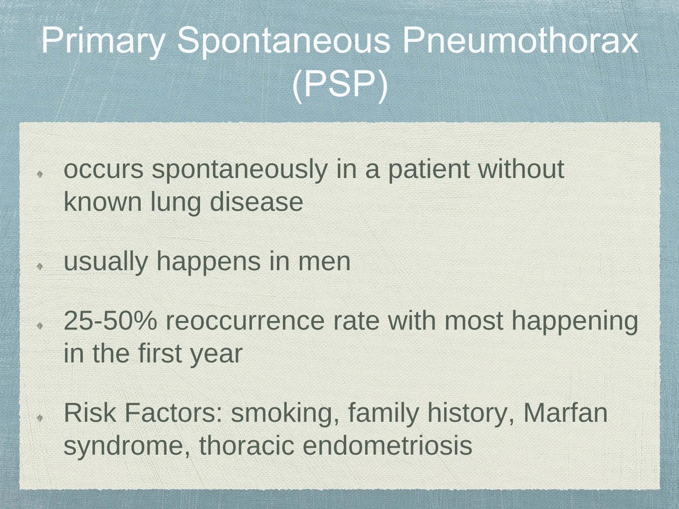
occurs spontaneously in a patient without
known lung disease
usually happens in men
25-50% reoccurrence rate with most happening
in the first year
Risk Factors: smoking, family history, Marfan
syndrome, thoracic endometriosis

caused as a complication of underlying lung
disease (COPD, CF, lung CA, necrotizing
pneumonia, PCP, TB)
Caused by an invasive procedure:
Biopsy
Central line placement
Thoracentesis
Bronchoscopy with transbronchial biopsy
Pleural biopsy
Mechanical ventilation

Treatment of
Pneumothoraces

Oxygen
Observation in asymptomatic patients with
<3cm between lung and chest wall on CXR
Removal of air from pleural space
needle aspiration (primary spontaneous PTX)
chest tube placement

If persistent airleak for >3 days in PSP, then
recommended VATS and pleurodesis, blood
patch

http://dxline.org/medic/term/video-assisted-resection/

PSP: 25-50% over 1-5 yr follow-up period with
highest risk within the first 30 days
SSP: reoccurrence is common and frequently life
threatening. One study with 50% reoccurrence
over 3 yrs.
Both PSP and SSP should undergo procedure
to prevent reoccurrences: VATS, thoracotomy
or chemical pleurodesis.

A. Pneumothorax
B. Pleural effusion
C. Both a pneumothorax
and pleural effusion
D. Pneumonia
http://www.gettyimages.no/detail/photo/hydropneumothorax-x-ray-high-res-stock-photography/556471079

C. Pneumothorax and Pleural Effusion
Also known as a Hydropneumothorax
Presence of both air and fluid in the
pleural space.
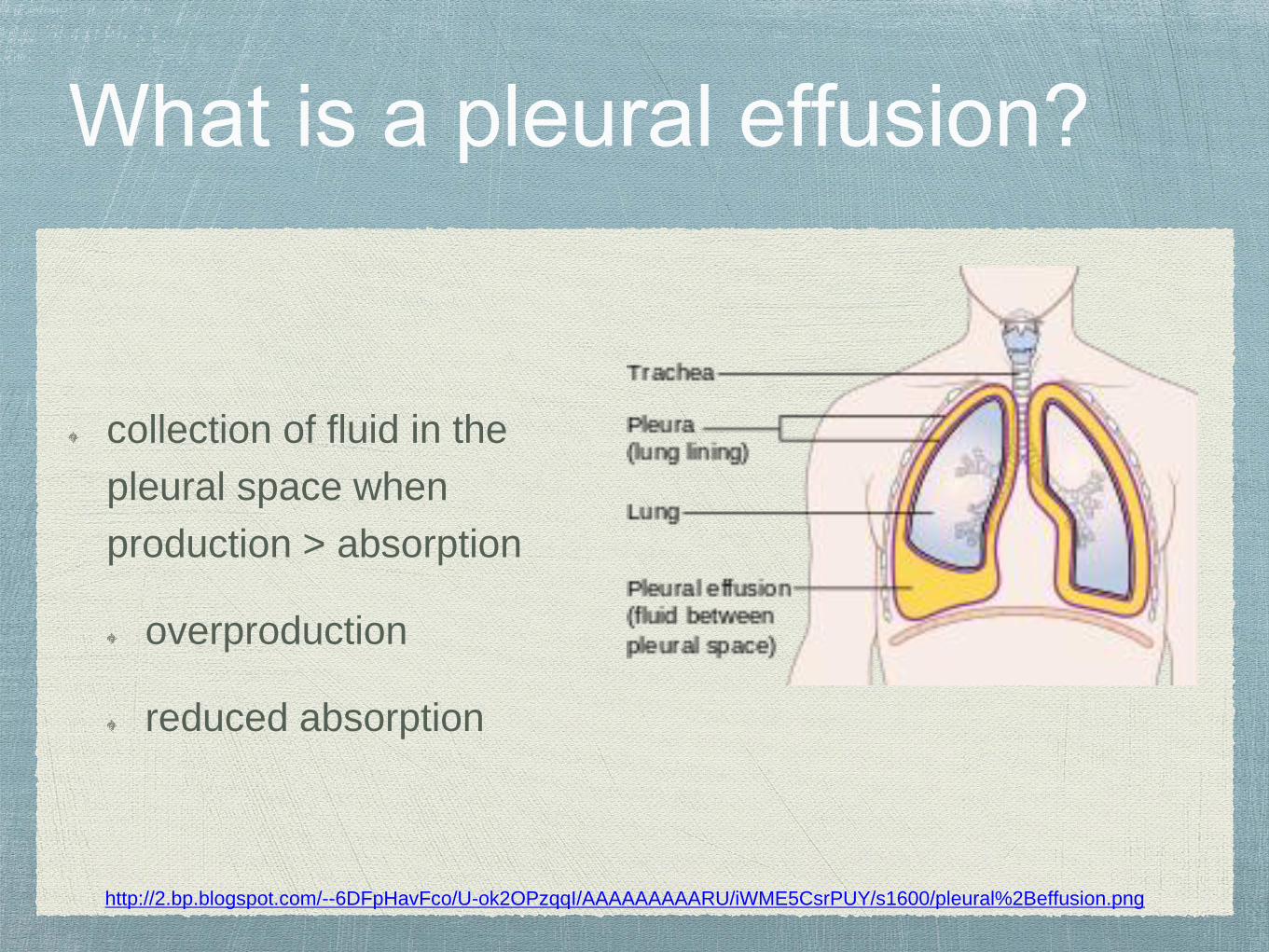
collection of fluid in the
pleural space when
production > absorption
overproduction
reduced absorption
http://2.bp.blogspot.com/--6DFpHavFco/U-ok2OPzqqI/AAAAAAAAARU/iWME5CsrPUY/s1600/pleural%2Beffusion.png

A. 0.3ml/kg body mass
B. 0.5ml/kg body mass
C. The pleural space does not contain any
fluid
D. 0.4ml/kg body mass

A. 0.3ml/kg body mass
Main function of pleural fluid is to guarantee close
apposition of visceral and parietal membranes
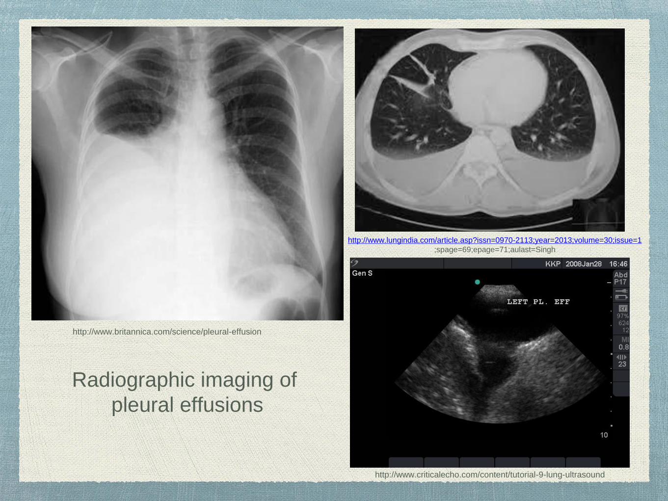
Radiographic imaging of
pleural effusions
http://www.britannica.com/science/pleural-effusion
http://www.lungindia.com/article.asp?issn=0970-2113;year=2013;volume=30;issue=1
;spage=69;epage=71;aulast=Singh
http://www.criticalecho.com/content/tutorial-9-lung-ultrasound

often can be asymptomatic
chest pain (sharp, worse with cough or taking a deep
breath, sometimes an ache)
shortness of breath
cough
fever
hiccups

initially made by imaging
pleural fluid analysis
thoracentesis
pH, LDH (pleural and serum), protein
(pleural and serum), glucose, cell count
and differential, cultures, cytology

Thoracentesis
http://www.ecinsw.com.au/pleural-tap-thoracentesis
New finding, need to determine a cause
2 cases when not required:
small amount of fluid and diagnosis
determined
clinical signs of heart failure

There are no absolute
contraindications to perform a
thoracentesis.

Pleural Fluid Analysis

Appearance (color, character, odor)
Characterization (Transudates, Exudates)
Light’s Criteria (1 of 3 positive then exudate)
pleural fluid protein/serum protein ratio > 0.5
or
Pleural fluid LDH/serum LDH ratio >0.6 or
Pleural fluid LDH > 2/3 the upper limits of
labs normal serum LDH

Pale yellow
Bloody (malignancy, injury, pulmonary infarct)
Milky (chylothorax, cholesterol effusion)
Brown (long standing blood, rupture of liver abscess)
Black (aspergillus, metastatic melanoma, perforation during
charcoal administration, etc)
Yellow-green (rheumatoid)
Dark green (biliothorax)

A. Transudative
B. Exudative
C. Pseudoexudative
D. Malignant Pleural
effusion

protein ratio: 5/2.3= 2.17
LDH ratio: 2500/150= 16.6
Pleural fluid LDH > 2/3 the upper limits of
labs normal serum LDH (usually about
200)

Imbalances in hydrostatic and oncotic pressures in the chest.
Causes:
CHF
Hepatic hydrothorax
Nephrotic syndrome
Iatrogenic
Urinothorax
Peritoneal dialysis

Transudative
can appear pseudo-
exudative secondary to
diuresis
Bilateral in 90% of cases
https://www.mypacs.net/cases/2695717.html

Transudative
Usually right sided
signs of ascites and/or
cirrhosis
Management: no chest tube
(unless infection), therapeutic
thoracentesis, salt restriction,
diuretics, TIPS, pleurodesis
http://posterng.netkey.at/esr/viewing/index.php?module=viewing_poster&task=viewsection&pi=128349&ti=436660&searchkey=
Usually result from pleural and lung inflammation or impaired lymphatic
drainage of pleural space.
Causes:
infection
malignancy
immunologic
lymphatic abnormalities
noninfectious inflammation
iatrogenic

Parapneumonic (Uncomplicated vs
Complicated)
Uncomplicated: usually resolves with abs
Complicated: requires drainage or surgery
Empyema: presence of pus and/or bacteria in
pleural fluid
A. 5-10%
B. 10-20%
C. 20-40%
D. Less than 5%

B. 10-20%

pus or bacterial organisms
in the gram stain
mortality 10-20%
1/3rd fail medical
management and require
surgery
25% prolonged
hospitalization
http://misc.medscape.com/pi/iphone/medscapeapp/html/A298485-business.html

Antibiotics
Chest tube +/-(tPA/DNase)
Small bore chest tube (10-14F)
pH <7.20
glucose <60
Surgery
VATS
Thoracotomy
Decortication
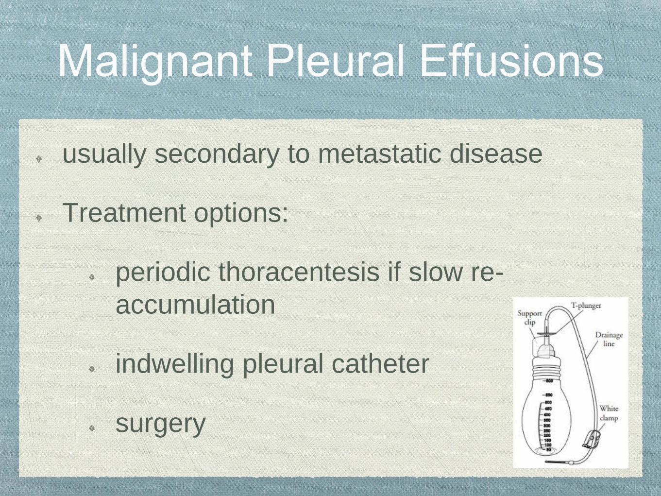
usually secondary to metastatic disease
Treatment options:
periodic thoracentesis if slow re-
accumulation
indwelling pleural catheter
surgery

Benign pleural effusion
Pleural plaques
Diffuse pleural fibrosis
Rounded atelectasis
Mesothelioma
Fibrous pleural tumor http://whatismesotheliomasymptoms.com/pleural-mesothelioma-what-is-malignant-pleural-mesothelioma/

PSP and SSP should undergo procedure to
prevent reoccurrences
If pleural effusion is undetermined, thoracentesis
should be completed with pleural fluid analysis
Complication pleural effusions and empyema
should be treated with abx, chest tube, +/-
surgery

Questions??

Cardenas, Andres, Kelleher, Barry and Chopra, Sanjiv. (2016). Hepatic
Hydrothorax. Retrieved from: UpToDate.
Ferri, Fred F. (2011). Ferri’s Clinical Advisor. Philadelphia, PA: Elsevier.
Heffner, John E. (2016). Diagnostic evaluation of a pleural effusion in
adults: initial testing. Retrieved from: UpToDate.
Light, Richard W. (2016). Primary spontaneous pneumothorax in adults.
Retrieved from: UpToDate.
Light, Richard W. (2016). Secondary spontaneous pneumothorax in adults.
Retrieved from: UpToDate.
Swanson, Karen L. (2016). Pleural Diseases I & II, 2016 CHEST Board
Review.

–Johnny Appleseed
“Type a quote here”