Embed Size (px)
Citation preview
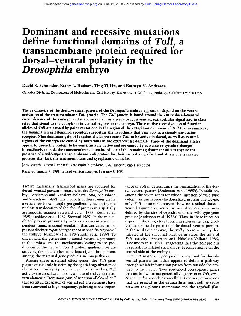
Dominant and recessive mutations define functional domains of Toll, a transmembrane protein required for dorsal-ventral polarity in the Drosophila emb ryo David S. Schneider, Kathy L. Hudson, Ting-Yi Lin, and Kathryn V. Anderson
Genetics Division, Department of Molecular and Cell Biology, University of California, Berkeley, California 94720 USA
The asymmetry of the dorsal-ventral pattern of the Drosophila embryo appears to depend on the ventral activation of the transmembrane Toll protein. The Toll protein is found around the entire dorsal-ventral circumference of the embryo, and it appears to act as a receptor for a ventral, extracellular signal and to then relay that signal to the cytoplasm in ventral regions of the embryo. Three of five recessive loss-of-function alleles of Toll are caused by point mutations in the region of the cytoplasmic domain of Toll that is similar to the mammalian interleukin-1 receptor, supporting the hypothesis that Toll acts as a signal-transducing receptor. Nine dominant gain-of-function alleles that cause Toll to be active in dorsal, as well as ventral, regions of the embryo are caused by mutations in the extracellular domain. Three of the dominant alleles appear to cause the protein to be constitutively active and are caused by cysteine-to-tyrosine changes immediately outside the transmembrane domain. All six of the remaining dominant alleles require the presence of a wild-type transmembrane Toll protein for their ventralizing effect and all encode truncated proteins that lack the transmembrane and cytoplasmic domains.
[Key Words: Dorsal-ventral; Drosophila embryo; Toll interleukin-1 receptor]
Received January 7, 1991; revised version accepted February 8, 1991.
Twelve maternally transcribed genes are required for dorsal-ventral pattern formation in the Drosophila em- bryo (Anderson and Nfisslein-Volhard 1984; Schfipbach and Wieschaus 1989). The products of these genes create a ventral-to-dorsal morphogen gradient by regulating the nuclear translocation of the dorsal protein in a spatially asymmetric manner (Steward et al. 1988; Roth et al. 1989; Rushlow et al. 1989; Steward 1989). In the nuclei, dorsal protein presumably acts as a concentration-de- pendent transcriptional regulator that activates or re- presses distinct zygotic target genes in specific regions of the embryo (Rushlow et al. 1987; Roth et al. 1989). To understand the generation of dorsal-ventral asymmetry in the embryo and the mechanisms leading to the pro- duction of the nuclear dorsal protein gradient, we are studying the biochemical functions of, and interactions among, the maternal gene products in this pathway.
Among these maternal effect genes, the Toll gene plays a crucial role in defining the spatial organization of the pattern. Embryos produced by females that lack Toll activity are dorsalized, lacking all lateral and ventral pat- tern elements. Dominant gain-of-function alleles of Toll that result in expansion of ventral pattern elements have been recovered at high frequency, pointing to the impor-
tance of Toll in determining the organization of the dor- sal-ventral pattern (Anderson et al. 1985bl. In addition, among the seven genes for which injection of wild-type cytoplasm can rescue the dorsalized mutant phenotype, only Toll- mutant embryos show no residual dorsal- ventral asymmetry, with the site of ventral structures defined by the site of deposition of the wild-type gene product (Anderson et al. 1985al. Thus, in these injection experiments, a high local concentration of the Toll prod- uct can define the polarity of the dorsal-ventral pattern. In the wild-type embryo, the Toll protein is evenly dis- tributed at the syncytial blastoderm stage, the time of Toll activity (Anderson and N/isslein-Volhard 1986; Hashimoto et al. 1991), suggesting that the Toll protein is spatially regulated such that it becomes active on the ventral side of the embryo.
The 12 maternal gene products required for dorsal- ventral pattern formation appear to define a pathway through which information passes from outside the em- bryo to the nuclei. Two sequenced dorsal-group genes that are known to act genetically upstream of Toil, east- er and snake, encode extracellular-type serine proteases that are present in the extracellular perivitelline space between the plasma membrane and the eggshell (De-
GENES & DEVELOPMENT 5:797-807 © 1991 by Cold Spring Harbor Laboratory Press ISSN 0890-9369/91 $3.00 797
Cold Spring Harbor Laboratory Press on June 13, 2018 - Published by genesdev.cshlp.orgDownloaded from
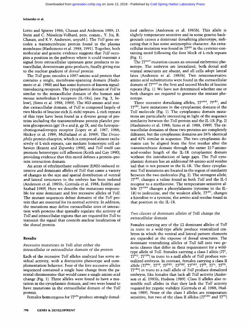
Schneider et al.
Lotto and Spierer 1986; Chasan and Anderson 1989; D. Stein and C. Nfisslein-Volhard, pers. comm.; Y. Jin, R. Chasan, and K.V. Anderson, unpubl.). The Toll gene en- codes a transmembrane protein found in the plasma membrane (Hashimoto et al. 1988, 1991). Together, both molecular and genetic evidence suggests that Toll occu- pies a position in the pathway where it could transmit a signal from extracellular upstream gene products to in- tracellular, downstream gene products, finally giving rise to the nuclear gradient of dorsal protein.
The Toll gene encodes a 1097-amino acid protein that contains a single, membrane-spanning domain (Hashi- moto et al. 1988) and has some features of known signal- transducing receptors. The cytoplasmic domain of Toll is similar to the intracellular domain of the human and mouse interleukin-1 receptors (IL-1Rs), (see Fig. 2, be- low); (Sims et al. 1988, 1989). The 803-amino acid resi- due extracellular domain, of Toll is composed largely of two blocks of leucine-rich (L-rich) repeats. L-rich repeats of this type have been found in a diverse group of pro- teins including the transmembrane protein platelet pro- tein glycoprotein (gp) lb ~ and f3, gp IX, and the lutropin- choriogonadotropin receptor (Lopez et al. 1987, 1988; Hickey et al. 1989; McFarland et al. 1989). The Droso- phila protein chaoptin, which is composed almost exclu- sively of L-rich repeats, can mediate homotypic cell ad- hesion (Krantz and Zipursky 1990), and Toll itself can promote heterotypic cell adhesion (Keith and Gay 1990), providing evidence that this motif defines a protein-pro- tein interaction domain.
An array of ethylmethane sulfonate (EMS)-induced re- cessive and dominant alleles of Toll that cause a variety of changes in the size and spatial distribution of ventral and lateral structures in the embryo has been isolated (Anderson et al. 1985b; Gerttula et al. 1988; Erddyi and Szabad 1989). Here we describe the mutations responsi- ble for nine dominant and five recessive alleles of Toll. The mutant sequences define domains of the Toll pro- tein that are essential for its normal activity. In addition, the mutations may define extracellular sites of interac- tion with proteins that spatially regulate the activity of Toll and intracellular regions that are required for Toll to transmit the signal that controls nuclear localization of the dorsal protein.
R e s u l t s
Recessive mutations in Toll alter either the intracellular or extracellular domain of the protein
Each of the recessive Toll alleles analyzed has some re- sidual activity, with a distinctive phenotype and com- plementation behavior. Four of the five recessive alleles sequenced contained a single base change from the pa- rental chromosome that would cause a single amino acid change (Fig. 1). Three alleles were found to have a mu- tation in the cytoplasmic domain, and two were found to have mutations in the extracellular domain of the Toll protein.
Females homozygous for T1 r26 produce strongly dorsal-
ized embryos (Anderson et al. 1985b). This allele is slightly temperature sensitive and in some genetic back- grounds causes a dominant dorsalizing phenotype, indi- cating that it has some antimorphic character. An extra- cellular mutation was found in Zl r26 in the cysteine-con- taining motif following the first block of L-rich repeats (Fig. 1).
The T1 rm9 mutation causes an unusual embryonic phe- notype. The embryos are lateralized; both dorsal and ventral structures are absent, and all cells adopt lateral fates (Anderson et al. 1985b). Two nonconservative amino acid substitutions were found in the extracellular domain of Zl rrn9 in the first and second blocks of leucine repeats (Fig. 1). We have not determined whether one or both changes are required to generate the mutant phe- notype.
Three recessive dorsalizing alleles, ZI r444' Zl rB1, and T1 rs2, have mutations in the cytoplasmic domain of the Toll molecule (Fig. 1). The cytoplasmic domain muta- tions are particularly interesting in light of the sequence similarity between the Toll protein and the IL-1R (Fig. 2) (Hashimoto et al. 1988; Sims et al. 1988, 1989). The ex- tracellular domains of these two proteins are completely different, but the cytoplasmic domains are 26% identical and 43% similar in sequence. The two cytoplasmic do- mains can be aligned from the first residue after the transmembrane domain through the entire 217-amino- acid-residue length of the IL-1R cytoplasmic domain without the introduction of large gaps. The Toll cyto- plasmic domain has an additional 68-amino-acid residue tail that is not present in the IL-1R. The three cytoplas- mic Toll mutations are located in the region of similarity between the two molecules (Fig. 2). The strongest allele, T1 "82, changes a valine found in both Toll and the IL-1 receptor to a methionine. The temperature-sensitive al- lele Zl r444 changes a phenylalanine (tyrosine in the IL- 1R) to isoleucine, and the weakest allele, T1 rB1, changes a histidine to a tyrosine, the amino acid residue found in that position in the IL-1R.
Two classes of dominant alleles of Toll change the extracellular domain
Females carrying any of the 12 dominant alleles of Toil in trans to a wild-type allele produce ventralized em- bryos in which the ventral and lateral pattern elements are expanded at the expense of dorsal structures. The dominant ventralizing alleles of Toll fall into two ge- netic classes that differ in their requirement for a wild- type allele of Toll. Females carrying a class I allele (T11, T19Q, TF °b) in trans to a null allele of Toll produce ven- tralized embryos. In contrast, females carrying a class II allele (T184c, TI sB, T1DB1, T1DB2, TI DB3, T12b, TI 3~, T12~, T118a) in trans to a null allele of Toll produce dorsalized embryos, like females that lack all Toll activity (Ander- son et al. 1985b; Hudson 1989). Class II alleles also re- semble null alleles in that they lack the Toll activity required for zygotic viability (Gerttula et al. 1988; Hud- son 1989). None of the class I alleles are temperature sensitive, but two of the class II alleles (T1 DB1 and TF b)
798 GENES & DEVELOPMENT
Cold Spring Harbor Laboratory Press on June 13, 2018 - Published by genesdev.cshlp.orgDownloaded from
Toll molecular lesions
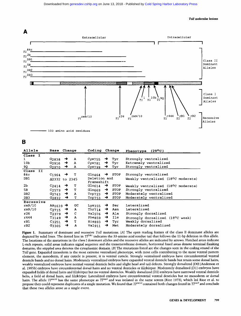
A
84c T1
2b T1
DBI T1
T15B
DB2 T1
DB3 T1
rm9110 r26 T1 T1
Extracellular
I////////////////////////////////////. i
r rB2 T1 T1 T1
I00 amino acid residues
Intracellular
I I I
,.6¢I///~ - : ~ . . . . . _ ........ - ........
T1
rm9/10 T1
1
Class II Dominant Alleles
Class I Dominant Alleles
Alleles
B
Allele Base Change
Class I 1 G2838 ~ A 10b G2916 ~ A 9Q G2970 ~ A Class II 84c C1964 ~ T
DBI A2332 to 2345
2b C2414 ~ T
5B C2579 ~ T DB2 G2743 4 A DB3 G2832 ~ T
Coding Change
Cys7 55 "~ Tyr Cys781 "~ Tyr Cys7 99 -) Tyr
Gln464 ~ STOP Deletion and Frameshift Gln614 ~ STOP
Gln669 ~ STOP TrP723 ~ STOP TrP753 ~ STOP
Pheno t~ri~ e (29°C)
Strongly ventralized Extremely ventralized Strongly ventralized
Strongly ventralized
Weakly ventralized (18°C moderate)
Weakly ventralized (18°C moderate) Strongly ventralized Moderately ventralized Moderately ventralized
Recessive rm9/10 AAI639 -~ GC Lys355 -) Ser rm9/10 C2715 "~ A Thr714 -) Asn r26 T2379 4 C Va1576 -~ Ala r444 T3149 -) A Phe859 -) Ile rBl C3251 -) T His893 4 Tyr rB2 G3305 "~ A Valgll -~ Met
Lateralized Lateralized
Strongly dorsalized
Strongly dorsalized (18°C weak) Weakly dorsalized Moderately dorsalized
Figure 1. Summary of dominant and recessive 7"oll mutations. (A} The open reading frames of the class II dominant alleles are depicted by solid lines. The dotted line in T1DB1 indicates the 33-amino acid residue tail that follows the 11-bp deletion in this allele. The locations of the mutations in the class I dominant alleles and the recessive alleles are indicated by arrows. Hatched areas indicate L-rich repeats; solid areas indicates signal sequence and the transmembrane domain; horizontal lined areas denote terminal flanking domains; the stippled area denotes the cytoplasmic domain. {B} The mutations listed are the changes seen in the coding strand of the T o l l gene. Expanded mesoderm is the most extreme ventralized phenotype, with most cells contributing to the most ventral pattern dement , the mesoderm; if any cuticle is present, it is ventral cuticle. Strongly ventralized embryos have circumferential ventral denticle bands and no dorsal hairs. Moderately ventralized embryos have expanded ventral denticle bands but retain some dorsal hairs; weakly ventralized embryos have normal ventral denticle belts and slight head and tail defects. Strongly dorsalized (D0) {Anderson et al. 1985b) embryos have circumferential dorsal hairs and no ventral denticles or filzk6rper. Moderately dorsalized ID1} embryos have expanded fields of dorsal hairs and filzk6rper but no ventral denticles. Weakly dorsalized {D2) embryos have narrowed ventral denticle belts, a field of dorsal hairs, and filzk6rper. Lateralized embryos have circumferential ventral denticles but no mesoderm or dorsal hairs. The allele T F m~° has the same phenotype as T1 =~9 and was isolated in the same screen {Rice 1973}, which led Rice et al. to propose they could represent duplicates of a single mutation. We found that TI ~ 1 ° contained both changes found in T1 rm9 and conclude that these two alleles arose as a single event.
GENES & DEVELOPMENT 799
Cold Spring Harbor Laboratory Press on June 13, 2018 - Published by genesdev.cshlp.orgDownloaded from

Schneider et al.
Tlr444-~ I
Toll 829 ~i~Q T, , ,K t l t L A H N L L L W , , E E D i~ ]KD, , , K F D A F IIS~i H K - D~i I E~L - -~V P ~ H G - V P E V 393 I I - I R : H 3 4 0 K I I D I 1 V I L W I - [ Y { R D S C Y D {b ~ L P I I K A - G I K I T ] Y D A Y I L K T V G T S 13 I F K
II-IR: M 341 K VID IlVlL WI-IYIR D S C S G IF L PIS K A GIKJTIY D A Y I L K T L G F S D T F K L P E V 398
T2 r B 1 - ) y T2 rB2"~ M
Toil 885 P ~G . . . . , , , , , , R ~ R Q C H E ~ W L ~ G H P N G D R ~ R,, M R S ~ A ! ~ ! ~ S QNF I KSE~i G ~ R L S S ~F R Q IA ~ M Y H R 458940 n-iR: H 398 L ocqYiKl~,irlZlY Y SDWVlEIVWNEN K VRSTSGr S II-IR: M 399 L Q F GIYIKILJF~IIY G D Y E D T IIEIV T N E N K V R D M G G F S G Q S S Q I I Y 459
R R ff-- . . . . . . . . . . 0 II-IR: H 459 V D G KVV L ~1~1~1~1~1~1~1~1 ~ - ~ ~ ~ T ~ ~ T ~ ~ 518 II-IR: M 460 I E G K I V L E L E KIIIQIDIYIE K MIPlDISIIIQ -I F I KIQ K H G~I S Q E R P Q S A K T R N 519
T o l l 9 9 1 A . R P V . I G . G
II-IR: H 519 V R S S S K H Q L S . . . . . K R E A H V 552 II-IR: M 520 A R S L 5 K H R L T L DIP V RID - K A A T H L 557
Figure 2. Toll and IL-1R sequence similarity. The sequence of the cytoplasmic domain of Toll is aligned with the cytoplasmic domains of the h u m a n and mouse IL-1Rs (IL-1R H and IL-1R M, respectively}. Similarities between the cytoplasmic domain of Toll and the IL-1R are boxed. Toll has an additional 68 amino acids in its cytoplasmic domain following the region of similarity. Amino acid similarities were defined according to Miyata, where I ~ V~ L~ M, D ~ E ~ Q ~ N, F ~ Y -~ W, H ~ K ~ R, and G -~ A ~ S
T ~ P {Miyata et al. 1979). The amino acid changes found in the cytoplasmic domain of Toll alleles are marked.
are cold sensitive, producing more severe phenotypes at 18°C than at 29°C {Fig. 3C, D). Class I and class II alleles also differ in their dependence on upstream genes. Dou- ble-mutant females that lack the activity of genes up- stream of Toll and carry a class I Toll allele produce embryos that make ventral or lateral structures in a dor- soventrally symmetric pattern, whereas double-mutant flies carrying class II alleles of Toll and upstream muta- tions produce dorsalized embryos {Anderson et al. 1985b; K.V. Anderson, unpubl.). In summary, class I alleles of Toll are cis active and partially independent of the activ- ity of upstream genes, whereas class II alleles are cis inactive, can alter the activity of the wild-type Toll prod- uct in trans, and require upstream genes for activity.
In general, the class I alleles produce more strongly ventralized phenotypes than most class II alleles, al- though the range of phenotypes overlaps. The most ex- treme ventralized phenotype is seen in embryos laid by mothers carrying the class I allele T1 l°b (Erd61yi and Szabad 1989}. In these embryos the most ventrally de- rived tissue, the mesoderm, is expanded and laterally derived epidermal structures are either absent or greatly reduced {Hudson 1989}. The ventralized phenotypes of embryos produced by mothers carrying the class I alleles T11 or T19° or the class II alleles T184c or T15B are similar {Fig. 3B)(Anderson et al. 1985b). Females carrying one of these four alleles produce ventralized embryos with ex- panded ventral epidermis and the complete loss of dorsal epidermis, but no expansion of the mesoderm. The other class II alleles cause a weaker ventralization of the em- bryonic pattern, with some dorsal pattern elements still present. The weakest phenotype is caused by the cold- sensitive allele TI°BI; at 29°C, 50% of the embryos hatch into larvae and the remainder do not undergo head invo- lution and may not shorten the germ band {Fig. 3D).
Nine of the dominant alleles were sequenced, and all contained mutations in the extracellular domain of the
Toil molecule {Fig. 1}. The sequence changes found in the dominant alleles fall into two molecular classes that coincide with the two genetically defined classes.
Each of the class I alleles changed a cysteine to a ty- rosine in the sequence after the second block of L-rich repeat domain, immediately adjacent to the transmem- brane domain (Fig. 1). This clustering of mutations prompted us to examine the carboxy-terminal flanking sequences of other L-rich repeat-containing proteins more closely. In Toil, both blocks of L-rich repeats are followed by a cysteine-containing motif, which is also found in a subset of proteins that have extracellular L- rich repeats including L-rich glycoprotein {Takahashi et al. 1985}, the platelet transmembrane proteins gp lb and 13, gp IX (Lopez et al. 1987, 1988; Hickey et al. 1989}, and the lutropin-choriogonadotropin receptor (McFar- land et al. 1989}. We found that the similarity between these motifs extends further than described previously {Hickey et al. 1989; Keith and Gay 1990} and that the motifs contain four, rather than only two, conserved cys- teine residues {Fig. 4). Each of the three class I alleles replaced one of the four cysteine residues in this motif with a tyrosine residue (Fig. 4J.
In each of the six class II alleles sequenced, premature stop codons or small deletions that introduced prema- ture stop codons were found (Fig. 1). The truncated poly- peptides produced by these alleles would be from 464 to 753 amino acid residues in length and would include one or both blocks of L-rich repeats. None of the truncated proteins would contain the transmembrane domain. No simple relationship between the length of a truncated class II polypeptide and its phenotypic strength has emerged. For instance, the two strongest class II Toll alleles, T15B and T184c, encoded truncated polypeptides of very different lengths.
Protein blots confirmed that TlCl'ssI1/T1 + heterozy- gores synthesized both full-length and truncated Toll
800 GENES & DEVELOPMENT
Cold Spring Harbor Laboratory Press on June 13, 2018 - Published by genesdev.cshlp.orgDownloaded from

Toil molecular lesions
Figure 3. Cuticular phenotypes produced by dominant ventralizing Toll alleles and by injection of dominant allele transcripts. {A) Wild-type, first-instar larva. (B) The cuticle of a strongly ventralized embryo produced by a T18aC/TM3 female. (C) The cuticle of a moderately ventralized embryo produced by a TIDB1/TM3 female at 18°C. (D) The cuticle of a weakly ventralized embryo produced by a TID~I/TM3 female at 29°C. (E) The cuticle of a wild-type embryo injected dorsally at 25% egg length with a transcript encoding a T1 t~u~c~ (66 ~g/ml). Both head and tail structures are ventralized. (F) The cuticle of a wild-type embryo injected at 25% egg length on the dorsal surface with the class I TF °b cDNA transcript (66 ~g/ml). Only the posterior end of the embryo was ventralized in these injections. (G) The cuticle of a strongly dorsalized embryo produced by a eaa/ea s°2erxl female. (H) The cuticle pattern of an embryo produced by an eaa/ea s°ee~xl female that was injected with a T1 ~°b transcript at 25% egg length. This embryo differentiated both the dorsolaterally derived filzk6rper and the ventrolaterally derived ventral denticle belts of the sixth, seventh, and eighth abdominal segments.
proteins (Fig. 5). The sizes of the observed proteins were consistent with the sizes predicted from their sequences. The truncated proteins examined were all -25% as abundant as the full-length Toll protein, perhaps because they are not as stable as the transmembrane protein.
Inject ion of d o m i n a n t allele transcripts can ventral ize the embryon i c pat tern
To test whether the coding changes found in the domi- nant alleles were responsible for the mutant phenotypes, we assayed the activity of both class I and class II mutant transcripts injected into wild-type embryos. This assay relied on the ability of injected transcripts of a full- length Toll cDNA to rescue the dorsalized phenotype of embryos laid by T1- females (C. Hashimoto, unpubl.). Injection of high concentrations of wild-type transcripts into wild-type embryos never produced a ventralizing phenotype (Table lJ.
We replaced segments of the Toll cDNA construct with restriction fragments containing the point muta- tions of class I dominant alleles and assayed the activity of transcripts of mutant alleles by injection into cleav- age-stage, wild-type embryos (Table 1). Transcripts con-
taining the T11 o r TI lOb cysteine-to-tyrosine changes caused ventralization of the embryonic pattern, confirm- ing that these changes are sufficient to produce a domi- nant ventralizing phenotype (Fig. 3F).
The extent of ventralization produced by the class I transcripts depended on the concentration of the injected transcripts. Injection of high concentrations (>66 t~g/ml) of T1 ~°b and T11 (Table 1) transcripts into wild-type em- bryos induced the formation of local cup-shaped invagi- nations at the time of gastrulation at the site of deposi- tion (Table 1). We believe that these local furrows corre- spond to local mesodermal invaginations. Embryos that had large local invaginations differentiated to have large holes in their cuticles, with only ventral cuticular struc- tures present in the surrounding cuticle. We interpreted these embryos as having expanded mesoderm, similar to embryos produced by females carrying the T1 l°b allele. Injections of lower concentrations (13 t~g/ml) of T1 l°b and T1 ~ transcripts produced more weakly ventralized embryos that resembled those produced by T11 females, with ventral denticle bands encircling the circumference of the larval cuticle. Thus, although TI~°b/+ females produce more strongly ventralized embryos than T1U+ females, the products of both T1 l°b and T1 ~ caused an
GENES & DEVELOPMENT 801
Cold Spring Harbor Laboratory Press on June 13, 2018 - Published by genesdev.cshlp.orgDownloaded from

Schneider et al.
T114 Y
~ ~ ~!i~ilimi~iii~~mnmm, m ~ i m ] ~ ~ ~ o ~ v o v ~ ~ ~ • ~ . v
gp IX I H ~::i::~i::i:: ~::i! L R T P . . . . . . . . . . . . . . . E A L trk b ~ T !~i~!~!~::S ii~!~iD~ T -~'iL~IT K S S P D . . . . . . . . . . . . . . T
s H i::i~ii!i~:-~:~:~ r R N ~lgl I< ~ ~ r S r s . . . . . . . . . . . . . ~ LHR ~Y i[~S H iiii:i:~:ii::i~i~ F kT F S
Tll0b-~ y TI9Q-) y
Figure 4. L-rich repeat terminal-flanking sequence similarity. The sequences of the regions carboxy-terminal to the blocks of leucine repeats in the extracellular domain in the Toll protein are aligned with leucine repeat carboxy-terminal-flanking sequences found in human blood platelet glycoprotein lb a and [5 (gplb a and f~) (Lopez et al. 1987, 1988), human blood platelet glycoprotein IX (gp IX) (Hickey et al. 1989), the protein tyrosine kinase receptor trk b (Klein et al. 1989), L-rich glycoprotein (LRG) (Takahashi et al. 1985), and the lutropin receptor (LHR} (McFarland et al. 1989). Amino acid residues are grouped on the basis of similarities between their physical and chemical properties, which were defined according to Miyata, I ~ V ~ L ~ M, D ~ E ~ Q ~ N, F ~ Y ~ W, H ~- K ~ R, and G ~ A ~ S ~ T ~ P (Miyata et al. 1979). Similar amino acids are shown in white with a black background. The four cysteine residues found in these repeats are highlighted with a dotted background. The mutations responsible for the dominant phenotypes of class I Toll alleles are in the second Toll terminal repeat at the positions shown.
expansion of the ventrally derived mesoderm at high concentrat ions and at lower concentrat ions expanded the laterally derived ventral epidermis wi thout detect- ably expanding the mesoderm.
When wild-type Toll m R N A is injected into T1- em- bryos the dorsal-ventral pat tern is rescued along one- third to one-half of the anter ior-poster ior length of the embryo (Anderson et al. 1985a; C. Hashimoto, unpubl.). The ventralizing effects produced by injection of T11 or T11°9 transcripts into wild-type embryos were also spa- tially restricted. Local invaginations at the site of injec- tion were seen when the transcripts were deposited ei- ther dorsally or ventral ly at the periphery of the embryo. When the transcripts were placed dorsally, a furrow in-
84c DB1 58 + - TI TI TI + - + + +
2 0 0 - -
9 3 - -
9 - - i i ,
Figure 5. Class II allele protein expression. Extracts of O- to 4-hr embryos were separated on 7.5% polyacrylamide gels and transferred to nitrocellulose. Blots were probed with a rabbit polyclonal antibody raised against the amino-terminal domain of Toll (Materials and methods). The product of T1 Sac migrates at 63 kD, the product of T1 °B~ at 82 kD, and the product of T15B at 84 kD, the same order of size predicted from the sequences. Each truncated product migrates -10 kD larger than predicted from its sequence, probably due to glycosylation. Wild-type Toll protein is glycosylated and therefore migrates at 135 kD rather than the 125 kD predicted from the open reading frame (Hash- imoto et al. 1991).
vaginated at that site - 2 min before the invagination of the ventral furrow. No interference wi th the invagina- tion of the normal ventral furrow was detected. When the transcripts were injected ventrally, a cup-shaped in- vagination at the injection site slightly preceded, and then appeared to be superimposed on, normal ventral furrow invagination. Injection at 25% egg length (0% egg length = posterior pole) prevented germ-band extension but did not affect the dorsal-ventral position of the ceph- alic fold (67% egg length). Larval cuticles of embryos injected at 25% egg length frequently had wild-type head structures but ventralized posteriors (Fig. 3F). No clear differences were seen in the cuticular patterns between embryos injected ventrally and those injected dorsally.
To test the activity of transcripts encoding truncated products like the class II alleles, we synthesized a Toll cDNA (TI t . . . . 1) that has a stop codon 5 amino acid res- idues amino-terminal to that found in T1 sS. Injection of high concentrations (330 i~g/ml) of a T1 tr'''cl transcript into wild-type embryos at any dorsal-ventral position caused a moderate ventral izat ion of the embryonic pat- tern, wi th expanded ventral denticle bands and the loss of some dorsal pattern elements, but some dorsal struc- tures were always differentiated (Fig. 3E). No local ven- tral furrow was ever induced in these injections (Table 1). In contrast wi th the local effects seen wi th class I transcript injections, when the T1 tru'cl transcript was injected at 25% egg length, the cephalic fold was shifted to the dorsal side of the embryo and germ-band extension was blocked. Both head and tail cuticle patterns were ventralized in these embryos {Fig. 3E). The global ven- tralization produced by class II, but not class I, tran- scripts suggests that the class II, but not the class I, pro- teins diffuse throughout the extracellular perivitelline space of the embryo.
802 GENES & DEVELOPMENT
Cold Spring Harbor Laboratory Press on June 13, 2018 - Published by genesdev.cshlp.orgDownloaded from

Toil molecular lesions
Table 1. Phenotypes of embryos in jec ted wi th transcripts encoding d o m i n a n t Toll alleles
Local Concentration Injection furrow
Transcript Recipient (~g/ml) position (%) n a
Differentiation pattern (%)
rues.
hatch FK + VD VD exp. n c
Tl l Ob
r l l Ob
7.110b r l 10b
Tl l Ob T1 lOb
TP TF I"11
TF
Tltruncl
rltruncl
/+
T/+
wild type 330 dorsal 82 wild type 330 ventral 90 wild type 66 dorsal 21 wild type 66 ventral 78 wild type 13 dorsal 0 wild type 13 ventral 11
wild type 200 dorsal 76 wild type 200 ventral 52 wild type 40 dorsal 10 wild type 40 ventral 0
wild type 330 dorsal 0 wild type 330 ventral 0
wild type 1000 dorsal 0 wild type 1000 ventral 0
49 52 68 81 47 18
46 46 10 11
20 25
42 37
0 2 10 88 21 0 5 95 47 4 13 34 49 63
20 17 17 46 81 48 21 21 12 18 50 28 11 11 44
30 23 47 47 45 12 43 51 73 22 5 40 92 4 4 26
0 18 82 25 4 35 61 29
100 33 100 21
Differentiation pattern (%)
m e s ,
no FK FK FK + VD VD exp.
T1 wb T1- 330 80 35 T1 wb T1- 66 64 44 T1 wb T1- 13 71 51 TF ob T1 2 0 46 15 TF °b T1 0.4 100
T1 l°b ea - 330 92 25 T1 wb ea - 66 72 75 TF °b ea - 13 0 58 6
TF T1- 200 87 137
T1 mmcl T1- 2000 0 41 100
TI t~mcl ea - 330 0 65 100
9 91 1! 12 88 16
2 38 6 54 48 70 15 27
17
4 54 42 24 4 51 20 25 59
50 38 6 18
11
Local furrow refers to the formation of a local mesoderm invagination during gastrulation at the site of injection. The columns headed ng and n¢ record the number of embryos observed at gastrulation or the number of larval cuticles counted, respectively. Embryos that did not hatch but did not have defects in dorsal-ventral pattern formation are not included here. Hatching was not scored for T11 injections. Cuticles recorded in the FK column were moderately dorsalized and contained only dorsolaterally derived filzk6rper but not the more ventrally derived ventral denticles. Cuticles recorded in the FK + VD column contained filzk6rper material and some ventral denticles. In injections into wild-type embryos the FK + VD column includes cuticles that were moderately ventralized. For injections into dorsalized embryos, the FK + VD column lists both moderately dorsalized, essentially wild-type and moderately ventralized cuticles, which could not be unambiguously distinguished from one another. Cuticles recorded in the VD column contained ventral denticles but no filzk6rper material and, therefore, were more strongly ventralized than those in the FK + VD column. Cuticles recorded in the mes. exp. (mesoderm expanded) column contained larges holes in their cuticles, which resemble those found in extremely ventralized embryos in which the mesoderm has expanded; only ventral cuticle was seen surrounding the holes. Transcripts were injected onto the dorsal side of T1- or e a - embryos.
We also assayed the act ivi ty of the d o m i n a n t tran- scripts in jec ted in to dorsal ized embryos tha t lacked the act iv i ty of T o l l or the ups t r eam dorsal group gene e a s t e r
(ea) (Table 1). At an appropriate concent ra t ion , in jec t ion of class I t ranscr ipts near the per iphery of embryos pro- duced by T 1 - females or e a - females p r o m o t e d the de- v e l o p m e n t of a normal , a s y m m e t r i c dorsa l -ven t ra l pat- tern (Fig. 3H). In bo th k inds of recipient , the m o s t vent ra l s t ruc tures developed at the site of RNA deposi t ion. The dose-response curve for rescue was very steep, w i t h h igh
concen t ra t ions of t ranscr ipt ven t ra l i z ing the embryos, and only a l im i t ed concen t r a t i on range genera t ing a wi ld- type pa t te rn (Table 1). Five t imes m o r e d o m i n a n t T o l l R N A was requi red to rescue lateral and vent ra l s t ruc tures in ea - embryos t han in T 1 - embryos, suggest- ing that the ea + ac t iv i ty presen t in the T 1 - embryo po- ten t ia tes the act iv i ty of the product of the d o m i n a n t al- lele. As expected f rom the c is inac t iv i ty and ups t r eam gene dependence of the class II alleles, in j ec t ion of class II t ranscripts in to embryos produced by T 1 - a n d e a -
GENES & DEVELOPMENT 803
Cold Spring Harbor Laboratory Press on June 13, 2018 - Published by genesdev.cshlp.orgDownloaded from

Schneider et al.
females did not rescue any lateral or ventral structures (Table 1).
Discussion
Our current hypothesis is that the Toll protein either directly transduces a signal across the plasma membrane of the embryo or is part of a signal transduction complex. To investigate this hypothesis, we have sequenced mu- tant Toll alleles that cause well-defined changes in the embryonic dorsal-ventral pattem. The sequence changes define functional domains of the Toll protein that are consistent with the idea that it acts as a receptor for an extracellular, spatially asymmetric signal and passes on a signal through its cytoplasmic domain. Recessive mu- tations that lower the activity of Toll alter either the extracellular or cytoplasmic domains of the protein. Dominant gain-of-function alleles, which cause Toll to be active in dorsal as well as ventral parts of the embryo, are all caused by mutations in its extracellular domain. The two genetic classes of dominant Toll alleles corre- spond to two distinct classes of molecular lesions, iden- tifying two regions of the extracellular domain that are of particular importance in controlling the activity of the Toll protein.
Class I dominant Toll alleles encode constitutively active products
The class I dominant alleles define the juxtamembrane cysteine-containing motif adjacent to the L-rich repeats as a domain that can control the activity of the Toll molecule. The 18 extracellular cysteine residues in Toll do not form disulfide bonds with other proteins (Hashi- moto et al. 1991), suggesting that the cysteines changed in these alleles normally participate in intramolecular disulfide bonds. The L-rich repeat carboxy-terminal flanking sequence of L-rich glycoprotein contains only two cysteine residues that form an intramolecular disul- fide bond (Takahashi et al. 1985). The pattern of se- quence similarity between the proteins suggests that di- sulfide bonds form between Cys 1 and Cys 3 and be- tween Cys 2 and Cys 4 in this domain of Toll. The conformational changes that lead to abnormal activity of class I Toll alleles could be due either to the substitution of bulky tyrosine residues for cysteine residues or to in- terference with normal disulfide bond formation. Both factors may be important. We predict that the T1 l°b and TP mutations disrupt the same disulfide bond, but they do not cause identical phenotypes.
Like Toil, the Caenorhabditis elegans lin-12 gene en- codes a transmembrane protein that is believed to func- tion as a signal transducer (Yochem et al. 1988). Domi- nant mutations of lin-12 that increase the activity of the protein change single extracellular amino acids adjacent to the transmembrane domain (Greenwald and Seydoux 1990). This suggests that although these two proteins share no sequence homology, the conformation of the region just outside the transmembrane domain may be important in defining the activity of the cytoplasmic do-
mains of both Toil and lin-12. In addition to producing similar mutant phenotypes, these domains may also be important in regulating the activity of the wild-type pro- teins. The similar activity of these two domains in Toll and lin- 12 suggests that extracellular juxtamembrane do- mains could have similar functions in other signal-trans- ducing molecules.
Because the class I alleles are active in the absence of upstream genes, it is likely that the class I alleles of Toll send a constitutive signal to the cytoplasm at all dorsal- ventral positions. The injection experiments reported here confirm this hypothesis by demonstrating that class I transcripts can induce ventral structures on the dorsal side of the wild-type embryo or in an embryo that lacks the activity of the upstream gene easter.
When the class I transcripts are injected locally into T1- or ea- embryos, ventral structures differentiate at the injection site and a complete, normally proportioned dorsal-ventral pattern can develop (Fig. 3H). The asym- metric dorsal-ventral pattern of the injected ea- em- bryos contrasts with the dorsoventrally symmetric lat- eralized embryos produced by Tll-ea - double-mutant females (Anderson et al. 1985b). This difference could be accounted for if in TP-ea- embryos the TP product is constitutively active at all dorsal-ventral positions, pro- ducing a lateralized phenotype, while in ea- embryos injected with a high concentration of T11 RNA, the con- stitutively active, nondiffusing TP product is confined to one side of the embryo and can then generate a normal, asymmetric pattern.
The TP-ea + embryo is ventralized and has normal polarity, with a ventral furrow forming on the normal ventral side. The asymmetry seen in the T11 (ea +) em- bryo must be the result of an enhancement of the activ- ity of the TP product above its baseline constitutive level by a ventral signal that depends on easter activity. This kind of enhancement was seen in the TP transcript injections, where the activity of the TP product was ap- proximately fivefold lower in the absence of ea + (Table 1). Thus, in the wild-type embryo it is likely that asym- metric activation of the uniformly distributed Toll prod- uct by an easter-dependent upstream ventral signal gives rise to the normal pattern.
It is interesting that two very different means of achieving localized Toll activity, local activation by an upstream signal or local injection of a nondiffusible, con- stitutively active receptor, can both result in a normal dorsal-ventral pattern. It seems likely that the spatial distribution of active Toll molecules is different in the two cases, suggesting that mechanisms act downstream of Toll to regulate proportioning of the dorsal-ventral pattern.
Truncated class H dominant products activate the wild-type Toll protein in trans
The class II dominant alleles of Toll encode stable trun- cated polypeptides (Fig. 5) that lack both the transmem- brane and cytoplasmic domains and are therefore pre- sumably secreted into the extracellular perivitelline
804 GENES & D E V E L O P M E N T
Cold Spring Harbor Laboratory Press on June 13, 2018 - Published by genesdev.cshlp.orgDownloaded from

Toil molecular lesions
space. The truncated, secreted products of the class II alleles are active only in the presence of both the trans- membrane Toll protein and the products of the genes upstream of 7"oi1. Thus, the amino- terminal 464 amino acids of 7"oll must include a domain that, together wi th the normal upst ream gene products, can control the ac- t ivity of wild-type Toll protein.
It is possible to explain the phenotypes of the class II alleles by three different kinds of interactions: The amino- terminal domain of Toi1 could bind the trans- membrane Toll, it could bind to an inhibi tor of To11, or it could bind to an activator of To11. In the first model, the truncated Toll proteins could increase the activity of the wild-type Toi1 gene product by interacting directly wi th the wild-type t ransmembrane protein. Many t ransmem- brane signal transducers require ligand-induced mult i - merizat ion to be activated (Ullrich and Schlessinger 1990). If Toll activation also requires mul t imerizat ion, the truncated molecules may facilitate this process. In a second model, the activity of the full-length Toll protein is increased because truncated Toll molecules sequester an inhibi tor of wild-type To11. This model predicts that there is an upst ream inhibi tor of Toi1, but no such mol- ecule has been identified genetically. In a third model, the dominant ventral izing activi ty of class II alleles de- pends on the secretion and solubil i ty of their gene prod- ucts. If the upstream gene products create a diffusible asymmetr ic activating signal on the ventral side of the embryo, truncated TolI molecules could bind to the ac- tivator and promote its diffusion to the dorsal side of the embryo, where the activator could be released to activate wild-type Toli protein.
Biochemical and molecular genetic evidence indicates that t runcated and secreted forms of many cell-surface receptors are produced natural ly (Goodwin et al. 1990). The truncated receptors could decrease receptor activity by competing for ligand wi th the t ransmembrane form or by interfering wi th the signal transduction process (Basu et al. 1989; Taira et al. 1989). However, in contrast wi th these dominant negative interactions, the Toll class II alleles indicate that a truncated protein can induce the ectopic activi ty of a full-length receptor.
The Toll cytoplasmic domain: Homology to the IL-1R
The sequence s imilar i ty between the cytoplasmic do- mains of Toll and the IL-1R and the mapping of the loss- of-function alleles of Toll to the region of s imilar i ty sug- gest the hypothesis that Toll and the IL-1R t ransmit their signals by s imilar mechanisms . The m e c h a n i s m of signal t ransduction by the IL-1R is not yet clear, al- though studies have implicated increases in cAMP or diacylglycerol as steps in the pathway (Rosoff et al. 1988; Shirakawa et al. 1988; Zhang et al. 1988). A number of new recessive Toll alleles that have been isolated re- cently (N. Machin, pers. comm.) may help to evaluate the significance of the sequence s imilar i ty and to define functional regions wi th in both cytoplasmic domains.
The potential homology between Toll and the IL-1R is particularly tantal izing because the proteins that act
downstream of these two t ransmembrane proteins may act in s imilar signal t ransduction pathways. Toll indi- rectly controls the activity of the dorsal protein by con- trolling its nuclear localization (Roth et al. 1989; Rushlow et al. 1989; Steward 1989). Similarly, in some cell lines, IL-1 causes the transcript ion factor NF-KB to be translocated from the cytoplasm into the nucleus, where it is then active (Shirakawa et al. 1989). The se- quence s imilar i ty of dorsal to the DNA-binding p50 sub- uni t of NF-KB (Ghosh et al. 1990; Kieran et al. 1990), the regulation of both transcription factors at the level of nuclear translocation, and the sequence s imilar i ty of Toll and the IL-1R suggest that the intermediates in the two pathways may be also similar.
Mater ia l s and m e t h o d s
Mutant alleles
Most Toll alleles have been described previously (Anderson et al. 1985b; Gerttula et al. 1988; Erd61yi and Szabad 1989). The dominant alleles T1DB1 (Dominant Berkeley 1), T1Ds2, and T1Ds3 were fortuitously isolated in F2 screens for maternal-effect mu- tations (S. Wasserman, D. Morisato, and K.V. Anderson, un- publ.).
Sequencing of mutant alleles
The sequences of the Toil alleles were determined by the dideoxy chain-termination technique (Sanger 1977), using the Sequenase system {U.S. Biochemical Corporation). The se- quences of the parental chromosomes of all the To11 alleles, with the exception of TF e6' T11, T1 l°b, and T12b, were also de- termined. Genomic DNA, including the To11 open reading frame and a 106-bp intron between eDNA nucleotides 1793 and 1794 (Hudson 1989), was sequenced by using oligonucleotide primers spaced every 200 bp.
Two mutant Toll alleles were cloned from genomic libraries. The T18~c allele was cloned from an EMBL 4 library of partial Sau3A-digested genomic DNA from TlS4C/TM3 flies. The T19Q allele was cloned from a XFIX (Stratagene) library of XhoI-di- gested genomic DNA obtained from T19Q/TM3 flies. Alleles of Toll cloned from the TM3 balancer chromosome were identi- fied by the presence of a polymorphic BamHI restriction site that is not present in the T184~ and TI 9Q alleles. The open read- ing frames of these two dominant alleles were sequenced com- pletely.
The remaining alleles were sequenced from mutant genomic DNA amplified by the polymerase chain reaction {PCR){Saiki et al. 1988). The recessive alleles were sequenced from PCR-am- plified DNA that was cloned into pBluescript (Stratagene). The 3.4-kb Toll open reading frame-containing DNA was amplified in two fragments from DNA obtained from flies homozygous for each recessive allele. The entire open reading frame of each of the recessive alleles was sequenced. Mutant sequences were confirmed by directly sequencing PCR products. The sequences of T1 l°b, T11, T1DB1, T15B, T12B, T1DB2, and T1DB3 were obtained by directly sequencing DNA amplified from genomic DNA iso- lated from flies carrying the mutant alleles in trans to a defi- ciency (T1 l°b, T1 l, and T1DBI) or in trans to a balancer (TM3) chromosome (TI 2b, T15B, T1DB2, and T1DB3). TF °b and T11 were sequenced from nucleotide 2535 to nucleotide 3040, T1DB1 and T1 eb were sequenced from nucleotide 2285 to nucleotide 2475, T1DSe and T1DB3 were sequenced from nucleotide 2555 to 2872.
GENES & DEVELOPMENT 805
Cold Spring Harbor Laboratory Press on June 13, 2018 - Published by genesdev.cshlp.orgDownloaded from

Schneider et al.
T1 sB was sequenced from nucleotide 1248 to nucleotide 2451 and from nucleotide 2540 to 2930.
Transcripts of mutant alleles
A full-length, wild-type Toll cDNA was constructed (C. Hash- imoto, unpubl.) in a pGEM-2 vector (Promega). cDNAs includ- ing the point mutations in the dominant alleles T/1 and TP °b were constructed by replacing a PflmI-StuI restriction fragment in the wild-type cDNA with a fragment (nucleotide 2800 -3483 in the cDNA sequence) amplified from mutant DNA by PCR. The reconstructed dominant alleles were sequenced over the PflmI-StuI restriction fragment to ensure that the DNA did not contain any PCR-derived artifacts.
A Toll allele containing a stop codon in the extracellular do- main (T1 t~c~) was constructed by inserting an XbaI linker (New England Biolabs) containing stop codons in all three read- ing frames into the open reading frame of the Toll eDNA. The pGEM-2 vector containing the full-length, wild-type eDNA was linearized with StuI, which cuts in the cytoplasmic domain of Toll. To place the linkers in the extracellular domain of Toil, ExoIII deletions were performed on this linearized plasmid es- sentially as described previously (Henikoff 1987). Nonphospho- rylated XbaI linkers were ligated to the deleted plasmids, and the ligation reactions were precipitated to remove unligated linkers. The ligated DNA was resuspended in 40 mM Tris-HC1 {pH 7.5), 20 mM MgCI2, and 50 mM NaC1, heated to 65°C, and cooled slowly to allow the free ends to anneal. This recircular- ized DNA was used to transform Esch erichia coli MC 1061 cells. T1 tr~c~ contains an XbaI linker after nucleotide 2559 in the Toll eDNA sequence and an 1825-bp deletion from nucleotide 2559 to 4384.
Transcript injection
pGEM-2 templates were linearized and SP6 transcripts were generated essentially as described (Krieg and Melton 1987), in the presence of 500 ~M TTP, CTP, ATP (Pharmacia), cap analog {5'GpppG3'; Pharmacia), 50 ~M GTP, and 2.5 nM [~-32p]CTP, at 800 Ci/mmole (Amersham). Mter 2 hr, the reaction mixture was diluted with one volume of 100 mM NaC1, 30 mM EDTA, 20 mM Tris (pH 7.5), and 1% SDS and passed over a 1-ml Sephadex G-50 spin column, equilibrated in 0.3 M sodium acetate and 0.1% SDS, to remove unincorporated nucleotides. The reaction was then extracted with phenol and precipitated twice with ethanol to remove residual SDS. Transcripts were resuspended in injection buffer (Anderson and Nfisslein-Volhard 1984). The concentration of the transcribed RNA was determined by cal- culating the percentage incorporation of the radiolabeled CTP.
Protein analysis
Staged 0- to 4-hr embryos were collected, dechorionated, and homogenized in a solution of 10 mM Tris-HC1 (pH 8.0), and 1 mM EDTA. Wild-type embryo extracts were obtained from Or- egon-R flies. T1- embryos were obtained from Df(3R) Tlr°XB3/ Df(3R)T19°ax females, which make no Toll RNA (Hashimoto et al. 1988). Ventralized embryos were collected from TldasSU/Tl + flies. Extracts were centrifuged at 10,000g for 10 min to remove insoluble material, and 100 ~g of protein was loaded per lane. Proteins were blotted as described (Hashimoto et al. 1991 ). Blots were probed with a 1 :40 dilution of affinity-purified rabbit polyclonal primary antibody raised against the amino-terminal portion of Toll (Hashimoto et al. 1991), and 1 : 10,000 dilution of HRP-linked goat-anti-rabbit secondary antibody (Bio-Rad). Toll protein was visualized by using an ECL kit (Amersham).
A c k n o w l e d g m e n t s
We thank Arend Sidow for advice concerning direct sequencing of PCR products and Chris Kaiser for help in sequence analysis. We also thank Becky Chasan, Chip Ferguson, Linda Hicke, Bruce Kimmel, Sylvia Sanders, and Mike Simon and the mem- bers of the Anderson laboratory for helpful comments on the manuscript. This work was supported by grants from the Na- tional Institutes of Health (GM 35437), the National Science Foundation (DCB 8452030), and the American Cancer Society (NP726) to K.V.A.
The publication costs of this article were defrayed in part by payment of page charges. This article must therefore be hereby marked "advertisement" in accordance with 18 USC section 1734 solely to indicate this fact.
R e f e r e n c e s
Anderson, K.V. and C. Nfisslein-Volhard. 1984. Information for the dorsal-ventral pattern of the Drosophila embryos is stored as maternal mRNA. Nature 311: 223-227.
• 1986. Dorsal-group genes of Drosophila. In Gametoge- nesis and the early embryo (ed. J. Gall), pp. 177-194. Alan g. Liss, New York.
Anderson, K.V., L. Bokla, and C. Nfisslein-Volhard. 1985a. Es- tablishment of dorsal-ventral polarity in the Drosophila em- bryo: The induction of polarity by the Toll gene product. Ceil 42: 791-798.
Anderson, K.V., G. Jfirgens, and C. Nfisslein-Volhard. 1985b. Establishment of dorsal-ventral polarity in the Drosophila embryo: Genetic studies on the role of the Toll gene product. Cell 42: 779-789.
Basu, A., M. Raghunath, S. Bishayee, and M. Das. 1989. Inhibi- tion of tyrosine kinase activity of the epidermal growth fac- tor (EGF) receptor by a truncated receptor form that binds to EGF: Role for interreceptor interaction in kinase regulation. Mol. Cell. Biol. 9: 671-677.
Chasan, R. and K.V. Anderson. 1989. The role of easter, an apparent serine protease, in organizing the dorsal-ventral pattern of the Drosophila embryo. Cell 56: 391--400.
DeLotto, R. and P. Spierer. 1986. A gene required for the spec- ification of dorsal-ventral pattern in Drosophila appears to encode a serine protease. Nature 323: 688-692.
Erd41yi, M. and J. Szabad. 1989. Isolation and characterization of dominant female sterile mutations of Drosophila melano- gaster. I. Mutations on the third chromosome• Genetics 122:111-127.
Gerttula, S., Y. Jin, and K.V. Anderson. 1988. Zygotic expression and activity of the Drosophila Toll gene, a gene required maternally for embryonic dorsal-ventral pattern formation. Genetics 119: 123-133.
Ghosh, S., A.M. Gifford, L.R. Riviere, P. Tempst, G.P. Nolan, and D. Baltimore. 1990. Cloning of the pS0 DNA binding subunit of NF-KB: Homology to rel and dorsal. Cell 62: 1019-1029.
Goodwin, R.G., D. Friend, S.F. Ziegler, R. Jerzy, B.A. Falk, S. Gimpel, D. Cosman, S.K. Dower, C.J. March, A.E. Namen, and L.S. Park. 1990. Cloning of the human and murine in- terleukin-7 receptors: Demonstration of a soluble form and homology to a new receptor superfamily. Cell 60:941-951.
Greenwald, I. and G. Seydoux. 1990. Analysis of gain-of-func- tion mutations of the lin-12 gene of Caenorhabditis elegans. Nature 346: 197-199.
Hashimoto, C., K.L. Hudson, and K.V. Anderson. 1988. The Toll gene of Drosophila, required for dorsal-ventral embryonic polarity, appears to encode a transmembrane protein. Cell
806 GENES & DEVELOPMENT
Cold Spring Harbor Laboratory Press on June 13, 2018 - Published by genesdev.cshlp.orgDownloaded from

Toil molecular lesions
52: 269-279. Hashimoto, C., S. Gerttula, and K.V. Anderson. 1991. Plasma
membrane localization of the Toll protein in the syncytial Drosophila embryo: Importance of transmembrane signaling for dorsal-ventral pattern formation. Development (in press).
Henikoff, S. 1987. Unidirectional digestion with exonuclease III in DNA sequence analysis. Methods Enzymol. 155: 156- 165.
Hickey, M.J., S.A. Williams, and G.J. Roth. 1989. Human plate- let glycoprotein IX: An adhesive prototype of leucine-rich glycoproteins with flank-center-flank structures. Proc. Natl. Acad. Sci. 86: 6773-6777.
Hudson, K.L. 1989. Ph.D. thesis. University of California, Berkeley.
Keith, F.J. and N.J. Gay. 1990. The Drosophila membrane re- ceptor Toll can function to promote cellular adhesion. EMBO J. 9: 4299-4306.
Kieran, M., V. Blank, F. Logeat, J. Vanderkerckhove, F. Lottspe- ich, O. Le Bail, M.B. Urban, P. Kourilsky, P.A. Baeuerle, and A. isra~l. 1990. The DNA binding subunit of NF-KB is iden- tical to factor KBF1 and homologous to the re/ oncogene product. Cell 62: 1007-1018.
Klein, R., L.F. Parada, F. Coulier, and M. Babacid. 1989. trkB, a novel tyrosine protein kinase receptor expressed during mouse neural development. EMBO J. 8: 3701-3709.
Krantz, D.E. and S.C. Zipursky. 1990. Drosophila chaoptin, a member of the leucine-rich repeat family, is a photoreceptor cell-specific adhesion molecule. EMBO J. 9: 1969-1977.
Krieg, P.A. and D.A. Melton. 1987. In vitro RNA synthesis with SP6 RNA polymerase. Methods Enzymol. 155: 397-415.
Lopez, J.A., D.W. Chung, K. Fujikawa, F.S. Hagen, T. Papayan- nopoulou, and G.J. Roth. 1987. Cloning of the r~ chain of human platelet glycoprotein Ib: A transmembrane protein with homology to leucine-rich a2-glycoprotein. Proc. Natl. Acad. Sci. 84: 5615-5619.
Lopez, J.A., D.W. Chung, K. Fujikawa, F.S. Hagen, E.W. Davie, and G.J. Roth. 1988. The ~ and B chains of human platelet glycoprotein Ib are both transmembrane proteins containing a leucine-rich amino acid sequence. Proc. Natl. Acad. Sci. 85: 2135-2139.
McFarland, K.C., R. Sprengel, H.S. Phillips, M. K6hler, N. Ro- semblit, K. Nikolics, D.L. Segaloff, P.H. Seeburg. 1989. Lutropin-choriogonadotropin receptor: An unusual member of the G protein-coupled receptor family. Science 245: 494- 499.
Miyata, T., S. Miyazawa, and T. Yasonaga. 1979. Two types of amino acid substitution in protein evolution. J. Mol. Evol. 12: 219-236.
Rice, T.B. 1973. Ph.D. thesis. Yale University, New Haven, Ct. Rosoff, P.M., N. Savage, and C.A. Dinarello. 1988. lnterleukin-1
stimulates diacylglycerol production in T lymphocytes by a novel mechanism. Cell 54: 73-81.
Roth, S., D. Stein, and C. Nfisslein-Volhard. 1989. A gradient of nuclear localization of the dorsal protein determines dor- soventral pattern in the Drosophila embryo. Cell 59: 1189- 1202
Rushlow, C., M. Frasch, H. Doyle, and M. Levine. 1987. Mater- nal regulation of zerknfillt: A homoeobox gene controlling differentiation of dorsal tissues in Drosophila. Nature 330: 583-586.
Rushlow, C.A., K. Han, J.L. Manley, and M. Levine. 1989. The graded distribution of the dorsal morphogen is initiated by selective nuclear transport in Drosophila. Cell 59: 1165- 1177.
Saiki, R.K., U.B. Gyllensten, and H.A. Erlich. 1988. The poly- merase chain reaction. In Genome analysis: A practical ap-
proach (ed. K.E. Davies), pp. 141-152. IRL Press, Oxford. Sanger, F., S. Nicklen, and A.R. Coulson. 1977. DNA sequenc-
ing with chain- terminating inhibitors. Proc. Natl. Acad. Sci. 74: 5463-5467.
Schiipbach, T. and E. Wieschaus. 1989. Female sterile muta- tions on the second chromosome of Drosophila melano- gasier. I. Maternal effect mutations. Genetics. 121:101-117.
Shirakawa, F., U. Yamashita, M. Chedid, and S.B Mizel. 1988. Cyclic AMP-An intracellular second messenger for interleu- kin 1. Proc. Natl. Acad. Sci. 85:8201-8205.
Shirakawa, F., M. Chedid, J. Suttles, B.A. Pollok, and S.B. Mizel. 1989. Interleukin 1 and cyclic AMP induce K immunoglob- ulin light-chain expression via activation of an NF-KB-like DNA-binding protein. Mol. Cell. Biol. 9: 959-964.
Sims, J.E., R. Acres, C.E. Grubin, C.J. McMahan, J.M. Wignall, C.J. March, and S.K. Dower. 1989. Cloning of the interleukin 1 receptor from human T cells. Proc. Natl. Acad. Sci. 86: 8946-8950.
Sims, J.E., C.J. March, D. Cosman, M.B. Widmer, H.R. MacDon- ald, C.J. McMahan, C.E. Grubin, J.M. Wignal, J.L. Jackson, S.M. Call, D. Friend, A.R. Alpert, S. Gillis, D.L. Urdal, and S.K. Dower. 1988. cDNA expression cloning of the IL-1 re- ceptor, a member of the immunoglobulin superfamily. Sci- ence 241: 585-589.
Steward, R. 1989. Relocalization of the dorsal protein from the cytoplasm to the nucleus correlates with its function. Cell 59: 1179-1188.
Steward, R., S.B. Zusman, L.H. Huang, and P. Schedl. 1988. The dorsal protein is distributed in a gradient in early Drosophila embryos. Cell 55: 487--495.
Taira, M., M. Taira, N. Hashimoto, F. Shimada Y. Suzuki, A. Kanatsuka, F. Nakamura, Y. Ebina, M. Tatibana, H. Makino, and S. Yoshida. 1989. Human diabetes associated with a de- letion of the tyrosine kinase domain of the insulin receptor. Science 245: 63--68.
Takahashi, N., Y. Takahashi, and F.W. Putnam. 1985. Periodic- ity of leucine and tandem repetition of 24-amino acid seg- ment in the primary structure of leucine-rich ~2-glyco- protein of human serum. Proc. Natl. Acad. Sci. 82: 1906- 1910.
Ullrich, A. and J. Schlessinger. 1990. Signal transduction by receptors with tyrosine kinase activity. Cell 61: 203-212.
Yochem, J., K. Weston, and I. Greenwald. 1988. The Caenorhab- ditis elegans lin-12 gene encodes a transmembrane protein with overall similarity to Drosophila Notch. Nature 335: 547-550.
Zhang, Y., J.-X. Lin, Y.K. Yip, and J. Vil~ek. 1988. Enhancement of cAMP levels and of protein kinase activity by tumor ne- crosis factor and interleukin 1 in human fibroblasts: Role in the induction of interleukin 6. Proc. Natl. Acad. Sci. 85: 6802-6805.
GENES & DEVELOPMENT 807
Cold Spring Harbor Laboratory Press on June 13, 2018 - Published by genesdev.cshlp.orgDownloaded from

10.1101/gad.5.5.797Access the most recent version at doi: 5:1991, Genes Dev.
D S Schneider, K L Hudson, T Y Lin, et al. Drosophila embryo.a transmembrane protein required for dorsal-ventral polarity in the Dominant and recessive mutations define functional domains of Toll,
References
http://genesdev.cshlp.org/content/5/5/797.full.html#ref-list-1
This article cites 40 articles, 16 of which can be accessed free at:
License
ServiceEmail Alerting
click here.right corner of the article or
Receive free email alerts when new articles cite this article - sign up in the box at the top
Copyright © Cold Spring Harbor Laboratory Press
Cold Spring Harbor Laboratory Press on June 13, 2018 - Published by genesdev.cshlp.orgDownloaded from