Embed Size (px)
Citation preview

synaptotagmin Mutants Reveal Essential Functions for the C2BDomain in Ca21-Triggered Fusion and Recycling of SynapticVesicles In Vivo
J. Troy Littleton,4 Jihong Bai,1 Bimal Vyas,1 Radhika Desai,1 Andrew E. Baltus,4 Martin B. Garment,2Stanley D. Carlson,2 Barry Ganetzky,3 and Edwin R. Chapman1
Departments of 1Physiology and 2Entomology, and 3Laboratory of Genetics, University of Wisconsin, Madison, Wisconsin53706, and 4Center for Learning and Memory and Department of Biology, Massachusetts Institute of Technology,Cambridge, Massachusetts 02139
Synaptotagmin has been proposed to function as a Ca21
sensor that regulates synaptic vesicle exocytosis, whereas thesoluble N-ethylmaleimide-sensitive factor attachment proteinreceptor (SNARE) complex is thought to form the core of aconserved membrane fusion machine. Little is known concern-ing the functional relationships between synaptotagmin andSNAREs. Here we report that synaptotagmin can facilitateSNARE complex formation in vitro and that synaptotagminmutations disrupt SNARE complex formation in vivo. Synapto-tagmin oligomers efficiently bind SNARE complexes, whereasCa21 acting via synaptotagmin triggers cross-linking of SNAREcomplexes into dimers. Mutations in Drosophila that delete the
C2B domain of synaptotagmin disrupt clathrin AP-2 bindingand endocytosis. In contrast, a mutation that blocks Ca21-triggered conformational changes in C2B and diminishesCa21-triggered synaptotagmin oligomerization results in apostdocking defect in neurotransmitter release and a decreasein SNARE assembly in vivo. These data suggest that Ca21-driven oligomerization via the C2B domain of synaptotagminmay trigger synaptic vesicle fusion via the assembly and clus-tering of SNARE complexes.
Key words: exocytosis; synaptotagmin; SNARE; Ca21; syn-aptic vesicle; membrane fusion; C2 domain; Drosophila
Neuronal exocytosis is precisely controlled by Ca21 ions (Katz,1969) and is extremely rapid (Llinas et al., 1981). The speed ofexocytosis dictates that a small number of molecular rearrange-ments couple Ca21 influx to the catalysis of bilayer fusion.Recent studies have established that cycles of Ca21-triggeredexocytosis require the assembly and disassembly of the solubleN-ethylmaleimide-sensitive factor attachment protein receptor(SNARE) complex (Sollner et al., 1993; Littleton et al., 1998) (forreview, see Rothman, 1994; Scheller, 1995; Jahn and Sudhof,1999). In synapses, this complex is composed of the target mem-brane SNAREs (t-SNAREs) syntaxin (Bennett et al., 1992) andsynaptosomal associated protein of 25 kDa (SNAP-25) (Oyler etal., 1989), and the vesicle membrane SNARE (v-SNARE)synaptobrevin/vesicle-associated membrane protein (VAMP)(Trimble et al., 1988). The core of the ternary complex(Fasshauer et al., 1998; Poirier et al., 1998a) is a parallel four-helix bundle (Sutton et al., 1998) that, upon assembly, brings thevesicle and target membranes together, potentially driving bilayerfusion (Poirier et al., 1998b; Hanson et al., 1997; Sutton et al.,1998). Consistent with this model, SNAREs reconstituted into
proteoliposomes can assemble and catalyze membrane fusion invitro (Weber et al., 1998).
Although there is evidence that the SNARE complex serves asthe core of the fusion machinery, it is unclear how SNARE-mediated fusion is regulated by Ca21. The synaptic vesicle pro-tein synaptotagmin I (Matthew et al., 1981; Perin et al., 1990)binds Ca21 (Brose et al., 1992) and has been shown, via geneticstudies, to be essential for efficient and rapid excitation–secretioncoupling in vivo (Littleton et al., 1993, 1994; Nonet et al., 1993;DiAntonio and Schwarz, 1994; Geppert et al., 1994). Whetherthis effect is attributable to a loss of Ca21 sensing (Geppert et al.,1994; Littleton et al., 1994), failure of vesicles to be recycled(Jorgensen et al., 1995), failure to dock efficiently at release sites(Reist et al., 1998), failure of the release machinery to be seques-tered near Ca21 channels (Sheng et al., 1997), or combinations ofthese defects in knock-out animals, remains the subject of debate.However, mutations in synaptotagmin can alter the [Ca21]–re-sponse curve for secretion (Littleton et al., 1994), and disruptionof the synaptotagmin I gene in mice selectively inhibits the fastsynchronous component of exocytosis (Geppert et al., 1994).Furthermore, the equilibrium and kinetic Ca21-binding proper-ties of synaptotagmin are consistent with the Ca21 requirementand speed of secretion (Davis et al., 1999). These data support amodel in which synaptotagmin functions as a Ca21 sensor forsecretion, albeit via an unknown mechanism.
Synaptotagmin spans the vesicle membrane once and bindsCa21 via two C2 domains designated C2A and C2B (Sudhof andRizo, 1996; Desai et al., 2000). One way to better define thefunction of synaptotagmin would be to generate animals that areselectively defective in the Ca21-sensing ability of each C2 do-main. Here, we use a genetic approach to determine whether the
Received Oct. 9, 2000; revised Nov. 16, 2000; accepted Dec. 11, 2000.This work was supported by National Institutes of Health Grants GM 56827–01,
GM43100, NS40296–01, and NS15390, American Heart Association Grant9750326N, and the Milwaukee Foundation. J.T.L. was sponsored through a MerckHelen Hay Whitney Foundation fellowship, and E.R.C. is a fellow of the PewCharitable Trust. We thank R. Jahn, S. Engers, and H. Jackle for generous gifts ofantibodies, A. Brunger, G. Schiavo, T. Sudhof, R. Scheller, and M. Wilson for cDNAclones, D. Fasshauer for purified SNARE complexes, R. Roy for assistance withexperiments, and D. Gaston for molecular modeling.
Correspondence should be addressed to Edwin R. Chapman, Department ofPhysiology, SMI 129, University of Wisconsin, 1300 University Avenue, Madison,WI 53706. E-mail: [email protected] © 2001 Society for Neuroscience 0270-6474/01/211421-13$15.00/0
The Journal of Neuroscience, March 1, 2001, 21(5):1421–1433

Ca21-sensing ability of the C2B domain of synaptotagmin func-tions in synaptic transmission. Furthermore, we investigate thebiochemical relationship between synaptotagmin and SNAREdynamics and propose a molecular model by which synaptotag-min may regulate SNARE-catalyzed membrane fusion.
MATERIALS AND METHODSIndividual recombinant proteins. cDNA encoding rat synaptotagmin I(Perin et al., 1990; Osborne et al., 1999) and human SNAP-25B (Barkand Wilson, 1994) were kindly provided by T. C. Sudhof (Dallas, TX), G.Schiavo (London, UK), and M. Wilson (Albuquerque, NM), respec-tively. cDNA encoding rat syntaxin 1A (Bennett et al., 1992) and syn-aptobrevin II /VAMP II (Elferink et al., 1989) were kindly provided byR. Scheller (Stanford, CA). Soluble forms of syntaxin, SNAP-25B, andsynaptobrevin were prepared by subcloning into pTrcHisA (Invitrogen,San Diego, CA), resulting in fusion proteins with T7 and His6 tags attheir N termini. His-tagged proteins were expressed and purified asdescribed (Chapman et al., 1995, 1996). Wild-type, AD1, and AD3mutant rat and Drosophila synaptotagmins were generated by PCR,subcloned into pGEX-2T, expressed, purified, and cleaved from theGST-fusion moiety with thrombin, as described (Chapman et al., 1996).All constructs were confirmed by DNA sequencing.
There are two reported rat synaptotagmin I sequences: one with aaspartate at position 374 (D374; Perin et al., 1990) and another with aglycine at this position (G374; Osborne et al., 1999). We have confirmedthat both forms of synaptotagmin are expressed in rats and have ob-served that the G374, but not the D374, form clusters in response toCa 21 (Davis et al., 1999; Desai et al., 2000). The basis for this sequencevariability is under investigation. To simplify interpretation ofsynaptotagmin-SNARE coimmunoprecipitation experiments, the D374form was used. In the assembly experiments shown in Figure 2, the D374form was used, but similar results were observed with the G374 form aswell as with the AD3 mutant (data not shown). The G374 form was usedin the oligomerization assays shown in Figure 3.
Midi SNARE complexes. Midi SNARE complexes were composed ofresidues 180–262 of syntaxin 1A, full-length SNAP-25A, and residues1–96 of synaptobrevin II. The components used to assemble midi com-plexes differ from those used in all other experiments. To obtain highlevel expression of SNAP-25, rat SNAP-25A was subcloned into thevector pET28a (Novagen, Madison, WI) via NheI and XhoI restrictionsites resulting in an N-terminal His6-tag. In addition, four cysteines (cys84, 85, 90, and 92) were replaced with serines using the overlappingprimer method (Chapman and Jahn, 1994). These mutations facilitatedexpression and purification and had no apparent effects on the structureor binding properties of SNAP-25 (Fasshauer et al., 1999). The cytoplas-mic domain of synaptobrevin II was generated as described (Fasshauer etal., 1997), and syntaxin fragment 180–262 was expressed using pET15b.SDS-resistant midi–SNARE complexes were assembled and purified tohomogeneity as described (Fasshauer et al., 1997, 1998).
Immunoprecipitation and antibodies. Mouse monoclonal antibodies di-rected against rat synaptotagmin I (41.1), syntaxin (HPC-1), SNAP-25(71.2), and synaptobrevin (69.1) were kindly provided by S. Engers andR. Jahn (Gottingen, Germany), and the anti-T7 tag antibody was fromNovagen. Monoclonal antisera against Drosophila syntaxin (8C3) wasused at 1:2000, polyclonal DSYT2 against Drosophila synaptotagmin I at1:2000 (Littleton et al., 1993), and polyclonal anti-Drosophila a-adaptinat 1:2000 (Gonzalez-Gaitan et al., 1996).
All immunoprecipitation and bead-binding experiments were per-formed at 4°C. Immunoprecipitation of recombinant SNAREs andSNARE complexes was performed as described (Chapman et al., 1995).Briefly, recombinant individual SNAREs or SNARE complexes wereincubated with recombinant synaptotagmin in Tris-buffered saline (TBS;20 mM Tris, pH 7.4, 150 mM NaCl) plus 0.5% Triton X-100 in thepresence of 2 mM EGTA or 1 mM Ca 21 for 2 hr. Syntaxin, synaptobre-vin, or SNAP-25 was immunoprecipitated by incubating the samples withHPC-1 (5 ml), 69.1 (1.5 ml), or 71.1 (5 ml) ascites, respectively, for 2 hrand 12 ml of Protein G Sepharose Fast-flow (Amersham PharmaciaBiotech) for 1 hr. The immunoprecipitates were washed three times andanalyzed by SDS-PAGE and either immunoblotting or staining withCoomassie blue. As a control for nonspecific precipitation of synapto-tagmin, samples were also prepared lacking SNAREs. In each case, theimmunoprecipitating antibodies did not bind synaptotagmin, and, underthe conditions of the binding assays, synaptotagmin did not precipitate inthe absence of SNAREs. Thus, for experiments shown in the Figures
(2SNARE) samples also lacked immunoprecipitating antibodies. Coim-munoprecipitation of synaptotagmin was quantified using a Bio-Rad(Hercules, CA) GS-670 Imaging Densitometer. Generation of Drosophilahead homogenates for AP-2 binding assays was as previously described(Littleton et al., 1998).
[Ca 21] determination. For Ca 21 titration experiments, [Ca 21]free wasdetermined using a Microelectrode MI-600 Ca 21 electrode, MI-402microreference electrode (Bedford, NH), and World Precision Instru-ments (Sarasota, FL) Ca 21 standards (pCa 21 range of 1–8). Ca 21
concentrations ,100 mM were buffered using 2 mM EGTA.In vitro assembly of SDS-resistant 7S SNARE complexes. His6-tagged
synaptobrevin II (1–96), SNAP-25B (1–206), and syntaxin 1A (1–265)were incubated at 0.5 mM with constant mixing for 0, 5, 15, or 120 min at25°C with or without 2 mM recombinant cytoplasmic domain of synap-totagmin Ia. All assembly reactions were performed using freshly puri-fied proteins in 150 ml of TBS supplemented with either 2 mM EGTA, 1mM Mg 21, or 1 mM Ca 21. In some experiments, assembly was performedin the presence of 1 mM DTT. Assembly reactions were stopped by adding15 ml of 33 SDS sample buffer, containing 10% b-mercaptoethanol, to 30ml of the reaction mixture. Samples were loaded onto discontinuous 9–15%SDS-PAGE mini-gels (Bio-Rad) without boiling (except where indicated)and separated at 15 mA per gel. The gels were immunoblotted usingmonoclonal antibodies directed against syntaxin, synaptobrevin, or synap-totagmin; immunoreactive bands were visualized using enhanced chemilu-minescence. Synaptotagmin consistently enhanced 7S assembly, but thiseffect was variable, ranging from 1.5- to 4-fold. A representative experimentshowing a threefold enhancement at 5 min is shown in Figure 2A.
Interaction of synaptotagmin oligomers with midi–SNARE complexes.Fifteen micrograms of GST-tagged synaptotagmin (amino acids 96–421;G374-version) immobilized on beads was incubated with 10 mM solublesynaptotagmin (amino acids 96–421; G374-version) for 1.5 hr in 150 mlof HEPES-buffered saline (HBS; 50 mM HEPES, pH 7.4, 100 mM NaCl)plus 0.5% Triton X-100 in either 2 mM EGTA or 1 mM Ca 21. Beads werewashed three times in binding buffer plus 2 mM EGTA or 1 mM Ca 21,and then incubated with 2 mM midi–SNARE complex for 1.5 hr. Beadswere washed three times as described above, and 25% of the samples weresubjected to SDS-PAGE; gels were stained with Coomassie blue.
To determine whether SNARE complexes can inhibit the binding ofsoluble synaptotagmin to immobilized synaptotagmin, oligomerizationassays were performed as described above. However, free soluble synap-totagmin was not removed by washing, and the concentration of SNAREcomplex was titrated (1, 3, and 6 mM). For analysis, samples were boiled,separated by SDS-PAGE, bound synaptotagmin was visualized by stain-ing with Coomassie blue, and SNAREs were detected by immunoblotanalysis.
Isolation of Drosophila SNARE complexes. Flies of the indicatedgenotype were frozen in liquid nitrogen, vortexed, and 10 heads for eachgenotype were homogenized in 50 ml of SDS sample buffer on ice. Thesamples were briefly centrifuged to pellet cuticle, and 20 ml of thesupernatant was resuspended in 30 ml of SDS sample buffer. Sampleswere loaded onto discontinuous 9 and 15% SDS-PAGE gels withoutboiling and separated at 15 mA per gel. The gels were immunoblottedwith anti-syntaxin monoclonal antibody 8C3 at 1:2000 dilution. Immu-noreactive bands were visualized using ECL.
EM. Transmission electron microscopy quantification at photoreceptorsynapses was done as previously described (Littleton et al., 1998). Thenumber of vesicles per T-bar was determined by counting vesicles thatwere under the arms of an active zone T-bar and within 40 nM of thepresynaptic membrane. Error measurements are reported in SD.
Drosophila genetics. Flies were cultured on standard medium at 23°C.
RESULTSSynaptotagmin drives SNARE complex assembly, andCa21-synaptotagmin drives the cross-linking ofSNARE complexes into dimersTo define the biochemical relationship between synaptotagminactivity and SNARE complex assembly, we undertook a detailedanalysis of the interaction dynamics between these two essentialelements of the vesicle fusion machinery. Direct interactionsbetween synaptotagmin and t-SNAREs have been previouslydemonstrated; synaptotagmin binds, in a stoichiometric andCa21-promoted manner, to both syntaxin and SNAP-25 (Chap-man et al., 1995; Schiavo et al., 1997; Davis et al., 1999; Gerona
1422 J. Neurosci., March 1, 2001, 21(5):1421–1433 Littleton et al. • Synaptotagmin Function in Exocytosis

et al., 2000). To further explore the interaction of synaptotagminwith SNAREs, we assembled syntaxin and SNAP-25 with synap-tobrevin to form SDS-resistant ternary SNARE complexes. Be-cause synaptotagmin binds solely to the H3-domain of syntaxin(Chapman et al., 1995; Kee and Scheller, 1996; Davis et al., 1999),we used “midi” SNARE complexes composed of residues 180–263 of syntaxin 1A, full-length SNAP-25A, and residues 1–96 ofsynaptobrevin II. Assembly of the midi complex is confirmed inFigure 1A (lef t panel), where the complex runs at ;67 kDa onSDS polyacrylamide gels and, after boiling, dissociates into thethree individual SNARE proteins. Midi complexes were incu-bated with increasing concentrations of recombinant rat synap-totagmin in either EGTA or Ca21 and then immunoprecipitatedwith anti-synaptobrevin antibodies. Immunoprecipitates wereboiled and analyzed by SDS-PAGE and Coomassie staining. Asshown in Figure 1A (middle and right panels), synaptotagminbound to midi complexes in both EGTA and in Ca21. Underthese conditions, Ca21 increased the affinity of synaptotagminfor the midi complex by approximately an order of magnitude. Inthe presence of Ca21, the EC50 for synaptotagmin binding toSNARE complexes was 1–2 mM. At saturation, the stoichiometrywas 0.5 moles of synaptotagmin per mole of complex, suggestingthat one copy of synaptotagmin binds two copies of the SNAREcomplex. Furthermore, the [Ca21]1/2 for the interaction of syn-aptotagmin with the SNARE complex was ;100 mM Ca 21 (Fig.1B), consistent with the Ca21 dependence for secretion in retinalbipolar neurons (194 mM Ca21; Heidelberger et al., 1994) andwithin a factor of 10 of the Ca21 dependence for secretion at thecalyx of Held (10–20 mM Ca21; Bollman et al., 2000; Schneggen-burger and Neher, 2000). Binding was selectively promoted byCa21 versus other divalent cations (Fig. 1C).
Isolated t-SNAREs have a distinct and less ordered conforma-tion than t-SNAREs that are assembled into the four helix bundle
that constitutes the core of the SNARE complex (Fasshauer et al.,1997; Sutton et al., 1998; Fiebig et al., 1999). The observation thatsynaptotagmin binds to isolated t-SNAREs, as well as to assem-bled SNARE complexes, efficiently and in a Ca21-regulatedmanner, indicates that isolated t-SNAREs are ordered into theirternary “SNARE-complex conformations” after complex forma-tion with synaptotagmin. This model predicts that synaptotagmin,via its “ordering” of t-SNAREs, would facilitate assembly ofSNARE complexes. To test this prediction, we incubated purifiedSNAREs (syntaxin, SNAP-25, and synaptobrevin) with and with-out rat synaptotagmin in the presence and absence of Ca21, forincreasing periods of time. SDS-resistant SNARE complexeswere detected using antibodies directed against syntaxin (Fig.2A), synaptobrevin (Fig. 2C), or SNAP-25 (data not shown).These complexes were disassembled into monomeric SNAREsafter boiling in SDS (Fig. 2A,C). Consistent with previous re-ports, SDS-resistant SNARE complexes formed in the absence ofsynaptotagmin and Ca21 (Fig. 2A; Hayashi et al., 1994). Underthese conditions, Ca21 had no apparent effect on the rate orextent of SDS-resistant SNARE complex assembly. However,addition of synaptotagmin to mixtures of isolated SNAREs ac-celerated SNARE complex assembly (Fig. 2A,B). This effect ismarked at early time points; at 5 min, synaptotagmin drove athreefold enhancement of SNARE complex assembly, and by 120min equal amounts of SNARE complex accumulated in thepresence and absence of synaptotagmin (Fig. 2C). Surprisingly,the ability of synaptotagmin to drive complex assembly wasCa21-independent, despite the fact that Ca21 promotes bindingof synaptotagmin to t-SNAREs. Therefore, in addition to thekinetics experiments shown in Figure 2A, we also conductedsynaptotagmin-titration experiments. In all kinetic and titrationexperiments, the ability of synaptotagmin to enhance the rate ofSNARE complex assembly was independent of Ca21 (data not
Figure 1. Interaction of synaptotagminwith assembled SNARE complexes. A,Left panel, The “midi” SNARE complex isSDS-resistant. Midi SNARE complex (1.5mg) was dissociated into its componentparts (residues 1–96 of synaptobrevin,1–206 of SNAP-25, and 180–262 of syn-taxin) by boiling in SDS sample buffer.Middle panels, Increasing concentrations ofsynaptotagmin were incubated with midicomplexes (2 mM) in the presence of EGTAor Ca 21 in a 75 ml reaction volume. Synap-totagmin binding was assayed by coimmu-noprecipitation using anti-synaptobrevinantibodies. Proteins were separated by SDS-PAGE and visualized with Coomassie blue.Forty percent of the bound material wasloaded onto the gel. Right panel, Coimmu-noprecipitated synaptotagmin was quanti-fied by densitometry. The level of bindingin EGTA (open circles) and Ca 21 (closedcircles) was normalized to the maximumlevel of binding and plotted versus [synap-totagmin]. In the presence of Ca 21, theEC50 was 1.7 mM; at saturation the stoichi-ometry was 0.5 mol of synaptotagmin per
mole of midi complex. B, Left panel, Synaptotagmin (3 mM) was mixed with midi–SNARE complex (2 mM) in 75 ml of HBS–0.5% Triton X-100 plusEGTA (2 mM) or the indicated concentration of Ca 21 for 2 hr at 4°C. SNARE complexes were immunoprecipitated with an anti-synaptobrevin antibody.Proteins were separated by SDS-PAGE and stained with Coomassie blue. Forty percent of the bound material was loaded onto the gel; total correspondsto 10% of the binding reaction. Right panel, Coimmunoprecipitated synaptotagmin was quantified by densitometry, normalized, and plotted versus thefree Ca 21 concentration. The [Ca 21]1/2 was ;100 mM. C, Synaptotagmin-midi–SNARE complex formation was monitored as described in B in the presenceof the indicated divalent cations (1 mM Mg 21; 200 mM Ca 21, Ba 21, Sr 21). The synaptotagmin and midi–SNARE complex concentrations were 2 mM.Synaptotagmin binding was normalized (binding in 2 mM EGTA and 200 mM Ca 21 were set at 0 and 100% binding, respectively), and the means fromtriplicate determinations are plotted. Error bars represent the SD from triplicate determinations.
Littleton et al. • Synaptotagmin Function in Exocytosis J. Neurosci., March 1, 2001, 21(5):1421–1433 1423
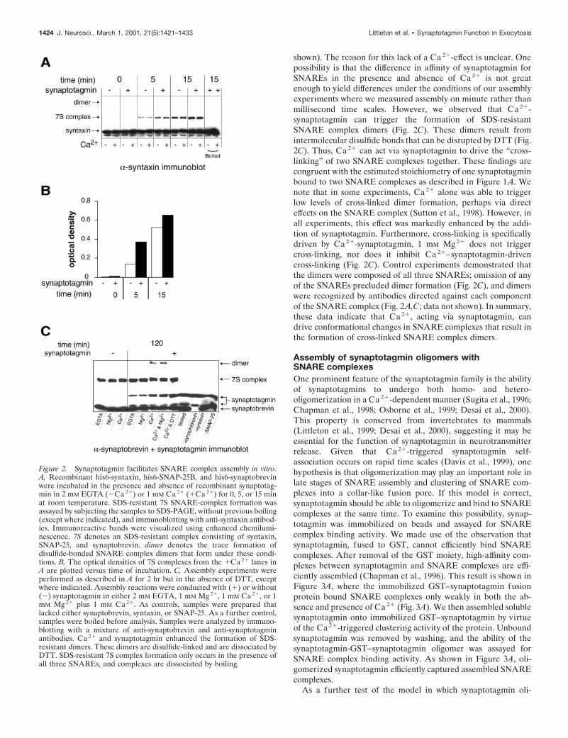
shown). The reason for this lack of a Ca21-effect is unclear. Onepossibility is that the difference in affinity of synaptotagmin forSNAREs in the presence and absence of Ca21 is not greatenough to yield differences under the conditions of our assemblyexperiments where we measured assembly on minute rather thanmillisecond time scales. However, we observed that Ca21-synaptotagmin can trigger the formation of SDS-resistantSNARE complex dimers (Fig. 2C). These dimers result fromintermolecular disulfide bonds that can be disrupted by DTT (Fig.2C). Thus, Ca21 can act via synaptotagmin to drive the “cross-linking” of two SNARE complexes together. These findings arecongruent with the estimated stoichiometry of one synaptotagminbound to two SNARE complexes as described in Figure 1A. Wenote that in some experiments, Ca21 alone was able to triggerlow levels of cross-linked dimer formation, perhaps via directeffects on the SNARE complex (Sutton et al., 1998). However, inall experiments, this effect was markedly enhanced by the addi-tion of synaptotagmin. Furthermore, cross-linking is specificallydriven by Ca21-synaptotagmin, 1 mM Mg21 does not triggercross-linking, nor does it inhibit Ca21–synaptotagmin-drivencross-linking (Fig. 2C). Control experiments demonstrated thatthe dimers were composed of all three SNAREs; omission of anyof the SNAREs precluded dimer formation (Fig. 2C), and dimerswere recognized by antibodies directed against each componentof the SNARE complex (Fig. 2A,C; data not shown). In summary,these data indicate that Ca21, acting via synaptotagmin, candrive conformational changes in SNARE complexes that result inthe formation of cross-linked SNARE complex dimers.
Assembly of synaptotagmin oligomers withSNARE complexesOne prominent feature of the synaptotagmin family is the abilityof synaptotagmins to undergo both homo- and hetero-oligomerization in a Ca21-dependent manner (Sugita et al., 1996;Chapman et al., 1998; Osborne et al., 1999; Desai et al., 2000).This property is conserved from invertebrates to mammals(Littleton et al., 1999; Desai et al., 2000), suggesting it may beessential for the function of synaptotagmin in neurotransmitterrelease. Given that Ca21-triggered synaptotagmin self-association occurs on rapid time scales (Davis et al., 1999), onehypothesis is that oligomerization may play an important role inlate stages of SNARE assembly and clustering of SNARE com-plexes into a collar-like fusion pore. If this model is correct,synaptotagmin should be able to oligomerize and bind to SNAREcomplexes at the same time. To examine this possibility, synap-totagmin was immobilized on beads and assayed for SNAREcomplex binding activity. We made use of the observation thatsynaptotagmin, fused to GST, cannot efficiently bind SNAREcomplexes. After removal of the GST moiety, high-affinity com-plexes between synaptotagmin and SNARE complexes are effi-ciently assembled (Chapman et al., 1996). This result is shown inFigure 3A, where the immobilized GST–synaptotagmin fusionprotein bound SNARE complexes only weakly in both the ab-sence and presence of Ca21 (Fig. 3A). We then assembled solublesynaptotagmin onto immobilized GST–synaptotagmin by virtueof the Ca21-triggered clustering activity of the protein. Unboundsynaptotagmin was removed by washing, and the ability of thesynaptotagmin-GST–synaptotagmin oligomer was assayed forSNARE complex binding activity. As shown in Figure 3A, oli-gomerized synaptotagmin efficiently captured assembled SNAREcomplexes.
As a further test of the model in which synaptotagmin oli-
Figure 2. Synaptotagmin facilitates SNARE complex assembly in vitro.A, Recombinant his6-syntaxin, his6-SNAP-25B, and his6-synaptobrevinwere incubated in the presence and absence of recombinant synaptotag-min in 2 mM EGTA (2Ca 21) or 1 mM Ca 21 (1Ca 21) for 0, 5, or 15 minat room temperature. SDS-resistant 7S SNARE-complex formation wasassayed by subjecting the samples to SDS-PAGE, without previous boiling(except where indicated), and immunoblotting with anti-syntaxin antibod-ies. Immunoreactive bands were visualized using enhanced chemilumi-nescence. 7S denotes an SDS-resistant complex consisting of syntaxin,SNAP-25, and synaptobrevin. dimer denotes the trace formation ofdisulfide-bonded SNARE complex dimers that form under these condi-tions. B, The optical densities of 7S complexes from the 1Ca 21 lanes inA are plotted versus time of incubation. C, Assembly experiments wereperformed as described in A for 2 hr but in the absence of DTT, exceptwhere indicated. Assembly reactions were conducted with (1) or without(2) synaptotagmin in either 2 mM EGTA, 1 mM Mg 21, 1 mM Ca 21, or 1mM Mg 21 plus 1 mM Ca 21. As controls, samples were prepared thatlacked either synaptobrevin, syntaxin, or SNAP-25. As a further control,samples were boiled before analysis. Samples were analyzed by immuno-blotting with a mixture of anti-synaptobrevin and anti-synaptotagminantibodies. Ca 21 and synaptotagmin enhanced the formation of SDS-resistant dimers. These dimers are disulfide-linked and are dissociated byDTT. SDS-resistant 7S complex formation only occurs in the presence ofall three SNAREs, and complexes are dissociated by boiling.
1424 J. Neurosci., March 1, 2001, 21(5):1421–1433 Littleton et al. • Synaptotagmin Function in Exocytosis

gomerizes and binds to SNAREs at the same time, we deter-mined whether SNARE complexes act as competitive inhibitorsof synaptotagmin oligomerization. For these experiments wemonitored the Ca21-dependent binding of soluble synaptotagminto immobilized synaptotagmin in the presence of increasing con-centrations of SNARE complexes. As shown in Figure 3B, addi-tion of SNAREs did not impede oligomerization. Our biochem-
ical observations are consistent with a role for synaptotagminoligomerization and SNARE binding in triggering vesicle fusion.To directly test this model, we characterized mutations in Drosoph-ila synaptotagmin I that block Ca21-dependent oligomerization.
The C2B domain of synaptotagmin is required for bothexocytosis and endocytosis of synaptic vesiclesin vivoA collection of 20 different alleles of synaptotagmin I (syt) havebeen generated in Drosophila (Littleton et al., 1993, 1994; Di-Antonio and Schwarz, 1994), providing useful experimental ma-terial to determine the mechanism by which synaptotagmin func-tions in synaptic vesicle cycling. Many of these mutations in syt,including P-element insertions, enhancer/promoter deletions, andearly stop codons in the open reading frame (DiAntonio andSchwarz, 1994; Littleton et al., 1994), disrupt synaptic function bydecreasing the levels of wild-type synaptotagmin at synapses.However, several syt alleles display intragenic complementation(Littleton et al., 1994). This form of complementation is oftenobserved for genes that encode proteins that are part of multi-meric complexes and that contain multiple distinct functionaldomains. Thus, intragenic complementation for syt mutationssuggests the presence of several independent domains withinsynaptotagmin that mediate distinct steps in neurotransmitterrelease. Two of the syt alleles involved in intragenic complemen-tation are AD1 and AD3. Flies containing various heteroalleliccombinations with AD1 and AD3 display defects including asevere lack of coordination and dramatically decreased viability.Previous electrophysiological analysis of heteroallelic combina-tions involving AD1 and AD3 (Littleton et al., 1994) demon-strated a profound decrease in synaptic exocytosis. At low Ca21
concentrations, evoked release is virtually abolished in AD3mutants. By raising extracellular Ca21 to 6 mM, exocytosis inAD3 mutants can be partially rescued (Fig. 4C). In contrast, AD1mutants have severe defects in synaptic transmission that cannotbe rescued by higher levels of extracellular Ca21 (Fig. 4C). Inaddition, recordings from synaptotagmin alleles that are viablewith AD1 (T7 and T41) and AD3 (T7, T41, T11, D2, D3, D37,D45) show either the AD1 or AD3 phenotype regardless of theother allele with which AD1 or AD3 are paired. Indeed, the samesynaptotagmin mutants (T41, T7) behave dramatically differentwhen paired with AD1 or AD3. Thus, the AD1 and AD3 allelesconfer the dominant phenotype to any synaptotagmin allele withwhich they are paired (even when paired with the same alleles—T7, T41), leading us to focus on the molecular defects in the AD1and AD3 mutants. Immunolocalization studies reveal that themutant synaptotagmins are targeted to synapses in AD1 and AD3mutants (data not shown). The amount of synaptotagmin that ispresent at mutant synapses is difficult to quantify precisely be-cause we do not know how these mutations affect the ability ofour anti-synaptotagmin I antibody to detect the mutated proteinin vivo. However, AD1 and AD3 mutants over a deletion thatcompletely removes synaptotagmin are far less severe phenotyp-ically than null mutants such as T77 and AD4 over deletion(Littleton et al., 1994) and survive much longer as larva than donull mutants. These observations directly demonstrate that theAD1 and AD3 mutant synaptotagmin proteins are made and havepartial function at synapses, allowing these mutants to surviveand function more efficiently than mutants that completely re-move synaptotagmin and die as embryos. Thus, an altered func-
Figure 3. Oligomerized synaptotagmin binds to assembled SNAREcomplexes. A, GST and GST–synaptotagmin were immobilized on beads(15 mg per data point) and assayed for binding to midi–SNARE com-plexes (2 mM) in 2 mM EGTA (2Ca 21) or 1 mM Ca 21 (1Ca 21) in 150ml of HBS using a cosedimentation assay, as described in Materials andMethods. To leave SNARE complexes intact, samples were subjected toSDS-PAGE without previous boiling. Coomassie staining revealed onlylow levels of SNARE binding to immobilized synaptotagmin in eithercondition. Immobilized synaptotagmin was then preincubated with solu-ble synaptotagmin (10 mM) in EGTA or Ca 21. Beads were washed threetimes to remove unbound soluble synaptotagmin, and the soluble- immo-bilized synaptotagmin oligomers were assayed for binding to midi–SNARE complexes. Twenty-five percent of the bound material wasloaded onto the gel; the lef t two lanes correspond to 0.3 and 0.5 mg ofsoluble synaptotagmin and midi–SNARE complex, respectively. Coomas-sie staining revealed efficient binding of soluble synaptotagmin to immo-bilized synaptotagmin. Furthermore, midi–SNARE complexes efficientlybound to the soluble-immobilized synaptotagmin oligomers. These resultsdemonstrate that synaptotagmin, which has oligomerized, is capable ofbinding SNARE complexes. *Denotes proteolytic fragments from GST–synaptotagmin. Note, Ca 21 induces a shift in the mobility of synaptotag-min that has not been boiled. Therefore, soluble and GST–synaptotagminare indicated with double arrows. B, SNARE complexes do not inhibitsynaptotagmin oligomerization. GST (12 mg per data point) andGST–synaptotagmin (8 mg per data point) were immobilized on beads.Soluble synaptotagmin (1.5 mM; 1) and midi–SNARE complex (6 mM; 1)or the indicated [SNARE complex] were incubated with the beads in 2mM EGTA (2) or 1 mM Ca 21 (1) for 1.5 hr. Samples were also preparedthat lacked SNARE complexes (2) or soluble synaptotagmin (2). Boundmaterial was boiled in SDS sample buffer and subjected to SDS-PAGE.Twenty-five percent of the bound material was loaded onto the gel; totalcorresponds to the mixture of 0.3 and 0.7 mg of soluble synaptotagmin andmidi–SNARE complex. Gels were stained with Coomassie blue to visu-alize bound synaptotagmin. Staining of disassembled SNARE complexeswas poor, therefore SNARE binding was detected by immunoblottingwith anti-SNAP-25 and anti-syntaxin antibodies. Immunoreactive bandswere visualized using enhanced chemiluminescence.
Littleton et al. • Synaptotagmin Function in Exocytosis J. Neurosci., March 1, 2001, 21(5):1421–1433 1425

tion of the mutant synaptotagmins, rather than a loss of theprotein at synapses, is likely the cause of the electrophysiologicaldefects. We cannot completely rule out some contribution to thephenotype from altered protein levels that are beyond our detec-tion. Sequence analysis of AD1 and AD3 revealed that the AD1phenotype is caused by a premature stop codon that deletes theC2B domain. AD3 results from a Y to N substitution in C2B(DiAntonio and Schwarz, 1994) at a residue (364) that is highlyconserved in all synaptotagmin isoforms from Caenorhabditiselegans to humans (Fig. 4A). The crystal structure of the cyto-plasmic domain of rat synaptotagmin III (Sutton et al., 1999)indicates that the AD3 mutation lies near two conserved aspartateresidues that may function as Ca21 ligands (Fig. 4B). These twomutants allow us to investigate the in vivo roles of the C2Bdomain of synaptotagmin I in synaptic function.
We first examined morphological defects in AD1 and AD3mutants to determine where in the synaptic vesicle cycle eachmutant is blocked. For this analysis we examined the first opticneuropil containing 800 highly stereotypic optic cartridges withdefined synaptic contacts between photoreceptor axons and lam-inar neurons that can be readily identified by the presence ofpresynaptic T-bars. We focused specifically on the histaminergicsynapses between photoreceptors (R1–R6) and postsynaptic lam-inar neurons (L1 and L2). Electroretinogram (ERG) recordings,in which synaptic transmission between photoreceptors and sec-ond order neurons in the lamina is indicated by the on and offtransients in response to a light flash, revealed that both AD1 andAD3 heteroallelic mutants lack these transients (Fig. 5). Thus,these mutations disrupt synaptic transmission at photoreceptorsynapses as well as at neuromuscular junctions.
To examine the morphological correlates of the block in syn-aptic transmission in AD1 and AD3, electron microscopy wasperformed on the photoreceptor synapses after 10 min of con-stant light stimulation before fixation to drive continuous vesiclecycling. A total of 168 micrographs were examined from cn,AD1/T41, AD1/T7, and AD3/T11 flies (n 5 3–10 flies for eachgenotype). The overall architecture of the lamina was normal insyt mutants. The most dramatic difference was a decrease ( p ,0.05, unpaired Student’s t test) in the number of synaptic vesiclesin photoreceptor terminals of AD1/T7 and AD1/T41 mutantscompared with controls (AD1 cn/T41 cn, 25 1 14 SD; AD1 cn/T7cn, 27 6 20 SD; AD3 cn/T11 cn, 88 6 28 SD; cn controls, 96 6 37SD (Fig. 5, compare A, B), suggesting a defect in endocytosis inAD1 heteroallelic mutants. Although we cannot rule out that theloss of synaptic vesicles in AD1 mutants is caused by a defect invesicle biogenesis from an internal compartment as opposed to adirect defect in endocytosis, the lack of any vesicle biogenesisdefect in synaptotagmin null mutants (Reist et al., 1998) arguesagainst this alternative interpretation. In contrast, AD3 mutantsdid not have depleted nerve terminals compared with controls,indicating that endocytosis in not disrupted in this mutant. In-deed, synaptic vesicles could be clearly visualized in contact withthe presynaptic membrane under T-bars (Fig. 5C), indicating thatAD3 mutant vesicles can undergo docking but are defective at alater step in exocytosis (2.6 6 1.4 SD docked vesicles in AD3cn/T11 cn compared with 2.3 6 0.9 in cn controls).
The morphological and electrophysiological analysis of AD1and AD3 suggest that fundamentally different processes are af-fected in the two mutants. AD1 terminals are relatively depletedof synaptic vesicles compared with controls (Fig. 5B), and synap-
Figure 4. Mutations in the C2B domainof Drosophila synaptotagmin I. A, Align-ment of the C2B domain sequence sur-rounding the Y364N change found inthe AD3 mutant (DiAntonio andSchwartz, 1994). The five putative Ca 21
ligands are highlighted in gray, whereasthe AD3 change is indicated in black.Y364 is conserved among all synapto-tagmin isoforms from C. elegans to hu-mans. B, Predicted structure of the AD1and AD3 mutant proteins based on thecrystal structure of synaptotagmin III(see Fig. 9 for details). The location ofthe Y to N change in AD3 is indicatedby the arrow. The AD1 mutations resultin a premature stop codon deleting theC2B domain. C, The electrophysiologicaldefects observed in AD3 and AD1 het-eroallelic combinations (Littleton et al.,1994) are plotted against the responses ofthe control cn bw sp line. Recordingswere made in 0.4 or 6.0 mM Ca 21 in Jan’sRinger’s solution. Excitatory junctionalpotential (EJP) amplitude at muscle fiber6 in segments A3–A5 is plotted vs theextracellular Ca 21 concentration. At lowCa 21, both AD1 and AD3 exhibit a pro-found block in evoked secretion. Athigher Ca 21 levels, the defects in AD3mutants can be partially rescued, whereasAD1 mutants continue to have dramati-cally abnormal synaptic responses. TheseEJP responses have not been correctedfor nonlinear summation. Thus, both synaptotagmin mutants still have significant defects compared with control responses even in high calcium, where thecontrol responses already saturated at these calcium levels. Dominant defects from the AD1 and AD3 alleles when paired with a wild-type allele ofsynaptotagmin have not been observed (Littleton et al., 1994).
1426 J. Neurosci., March 1, 2001, 21(5):1421–1433 Littleton et al. • Synaptotagmin Function in Exocytosis

tic transmission cannot be rescued by high extracellular Ca21
(Fig. 4C). AD3 mutants have a defect in exocytosis, not endocy-tosis, (Fig. 5C; see Fig. 7A), and release can be partially rescuedby high extracellular Ca21 (Fig. 4C). Thus, the morphologicallydocked vesicles in AD3 mutants are also physiologically compe-tent for release but require significantly higher Ca21 concentra-tions. We therefore examined the biochemical defects caused byAD1 and AD3 to determine which activities of the C2B domainare required for endocytosis and exocytosis, respectively. For thisanalysis we generated GST fusion proteins containing the cyto-plasmic domains from wild-type Drosophila synaptotagmin, AD1(C2A domain alone lacking C2B), and AD3. To determinewhether AD1 and AD3 are able to penetrate membranes in the
presence of Ca21, we tested the immobilized recombinant cyto-plasmic domains for their ability to bind liposomes (25% phos-phatidyl serine, 75% phosphatidyl choline) with or without Ca21
(Fig. 6A). Wild-type, AD1, and AD3 fusion proteins all showedrobust Ca21-dependent phospholipid binding, an activity previ-ously shown to be mediated by the C2A domain of synaptotagminIa (Bai et al., 2000; Desai et al., 2000). We next examined bindingto the t-SNARE, syntaxin 1A, whose Ca21 dependence is alsomediated by Ca21 ligands in the 2A domain of synaptotagmin Ia(Bai et al., 2000; Desai et al., 2000). Recombinant Drosophilasyntaxin 1 was able to interact with wild-type, AD1, and AD3fusion proteins (Fig. 6B). We also examined the interaction ofsynaptotagmin with the clathrin adapter AP-2. Binding of theAP-2 complex from Drosophila head extracts was detected with anantibody generated against a-adaptin (Gonzalez-Gaitan et al.,1996). Whereas wild-type and AD3 synaptotagmin bound AP-2in the absence or presence of Ca21, AD1 synaptotagmin did notbind AP-2 under either condition (Fig. 6B). Thus, as reported formammalian synaptotagmin I, AP-2 binding to Drosophila synap-totagmin is also mediated through the C2B domain (Zhang et al.,1994). We conclude that AP-2 binding to synaptotagmin andsubsequent clathrin recruitment is altered in AD1 mutants, lead-ing to defective endocytosis and a relative depletion of synaptic
Figure 5. Ultrastructural analysis of stimulated synapses in C2B mutants.Ultrastructural defects in control cn (A), AD3 cn/T11 cn (B), and AD1cn/T41 cn (C) photoreceptor synapses were examined by driving photo-receptors with constant light stimulation for 10 min, followed by rapidfixation. Both AD1 and AD3 mutants lack the on–off transients measuredduring ERG recordings in the retina (shown on the right), demonstratingthat synaptic transmission is disrupted at these photoreceptor synapses.AD1 mutants show a decrease in the overall number of synaptic vesicles,whereas AD3 synapses do not show a depletion of synaptic vesicles, butrather a defect in the ability of docked synaptic vesicles to fuse. Quanti-fication of vesicles per photoreceptor synapse for each of the genotypeswas: AD1 cn/T41 cn, 25 6 14 SD; AD1 cn/T7 cn, 27 6 20 SD; AD3 cn/T11cn, 88 6 28 SD; cn controls, 96 6 37 SD. Quantification of vesicles per T-barfor each of the genotypes was: AD1 cn/T41 cn, 1.4 6 0.9 SD; AD1 cn/T7 cn,1.9 6 0.9 SD; AD3 cn/T11 cn, 2.6 6 1.4 SD; cn controls, 2.3 6 0.9 SD.
Figure 6. Synaptotagmin AD1 mutants fail to bind AP-2. A, Ca 21-dependent phospholipid binding of immobilized recombinant wild-type(WT ), AD3, or AD1 synaptotagmin I proteins. Both AD1 and AD3recombinant proteins showed robust Ca 21-stimulated phospholipid bind-ing. Phospholipid binding assays were conducted as previously described(Littleton et al., 1999). B, Binding of recombinant syntaxin (5 mM) andnative AP-2 a-adaptin (0.2 mg of Drosophila head membranes) to 30 mgof recombinant WT, AD3, or AD1 Drosophila synaptotagmins in 2 mMEGTA or 1 mM Ca 21 for 2 hr at 4°C. For detection of recombinantsyntaxin binding to synaptotagmins, Western analysis with the monoclo-nal anti-syntaxin antisera 8C3 was performed. For analysis of AP-2binding, fly head membranes were prepared as previously described(Littleton et al., 1998), and AP-2 binding was detected with a polyclonalantibody generated against a-adaptin (Gonzalez-Gaitan and Jackle,1996). Immunoreactive bands were visualized by enhanced chemilumines-cence. Both AD1 and AD3 mutant proteins showed Ca 21-dependentbinding to syntaxin. However, only AD3 showed an interaction with AP-2.
Littleton et al. • Synaptotagmin Function in Exocytosis J. Neurosci., March 1, 2001, 21(5):1421–1433 1427

vesicles in stimulated synapses. Although complete removal ofthe C2B domain would also be expected to disrupt the exocytoticactivities mediated by C2B, the loss of vesicles in the AD1 mutantdominates the morphological and electrophysiological phenotype.A similar morphological depletion of synaptic vesicles has beenobserved in a C. elegans synaptotagmin mutant that also deletesthe C2B domain (Jorgensen et al., 1995). These endocytoticdefects preclude the investigation of the role of the C2B domainin exocytosis in AD1 mutants. The lack of any defect in AP-2binding by the AD3 mutant protein and the corresponding lack ofan endocytotic defect by morphological or electrophysiologicalcriteria in AD3 mutants defines a second function for the C2Bdomain of synaptotagmin in synaptic vesicle exocytosis that isdisrupted in AD3 mutants.
The AD3 mutation selectively impairs Ca21-drivenconformational changes and Ca21-triggeredoligomerization of the C2B domain of synaptotagminSynaptic vesicles in AD3 mutants are capable of translocating anddocking at active zones during synaptic stimulation, as shown inFigure 7A. Thus, it is likely that the defect in AD3 mutants liessomewhere after docking. This interval encompasses both prim-ing and fusion, although it is unknown what molecular eventsoccur during this period. One possibility is that individualSNAREs assemble into various stages of “loose” and “tight”states of SNARE complexes (Xu et al., 1999), generating a
potential fusion pore that can be triggered to open by Ca21. Wewere thus interested in determining whether there were defects ina specific stage of SNARE assembly in the AD3 mutant. Onepossibility is that synaptotagmin is required to trigger SNAREcomplex assembly. Another possibility is that synaptotagminbinds preassembled SNARE complexes and prevents them frommediating full fusion until arrival of a Ca21 signal. To explorethese possibilities, we examined 7S complexes in head extracts ofAD3 mutants. SNARE complexes do not form in SDS (Littletonet al., 1998), but once the complex is formed, they are resistant todissociation by SDS unless the sample is boiled (Hayashi et al.,1994). To assay complex formation in flies, supernatants fromSDS-solubilized control and AD3 mutant fly heads were sepa-rated on SDS-PAGE gels and probed with an anti-syntaxin anti-serum. Monomeric 35 kDa syntaxin can be detected, as well asthe 73 kDa SNARE complex containing syntaxin, synaptobrevin,and SNAP-25. Because 7S SNARE complexes do not form inSDS, the complex we detect corresponds to that present in vivo.AD3 mutants show a dramatic decrease in the amount of 7Scomplex (Fig. 7B), consistent with a defect in the ability ofsynaptotagmin to trigger SNARE complex formation rather thana defect in triggering fusion after SDS-resistant SNARE complexassembly. The block in SNARE complex assembly in AD3 mu-tants suggests an activity mediated by the C2B domain of synap-totagmin that also acts late in the exocytotic pathway to triggerSNARE assembly and consequent fusion. We therefore under-
Figure 7. Synaptotagmin AD3 mutantsdecrease SNARE complex assembly invivo. A, Enlarged image of an activezone in AD3/T11 mutant photoreceptorterminals demonstrating docked vesicles(arrowheads) under a T-bar that havenot fused. B, 7S complexes from 10 con-trol (CS) or synaptotagmin AD3/T11mutants were isolated. Syntaxin ispresent in a 35 kDa monomeric formand in a 73 kDa complex with SNAP-25and synaptobrevin in wild-type flies. Asevere reduction in the amount of 7Scomplex was found in AD3/T11 synapto-tagmin mutants. C, Both wild-type andAD3 recombinant synaptotagmins areable to bind SNARE complexes in aCa 21-stimulated manner. Either 3 mMwild-type (sytWT ) or AD3 mutant syn-aptotagmin (sytAD3 ) was incubated with3 mM midi–SNARE complex for 1.5 hrin either 2 mM EGTA (E) or 1 mMCa 21. Midi–SNARE complex was im-munoprecipitated, and samples wereseparated by SDS-PAGE and stainedwith Coomassie blue. As a control,samples were prepared that lackedmidi–SNARE complex and immuno-precipitating antibodies. Thirty percentof the immunoprecipitated material wasloaded onto the gel; total corresponds to6% of the binding reaction. Note: theasterisk indicates a proteolytic fragmentpresent in preparations of soluble AD3mutant rat synaptotagmin. D, Both wild-type and AD3 synaptotagmins are ableto bind the mammalian synprint pep-tide. Ten micrograms of GST or GSTfused to the cytoplasmic domain of WTor AD3 mutant synaptotagmin were immobilized on beads and incubated with 1 mM T7-tagged synprint for 2 hr in 2 mM EGTA (E) or 1 mM Ca 21.Samples were washed, and bound material was subjected to SDS-PAGE and immunoblot analysis using an anti-T7 tag antibody and enhancedchemiluminescence. Twelve percent of the bound material was loaded onto the gel; total corresponds to 3.5% of the binding reaction.
1428 J. Neurosci., March 1, 2001, 21(5):1421–1433 Littleton et al. • Synaptotagmin Function in Exocytosis

took an investigation of the defects resulting from the Y364Nchange in C2B in the AD3 mutant to uncover a biochemical linkbetween synaptotagmin, assembly of the SNARE complex, andvesicle fusion.
For this analysis, we engineered the AD3 mutation into ratsynaptotagmin I (corresponding to amino acid 311 in the ratsequence) to be able to use the biochemical reagents available forthis organism. One possibility to explain the AD3 phenotype isthat the mutant protein fails to interact with the SNARE complexto trigger Ca21-induced conformational changes required forfusion. However, both wild-type and AD3 mutant synaptotagminshowed comparable binding to preassembled SNARE complexes(Fig. 7C). Another possibility is that the interaction of synapto-tagmin with the synprint domain of the presynaptic Ca21 channelis affected, resulting in an alteration in Ca21 entry that couldblock the interaction of a second Ca21 sensor with the SNAREcomponents. Although the C2B domain is required for the inter-action with synprint, the AD3 mutation does not affect thisinteraction (Fig. 7D).
The AD3 mutation lies in close proximity to a set of fiveconserved amino acid residues that coordinate divalent cations ina number of C2 domains (Fig. 8A). These structural data suggestthat the AD3 mutation may impair the putative Ca21-bindingproperties of C2B. To test this hypothesis, the C2B domain ofwild-type and AD3 mutant rat synaptotagmin were immobilizedas GST fusion proteins (the isolated C2B domain of Drosophila is
insoluble and could not be produced in sufficient quantities toperform this analysis). Fusion proteins were incubated with in-creasing concentrations of chymotrypsin in the presence ofEGTA or Ca21, and the proteolysis patterns were analyzed bySDS-PAGE. As shown in Figure 8B, the degradation patterns inEGTA versus Ca21 were distinct, demonstrating that C2B un-dergoes a conformational change after binding Ca21. A protease-resistant fragment accumulated in the presence of Ca21, suggest-ing that Ca 21 binds to and stabilizes this domain. This result isanalogous to the data from limited proteolysis of the C2A domain(Davletov and Sudhof, 1994). In contrast, a different proteol-ysis pattern was observed with the AD3 mutant C2B domainand the presence of Ca 21 has no effect on this pattern. Theseresults suggest that the AD3 mutation impairs Ca 21-drivenconformational changes in C2B. We note that the ability of theAD3 mutant to bind SNARE complexes, AP-2, and the syn-print peptide, as described above, demonstrates that the C2Bdomain is not misfolded, but rather, exhibits a selective loss offunction.
The C2B domain of synaptotagmin mediates Ca21-triggeredoligomerization as previously described (Chapman et al., 1996;Sugita et al., 1996; Desai et al., 2000). To determine whether thisoligomerization activity is affected by the observed alteration inCa21-dependent conformational changes in the AD3 C2B do-main, oligomerization of the cytoplasmic domains of wild-typeand AD3 Drosophila synaptotagmin were assayed at increasing
Figure 8. The AD3 mutation blocksCa 21-driven conformational changeswithin the C2B domain of synaptotagminand disrupts Ca 21-triggered oligomer-ization activity. A, Crystal structure ofthe C2B domain of synaptotagmin III.This image was modified from Sutton etal. (1999); the structure of the C2B do-main of synaptotagmin I has not beenreported, however, all known C2B do-mains share similar structures. The ty-rosine that is mutated to an asparaginein the AD3 mutant allele of Drosophilasynaptotagmin is indicated, as are fiveputative Ca 21 ligands and a singlebound Mg 21 ion. B, The C2B domain ofWT (GST-C2BWT ) and AD3 (GST-C2BAD3 ) rat synaptotagmin Ib were im-mobilized as a GST fusion proteins (20mg/data point) and subjected to limitedproteolysis in the presence of 2 mMEGTA (2) or 1 mM Ca 21 (1) at theindicated [chymotrypsin] for 60 min atrt. Samples were boiled in SDS samplebuffer, analyzed by SDS PAGE, andstained with Coomassie blue. C, Ca 21-triggered synaptotagmin oligomeriza-tion is impaired by the AD3 mutation.Eight micrograms of GST or GST fusedto the cytoplasmic domain of wild-type(sytWT ) or AD3 mutant (sytAD3 ) Dro-sophila synaptotagmin was immobilized
on beads. Beads were incubated with 1.5 mM soluble WT or AD3 mutant Drosophila synaptotagmin for 1.5 hr in 150 ml of TBS plus 0.5% Triton X-100and either 2 mM EGTA (2), 1 mM Ca 21 (1), or the indicated concentration of Ca 21. Beads were washed three times with binding buffer and boiledin SDS sample buffer. Three percent of the soluble synaptotagmin from the binding assay (lef t two lanes) and 25% of the bound material (remaining lanes)were subjected to SDS-PAGE and visualized by staining with Coomassie blue. Syt, Cytoplasmic domain of synaptotagmin I. Note: the asterisk indicatesa proteolytic fragment present in preparations of GST-fused AD3 mutant synaptotagmin. D, Data from two oligomerization assays (as described in C)were quantified by densitometry, normalized to the pixel intensity in the “total” lanes, and plotted versus the free [Ca 21]. Closed circles, Wild-typesynaptotagmin; open circles, AD3 synaptotagmin. E, Soluble Drosophila synaptotagmin I (5 mM) was incubated with 30 mg of either wild-type synaptotagminIV, AD3 synaptotagmin IV, or synaptotagmin IV containing a KK to AA substitution (Chapman et al., 1998) at amino acids 385 and 386. Binding ofsynaptotagmin I was visualized by Western analysis with anti-synaptotagmin I DSYT2 antisera (Littleton et al., 1993). ● denotes binding reactions lackingsoluble synaptotagmin.
Littleton et al. • Synaptotagmin Function in Exocytosis J. Neurosci., March 1, 2001, 21(5):1421–1433 1429

concentrations of Ca21. As shown in Figure 8, C and D, wild-type synaptotagmin efficiently bound to immobilized synaptotag-min in response to Ca 21. In contrast, Ca21-triggered oligomer-ization of the AD3 mutant was reduced to ;40% of the wild-typeclustering activity. These data suggest that the AD3 phenotyperesults from a defect in the Ca21-sensing ability of the C2Bdomain, causing a loss of Ca 21-induced synaptotagmin oligomer-ization and subsequent SNARE assembly in vivo. To obtainadditional evidence for this model, we tested whether the AD3 Yto N change also alters Ca21-dependent oligomerization whenintroduced into other isoforms of synaptotagmin. As shown inFigure 8E, when the AD3 change is engineered into Drosophilasynaptotagmin IV, Ca21-dependent hetero-oligomerization withwild-type synaptotagmin I is decreased by .80%. The AD3mutation disrupted oligomerization as effectively as the wellcharacterized K326,327A substitution within the C2B domain ofrat synaptotagmin I (Chapman et al., 1998; Desai et al., 2000;note: this mutation corresponds to K385, K386 in Drosophilasynaptotagmin IV).
Under our assay conditions, oligomerization of wild-type syn-aptotagmin was half-maximal at ;6 mM Ca 21. This value is
considerably lower than the Ca21 dependence for exocytosis inretinal bipolar neurons (Heidelberger et al., 1994) but is consis-tent with the Ca21 dependence for exocytosis at the axosomaticsynapse formed by the calyx of Held (Bollman et al., 2000;Schneggenburger and Neher, 2000).
DISCUSSION
Mutations in synaptotagmin I result in profound defects in neu-rotransmitter release. In Drosophila, these defects include a se-vere reduction in evoked release, an increase in spontaneousfusion, and delays in the onset of vesicle fusion (Littleton et al.,1993, 1994; DiAntonio and Schwarz, 1994). In mice, disruption ofthe synaptotagmin I gene results in the selective loss of the rapidsynchronous component of exocytosis without affecting the fre-quency of spontaneous fusion events (Geppert et al., 1994). Incontrast, removal of the t-SNARE syntaxin eliminates bothevoked and spontaneous fusion (Schulze et al., 1995), andtemperature-sensitive mutations in syntaxin that block SNAREassembly result in paralysis and an accumulation of unfusedvesicles at release sites (Littleton et al., 1998). These results are
Figure 9. Model depicting the synap-totagmin-SNARE complex. A, The core ofthe SNARE complex, the Habc domain ofsyntaxin, the cytoplasmic domain of syn-aptotagmin, and a simulated lipid bilayerwere modified from Sutton et al. (1998),Fernandez et al. (1998), Sutton et al.(1999), and Heller et al. (1993), respec-tively, and rendered using MOLSCRIPT(Kraulis, 1991). The regions that interactare indicated with brackets; both C2 do-mains of synaptotagmin are required forhigh affinity binding to the base of theSNARE complex (Chapman et al., 1995;Davis et al., 1999; Gerona et al., 2000). Thetransmembrane anchors of syntaxin, syn-aptobrevin, and synaptotagmin were gen-erated by molecular modeling. B, Modelfor synaptotagmin-mediated assembly andclustering of SNARE complexes. One syn-aptotagmin can interact with two SNAREcomplexes; this interaction is depicted astwo grooves within synaptotagmin thatbind and assemble SNAREs (which areshown in an “end view” in which eachstrand of the four-helix bundle is depictedas a quarter of a circle).
1430 J. Neurosci., March 1, 2001, 21(5):1421–1433 Littleton et al. • Synaptotagmin Function in Exocytosis

consistent with mounting functional data indicating that assemblyof the SNARE complex is essential for vesicle fusion (Weber etal., 1998; Chen et al., 1999; Xu et al., 1999). The more variableeffects on secretion in synaptotagmin mutants are consistent withthe possibility that synaptotagmin plays a regulatory role in pro-moting evoked release through direct interactions with the fusionmachinery, without being absolutely necessary for vesicle fusion.Here, we provide evidence for two independent functions for theC2B domain of synaptotagmin I in synaptic vesicle cycling. AD1mutations, which lack the C2B domain, disrupt synaptotagmin—AP-2 interactions (Zhang et al., 1994) and lead to a fourfoldreduction in the total number of synaptic vesicles at mutantterminals during nerve terminal stimulation. These findings aresimilar to studies of synaptotagmin mutants in C. elegans (Jor-gensen et al., 1995). AD1 terminals do harbor some synapticvesicles surrounding active zones and have an elevated frequencyof spontaneous fusions when examined electrophysiologically.Thus, it is likely that additional endocytotic pathways are capableof maintaining the smaller pool of vesicles that are recycled in theimmediate vicinity of the active zone. Our observations are con-sistent with the distribution of Drosophila AP-2, which is absentnear active zones, but concentrated in the synaptic periphery(Gonzalez-Gaitan et al., 1996). The lack of a complete loss ofendocytosis in Drosophila synaptotagmin null mutants (Reist etal., 1998) also indicates that in unstimulated synapses, otherendocytotic pathways that bypass synaptotagmin I can refill nerveterminals given enough time. One possible candidate for mediat-ing this endocytotic trafficking in the absence of synaptotagmin Iis synaptotagmin IV, which is also present on synaptotagminI-containing vesicles and binds to AP-2 as effectively as synapto-tagmin I (Li et al., 1995; Littleton et al., 1999).
In contrast with the endocytotic defect manifested in AD1mutants, the AD3 mutant phenotype results from a postdockingdefect in synaptic vesicle exocytosis and a failure to assembleSNARE complexes in vivo. Our biochemical analysis revealedthat this mutation disrupts Ca21-induced conformationalchanges in the C2B domain and inhibits Ca21-induced oligomer-ization. Whether the failure to assemble SNARE complexesresults from a direct defect in the acceleration of SNARE for-mation by synaptotagmin or an alteration in additional down-stream SNARE interactions after synaptotagmin clustering re-quires further analysis. Nonetheless, these results indicate thatthe C2B domain of synaptotagmin must be able to bind Ca21,change conformation, and cluster to trigger coordinated vesiclefusion at nerve terminals. These results provide biochemicallysupported genetic evidence that synaptotagmin is indeed aCa 21 sensor for fast exocytosis, as proposed in previous stud-ies (Brose et al., 1992; DiAntonio and Schwarz, 1994; Geppertet al., 1994; Littleton et al., 1994). How oligomerization ofsynaptotagmin leads to vesicle fusion is unknown, but it islikely to involve direct effects on the SNARE complex. Wesuggest that clustering of SNARE complexes by Ca 21-synaptotagmin leads to rapid triggering of fusion via formationof SNARE-dependent fusion pores. The Ca 21-independentinteraction of synaptotagmin with SNAREs would allow thesevesicles to remain in a fusion-ready state and may contribute tothe suppression of spontaneous fusion events (DiAntonio andSchwartz, 1994, 1999; Littleton et al., 1994). The loss ofSNARE clustering activity in synaptotagmin mutants wouldprevent rapid, Ca 21-dependent vesicle fusion as has beenobserved in syt mutants. The partial rescue of release in sytmutants at very high Ca 21 levels could reflect the ability of
other Ca 21 sensors to trigger fusion under these conditions.These other Ca 21 sensors might be other members of thesynaptotagmin family, which contains seven members in Dro-sophila. Biochemical characterization of the Ca 21-dependentoligomerization properties of these synaptotagmins might un-cover other candidates for mediating fusion at high Ca 21
concentrations.Scale models reveal precisely how close complete assembly of
the SNARE complex would bring the vesicle and target mem-branes (Fig. 9A; Sutton et al., 1998). A number of experimentsindicate that SNARE complexes do not fully assemble beforefusion (Chen et al., 1999; Xu et al., 1999). Thus, final “zippering”of the complex may not occur until arrival of the Ca21 signal thattriggers fusion (Chen et al., 1999). In this case, Ca21-triggeredoligomerization of synaptotagmin and its association with par-tially assembled SNARE complexes could drive final assembly ofthe base of the complex (Chapman et al., 1995; Kee and Scheller,1996; Davis et al., 1999; Gerona et al., 2000) to accelerateSNARE-mediated membrane fusion and SDS-resistant SNAREcomplex assembly (Weber et al., 1998). The facilitation ofSNARE complex assembly, in vitro and in vivo, reported heresupports this model. Furthermore, Ca21-synaptotagmin triggersthe formation of disulfide-bonded SNARE complex dimers. Thisfinding, in conjunction with the 1:2 stoichiometry of saturatedsynaptotagmin-SNARE complexes, argues that Ca21-synaptotagmin can bring at least two SNARE complexes intoclose proximity. The ability of synaptotagmin to oligomerize andsimultaneously bind SNAREs suggests that synaptotagmin cancluster multiple SNARE complexes into a higher orderedassembly that might correspond to the exocytotic fusion pore.In this light we point out that SNARE complexes alone appearto cluster only weakly (Fasshauer et al., 1997; Hohl et al., 1998;but see also Poirier et al., 1998) and that viral fusion proteinsare homotrimers that must assemble into oligomers to formfunctional fusion pores (Blumenthal et al., 1996; Danieli et al.,1996). Similarly, a protein designated EEA1 has recently beenreported to cluster syntaxin-13 into oligomeric structures that arerequired for endosomal fusion (McBride et al., 1999). An analo-gous model for synaptotagmin in neuronal exocytosis is shown inFigure 9. Synaptotagmin binds to the membrane proximal regionof the SNARE complex while simultaneously penetrating intomembranes (Davis et al., 1999; Gerona et al., 2000; Fig. 9A). Wespeculate that these interactions, in conjunction with the ability ofsynaptotagmin to oligomerize and to facilitate SNARE complexassembly (Fig. 9B), leads to formation of an open fusion pore. Insupport of this model, we have recently shown that disruptingC2B-mediated oligomerization of synaptotagmin I in vitro can leadto postdocking vesicle fusion defects in cracked PC12 cells (Desaiet al., 2000).
Finally, we point out that the N terminus of synaptotagmincontains a novel Ca21-independent clustering domain that mayplay an important role in both endocytosis (von Poser et al., 2000)and exocytosis (Bai et al., 2000). We speculate that N-terminalclustering activity, in conjunction with the weak Ca21-independent components of C2B-C2B and synaptotagmin–SNARE interactions, may serve to poise synaptotagmin–SNARE complexes for rapid conformational changes, includingthe formation of a stable ring-like structure, in response to a risein intracellular Ca21.
In summary, our data indicate that the C2B domain of synap-totagmin must “sense” Ca21 and assemble into clusters, fordocked synaptic vesicles to undergo synchronous exocytosis.
Littleton et al. • Synaptotagmin Function in Exocytosis J. Neurosci., March 1, 2001, 21(5):1421–1433 1431

These data further suggest Ca21–synaptotagmin can regulateSNARE complex dynamics, thus providing a compelling connec-tion between the Ca21 sensor for exocytosis and the SNAREfusion machinery.
REFERENCESBai J, Earles C, Lewis J, Chapman ER (2000) Membrane-embedded
synaptotagmin interacts with cis and trans target membranes and as-sembles into oligomers via a novel mechanism. J Biol Chem275:25427–25435.
Bark IC, Wilson MC (1994) Human cDNA clones encoding two differentisoforms of the nerve terminal protein SNAP-25. Gene 139:291–292.
Bennett MK, Calakos N, Scheller RH (1992) Syntaxin: a synaptic pro-tein implicated in docking of synaptic vesicles at presynaptic activezones. Science 257:255–259.
Blumenthal R, Sarkar DP, Durell S, Howard DE, Morris SJ (1996)Dilation of the influenza hemagglutinin fusion pore revealed by kinet-ics of individual cell-cell fusion events. J Cell Biol 135:63–71.
Bollman JH, Sakmann B, Borst JGG (2000) Calcium sensitivity of glu-tamate release in a Calyx-type terminal. Science 289:953–957.
Brose N, Petrenko AG, Sudhof TC, Jahn R (1992) Synaptotagmin: aCa 21 sensor on the synaptic vesicle surface. Science 256:1021–1025.
Chapman ER, Jahn R (1994) Calcium-dependent interaction of the cy-toplasmic region of synaptotagmin with membranes. J Cell Biol269:5735–5741.
Chapman ER, Hanson PI, An S, Jahn R (1995) Ca 21 regulates theinteraction between synaptotagmin and syntaxin 1. J Biol Chem270:23667–23671.
Chapman ER, An S, Edwardson JM, Jahn R (1996) A novel function forthe second C2 domain of synaptotagmin: Ca 21-triggered dimerization.J Biol Chem 271:5844–5849.
Chapman E, Desai R, Davis A, Tornehl C (1998) Delineation of theoligomerization, AP-2 binding, and synprint binding region of the C2Bdomain of synaptotagmin. J Biol Chem 273:32966–32973.
Chen YA, Scales SJ, Patel SM, Doung YC, Scheller RH (1999) SNAREcomplex formation is triggered by Ca 21 and drives membrane fusion.Cell 97:165–174.
Danieli T, Pelletier SL, Henis YI, White JM (1996) Membrane fusionmediated by the influenza virus hemagglutinin requires the concertedaction of at least three hemagglutinin trimers. J Cell Biol 133:559–569.
Davis AF, Bai J, Fasshauer D, Wolowick MJ, Lewis JL, Chapman ER(1999) Kinetics of synaptotagmin responses to Ca 21 and assemblywith the core SNARE complex onto membranes. Neuron 24:363–376.
Davletov BA, Sudhof TC (1994) Ca 21-dependent conformationalchange in synaptotagmin I. J Biol Chem 269:28547–50.
Desai R, Vyas B, Earles C, Littleton JT, Kowalchyck J, Martin TFJ,Chapman ER (2000) The C2B-domain of synaptotagmin is a Ca 21
sensing module essential for exocytosis. J Cell Biol 150:1125–1135.DiAntonio A, Schwarz TL (1994) The effect on synaptic physiology of
synaptotagmin mutations in Drosophila. Neuron 12:909–920.Elferink LA, Trimble WS, Scheller RH (1989) Two vesicle-associated
membrane protein genes are differentially expressed in the rat centralnervous system. J Biol Chem 264:11061–11064.
Fasshauer D, Otto H, Eliason WK, Jahn R, Brunger AT (1997) Struc-tural changes are associated with soluble N-ethylmaleimide-sensitivefusion protein attachment receptor complex formation. J Biol Chem272:28036–28041.
Fasshauer D, Eliason WK, Brunger AT, Jahn R (1998) Identification ofa minimal core of the synaptic SNARE complex sufficient for reversibleassembly and disassembly. Biochemistry 37:10345–10353.
Fasshauer D, Antonin W, Margatti M, Pabst S, Jahn R (1999) Mixed andnon-cognate SNARE complexes. J Biol Chem 274:15440–15446.
Fernandez I, Ubach J, Dulubova I, Zhang X, Sudhof TC, Rizo J (1998)Three-dimensional structure of an evolutionarily conserved N-terminaldomain of syntaxin 1A. Cell 94:841–849.
Fiebig KM, Rice LM, Pollock E, Brunger AT (1999) Folding interme-diates of SNARE complex assembly. Nat Struct Biol 6:117–123.
Geppert M, Goda Y, Hammer RE, Li C, Roshal TW, Stevens CF, SudhofTC (1994) Synaptotagmin I: a major Ca 21 sensor for transmitterrelease at a central synapse. Cell 79:717–727.
Gerona RRL, Larsen EC, Kowalchyk JA, Martin TFJ (2000) The Cterminus of SNAP25 is essential for Ca 21-dependent binding of syn-aptotagmin to SNARE complexes. J Biol Chem 275:6328–6336.
Gonzalez-Gaitan M, Jackle H (1996) Role of Drosophila a-adaptin inpresynaptic vesicle recycling. Cell 88:767–776.
Hanson PI, Heuser JE, Jahn R (1997) Neurotransmitter release-fouryears of SNARE complexes. Curr Opin Neurobiol 7:310–315.
Hayashi T, McMahon H, Yamasaki S, Binz T, Hata Y, Sudhof TC,Niemann H (1994) Synaptic vesicle membrane fusion complex: actionof clostridial neurotoxins on assembly. EMBO J 13:5051–5061.
Heidelberger R, Heinemann C, Matthews G (1994) Ca 21 dependence ofthe rate of exocytosis in a synaptic terminal. Nature 371:513–515.
Heller H, Schaefer K, Schulten K (1993) Molecular dynamics simulation
of a lipid bilayer of 200 lipids in the gel and liquid crystal phases. J PhysChem 97:8343–8360.
Hohl TM, Parlati F, Wimmer C, Rothman JE, Sollner TH, Engelhardt H(1998) Arrangement of subunits in 20 S particles consisting of NSF,SNAPs, and SNARE complexes. Mol Cell 2:539–548.
Jahn R, Sudhof TC (1999) Membrane fusion and exocytosis. Annu RevBiochem 68:863–911.
Jorgensen EM, Hartwieg E, Schuske K, Nonet ML, Jin Y, Horvitz HR(1995) Defective recycling of synaptic vesicles in synaptotagmin mu-tants of Caenorhabditis elegans. Nature 378:196–199.
Katz B (1969) The release of neural transmitter substances. Liverpool:Liverpool UP.
Kee Y, Scheller RH (1996) Synaptotagmin-binding domains of syntaxin.J Neurosci 16:1975–1981.
Kraulis PJ (1991) MOLSCRIPT: a program to produce both detailedand schematic plots of protein structures. J Appl Crystal 24:946–950.
Li C, Ullrich B, Zhang JZ, Anderson RGW, Brose N, Sudhof TC (1995)Ca 21-dependent and independent activities of neural and non-neuralsynaptotagmins. Nature 375:594–599.
Littleton JT, Stern M, Schulze K, Perin M, Bellen HJ (1993) Mutationalanalysis of Drosophila synaptotagmin demonstrates its essential role inCa 21-activated neurotransmitter release. Cell 74:1125–1134.
Littleton JT, Stern M, Perin M, Bellen HJ (1994) Calcium dependenceof neurotransmitter release and rate of spontaneous vesicle fusions arealtered in Drosophila synaptotagmin mutants. Proc Natl Acad Sci USA91:10888–10892.
Littleton JT, Chapman ER, Kreber R, Garment MB, Carlson SD,Ganetzky B (1998) Temperature-sensitive paralytic mutations demon-strate that synaptic exocytosis requires SNARE complex assembly anddisassembly. Neuron 21:401–413.
Littleton JT, Serano TL, Rubin GM, Ganetzky B, Chapman ER (1999)Synaptic function modulated by changes in the ratio of synaptotagminI and IV. Nature 400:757–760.
Llinas R, Steinberg IZ, Walton K (1981) Presynaptic Ca 21 currents insquid giant synapse. Biophys J 33:289–322.
Matthew WD, Tsavaler L, Reichardt LF (1981) Identification of a syn-aptic vesicle-specific protein with a wide tissue distribution in neuronaland neurosecretory tissue. J Cell Biol 91:257–269.
McBride HM, Rybin V, Murphy C, Giner A, Teasdale R, Zerial M(1999) Oligomeric complexes link rab5 effectors with NSF and drivemembrane fusion via interactions between EEA1 and syntaxin 13. Cell98:377–386.
Nonet ML, Grundahl K, Meyer BJ, Rand JB (1993) Synaptic function isimpaired but not eliminated in C. elegans mutants lacking synaptotag-min. Cell 7:1291–1305.
Osborne SL, Herreros J, Bastiaens PI, Schiavo G (1999) Calcium-dependent oligomerization of synaptotagmins I and II. J Biol Chem274:59–66.
Oyler GA, Higgins GA, Hart RA, Battenberg E, Billingley M, Bloom FE,Wilson MC (1989) J Cell Biol 109:3039–3052.
Perin MS, Fried VA, Mignery GA, Jahn R, Sudhof TC (1990) Phospho-lipid binding by a synaptic vesicle protein homologous to the regulatoryregion of protein kinase C. Nature 345:260–263.
Poirier MA, Hao JC, Malkus PN, Chan C, Moore MF, King DS, BennettMK (1998a) Protease resistance of syntaxin/SNAP-25/VAMP com-plexes. Implications for assembly and structure. J Biol Chem273:11370–11377.
Poirier MA, Xiao W, Macosko JC, Chan C, Shin YK, Bennett MK(1998b) The synaptic SNARE complex is a parallel four-strandedhelical bundle. Nat Struct Biol 5:765–769.
Reist NE, Buchanan J, Li J, DiAntonio A, Buxton EM, Schwarz TL(1998) Morphologically docked synaptic vesicles are reduced in synap-totagmin mutants of Drosophila. J Neurosci 18:7662–7673.
Rothman JE (1994) Mechanisms of intracellular membrane transport.Nature 372:55–63.
Scheller RH (1995) Membrane trafficking in the presynaptic nerve ter-minal. Neuron 14:893–897.
Schiavo G, Stenbeck G, Rothman JE, Sollner TH (1997) Binding of thesynaptic vesicle v-SNARE, synaptotagmin, to the plasma membranet-SNARE, SNAP-25, can explain docked vesicles at neurotoxin-treatedsynapses. Proc Natl Acad Sci USA 94:997–1001.
Schneggenburger R, Neher E (2000) Intracellular calcium dependenceof transmitter release rate of a fast central synapse. Nature406:889–893.
Schulze K, Broadie K, Perin M, Bellen HJ (1995) Genetic and elec-trophysiological studies of Drosophila syntaxin-1A demonstrateits role in nonneuronal secretion and neurotransmission. Cell 80:311–320.
Sheng ZH, Yokoyama CT, Catterall WA (1997) Interaction of the syn-print site of N-type Ca 21-channels with the C2B domain of synapto-tagmin I. Proc Natl Acad Sci USA 94:5405–5410.
1432 J. Neurosci., March 1, 2001, 21(5):1421–1433 Littleton et al. • Synaptotagmin Function in Exocytosis

Sollner T, Whiteheart SW, Brunner M, Erdjument-Bromage H, Geroma-nos S, Tempst P, Rothman JE (1993) SNAP receptors implicated invesicle targeting and fusion. Nature 362:318–324.
Sudhof TC, Rizo J (1996) Synaptotagmins: C2-domain proteins thatregulate membrane traffic. Neuron 17:379–388.
Sugita S, Hata Y, Sudhof TC (1996) Distinct Ca 21-dependent propertiesof the first and second C2-domains of synaptotagmin. J Biol Chem271:1262–1265.
Sutton RB, Fasshauer D, Jahn R, Brunger AT (1998) Crystal structure ofa SNARE complex involved in synaptic exocytosis at 2.4 A resolution.Nature 395:347–353.
Sutton RB, Ernst JA, Brunger AT (1999) Crystal structure of the cyto-solic C2A-C2B domains of synaptotagmin III: implications for Ca 21-independent SNARE complex formation. J Cell Biol 147:589–598.
Trimble WS, Cowan DM, Scheller RH (1988) VAMP I: a synaptic
vesicle-associated integral membrane protein. Proc Natl Acad Sci USA85:4538–4542.
von Poser C, Zhang JZ, Mineo C, Ding W, Ying YS, Sudhof TC,Anderson RGW (2000) Synaptotagmin regulation of coated pit as-sembly. J Biol Chem 275:30916–30924.
Weber T, Zemelman BV, McNew JA, Westermann B, Gmachl M, ParlatiF, Sollner TH, Rothman JE (1998) SNAREpins: minimal machineryfor membrane fusion. Cell 92:759–772.
Xu T, Rammer B, Margittai M, Artalejo AR, Neher E, Jahn R (1999)Inhibition of SNARE complex assembly differentially affects kineticcomponents of exocytosis. Cell 99:713–722.
Zhang JZ, Davletov BA, Sudhof TC, Anderson RG (1994) Synaptotag-min I is a high affinity receptor for clathrin AP-2: implications formembrane recycling. Cell 78:751–760.
Littleton et al. • Synaptotagmin Function in Exocytosis J. Neurosci., March 1, 2001, 21(5):1421–1433 1433


