Embed Size (px)
Citation preview

1
DOI: 10.1002/((please add manuscript number))
Article type: Communication
Tripeptide Emulsifiers
Gary G. Scott†, Paul McKnight§, Tell Tuttle†* and Rein V. Ulijn†‡*
† Gary G. Scott, Dr Tell Tuttle and Prof. Dr Rein V. Ulijn
WestCHEM, Department of Pure and Applied Chemistry, University of Strathclyde, Glasgow
G1 1XL, U.K.
E-mail: [email protected]
§ Paul McKnight
Macphie of Glenbervie, Stonehaven, Scotland, AB39 3YG
‡ Prof. Dr. Rein V. Ulijn,
Advanced Science Research Center (ASRC), City University of New York, New York,
NY10031, USA.
E-mail: [email protected]
Keywords: self-assembly, tripeptide, emulsions, computational,
Traditional, surfactant based emulsions have applications in the food, cosmetic, encapsulation
and materials industries.1-8 The majority of the surfactants that are currently in use are based on
lipids that are extracted from natural sources,9-13 however, other surfactants, based on
polypeptides,14-16 copolymers17-19 and solid particles (Pickering emulsions)20-27 are also used.
The process by which traditional amphiphilic surfactants stabilize biphasic mixtures by
interfacial assembly and the consequent reduction of surface tension is well understood.
Although these surfactants are well-suited to stabilize emulsions, they are not always
biocompatible or biodegradable. In addition, they may not have sufficient stability at elevated
temperatures or extremes of pH,28-32, which can limit their utility in a variety of applications.
Therefore, it is desirable to identify a class of surfactants that can be tuned, or tailored, to match
the application for which they are used.
2
Peptide self-assembly is a viable approach for the formation of nanoscale objects.33 The use of
very short peptide sequences as self-assembling structures was established with the discovery
of the self-assembling diphenylalanine (FF) by Gazit.34 FF based nanostructures have since
been studied widely and have been demonstrated to be versatile materials showing a range of
unexpected properties,35-37 while they can be processed using techniques not normally
associated with biological materials, such as chemical vapor deposition and electrospinning.38,39
In addition, by inclusion of capping groups such as Fmoc/Boc, self-supporting hydrogels may
be formed.40,41 We have developed computational screening methods that enable the di- and
tripeptide sequences space to be searched for promising self-assembly candidates. This tool has
enabled the discovery of a number of new gels based purely on alpha peptides.42,43
Short peptides and derivatives thereof have also been explored as surfactants and emulsifiers.
For example, Zhang et al.33 first described short peptide sequences composed of block-like
sequences of hydrophobic and hydrophilic amino acids, giving rise to designer peptide
surfactants. We recently demonstrated that aromatic peptide amphiphiles (Fmoc and pyrene
peptides) are able to stabilize emulsions by the formation of nanofiber networks at the oil/water
interface. 44 We demonstrated that these fiber networks are able to stabilize organic droplets
(chloroform) within an aqueous solution formed upon brief shaking of the oil/water/peptide
mixture by hand, showing enhanced stability compared to SDS.41 The formation of interfacial
nanofibrous networks, rather than a surfactant bilayer provides a distinctly different way to
stabilize emulsions.
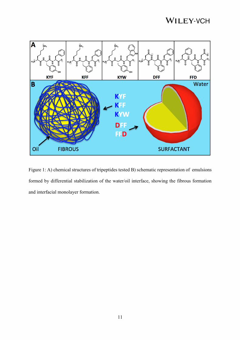
In this work, we demonstrate how the self-assembling ability of unprotected tripeptides42 can
be utilized at an oil/water interface to creating stabilized emulsions with properties that are
dictated by peptide sequence. Using a combined experimental/computational approach, we
show that tripeptides are able to predictably stabilize emulsions by either forming conventional,
3
surfactant-like stabilized droplets or through the formation of nanofibrous networks (Figure 1)
depending on their sequence.
The computational aspect of the work is focused on coarse grain molecular dynamics
simulations, primarily utilizing the MARTINI coarse grained force field45, which has recently
been demonstrated to be useful to model the molecular self-assembly of supramolecular
materials.42,43,46-49 Our previously reported methodology which relied on the aggregation
propensity of di- and tripeptides42,43 was used to identify various tripeptides that showed a high
tendency for aggregation in water.
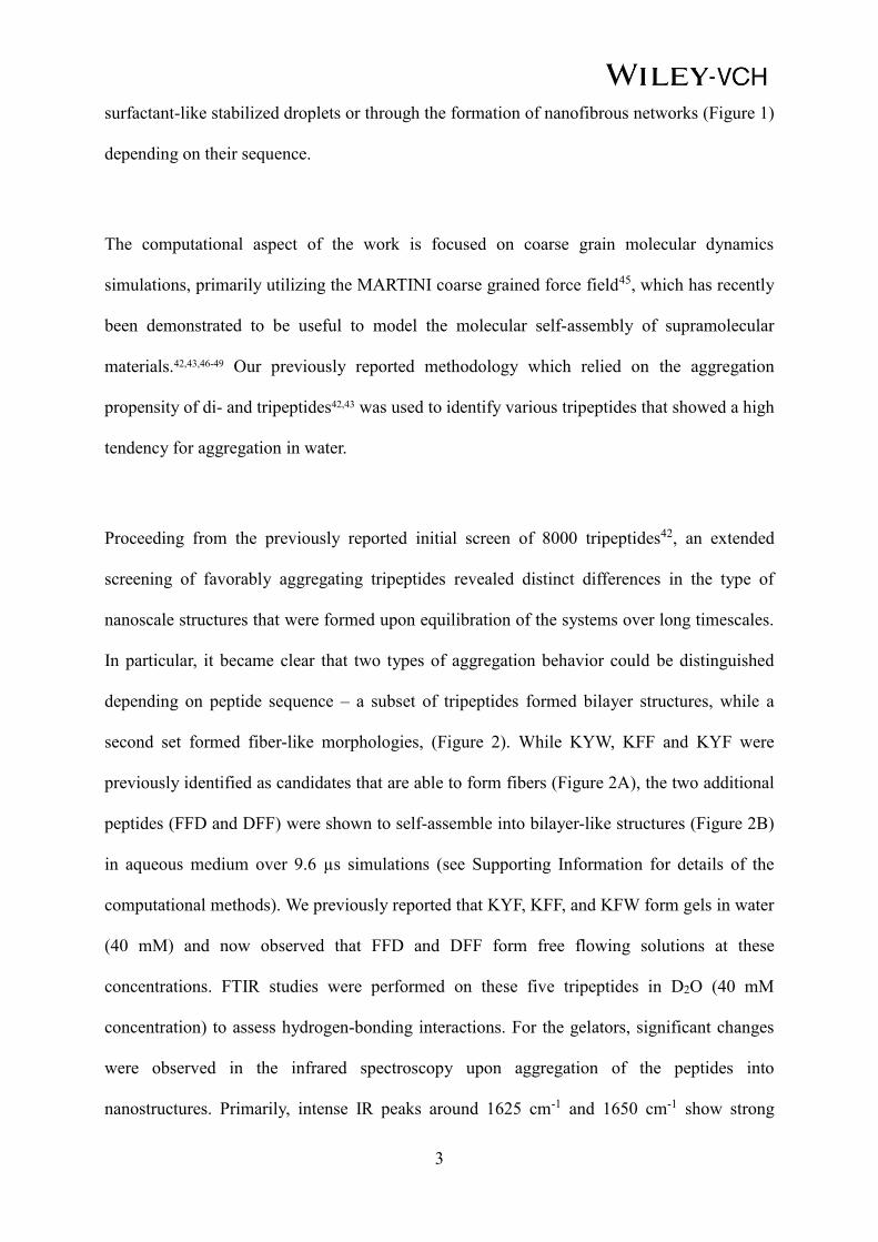
Proceeding from the previously reported initial screen of 8000 tripeptides42, an extended
screening of favorably aggregating tripeptides revealed distinct differences in the type of
nanoscale structures that were formed upon equilibration of the systems over long timescales.
In particular, it became clear that two types of aggregation behavior could be distinguished
depending on peptide sequence – a subset of tripeptides formed bilayer structures, while a
second set formed fiber-like morphologies, (Figure 2). While KYW, KFF and KYF were
previously identified as candidates that are able to form fibers (Figure 2A), the two additional
peptides (FFD and DFF) were shown to self-assemble into bilayer-like structures (Figure 2B)
in aqueous medium over 9.6 µs simulations (see Supporting Information for details of the
computational methods). We previously reported that KYF, KFF, and KFW form gels in water
(40 mM) and now observed that FFD and DFF form free flowing solutions at these
concentrations. FTIR studies were performed on these five tripeptides in D2O (40 mM
concentration) to assess hydrogen-bonding interactions. For the gelators, significant changes
were observed in the infrared spectroscopy upon aggregation of the peptides into
nanostructures. Primarily, intense IR peaks around 1625 cm-1 and 1650 cm-1 show strong
4
hydrogen bonding between the amide groups of the peptide chains.. Similarly, a larger broad
peak around 1560 cm-1 is indicative of the deprotonated carboxylate group, COO-. The shift
and broadening of this peak from the solution state to the nanofibrous state indicates an
introduction of a salt bridge between either the corresponding termini or the lysine side chain.
This proposes a head to tail interaction between the COO- and H3N+ termini of the peptides
with hydrogen bonding between the two the self-assembled structures giving an overall
extended stable structure. DFF and FFD do not show defined peaks thus no nanostructures are
present within the sample.
Having established differential self-assembly in aqueous media, the five tripeptides were
selected for extended simulations of 9.6 µs in a biphasic water/octane solution, using the
MARTINI coarse-grained force field45 (see Figure S1 in the Supporting Information). The
simulations show the assembly of the organic solvent as droplets with the peptides assembled
at the water/octane interface (see Figure S2 in Supporting Information). As expected, the
peptides assemble with the hydrophobic groups exposed to the organic core of the droplet thus
decreasing the interfacial tension between the two phases. Similarly, the hydrophilic groups act
as a barrier for the water phase (Figure 2C). It is not clear from the current simulation whether
these tripeptides may also form nanofibers around the interface due to the size limitations of
the model (300 tripeptides), which does not provide sufficient coverage, to encapsulate the
octane droplet. Nonetheless, the ability of the tripeptides to interact with both the organic and
aqueous phases is a positive indicator for these molecules to act as emulsifiers and the
simulations in water suggest two distinct behaviors for the K and D containing sequences.
Therefore, laboratory experiments were carried out to test whether stabilized emulsions could
be formed.
The five tripeptides were purchased at >98% purity. Each of the tripeptides were dissolved in
water, at 40 mM concentration, and the pH was altered using 0.5 M NaOH to a neutral pH ~7.5.
5
To create the emulsions, 100 μL rapeseed oil (with 1mg/mL concentration sudan II) was added
to each of the systems. Rapeseed oil was chosen as it is a food-regulated oil and tunable peptide-
based emulsions have potential applications in food science. Homogenization was carried out
on each sample for 5 seconds using a bench top homogenizer. Thereafter; the samples were
stored for 24hrs to ensure a stable emulsion was formed (see the Supporting Information for the
complete experimental details – Figure S3). Visual inspection of the resulting emulsions
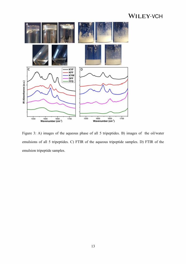
revealed KYF, KFF and KYW forming more stable emulsions compared to DFF and FFD. The
opacity of the samples (Figure 3B) differs between the surfactant like (FFD and DFF) and
fibrous (KYF, KFF, KFW) emulsifiers, with the more opaque emulsion suggesting a more
complete dispersion of oil within the aqueous phase.
To confirm that the tripeptides form self-assembled nanoscale network structures, rather than
aggregating in a surfactant-like manner at the interface, FTIR spectroscopy was carried out on
the emulsions in order to compare key interactions between aqueous and emulsions states which
are indicative of self-assembly of the peptides. In the biphasic systems where the emulsions are
formed, the samples that form fibers (KYF, KFF, KYW) have similar FTIR spectra to those
observed in the aqueous phase. This indicates that similar fibrous networks are forming and
therefore, these droplets are most likely stabilized by nanofiber networks (Figure 3D). In
contrast, comparison of the FTIR spectra for FFD and DFF between the aqueous and biphasic
systems, reveal the emergence of peaks, at approx. 1650 cm-1, in the emulsion state indicating
the formation of assembled structures, which stabilize the droplets. Since this is not observed
in the aqueous state, this suggests that the introduction of the oil induces the self-assembling
process for these peptides. This peak could reasonably represent a parallel arrangement of the
peptides, which are assembling in a similar manner to the traditional surfactant model with
additional lateral stabilization through H-bonding as previously seen in N-acyl amino acids
such as N-lauroyl-L-glutamic acid.50
6
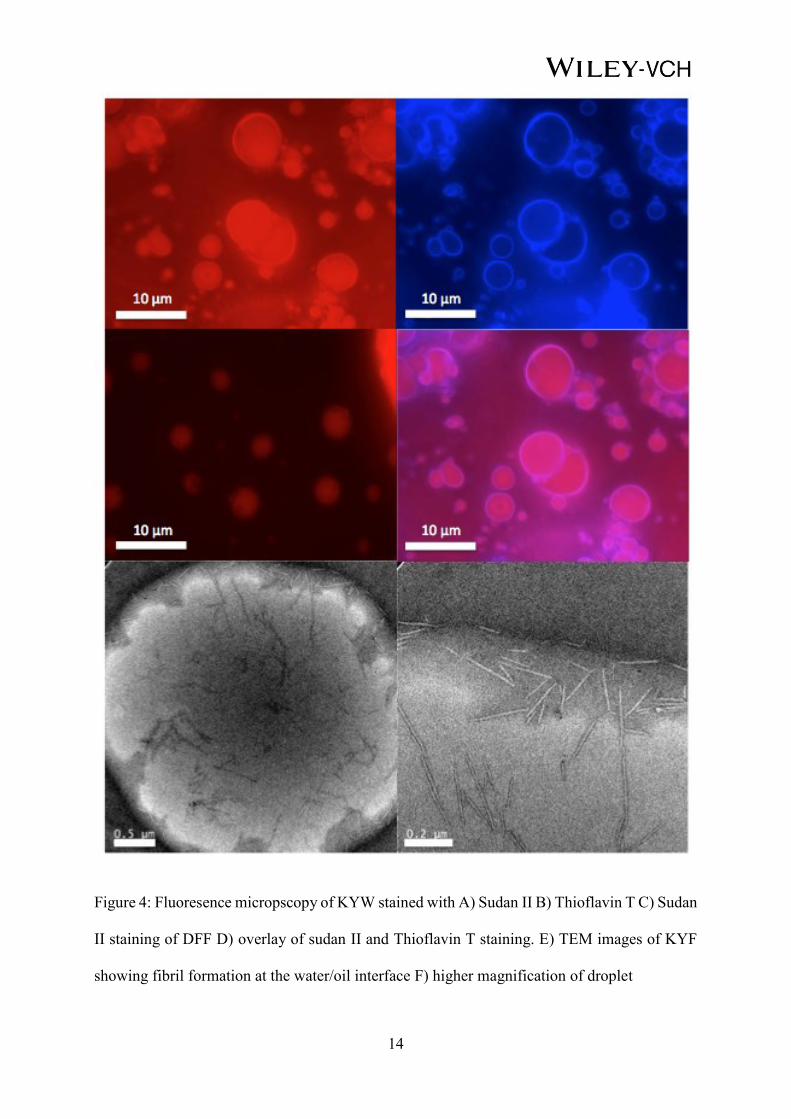
In order to visualize the interfacial assemblies, fluorescent microscopy was carried out on each
of the samples to identify the size and distribution of the droplets as well as to identify how the
peptides interact at the interface (see Supporting Information for the complete set of tripeptide
emulsions, Figure S4). Figure 4 shows the results of these studies for KYW. Labeling the
organic phase with Sudan II reveals stabilized organic droplets (Figure 4A). The introduction
of Thioflavin T,51,52 which labels the peptide region (β-sheet formation) shows that the KYW is
localized to the interface of the droplets (Figure 4B). This suggests that, in the case of KYW,
the tripeptide is self-assembling into fibrils at the interface to create a network which is
stabilizing the droplet, as previously observed for a range of Fmoc-dipeptides.44 For DFF,
smaller and fewer droplets were seen. Staining with Thioflavin T did not show any fluorescence
indicating that no β-sheet forming fibrils are forming. The combination of the two dyes (Figure
4D) further highlights the localization of the tripeptide to the surface of the droplet. In addition
to the fluorescence staining, transmission electron microscopy was carried out to visualize the
fibril assembling at the water/oil interface. Figure 4E/F shows the presence of fibrils at the
interface, for KYF, indicating that these fibrils stabilize the droplets. TEM image of KYW
stabilized emulsion droplet can be found in the Supporting information Figure S5. KFF could
not be imaged due to the weakness of the emulsion. Since DFF and FFD do not form fibrils
these were not imaged.
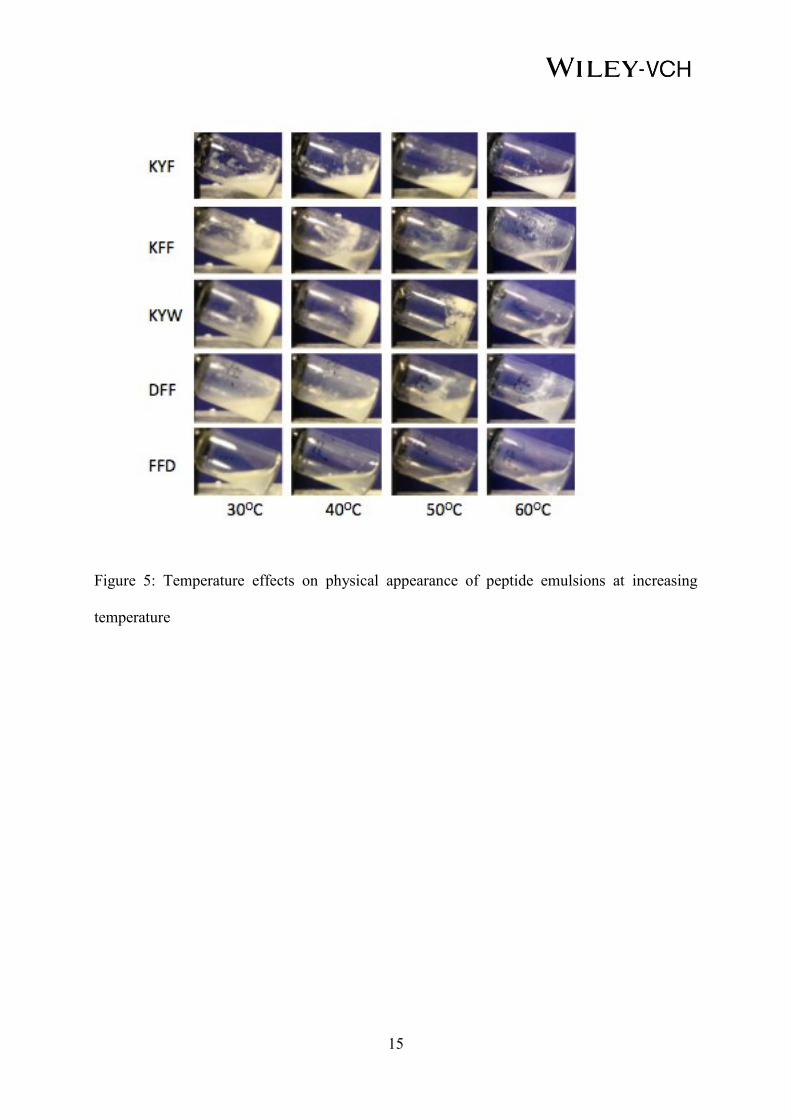
The ability to control separation of an emulsion through environmental triggers is a useful
property within a variety of application areas.53,54 In particular, the ability to disrupt emulsions
at various temperatures is a key property of interest for the application of emulsions in the food
industry.55,56 Therefore, the thermal stability of the emulsions was investigated for each of the
five tripeptides. Each sample was placed in an oil bath and the appearance of the emulsions
were monitored in 10°C intervals (Figure 5).
7
As the temperature is increased the emulsion separates into two different layers. This de-
emulsification is observed for all samples at 60°C apart from KYF, which remains in the
emulsified state at this temperature. This shows that KYF has a higher tolerance for heat than
the other samples and that there are opportunities to tune heat resistance through the
modification of the peptide sequence. Across the range in temperatures, it is clear that DFF and
FFD de-emulsify more easily, which correlates with the initial observations that these peptides
are not strong emulsifiers.. KFF begins to break down at approximately 40°C with KYW
breaking down at 50°C suggesting the strength of the nanofiber assembly is directly
proportional to the stability of the emulsion.
The thermal transitions from emulsion to separated solutions were investigated at the
supramolecular level by investigating changes in FTIR spectra at varying temperature. These
experiments were carried out in D2O/oil mixtures. For KYF and KYW, the peak at ~1625 cm-
1, associated with the H-bonded C=O stretch, begins to decrease in intensity towards the higher
temperatures, this is more noticeable for KYW (breakdown of the fibers at 70ºC) due to the
greater stability of KYF (no breakdown at 80ºC, see Supporting Information, Figure S6, for
additional information). Subtle differences are observed for KFF, although this is a weaker
emulsifier and as such increases in the temperature disrupts the molecular packing at an earlier
stage (50ºC, Figure 64, Supporting Information). Surfactant-like tripeptides FFD and DFF
showed no observable temperature dependence when analyzed by FTIR. The relative stability
of the tripeptide-based emulsions is also confirmed through the pH and concentration
dependence of the emulsions, which can be found in the Supporting Information, Figures S7
and S8.
In summary, we report the first examples of unprotected tripeptides that are capable of self-
assembling in a biphasic systems to stabilize emulsions. These tripeptides have shown varying
emulsion-stabilizing capability at both ambient and elevated temperatures giving a range of
8
properties that are tunable and dependent on sequence. In particular, we show that peptides that
have a preference for fibrillar assembly form more stable emulsions compared with the
traditional surfactant model. Due to their inherent biodegradability to natural amino acids, these
peptides are promising candidates for food and cosmetics applications.
Supporting Information
Supporting Information is available from the Wiley Online Library or from the author.
Notes
The University of Strathclyde has filed a patent application on technology related to the
processes described in this article. Several authors are listed as inventors on the patent
application.
Acknowledgements
This work was supported by ESPRC and Macphie of Glenbervie. Computational results were
obtained using the EPSRC-funded ARCHIE-WeST High Performance Computer (www.archie-
west.ac.uk; EPSRC grant no. EP/K000586/1). The research leading to these results has received
funding from the European Research Council under the European Union's Seventh Framework
Programme (FP7/2007-2013)/EMERgE/ERC Grant Agreement No. (258775).
Received: ((will be filled in by the editorial staff))
Revised: ((will be filled in by the editorial staff))
Published online: ((will be filled in by the editorial staff))
(1) Bais, D.; Trevisan, A.; Lapasin, R.; Partal, P.; Gallegos, C. J. Colloid Interface Sci.
2005, 290, 546.
(2) Bouchemal, K.; Briançon, S.; Perrier, E.; Fessi, H. Int. J. Pharmaceutics 2004, 280, 241.
9
(3) Garti, N. LWT - Food Sci. Technol. 1997, 30, 222.
(4) McClements, D. J.; Li, Y. Adv. Colloid Interface Sci. 2010, 159, 213.
(5) Miller, D. J.; Henning, T.; Grünbein, W. Colloids Surfaces A: Physicochem. Eng.
Aspects 2001, 183–185, 681.
(6) Sanguansri, P.; Augustin, M. A. Trends in Food Sci. Technol. 2006, 17, 547.
(7) Weiss, J.; Takhistov, P.; McClements, D. J. J. Food Sci. 2006, 71, R107.
(8) Lim, G. K.; Wang, J.; Ng, S. C.; Gan, L. M. Langmuir 1999, 15, 7472.
(9) Antonietti, M.; Wenzel, A.; Thünemann, A. Langmuir 1996, 12, 2111.
(10) Fang, Y.; Dalgleish, D. G. Colloids Surfaces B: Biointerfaces 1993, 1, 357.
(11) Holmberg, K. Curr. Opin. Colloid Interface Sci. 2001, 6, 148.
(12) Miller, K. W.; Small, D. M. J. Colloid Interface Sci. 1982, 89, 466.
(13) Veldhuizen, R.; Nag, K.; Orgeig, S.; Possmayer, F. Biochim. Biophys. Acta (BBA)-
Molecular Basis of Disease 1998, 1408, 90.
(14) Hanson, J. A.; Chang, C. B.; Graves, S. M.; Li, Z.; Mason, T. G.; Deming, T. J. Nature
2008, 455, 85.
(15) Morikawa, M. a.; Yoshihara, M.; Endo, T.; Kimizuka, N. Chem.-A Eur. J. 2005, 11,
1574.
(16) Malcolm, A. S.; Dexter, A. F.; Middelberg, A. P. J. Asia-Pacific J. Chem. Eng. 2007, 2,
362.
(17) Schlaad, H.; Antonietti, M. Eur. Phys. J. E: Soft Matter and Biological Physics 2003,
10, 17.
(18) Zhao, Y.; Sakai, F.; Su, L.; Liu, Y.; Wei, K.; Chen, G.; Jiang, M. Adv. Mater. 2013, 25,
5215.
(19) Zhang, J.; Coulston, R. J.; Jones, S. T.; Geng, J.; Scherman, O. A.; Abell, C. Science
2012, 335, 690.
(20) Binks, B. P. Curr. Opin. Colloid Interface Sci. 2002, 7, 21.
(21) Dickinson, E. Curr. Opin. Colloid Interface Sci. 2010, 15, 40.
(22) Fujii, S.; Read, E. S.; Binks, B. P.; Armes, S. P. Adv. Mater. 2005, 17, 1014.
(23) Melle, S.; Lask, M.; Fuller, G. G. Langmuir 2005, 21, 2158.
(24)Pickering, S. SU, J’. Soc. Client. Ind. London 1910, 29, 129.
(25) Tcholakova, S.; Denkov, N.; Lips, A. PCCP 2008, 10, 1608.
(26) Wolters, D.; Meyer‐ Zaika, W.; Bandermann, F. Macromol. Mat. Eng. 2001, 286, 94.
(27) Wang, Z.; van Oers, M. C. M.; Rutjes, F. P. J. T.; van Hest, J. C. M. Ange. Chem. Int.
Ed. 2012, 51, 10746.
(28) Chu, Z.; Feng, Y. Chem. Commun. 2011, 47, 7191.
(29) Fowler, C. I.; Muchemu, C. M.; Miller, R. E.; Phan, L.; O’Neill, C.; Jessop, P. G.;
Cunningham, M. F. Macromolecules 2011, 44, 2501.
(30) Liu, Y.; Jessop, P. G.; Cunningham, M.; Eckert, C. A.; Liotta, C. L. Science 2006, 313,
958.
(31) Minkenberg, C. B.; Florusse, L.; Eelkema, R.; Koper, G. J.; van Esch, J. H. J. Am. Chem.
Soc. 2009, 131, 11274.
(32) Ristenpart, W. D.; Bird, J.; Belmonte, A.; Dollar, F.; Stone, H. Nature 2009, 461, 377.
(33) Zhang, S. Nat. Biotechnol. 2003, 21, 1171.
(34) Reches, M.; Gazit, E. Science 2003, 300, 625.
(35) Ikeda, M.; Tanida, T.; Yoshii, T.; Hamachi, I. Adv. Mater. 2011, 23, 2819.
(36) Ryu, J.; Lim, S. Y.; Park, C. B. Adv. Mater. 2009, 21, 1577.
(37) Smith, A. M.; Williams, R. J.; Tang, C.; Coppo, P.; Collins, R. F.; Turner, M. L.; Saiani,
A.; Ulijn, R. V. Adv. Mater. 2007, 20, 37.
(38) Adler-Abramovich, L.; Aronov, D.; Beker, P.; Yevnin, M.; Stempler, S.; Buzhansky,
L.; Rosenman, G.; Gazit, E. Nat Nano 2009, 4, 849.
10
(39) Singh, G.; Bittner, A. M.; Loscher, S.; Malinowski, N.; Kern, K. Adv. Mater. 2008, 20,
2332.
(40) Jayawarna, V.; Ali, M.; Jowitt, T. A.; Miller, A. F.; Saiani, A.; E., G. J.; Ulijn, R. V.
Adv. Mater. 2006, 18, 611.
(41) Mahler, A.; Reches, M.; Rechter, M.; Cohen, S.; Gazit, E. Adv. Mater. 2006, 18, 1365.
(42) Frederix, P., W. J. M.; Scott, G. G.; Abul-Haija, Y. M.; Kalafatovic, D.; Pappas, C. G.;
Javid, N.; Hunt, N. T.; Ulijn, R. V.; Tuttle, T. Nat. Chem. 2015, 7, 30.
(43) Frederix, P. W. J. M.; Ulijn, R. V.; Hunt, N. T.; Tuttle, T. J. Phys. Chem. Lett. 2011, 2,
2380.
(44) Bai, S.; Pappas, C.; Debnath, S.; Frederix, P. W. J. M.; Leckie, J.; Fleming, S.; Ulijn, R.
V. ACS Nano 2014, 8, 7005.
(45) Marrink, S. J.; Risselada, H. J.; Yefimov, S.; Tieleman, D. P.; De Vries, A. H. J. Phys.
Chem. B 2007, 111, 7812.
(46) Lee, O.-S.; Cho, V.; Schatz, G. C. Nano Lett. 2012, 12, 4907.
(47) McCullagh, M.; Prytkova, T.; Tonzani, S.; Winter, N. D.; Schatz, G. C. J. Phys. Chem.
B 2008, 112, 10388.
(48) Guo, C.; Luo, Y.; Zhou, R.; Wei, G. ACS Nano 2012, 6, 3907.
(49) Guo, C.; Luo, Y.; Zhou, R.; Wei, G. Nanoscale 2014, 6, 2800.
(50) Sakamoto, K.; Yoshida, R.; Hatano, M.; Tachibana, T. J. Am. Chem. Soc. 1978, 100,
6898.
(51) Shimadzu, H.; Suemoto, T.; Suzuki, M.; Shiomitsu, T.; Okamura, N.; Kudo, Y.;
Sawada, T. J. Labelled Compd. Radiopharm. 2003, 46, 765.
(52) Gebbink, M. F.; Claessen, D.; Bouma, B.; Dijkhuizen, L.; Wösten, H. A. Nat. Rev.
Microbiol. 2005, 3, 333.
(53) Ngai, T.; Behrens, S. H.; Auweter, H. Chem. Commun. 2005, 331.
(54) Alava, C.; Saunders, B. R. Colloids Surf. Physicochem. Eng. Aspects 2005, 270, 18.
(55) Guzey, D.; McClements, D. J. Adv. Colloid Interface Sci. 2006, 128, 227.
(56) Gibbs, F. S. K.; Inteaz Alli, C. N.; Mulligan, B. Int. J. Food Sci. Nutr. 1999, 50, 213.
11
Figure 1: A) chemical structures of tripeptides tested B) schematic representation of emulsions
formed by differential stabilization of the water/oil interface, showing the fibrous formation
and interfacial monolayer formation.

12
Figure 2: Computational time coarse for the three different types of assemblies. i) Initial frame
(0 μs) ii) mid-point(~ 4.8 μs) and iii) final frame (9.6 μs) for A) cationic peptides B) anionic
peptides and C) biphasic systems
13
Figure 3: A) images of the aqueous phase of all 5 tripeptides. B) images of the oil/water
emulsions of all 5 tripeptides. C) FTIR of the aqueous tripeptide samples. D) FTIR of the
emulsion tripeptide samples.
14
Figure 4: Fluoresence micropscopy of KYW stained with A) Sudan II B) Thioflavin T C) Sudan
II staining of DFF D) overlay of sudan II and Thioflavin T staining. E) TEM images of KYF
showing fibril formation at the water/oil interface F) higher magnification of droplet
15
Figure 5: Temperature effects on physical appearance of peptide emulsions at increasing
temperature