Embed Size (px)
Citation preview

Document Diagnostic Imaging Guidelines: Getting to Yes Ma Magellan Health Services, Inc. April 2007

Diagnostic Imaging Guidelines:
Getting to YES!
© Magellan Health Services, Inc. 2007 Proprietary & Confidential All Rights Reserved Page 2
Proprietary Notice © 2007 Magellan Health Services, Inc. All rights re served. The information contained and compiled herein (the “Information”) is the property of Magellan Health Services, Inc. (“Magellan”) and is disseminated solely under license. It is never sold. The Information is protected by copyright, trade-secret law and other laws. The copying, reproduction, republication or transfer of the Information (in whole or part) by licensees or others without the express written permission of Magellan is unlawful and will subject the violator to civil and criminal penalties. The Information may include imaging exam indicators (“Indicators”). Indicators are provided solely to qualified medical professionals and solely for informational purposes. Treatment decisions and other medical decisions should be made only by qualified medical personnel in consultation with their patients and should not be based, in whole or part, upon the Indicators. Distribution or dissemination of Indicators (by licensees or others) other than to qualified medical personnel for any purpose whatsoever is strictly prohibited. Magellan does not warrant the Indicators or other Information as to completeness or accuracy and disclaims all warranties, express or implied, including any warranties of merchantability or fitness for any purpose.

Diagnostic Imaging Guidelines:
Getting to YES!
© Magellan Health Services, Inc. 2007 Proprietary & Confidential All Rights Reserved Page 3
Contents Proprietary Notice ...........................................................................................................................................................2 Dates of Revisions/Additions To Guidelines .............................................................................................................6 Diagnostic Imaging Guidelines, Getting to YES!.......................................................................................................8 Example of Decision Process ........................................................................................................................................9 Head and Neck Section ............................................................................................................................................... 11
Brain, MRI – Codes 70551, 70552, 70553 (Includes Pituitary gland) ................................................. 11 Brain, CT - Codes, 70450, 70460, 70470 (Includes Hearing Canal).................................................... 14 Head MRA w/o Contrast–70544; MRA w/ Contrast–70545................................................................ 16 Head MRA w/o & w/Contrast –70546 ................................................................................................. 16 Head, CT Angiogram – 70496.............................................................................................................. 18 Neck MRAngiogram w/o Contrast 70547; ........................................................................................... 19 Neck MRA w/Contrast – 70548 ........................................................................................................... 19 Neck MRAngiogram w/o & w/Contrast – 70549................................................................................. 19 Neck CTA 70498 .................................................................................................................................. 21 Orbit, Face, and Neck MRI – 70540..................................................................................................... 22 Orbit, Sella, and Posterior Fossa MRI – 70480, 70481, 70482 ............................................................ 22 Orbit, Face, Neck MRIw/Contrast – 70542.......................................................................................... 22 Orbit Face, Neck w/o & w/Contrast – 70543 ....................................................................................... 22 Temporal Bone, Mastoid, (Outer, Mid, or Inner) Ear CT –70480, 70481, 70482................................ 24 Temporomandibular Joint (TMJ) MRI - 70336.................................................................................... 26 Face, Neck and Orbit MRI – 70540...................................................................................................... 27 Face, Neck and Orbit CT w/Contrast– 70542....................................................................................... 27 Face, Neck and Orbit CTw/o & w/Contrast- 70543 ............................................................................. 27 Face and Sinus CT – 70486 70487 70488 ............................................................................................ 27 Paranasal Sinus CT – 70486 70487 70488 ........................................................................................... 28 Neck, Face, and Orbit MRI – 70540..................................................................................................... 30 Neck, Face and Orbit CT w/Contrast – 70542...................................................................................... 31 Neck, Face, and Orbit CT w/o & w/Contrast - 70543 .......................................................................... 31 Neck Soft Tissue CT – 70490, 70491, 70492....................................................................................... 31 Brain Functional MRI (fMRI) 70554,70555 ........................................................................................ 32
Chest and Cardiac Section........................................................................................................................................... 32 Chest CT - 71250 71260 71270............................................................................................................ 33 Chest MRI - 71550................................................................................................................................ 35 Chest MRA – 71555 ............................................................................................................................. 37 Chest CTA - 71275 ............................................................................................................................... 37 Cardiac/Coronary CTA – 0148T .......................................................................................................... 38 Breast MRI – uni lateral 77058, bi lateral 77059.................................................................................. 40 Nuclear Cardiac Imaging / Myocardial Perfusion Imaging –78460, 78461 78464, 78465.................. 42 MUGA / Gated Wall Motion Study - 78472 ........................................................................................ 45
Abdomen and Pelvis Section ...................................................................................................................................... 46 Abdomen CT – 74150 74160 74170..................................................................................................... 46 Abdomen MRI – 74181 ........................................................................................................................ 48 Abdomen MRI w/Contrast – 74182...................................................................................................... 48

Diagnostic Imaging Guidelines:
Getting to YES!
© Magellan Health Services, Inc. 2007 Proprietary & Confidential All Rights Reserved Page 4
Abdomen MRI w/o & w/Contrast - 74183 ........................................................................................... 48 MRCP S8037 ........................................................................................................................................ 50 Magnetic Resonance Cholangiopancreatography................................................................................. 50 CT Colonscopy (Virtual Colonoscopy) 0067 T.................................................................................. 52 Abdominal MRA – 74185, CTA – 74175 Abdominal CT 74160 ........................................................ 54 Abdomen/Pelvis CT Combo: Abdo – 74150, 74160, 74170, Pelvic – 72192, 72193, 72194.............. 56 Pelvic CT – 72192 72193 72194 .......................................................................................................... 58 Pelvic MRI - 72196............................................................................................................................... 60
Spinal Imaging Section................................................................................................................................................. 61 Cervical Spine MRI – 72141 72142 72156 .......................................................................................... 61 Cervical Spine CT – 72125, 72126, 72127.......................................................................................... 63 Thoracic Spine MRI – 72146, 72147, 72157, CT – 72128, 72129, 72130........................................... 64 Lumbar Spine MRI - 72148 72149 72158............................................................................................ 65 Lumbar Spine CT – 72131 72132 72133.............................................................................................. 67
Joint Imaging Section................................................................................................................................................... 69 Shoulder MRI – 73221.......................................................................................................................... 69 Joint MRI of Upper Extremity w/Contrast – 73722 ............................................................................. 69 Joint MRI of Upper Extremity w/o & w/Contrast CT – 73200 73201 73202 ...................................... 69 Elbow MRI Wrist MRI - 73221........................................................................................................... 71 Joint MRI of Upper Extremity w/Contrast- 73722............................................................................... 71 Joint MRI of Upper Extremity w/o & w/Contrast - 73723................................................................... 71 CT – 73200 73201 73202 ..................................................................................................................... 71 Knee MRI – 73721................................................................................................................................ 72 Joint MRI of lower Extremity w/Contrast- 73722................................................................................ 72 Joint MRI of Lower Extremity w/o & w/Contrast - 73723 .................................................................. 72 CT – 73700 73701 73702 ..................................................................................................................... 72 Hip MRI - 73721 72196...................................................................................................................... 74 Joint MRI of Lower Extremity w/Contrast – 73722............................................................................. 74 Joint MRI of Lower Extremity w/o & w/Contrast - 73723 .................................................................. 74 Pelvis (for bilateral hips)....................................................................................................................... 74 CT – 73700 73701 73702 ..................................................................................................................... 74 Ankle/Foot MRI - 73721 ...................................................................................................................... 75 Joint MRI of Lower Extremity w/Contrast – 73722............................................................................. 75 Joint MRI of Lower Extremity w/o & w/Contrast 73723..................................................................... 75 CT - 73700 73701 73702 ..................................................................................................................... 75
Non-Joint Extremity Imaging Section....................................................................................................................... 76 Upper Extremity (non-joint) MRI – 73220........................................................................................... 76 Upper Extremity (non-joint) w/o Contrast – 73218.............................................................................. 76 Upper Extremity (non-joint) w/Contrast - 73219 ................................................................................. 76 Lower Extremity (non-joint) MRI – 73720 .......................................................................................... 76 Lower Extremity (non-joint) w/o Contrast – 73718 ............................................................................. 76 Lower Extremity (non-joint) w/Contrast - 73719................................................................................. 76 Upper Extremity CT – 73200 73201 73202 ......................................................................................... 76 Lower Extremity CT – 73700 73701 73702......................................................................................... 76
Extremity MRA Imaging Section............................................................................................................................... 78 Lower Extremity MRA/MRV – 73725................................................................................................. 78

Diagnostic Imaging Guidelines:
Getting to YES!
© Magellan Health Services, Inc. 2007 Proprietary & Confidential All Rights Reserved Page 5
“Runoff” CT Angiography -- 75635..................................................................................................... 78 Bone Marrow MRI Imaging........................................................................................................................................ 79
Bone Marrow MRI – 77084.................................................................................................................. 79 PET (Positron Emission Tomography) Imaging Section....................................................................................... 80
PET Cardiac Scan - 78459.................................................................................................................... 82 PET Scan, Metabolic, Brain (Seizures and Tumors) 78608 ................................................................. 83 PET Scan, Metabolic, Brain (Alzheimer’s) 78608 ............................................................................... 84 PET Scan, Tumor imaging - Head and Neck 78811, 78812, 78813, 78814, 78815, 78816............... 86 PET Scan, Tumor Imaging - Lymphoma -- 78811, 78812, 78813, 78814, 78815, 78816................... 87 PET Scan, Tumor Imaging – Melanoma --78811, 78812, 78813, 78815, 78816................................. 88 PET Scan, Tumor Imaging – ColoRectal -- 78811, 78812, 78813, 78814, 78815, 78816 ................... 89 PET Scan, Tumor Imaging – Lung Cancer -- 78811, 78812, 78813.................................................... 90 Non-Small Cell or Solitary Pulmonary Lesion --78814, 78815, 78816 ............................................... 90 PET Scan, Tumor Imaging – Esophagus -- 78811, 78812, 78813, 78814, 78815, 78816 ................... 91 PET Scan, Tumor Imaging – Thyroid --78811, 78812, 78813, 78814, 78815, 78816......................... 92 PET Scan, Tumor Imaging – Cervical Cancer -- 78811, 78812, 78813, 78814, 78815, 78816........... 93 PET Scan, Tumor Imaging - Breast Imaging -- 78811, 78812, 78813. 78814, 78815, 78816............. 94

Diagnostic Imaging Guidelines:
Getting to YES!
© Magellan Health Services, Inc. 2007 Proprietary & Confidential All Rights Reserved Page 6
Dates of Revisions/Additions to Guidelines Getting To YES! Guideline Title 7/2006 4/2007 2007 Cover Page Proprietary Notice X Table of Contents X Dates of Revisions/Additions to Guidelines NEW Read Me First X Head and Neck
Brain, Pituitary, Posterior Fossa, IAC MRI X Brain, Pituitary, Posterior Fossa, IAC CT X Head MRA X Head CT X Neck MRA/CT X Neck CTA X Orbit MRI/CT X Temporal/Mastoid Bone MRI/CT X TMJ/Mandible MRI/CT X Face MRI/CT X Paranasal Sinus MRI/CT X Neck, Face and Orbit MRI X Neck, Face and Orbit CT X Brain Functional MRI (fMRI) NEW
Chest and Cardiac Chest CT X Chest MRI X Chest MRA/CTA. X Cardiac/Coronary CTA NEW Breast MRI X Nuclear Cardiology X MUGA X
Abdomen and Pelvis Abdominal CT X Abdominal MRI X MRCP X CT Colonscopy (Virtual Colonoscopy) NEW Abdominal MRA /CT X Abdominal/Pelvic Combo X Pelvic CT X Pelvic MRI X
Spinal Imaging Cervical MRI X Cervical CT X Thoracic MRI/CT X Lumbar MRI X Lumbar CT X
Joint Imaging/Miscellaneous Ortho Shoulder MRI/CT X Elbow /Wrist MRI/CT X Knee MRI/CT X Hip MRI/CT X

Diagnostic Imaging Guidelines:
Getting to YES!
© Magellan Health Services, Inc. 2007 Proprietary & Confidential All Rights Reserved Page 7
Getting To YES! Guideline Title 7/2006 4/2007 2007 Ankle/Foot MRI/CT X
Non-Joint Extremity MRI/CT Imaging Extremity (non joint) MRI/CT X
Extremity MRA/CTA Imaging Extremity MRA X
PET Imaging PET (Positron Emission Tomography) Imaging Section “Read Me First” X PET Cardiac Scan X PET Scan – Metabolic Brain (Seizures and Tumors) X PET Scan – Metabolic Brain (Alzheimer’s) X PET Scan – Tumor Imaging - Head and Neck X PET Scan – Tumor Imaging - Lymphoma X PET Scan – Tumor Imaging - Melanoma X PET Scan - Tumor Imaging - ColoRectal X PET Scan-Tumor Imaging - Lung Cancer, X Non-Small Cell or Solitary Pulmonary Lesions X PET Scan – Tumor Imaging - Esophagus X PET Scan – Tumor Imaging - Thyroid X PET Scan - Tumor Imaging - Cervical Cancer X PET Scan – Tumor Imaging - Breast Imaging X
X

Diagnostic Imaging Guidelines:
Getting to YES!
© Magellan Health Services, Inc. 2007 Proprietary & Confidential All Rights Reserved Page 8
Diagnostic Imaging Guidelines: Getting to YES! With due respect to Fisher and Ury, authors of the very popular book, Getting to Yes: Negotiating Without Giving In, this document is designed to establish a pathway to clinical consensus on the use of a single or combination of Diagnostic Imaging examinations.2 The reader/user is urged to keep in mind that the guidelines that follow are not only intended to aid in the arrival at clinical consensus with Magellan/NIA (National Imaging Associates) algorithms and/or Clinical reviewers but also that they may be used as examples of “mainstream” medicine in clinical practice. They are intended to be “filters” that cover a specific study’s use in the vast majority of cases. Magellan/NIA understands that there will be unusual cases that will meet appropriate indications that are not covered in the following document. Appropriate use is not limited to the following circumstances and may be discussed and recommended at a peer-to-peer level. “Procedure-based guidelines” do not represent a complete episode of care; the decision process to obtain an Imaging Study is assumed to be embedded in a continuum of care including pre-test assessment and the assumption that the test results will have a definite influence on post-test treatment. The reader will note that the following document, as written, will reflect these imperatives. Note the following algorithm graphically demonstrates the process.
2 Fisher R, Ury W, Getting to Yes: Negotiating Without Giving In; 2nd Ed Penguin USA Dec. 1991.

Diagnostic Imaging Guidelines:
Getting to YES!
© Magellan Health Services, Inc. 2007 Proprietary & Confidential All Rights Reserved Page 9
Example of Decision Process Courtesy of Dexter Campinha-Bacote, MD.

Diagnostic Imaging Guidelines:
Getting to YES!
© Magellan Health Services, Inc. 2007 Proprietary & Confidential All Rights Reserved Page 10
Magellan/NIA criteria are the result of combined experience, current practice and extensive literature review. All revisions are the result of a scientific process of clinical consensus, approved by the Magellan/NIA Chief Medical Officer, Chief Executive Officer and Board of Directors. In each case of application to an individual Health Plan they are also reviewed and approved by the plan Medical Director consistent with the plan’s internal quality and utilization management functions as well as all applicable accrediting agencies including NCQA, URAC and the several States Departments of Insurance. Magellan/NIA reviewers use these guidelines in the day-to-day operation of the call center. Any MRI, CT, nuclear cardiac, PET or other study not found in this document will be referred for individual medical (peer-to-peer) review. We have included occasional literature references in this document. We have endeavored to provide references to the latest or most classic information related to covered benefits as defined by typical health plan Technical Assessment (TEC) Committees. Some will be recent while others will point to classic works yet to be scientifically challenged.
PLEASE NOTE • Information provided during the review process is part of the patient’s medical record.
Accuracy is paramount, as information provided may have a lasting impact on your patient’s health ratings!
• General Medical Policy consists of medical guidelines and payment guidelines.
Medical guidelines detail when certain medical services are medically necessary, and whether or not they are investigational. (For more information concerning medical necessity and investigational criteria, please see the health plan’s specific policies.) Our medical guidelines are written to cover a given condition for the majority of people. Each individual's unique, clinical circumstances may be considered in light of current scientific literature. Medical guidelines are based on constantly changing medical science, and we reserve the right to review and update our policies periodically.
Payment guidelines provide claims payment editing logic for CPT®, HCPCS and ICD-9-CM coding. Payment guidelines are developed by clinical staff, and include yearly coding updates, periodic reviews of specialty areas based on input from specialty societies and physician committees and updated logic based on current coding conventions.
Benefits and eligibility are determined before medical guidelines and payment guidelines are applied. Therefore, concurrence with these medical policies does not guarantee authorization, certification, explanation of benefits, or a contract. Benefits are determined by the group contract and the subscriber certificate that is in effect at the time services are rendered.
Diagnostic Imaging Guidelines:
Getting to YES!
© Magellan Health Services, Inc. 2007 Proprietary & Confidential All Rights Reserved Page 11
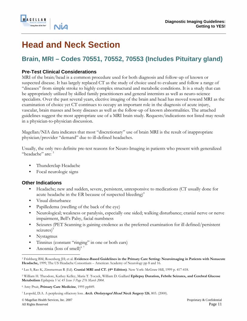
Head and Neck Section
Brain, MRI – Codes 70551, 70552, 70553 (Includes Pi tuitary gland)
Pre-Test Clinical Considerations MRI of the brain/head is a common procedure used for both diagnosis and follow-up of known or suspected disease. It has largely replaced CT as the study of choice used to evaluate and follow a range of “diseases” from simple stroke to highly complex structural and metabolic conditions. It is a study that can be appropriately utilized by skilled family practitioners and general internists as well as neuro-science specialists. Over the past several years, elective imaging of the brain and head has moved toward MRI as the examination of choice yet CT continues to occupy an important role in the diagnosis of acute injury, vascular, brain masses and bony diseases as well as the follow-up of known abnormalities. The attached guidelines suggest the most appropriate use of a MRI brain study. Requests/indications not listed may result in a physician-to-physician discussion. Magellan/NIA data indicates that most “discretionary” use of brain MRI is the result of inappropriate physician/provider “demand” due to ill-defined headaches. Usually, the only two definite pre-test reasons for Neuro-Imaging in patients who present with generalized “headache” are: 3
• Thunderclap Headache
• Focal neurologic signs
Other Indications • Headache; new and sudden, severe, persistent, unresponsive to medications (CT usually done for
acute headache in the ER because of suspected bleeding)4
• Visual disturbance
• Papilledema (swelling of the back of the eye)
• Neurological; weakness or paralysis, especially one sided; walking disturbance; cranial nerve or nerve impairment, Bell’s Palsy, facial numbness
• Seizures (PET Scanning is gaining credence as the preferred examination for ill defined/persistent seizures)5
• Nystagmus
• Tinnitus (constant “ringing” in one or both ears)
• Anosmia (loss of smell)6 7 3 Frishberg BM; Rosenberg JH; et al. Evidence-Based Guidelines in the Primary Care Setting: Neuroimaging in Patients with Nonacute Headache, 1999, The US Headache Consortium – American Academy of Neurology pp 8 and 16.
4 Lee S, Rao K, Zimmerman R (Ed). Cranial MRI and CT. (4th Edition). New York: McGraw Hill, 1999 p. 417-418.
5 William H. Theodore, Kathey Kelley, Maria T. Toczek, William D. Gaillard Epilepsy Duration, Febrile Seizures, and Cerebral Glucose Metabolism Epilepsia Vol. 45 Issue 3 Page 276 March 2004.
6 Amy Pruit, Primary Care Medicine, 1995 pp849.
7 Leopold, D.A. A perplexing olfactory loss. Arch. Otolaryngol Head Neck Surgery 126, 803. (2000).

Diagnostic Imaging Guidelines:
Getting to YES!
© Magellan Health Services, Inc. 2007 Proprietary & Confidential All Rights Reserved Page 12
• Primary or metastatic tumor, new or follow-up (within reason, MRI will identify multiple or complex lesions)
• Trauma (MRI is commonly ordered but CT may be superior for depressed fracture)8
• Stroke or TIA9
• Multiple Sclerosis and other white matter disease10 11
• Arnold Chiari Malformation
• Suspected (r/o) bleeding, or vascular abnormalities12 13
• Syrinx, congenital or acquired (abnormal skull formation)14 • Suspected (r/o) congenital abnormality/developmental delay • Meningitis or abscess15
• Vasculitis
• Encephalopathy16
• Aneurysm or AV malformation
• Hydrocephalus, primary or follow-up (usually CT)
• Craniosynostosis (recommend CT not MRI)
• AIDS17 18
• Endocrine abnormality
Investigation of the internal auditory (hearing) canal is most often performed to evaluate a known or suspected tumor such as an acoustic neuroma or cholesteatoma of the inner or middle ear. It is frequently ordered in conjunction with a CT or MRI of the brain or head. In general, if “suspected” we recommend only the head examination be obtained but if “known” then we suggest a study of the specific area.
• Documented sensorineural hearing loss
• Acoustic Neuroma
• Optic 8 Rothrock SG, Buchanan C, Green SM, et al. Cranial computed tomography in the emergency evaluation of adult patients without a recent history of head trauma: a prospective analysis. Acad Emerg Med 1997 Jul;4(7):654-61.
9 Raymond D. Adams & Maurice Victor, Principles of Neurology 1995.
10 Rovaris M, Filippi M. The value of new magnetic resonance techniques in multiple sclerosis. Current Opinion in Neurology 2000 Jun;13(3):294-54.
11 PRISMS (Prevention of Relapses and Disability by Interferon -1a Subcutaneously in Multiple Sclerosis) Study Group : Randomised double-
blind placebo-controlled study of interferon -1a in relapsing/remitting multiple sclerosis. Lancet. 1998: 352: 1498- 504.
12 Rothrock SG, Buchanan C, Green SM, et al. Cranial computed tomography in the emergency evaluation of adult patients without a recent history of head trauma: a prospective analysis. Acad Emerg Med 1997 Jul;4(7):654-61.
13 Wardlaw JM, White PM. The detection and management of unruptured intracranial aneurysms. Brain 2000 Feb;123 (Pt 2):205-21.
14 Pavlova NG, Konstantinove NN, Arutjunyan AV. Functional and biochemical criteria for investigation of brain development disorders. Int J Dev Neurosci 1999 Dec;17(8):839-48.
15 Moses S: Meningitis: Acute Bacterial Meningitis. 2001. Available at: http://www.fpnotebook.com/NEU112.htm. Accessed April 12, 2004.
16 Rosenberg S. Recent advances in the molecular biology of hepatitis C virus. J Mol Biol 2001; 313:451-64.
17 Rohit Bakshi, MD Medscape Neurology & Neurosurgery 2(1), 2000. © 2000 Medscape Portals, Inc. Accessed April 2004.
18 Raymond D. Adams & Maurice Victor, Principles of Neurology 1995, pp561.

Diagnostic Imaging Guidelines:
Getting to YES!
© Magellan Health Services, Inc. 2007 Proprietary & Confidential All Rights Reserved Page 13
General
• MRI is better than CT, especially for the rear of the brain (chronic dizziness, hearing loss).
• In many cases MRI will be recommended as a substitute if a CT is requested unless there are contraindications to MRI which may include motion, pacing devices and other metallic devices.19
• CT is less expensive than a MRI, a much faster procedure and easier to schedule. It is a very good test for initial study when the index of suspicion of complex disease is low.
• There is rarely a need both CT and MRI.
• For pituitary gland evaluation, MRI of the sella tursica or pituitary is the best study (microadenoma, prolactin tumor, others).20
• Under certain circumstances MRS studies can be used to differentiate tumor/recurrence from radiation effect and should be considered as an adjunctive study.21 MRS, however, is considered investigational by most plans.
• Orbits and/or sinuses are well demonstrated on brain MRI, and not as well on CT. Therefore if both are of serious concern, consider a MRI to cover both. Under certain circumstances an ENT specialist may specifically want the bone detail of a CT.
• Not recommended for short term symptoms such as dizziness, tension headaches, hypertension.
• Temporal Bone, IAC or mastoid MRI: There is no separate CPT (billing) code for this procedure; these areas are imaged in the exam for a Brain MRI.
Combination studies - May be useful if they meet th e following requirements:
• Brain/Cervical – Arnold Chiari, Multiple Sclerosis, demyelinating disease.
• Brain/Neck – confirmed carotid (blood vessel) occlusion of > 60% (but only if candidate for surgery); evaluation of known mass.
• Brain MRI/MRA – These are rarely performed in “tandem”. Appropriate use should be MRI first followed by MRA if positive findings on MRI. The exception would be a strong suspicion of aneurysm such as “thunderclap” headache or neurologic finding in a person with a first degree family history of aneurysm.
Post-Test Considerations
• Combined examinations (except for those listed above) are rarely indicated.
• Follow-up studies (Surveillance) are not recommended unless new signs/symptoms.
• Additional images for same or poor or contrast enhanced study should be the responsibility of the imaging provider.
• Any MRI combination to include Brain and Auditory or canals should be imaged as a single procedure. Brain to include IAC may need extra scans and can be compensated by billing with a –22 modifier, not by billing for a second study.
19 Imaging Handbook, Douglas J. Quint, M.D. 1997, pp397
20 Saeki, Naokatsu, Uchin Yoshio, Murai Hisayuki et al. MR imaging study of edema-like change along the optic tract in patients with pituitary region tumors. AJNR 2003;24:336-342.
21 Jordan HS, Bert R, Chew P, et al, and the Tufts-New England Medical Center Evidence-Based Practice Center. Magnetic resonance spectroscopy for brain tumors. EPC Technical Support of the CPTA Technology Assessment Program. Prepared for the Agency for Healthcare Research and Quality (AHRQ). Contract No. 290-02-0022, Task Order # 1. Rockville, MD: AHRQ; revised June 13, 2003.
Diagnostic Imaging Guidelines:
Getting to YES!
© Magellan Health Services, Inc. 2007 Proprietary & Confidential All Rights Reserved Page 14
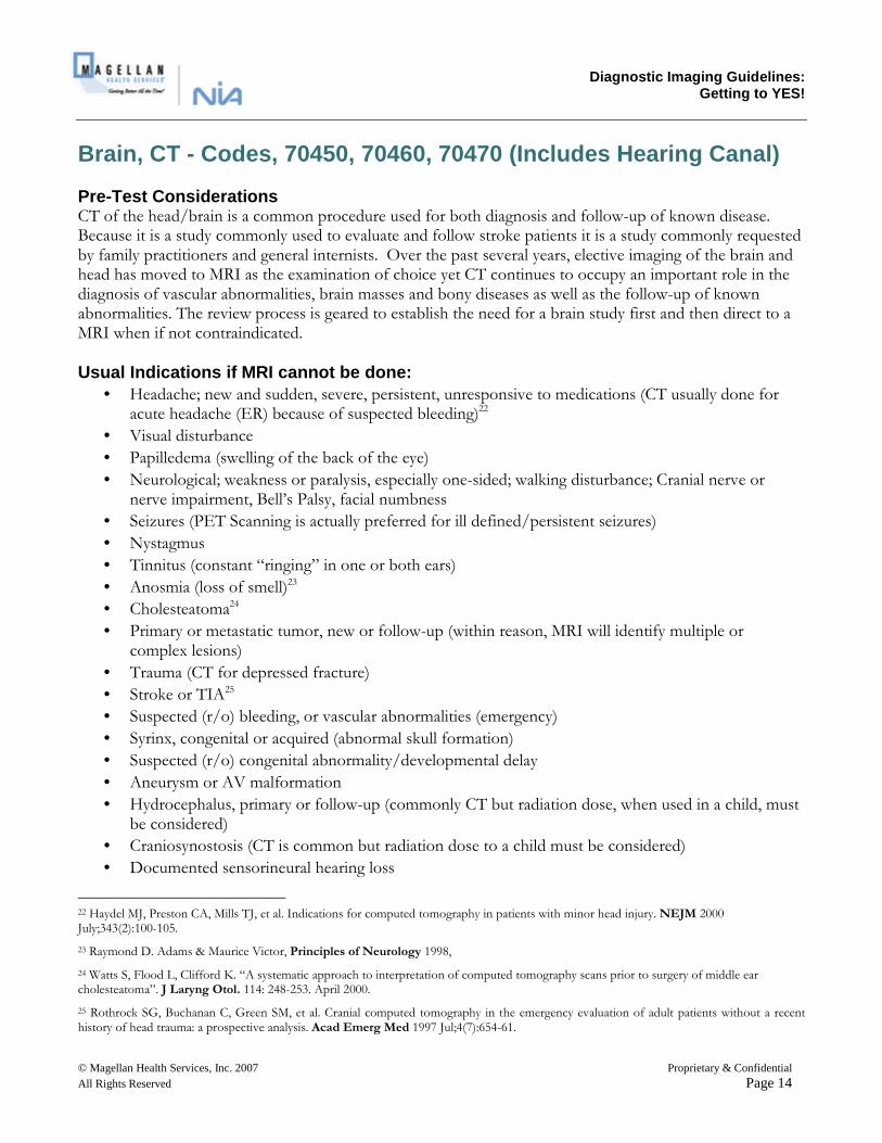
Brain, CT - Codes, 70450, 70460, 70470 (Includes He aring Canal)
Pre-Test Considerations CT of the head/brain is a common procedure used for both diagnosis and follow-up of known disease. Because it is a study commonly used to evaluate and follow stroke patients it is a study commonly requested by family practitioners and general internists. Over the past several years, elective imaging of the brain and head has moved to MRI as the examination of choice yet CT continues to occupy an important role in the diagnosis of vascular abnormalities, brain masses and bony diseases as well as the follow-up of known abnormalities. The review process is geared to establish the need for a brain study first and then direct to a MRI when if not contraindicated.
Usual Indications if MRI cannot be done: • Headache; new and sudden, severe, persistent, unresponsive to medications (CT usually done for
acute headache (ER) because of suspected bleeding)22
• Visual disturbance
• Papilledema (swelling of the back of the eye)
• Neurological; weakness or paralysis, especially one-sided; walking disturbance; Cranial nerve or nerve impairment, Bell’s Palsy, facial numbness
• Seizures (PET Scanning is actually preferred for ill defined/persistent seizures)
• Nystagmus
• Tinnitus (constant “ringing” in one or both ears)
• Anosmia (loss of smell)23
• Cholesteatoma24
• Primary or metastatic tumor, new or follow-up (within reason, MRI will identify multiple or complex lesions)
• Trauma (CT for depressed fracture)
• Stroke or TIA25
• Suspected (r/o) bleeding, or vascular abnormalities (emergency)
• Syrinx, congenital or acquired (abnormal skull formation)
• Suspected (r/o) congenital abnormality/developmental delay
• Aneurysm or AV malformation
• Hydrocephalus, primary or follow-up (commonly CT but radiation dose, when used in a child, must be considered)
• Craniosynostosis (CT is common but radiation dose to a child must be considered)
• Documented sensorineural hearing loss
22 Haydel MJ, Preston CA, Mills TJ, et al. Indications for computed tomography in patients with minor head injury. NEJM 2000 July;343(2):100-105.
23 Raymond D. Adams & Maurice Victor, Principles of Neurology 1998,
24 Watts S, Flood L, Clifford K. “A systematic approach to interpretation of computed tomography scans prior to surgery of middle ear cholesteatoma”. J Laryng Otol. 114: 248-253. April 2000.
25 Rothrock SG, Buchanan C, Green SM, et al. Cranial computed tomography in the emergency evaluation of adult patients without a recent history of head trauma: a prospective analysis. Acad Emerg Med 1997 Jul;4(7):654-61.
Diagnostic Imaging Guidelines:
Getting to YES!
© Magellan Health Services, Inc. 2007 Proprietary & Confidential All Rights Reserved Page 15
General
• MRI better than CT, especially for the rear of the brain (chronic dizziness, hearing loss).
• There are several reasons for CT rather than MRI which may include motion, pacing devices and metallic devices.
• CT is less expensive than a MRI, a much faster procedure and easier to schedule. It is a good test for initial study when complex disease is a lesser consideration.
• Rarely need both CT and MRI.
• For pituitary gland evaluation, MRI of the sella tursica (a bony structure in the middle of the brain) or pituitary is the best study (similarly, microadenoma, prolactin tumor, others).
• MRS/PET studies can differentiate tumor from radiation scarring effect.
• Orbits and/or sinuses are best seen on brain MRI, and not as well on CT. Therefore we suggest a MRI to cover both—except for ENT specialist who may specifically want the bone detail of a CT.
• Useful in minor head injury.26
Combination studies - May be useful if they meet th e following: • Brain/Cervical – Arnold Chiari
• Brain/Sinuses– if ordered by a Ear, Nose and Throat Doctor
Post Test Considerations
• Will the results of this study change the course of treatment?
• Any combination exams (except for those listed above) are discouraged.
• Follow-up studies (Surveillance) are not recommended unless new signs/symptoms.
• Additional images for same or poor or contrast enhanced study is the responsibility of the imaging provider to rectify.
26 Haydel MJ, Preston CA, Mills TJ, et al. Indications for computed tomography in patients with minor head injury. NEJM 2000 July;343(2):100-105.
Diagnostic Imaging Guidelines:
Getting to YES!
© Magellan Health Services, Inc. 2007 Proprietary & Confidential All Rights Reserved Page 16
Head MRA w/o Contrast–70544; MRA w/Contrast–70545 Head MRA w/o & w/Contrast –70546
Pre-Test Considerations MR Angiography performed on high field strength MRI units is an effective and definitive method for the evaluation of patients with known or strongly suspected vascular disease. At the time of writing it has not yet become a part of the management of simple stroke. “Patients with transient ischemic attacks or strokes typically undergo MRI as part of the initial work up to identify infarcted areas in the brain. An intracranial MRA can be easily appended to the MRI and for that reason is frequently ordered. However, an intracranial MRA is not ordinarily considered medically necessary as the initial study. The use of MRA in the work up of patients with signs/symptoms of vertebrobasilar syndrome must be considered on a case-by-case basis”27.
Because of the expense and sophistication of the examination, it is not a screening study. The pre-test requirement that the disease be “reasonably established” before performance of the MRA is essential. In the case of intracranial disease this may be the result of a previously performed abnormal CT or MRI. The use of MRA in evaluating flow in the carotid arteries, the circle of Willis, the anterior, middle or posterior cerebral arteries, the vertebral or basilar arteries, or the venous sinuses have been the most well researched applications. Numerous articles have demonstrated that MRA can image the vessels with a high degree of sensitivity and specificity. However, the appropriate use of MRA in this setting must be coordinated with the use of the competing technologies, Duplex ultrasonography and angiography. There is no mention in the literature that all three technologies should be used routinely in the work up of carotid artery disease. The intent of this Guideline is to stress the establishment of a reasonable cause to perform the exam, to assure that there has been sufficient pre-evaluation and to assess whether the patient is a candidate for remedial treatment.
Indications:
• To evaluate steno-occlusive disease for mid/large size intracranial arteries at facilities where intracranial angioplasty is an approved procedure (not usually reimbursed)
• Cerebral aneurysm28
• Intracranial vascular malformations29
• Cerebral sinus compression or pulsitile tinnitus30
General Considerations • Intracranial examination is usually performed to rule out a blood vessel malformation or aneurysm.
• Should not be ordered in conjunction with MRI unless there is proof from a previous exam that an abnormality is present and the course of therapy will be changed by the results.31
27 Aetna Clinical Policy BulletinNumber: 0094 Subject: Magnetic Resonance Angiography (MRA) and Magnetic Resonance Venography (MRV) March 2004.
28Christopher S. Ogilvy, M.D., Elizabeth S. Lustrin, , James H. Brown, Computerized Tomographic Angiography (CTA) Assists in the Evaluation of Patients with Intracranial Aneurysms MGH Interactive Neuro Web site 2002; accessed April 2004.
29 Liauw L, van Buchem MA, Spilt A, et al. MR angiography of the intracranial venous system. Radiology. 2000;214(3):678-682.
30 Imaging Handbook, Douglas J. Quint, M.D. 1997, pp397.

Diagnostic Imaging Guidelines:
Getting to YES!
© Magellan Health Services, Inc. 2007 Proprietary & Confidential All Rights Reserved Page 17
• May be ordered separately to rule out aneurysm in patient with family history (10% incidence).32
• Any combination studies, i.e., MRI/MRA will be questioned.
Post-Test Considerations
• Will the results of this study change the course of treatment?
• Any combination exams (except for those listed above) are discouraged.
• Follow-up studies (Surveillance) are not recommended unless new signs/symptoms.
• Additional images for same or poor or contrast enhanced study is the responsibility of the imaging provider to rectify.
31 ibid
32 ibid
Diagnostic Imaging Guidelines:
Getting to YES!
© Magellan Health Services, Inc. 2007 Proprietary & Confidential All Rights Reserved Page 18
Head, CT Angiogram – 70496
Pre-Test Considerations Intracranial CT angiography is rarely indicated and when a vascular examination is contemplated MRA is the superior technology. The requirement that vascular disease be “reasonably established” before performance of the CTA is essential. In the case of intracranial disease this may be the result of a previously performed abnormal CT or MRI. The use of CTA in evaluating flow in the carotid arteries, the circle of Willis, the anterior, middle or posterior cerebral arteries, the vertebral or basilar arteries, or the venous sinuses have been the most well researched applications. Numerous articles have demonstrated that CTA can image the vessels with a high degree of sensitivity and specificity. CTA is emerging as a study complementary to standard un-enhanced CT. It may be performed in fewer than five minutes following initial CT without moving the patient. CTA has good correlation with confirming studies such as digital subtraction angiography (DSA) and ultrasound (US). It is less invasive than DSA and less time-consuming and more readily available than either DSA or US. CTA evidence of occlusion at presentation correlates strongly and independently with clinical outcome. 33 The intent of this Guideline is to establish a reasonable cause to perform the exam, to assure that there has been sufficient pre-evaluation and to assess whether the patient is a candidate for remedial treatment.
Indications
• Intracranial examination is usually ordered to rule out a blood vessel malformation or aneurysm.
• Should not be ordered in conjunction with brain CT unless there is proof from a previous exam that an abnormality is present and patient is a candidate for remedial intervention. This bias is toward Aneurysm and away from Ischemic stroke.
• May be ordered separately to rule out aneurysm in patient with family history (10% incidence).34
• To evaluate steno-occlusive disease for mid/large size intracranial arteries if intervention is planned (not usually reimbursed).
• Intracranial vascular malformations.
• Cerebral sinus compression or pulsitile tinnitus.
• Combination CT/CTA studies are clinically discouraged.
Post-Test Considerations
• Will the results of this study change the course of treatment?
• Any combination exams (except for those listed above) are discouraged.
• Follow-up studies (Surveillance) are not recommended unless new signs/symptoms.
• Additional images for same or poor or contrast enhanced study is the responsibility of the imaging provider to rectify.
33 Verro P, Tanenbaum LN, Borden NM, et al. CT angiography in acute ischemic stroke: Preliminary results. Stroke 2002;33:276-278.
34 Imaging Handbook, Douglas J. Quint, M.D. 1997, pp397

Diagnostic Imaging Guidelines:
Getting to YES!
© Magellan Health Services, Inc. 2007 Proprietary & Confidential All Rights Reserved Page 19
Neck MRAngiogram w/o Contrast 70547; Neck MRA w/Contrast – 70548 Neck MRAngiogram w/o & w/Contrast – 70549
Pre-Test Considerations MR Angiography, performed on high field strength MRI units, is a very effective and definitive method for the evaluation of patients with a high index of suspicion for vascular disease established by physical findings and Ultrasound.
Because of the expense and sophistication of the examination, this should not be considered a screening study. The use of MRA in evaluating flow in the carotid arteries, the circle of Willis, the anterior, middle or posterior cerebral arteries, the vertebral or basilar arteries, or the venous sinuses have been the most well researched applications. Numerous articles have demonstrated that MRA can visualize vessels with a high degree of sensitivity and specificity. However, the appropriate use of MRA in this setting must be coordinated with the use of the competing technologies, Duplex ultrasonography and angiography. There is no mention in the literature that all three technologies should be used routinely in the work up of carotid artery disease. The disease should be “reasonably established” before performance of the MRA. In the case of extra Cranial (carotid) disease this may be the result of an abnormal ultrasound. The intent of the guideline is to establish reasonable cause to perform the study, to assure that there has been sufficient pre-evaluation and to assess whether the patient is a candidate for remedial treatment. Because this examination is frequently used in a “shotgun” manner in “combination” with a Brain MRI, the requesting physician is urged to narrow the focus of his/her suspicion. Usual Indications
• Suspected carotid stenosis35
• CervicoCranial arterial dissection
• For carotid body tumors, i.e., glomus tumor
• For post-op evaluation of carotid endarterectomy (arterial neck surgery) when it replaces catheter angiography if there are newly presenting signs/symptoms 36
General
• Patients should have had an abnormal Carotid Doppler as an initial study.37
• Repeat studies are considered duplicative unless there has been a significant change in the patient’s condition.38
35 Leclerc X, Pruvo JP. Recent advances in magnetic resonance angiography of carotid and vertebral arteries. Curr Opin Neurol. 2000;13(1):75-82.
36 Tierney L, McPhee S, Papadakis M (Ed). Current Medical Diagnosis and Treatment (40th Edition). New York: Lange Medical Books/McGraw-Hill p. 982.
37 Caplan LR: Carotid artery disease. N Engl J Med 1986; 315: 886-888
38 Karamessini MT, et al., CT angiography with three-dimensional techniques for the early diagnosis of intracranial aneurysms. Comparison with intra-arterial DSA and the surgical findings.Eur J Radiol. 2004 Mar;49(3):212-23.

Diagnostic Imaging Guidelines:
Getting to YES!
© Magellan Health Services, Inc. 2007 Proprietary & Confidential All Rights Reserved Page 20
Post-Test Considerations • Will the results of this study change the course of treatment?
• Any combination exams (except for those listed above) are discouraged,
• Follow-up studies (Surveillance) are not recommended unless the patient is presenting with new signs/symptoms.
• Additional images for same or poor or contrast enhanced study is the responsibility of the imaging provider to rectify.

Diagnostic Imaging Guidelines:
Getting to YES!
© Magellan Health Services, Inc. 2007 Proprietary & Confidential All Rights Reserved Page 21
Neck CTA 70498
Pre-Test Considerations CT Angiography is a very effective and definitive method for the evaluation of patients with known vascular disease though MRA is emerging as the procedure of choice. Because of the expense and sophistication of the examination, this should not be considered a screening study. The requirement that the disease be “reasonably established” before performance of the CTA is essential and in the case of extra Cranial (carotid) disease this may be an abnormal ultrasound. The intent of the guideline is to ensure reasonable cause to perform the study, to ensure that there has been sufficient pre-evaluation and to assess whether the patient is a candidate for remedial treatment. Because this examination is frequently used in a “shotgun” manner in “combination” with a Head CT, the requesting physician is urged to narrow the focus of his/her suspicion. Usual Indications
• Suspected carotid stenosis
• CervicoCranial arterial dissection
• For carotid body tumors, i.e., glomus tumor
• For post-op evaluation of carotid endarterectomy (arterial neck surgery) when it replaces catheter angiography39
General Considerations
• Patients should have an abnormal Carotid Doppler as an initial study.
• A previous CTA/MRA is generally considered duplicative unless there has been a significant change in the patient’s condition.40
Post-Test Considerations
• Will the results of this study change the course of treatment?
• Any combination exams (except for those listed above) are discouraged.
• Follow-up studies (Surveillance) are not recommended unless the patient is presenting with new signs/symptoms.
• Additional images for same or poor or contrast enhanced study is the responsibility of the imaging provider to rectify.
39 Tierney L, McPhee S, Papadakis M (Ed). Current Medical Diagnosis and Treatment (40th Edition). New York: Lange Medical Books/McGraw-Hill p. 982.
40 Leclerc X, Pruvo JP. Recent advances in magnetic resonance angiography of carotid and vertebral arteries. Curr Opin Neurol. 2000;13(1):75-82.

Diagnostic Imaging Guidelines:
Getting to YES!
© Magellan Health Services, Inc. 2007 Proprietary & Confidential All Rights Reserved Page 22
Orbit, Face, and Neck MRI – 70540 Orbit, Sella, and Posterior Fossa MRI – 70480, 7048 1, 70482 Orbit, Face, Neck MRIw/Contrast – 70542 Orbit Face, Neck w/o & w/Contrast – 70543
Pre-Test Considerations MRI of the face, etc. is an important and valuable procedure for examination of known disease. The two most common reasons for requesting this examination are for surgical planning and/or for evaluation of the status of the temporal-mandibular joints (TMJ). Therefore, because it is an expensive and sensitive study commonly used in conjunction with known disease, the presence of abnormal physical findings and/or associated abnormal diagnostic studies is usual. In the case of TMJ evaluation, a strong suspicion of temporal-mandibular joint disease would be an indication for approval if a trial of conservative therapy (80% effective) has failed. 41 42
Usual Indications
• Proptosis or a “bulging” eye
• Rapidly progressive vision changes
• Decreased range of motion of the eyes43
• Tumor (especially melanoma, only shows on MRI)44
• Hyperthyroidism, known or suspected
• Trauma to the eye
• Optic Neuritis
• Unilateral eye visual disturbance
• Papilledema with suspected pseudotumor45
General
• MRI is usually better than CT.
• CT is usually preferable for foreign body and other trauma.
• These studies are often ordered with brain and/or pituitary exams. MRI will show all with one examination, so combination studies are discouraged.
• MRI preferred for optic (vision) pathway abnormality.46
41 Anne D. Walling Review of Diagnosis and Treatment of TMJ Disorders. American Family Physician, Nov 15, 1998, On-line, Accessed April 2004.
42 American Academy of Otolaryngology. Head and Neck Surgery. Pain and the TMJ. On-line, accessed April 2004.
43 Kleinheinz J, Stamm T. Three dimensional magnetic resonance imaging of the orbit in craniofacial malformations and trauma. Orthodontics and Orthognathic Surgery 2000 Spring;15(1):648.
44 Werner JA et al., Functional anatomy of the lymphatic drainage system of the upper aerodigestive tract and its role in metastasis of squamous cell carcinoma. Head Neck. 2003 Apr;25(4):322-32.
45 MRI indications for the referring physician, Paul Rodriguez, MD., 1999 pp24.
46 Ibid
Diagnostic Imaging Guidelines:
Getting to YES!
© Magellan Health Services, Inc. 2007 Proprietary & Confidential All Rights Reserved Page 23
Post-Test Considerations • Have the results of this study changed the course of treatment?
• Any combination exams (except for those listed above) are discouraged.
• Follow-up studies (Surveillance) are not recommended unless the patient is presenting with new signs/symptoms.
• Additional images for same or poor or contrast enhanced study is the responsibility of the imaging provider to rectify.
Diagnostic Imaging Guidelines:
Getting to YES!
© Magellan Health Services, Inc. 2007 Proprietary & Confidential All Rights Reserved Page 24
Temporal Bone, Mastoid, (Outer, Mid, or Inner) Ear CT –70480, 70481, 70482
Pre-Test Considerations Investigation of the internal auditory canal is most often undertaken to evaluate a known or suspected infection, acoustic neuroma or cholesteatoma of the inner or middle ear. It is frequently ordered in conjunction with a CT or MRI of the brain or head. If suspected, only the head exam is recommended but if known, a specific area study is warranted. That is to say, upon suspicion either a brain CT or MRI is initially recommended, however if the condition is known a specific study is encouraged if it will change the course of treatment.47 48
Usual Indications
• Documented nystagmus
• Sensorineural hearing loss
• Ringing, or constant pulsatile sensation in or around the ear
• Blood vessel mass behind eardrum
• Skull base tumor
• Acoustic neuroma
• Ear infections, ear drainage
• Mastoiditis
• Cholesteatoma
• Congenital hearing loss, deformity
• Evaluation of known cochlear implants
• Conductive hearing loss49
General Considerations
• Temporal Bone/mastoid CT is a unique study usually obtained for ear infections, ear drainage, mastoiditis, cholesteatoma; rarely for 7th or 8th nerve tumor for which a MRI is the procedure of choice.
• MRI of the ear canal/posterior brain is usually for 7th or 8th nerve tumor, but also used for vertigo, dizziness. A good radiology facility will also do entire brain when this is ordered. This procedure defaults to a Brain MRI code.
47 Conn’s Current Therapy, N. Scott Litofsky, M.D., 1998, pp 970 48 CT scanning of middle ear cholesteatoma: what does the surgeon want to know? Yates et al. Br J Radiol.2002; 75: 847-852.
49 Daniels and others. Causes of unilateral sensorineural hearing loss screened by high-resolution fast spin echo magnetic resonance imaging: review of 1070 consecutive cases. Am. J. otol 21:173180, 2000

Diagnostic Imaging Guidelines:
Getting to YES!
© Magellan Health Services, Inc. 2007 Proprietary & Confidential All Rights Reserved Page 25
Not Usually Recommended
• Any combination exams
• The study be performed for occasional dizziness
• General loss of hearing due to age
Post-Test Considerations • Have the results of this study changed the course of treatment?
• Any combination exams (except for those listed above) are discouraged.
• Follow-up studies (Surveillance) are not recommended unless the patient is presenting with new signs/symptoms.
• Additional images for same or poor or contrast enhanced study is the responsibility of the imaging provider to rectify.
Diagnostic Imaging Guidelines:
Getting to YES!
© Magellan Health Services, Inc. 2007 Proprietary & Confidential All Rights Reserved Page 26
Temporomandibular Joint (TMJ) MRI - 70336
Pre-Test Considerations MRI of the temporal-mandibular joint (TMJ) is a valuable procedure for definition of known disease. Because it is an expensive and sensitive study best used in conjunction with known disease, positive physical findings and/or associated diagnostic studies are required. Therefore, because it is an expensive and sensitive study commonly used in conjunction with known disease, the presence of physical findings and/or associated diagnostic studies is usual. In the case of TMJ evaluation, a strong suspicion of temporal-mandibular joint disease would be an indication for approval if a trial of conservative therapy (80% effective) has failed. 50 51
Usual Indications
• Failed conservative therapy including TMJ splint or bite block and anti-inflammatory meds
• Pre-op evaluation
• Frozen jaw
General
• TMJ usually done for difficulty in the ability to open mouth, pain with chewing, etc. These studies are ordinarily ordered by an oral surgeon or ENT specialist.
• CT of the mandible may be most appropriate for jaw trauma or tumor rather than an MRI.
• A single code (70336) is used and will include a bilateral study with open and closed mouth views.
Post-Test Considerations • Have the results of this study changed the course of treatment?
• Any combination exams (except for those listed above) are discouraged.
• Follow-up studies (Surveillance) are not recommended unless the patient is presenting with new signs/symptoms.
• Additional images for same or poor or contrast enhanced study is the responsibility of the imaging provider to rectify.
50 Anne D. Walling Review of Diagnosis and Treatment of TMJ Disorders. American Family Physician, Nov 15, 1998, On-line, accessed April 2004
51 American Academy of Otolaryngology. Head and Neck Surgery. Pain and the TMJ. Online, accessed April 2004.

Diagnostic Imaging Guidelines:
Getting to YES!
© Magellan Health Services, Inc. 2007 Proprietary & Confidential All Rights Reserved Page 27
Face, Neck and Orbit MRI – 70540 Face, Neck and Orbit CT w/Contrast– 70542 Face, Neck and Orbit CTw/o & w/Contrast- 70543 Face and Sinus CT – 70486 70487 70488 Pre-Test Considerations MRI or CT of the face etc. is an important and valuable procedure for examination of known disease. The two most common reasons for requesting this examination are for surgical planning and/or for evaluation of the status of the temporal-mandibular joints (TMJ). Therefore, because it is an expensive and sensitive study commonly used in conjunction with known disease, the presence of physical findings and/or associated diagnostic studies is usual. In the case of TMJ evaluation, a strong suspicion of temporal-mandibular joint disease would be an indication for approval if a trial of conservative therapy (80% effective) has failed. 52 53
Usual Indications 54 • Sinus, nose or facial tumor/Trauma
• Osteomyelitis of a facial bone
• Parotid/Salivary Duct Stones
General • Usually ordered for trauma, tumor or palpable mass. CT is used for trauma. Either CT or MRI will
be useful for evaluation of tumor.
• Appropriately requested for specific, localized facial pain. • To evaluate prior to medical or surgical therapy, particularly antibiotic treatment.
Not Ordinarily Recommended
• For facial pain only
• Any combination examination
Post-Test Considerations • Have the results of this study changed the course of treatment?
• Any combination exams (except for those listed above) are discouraged.
• Follow-up studies (Surveillance) are not recommended unless the patient is presenting with new signs/symptoms.
• Additional images for same or poor or contrast enhanced study is the responsibility of the imaging provider to rectify.
52 Anne D. Walling Review of Diagnosis and Treatment of TMJ Disorders. American Family Physician, Nov 15, 1998, On-line, Accessed April 2004.
53 American Academy of Otolaryngology. Head and Neck Surgery. Pain and the TMJ. Online, accessed April 2004.
54 Curtin HD, Som PM, Bergeron RT Temporal bone trauma. Sem US, CT, MR 2001;22:219-228.
Diagnostic Imaging Guidelines:
Getting to YES!
© Magellan Health Services, Inc. 2007 Proprietary & Confidential All Rights Reserved Page 28
Paranasal Sinus CT – 70486 70487 70488
Pre-Test Consideration The performance of any radiographic evaluation of the sinuses should be approached with deliberation and purpose. In the treatment of sinusitis imaging should only be obtained after several courses of failed antibiotic therapy. This is frequently a disease of the young and the radiation exposure to the eye and thyroid must be a serious consideration.55 Imaging studies are not cost-effective in the initial assessment and treatment of patients with clinical findings suggestive of acute sinusitis. Radiographs, however, may be helpful in uncertain or recurrent cases. A normal sinus x-ray series has a negative predictive value of 90 to 100 percent, particularly for the frontal and maxillary sinuses. The positive predictive value of x-rays using opacification and air-fluid levels as end points is 80 to 100 percent, but the sensitivity is low since only 60 percent of patients with acute sinusitis have opacification or air-fluid levels.56 The treatment/imaging sequence for sinusitis should therefore begin with antibiotic therapy. If several (four or more) trials of antibiotic therapy have failed then either plain sinus films or a sinus CT may be performed. Only if endoscopy or surgery is planned should one consider a full sinus CT. Follow-up studies (Surveillance) are rarely recommended unless the patient is presenting with new signs/symptoms.
Sinus CT of the face, etc. is an important and valuable procedure for definition of known disease. Since it is frequently obtained as an “add-on” to a head or brain MRI or CT, when both are ordered they may be duplicative and unnecessary. A usual pre requisite to this examination will be a trial of conservative treatment.
Usual Indications 57 • Pre-operative after failure of conservative treatment
• After operation for sinus surgery if there are new signs/symptoms
• Sinus-nasal tumor
• Sinusitis in patients with AIDS58
• Osteomyelitis
• Mucocele
• Polyposis, multiple polyps
• Sinusitis; chronic, persistent, failed several courses of antibiotic therapy
• Asthma – when ordered by a Pulmonologist, may be ordered in conjunction with Chest CT
General • CT preferred over MRI to evaluate or rule out sinusitis or pre-op evaluation. Either is adequate for
tumor.
• Best study to evaluate the osteomeatal complex (seen only with CT).
• Frequently ordered by ENT for follow-up of treatment when refractory or new signs/symptoms.
55 Guidelines, American College of Allergy, Asthma & Immunology http://www.acaai.org/public/advice/sinus.htm; accessed March 2005.
56 Willett LR, Carson JL, Williams JW Jr. Current diagnosis and management of sinusitis. J Gen Intern Medicine 1994;9:38-45.
57 C. Douglas Phillips, MD Screening Sinus CT and Paranasal Sinus Imaging Appl Radiol 30(5):9-15, 2001. © 2001
58 www.webhealthcentre.com/centers/sinusiti.asp - 18k - Apr 11, 2004 On-line assessed April, 2004

Diagnostic Imaging Guidelines:
Getting to YES!
© Magellan Health Services, Inc. 2007 Proprietary & Confidential All Rights Reserved Page 29
• In general, unless there is a question of airway obstruction, (deviated septum, structural abnormality, polyp), should have a trial of conservative therapy before imaging.
• Soft tissue sinus disease is best imaged with MRI.59
Not Usually Recommended • For Allergy alone
• Nasal Stuffiness
• Any symptoms treated on less than four occasions
• Any combination of exams (except when ordered w/ Chest CT by Pulmonologist)
Post-Test Considerations
• Have the results of this study changed the course of treatment?
• Any combination exams (except for those listed above) are discouraged.
• Follow-up studies (Surveillance) are not recommended unless the patient is presenting with new signs/symptoms.
• Additional images for same or poor or contrast enhanced study is the responsibility of the imaging provider to rectify.
59 Velche-Haag B, Proust F, Laquerriere A, Dehesdin D, Freger P. Ewing's sarcoma of the ethmoid bone: case report. Neurochirurgie 2002; 48: 25–29

Diagnostic Imaging Guidelines:
Getting to YES!
© Magellan Health Services, Inc. 2007 Proprietary & Confidential All Rights Reserved Page 30
Neck, Face, and Orbit MRI – 70540 Pre-Test Considerations MRI of the soft tissues of the neck is a commonly requested procedure and SHOULD NOT BE CONFUSED with a request for a cervical spine. While occasionally done to evaluate infection or abscess, its most common use is the evaluation of known or suspected adenopathy. The adenopathy may be related to a lymphoma and this examination, while occasionally ordered as a stand-alone study, is most often obtained in conjunction with additional MRI studies.
Usual Indications 60 • Mass in neck (greater than six weeks duration)61
• Skull base mass
• Vocal cord lesion, hoarseness, paralysis
• Known suspicious lesion in mouth or throat
• Suspected or known tumor of larynx, pharynx, nasopharynx, parathyroid, or salivary glands 62
• Lymphadenopathy
• Tracheal Stenosis
• Branchial cleft cyst63
General Considerations
• Neck studies begin at the level of the external auditory canal (Ear) and go to the sternal notch (upper chest) so they include views of the nasopharynx, part of the facial bones, the pharynx, salivary glands, mandible etc.
• Almost never need both CT and MRI.
• Not the appropriate study for specific views of the vascular system.
• Initial thyroid imaging should be with ultrasound or nuclear medicine, unless known carcinoma. 64
Post-Test Considerations • Have the results of this study changed the course of treatment?
• Any combination exams (except for those listed above) are discouraged.
• Follow-up studies (Surveillance) are not recommended unless the patient is presenting with new signs/symptoms.
• Additional images for same or poor or contrast enhanced study is the responsibility of the imaging provider to rectify.
60 DeScheeper AM, ed. Imaging of soft tissue tumors. Berlin: Springer-Verlag, 1997. 61Essential Otolaryngology, Kim R. Jones, M.D., Ph.D., 1995, pp475.
62 Essential Otolaryngology, Helmut W. Gahbauer, M.D., Ken Yanagisawa, M.D., 1995, pp1060. 63 Weber AL et al., The thyroid and parathyroid glands. CT and MR imaging and correlation with pathology and clinical findings. Radiol Clin North Am. 2000 Sep;38(5):1105-29. 64 Toft AD 2001 Sublinical hyperthyroidism. N Engl J Med 345:512–516.

Diagnostic Imaging Guidelines:
Getting to YES!
© Magellan Health Services, Inc. 2007 Proprietary & Confidential All Rights Reserved Page 31
Neck, Face and Orbit CT w/Contrast – 70542 Neck, Face, and Orbit CT w/o & w/Contrast - 70543 Neck Soft Tissue CT – 70490, 70491, 70492 Pre-Test Considerations CT of the soft tissues of the neck represents a commonly requested procedure and SHOULD NOT BE CONFUSED with a request for the cervical spine. While occasionally obtained to evaluate infection or abscess, its most common use is the evaluation of known or suspected adenopathy (MRI is the superior examination). The adenopathy may be related to a lymphoma and this examination, while occasionally ordered as a stand-alone study, is most often obtained in conjunction with additional CT examinations.
Usual Indications
• Proven mass in neck in a patient who cannot have a MRI65
• Skull base mass
• Vocal cord lesion, hoarseness, paralysis
• Known suspicious lesion in mouth or throat
• Suspected or known tumor of larynx, pharynx, nasopharynx, parathyroid, or salivary glands 66
• Lymphadenopathy
• Tracheal Stenosis
General • Neck studies begin at the level of the external auditory canal and go to the sternal notch so they
include views of the nasopharynx, part of the facial bones, the pharynx, salivary glands, mandible etc.
• Almost never need both CT and MRI.
• Initial thyroid imaging should be with ultrasound or nuclear medicine, unless known carcinoma. If known and post treatment, PET may be the study of choice.67
Post-Test Considerations • Have the results of this study changed the course of treatment?
• Any combination exams (except for those listed above) are discouraged.
• Follow-up studies (Surveillance) are not recommended unless the patient is presenting with new signs/symptoms.
• Additional images for same or poor or contrast enhanced study is the responsibility of the imaging provider to rectify.
65 Nusynowitz ML. Thyroid imaging. Lippincotts Prim Care Pract 1999 Nov-Dec;3(6):546-55..
66 Eskey CJ, Robson CD & Weber AL. Imaging of Benign and malignant soft tissue tumours of the neck. Radiol. Clin. of North Amer. 2000; 38(5): 1091-1104.
67 Alnafisi NS, Driedger AA, Coates G, Moote DJ, Raphael SJ. FDG PET of recurrent or metastatic 131I-negative papillary thyroid carcinoma. J Nucl Med 2000;41:1010 - 1015

Diagnostic Imaging Guidelines:
Getting to YES!
© Magellan Health Services, Inc. 2007 Proprietary & Confidential All Rights Reserved Page 32
Brain Functional MRI (fMRI) – 70554, 70555 NOTE: Clear and unambiguous medical necessity indications for this technology have not been established and therefore many health plans do not consider this a “covered” benefit. Please check with your health plan prior to performance.
Pre-Test Considerations Before neurological surgery for seizure disorders or resection of brain tumors, localization of certain areas of the brain, such as speech centers, is important. For example, from 25 to 60 percent of patients who undergo left anterior temporal lobectomy develop dysnomia (language/naming difficulties). Most often these "eloquent" areas are assessed using the Wada test and direct electrical simulation. Both of these tests are invasive and require involvement of various specialists. The Wada test involves angiography and injection of amobarbital into the carotid artery. Direct electrical stimulation involves surgical placement of electrodes in the brain. Functional Magnetic Resonance Imaging (MRI) is used as a noninvasive alternative for evaluation of these eloquent brain areas. Functional MR imaging uses sequences based on T2-weighted blood oxygen. Images are collected as various activities are conducted. Laterality indices are calculated reflecting the interhemispheric difference between activated volumes in the left and right hemispheric regions of interest. These studies are often done on MR scanners with field strengths of 1.5 Tesla or greater.
Usual Indications The only indication currently accepted by some but not all health plans is to use this technology for brain mapping prior to surgical ablation for an epileptic focus/foci.Chest and Cardiac Section

Diagnostic Imaging Guidelines:
Getting to YES!
© Magellan Health Services, Inc. 2007 Proprietary & Confidential All Rights Reserved Page 33
Chest CT – 71250, 71260, 71270 Pre-Test Considerations CT of the chest is a common advanced imaging procedure used for both diagnosis and follow-up of known disease. Additionally, it is now widely used to replace ventilation/perfusion scanning in suspected pulmonary embolus. The most common current reasons for disapproval of the use of chest CT is its use as a screening examination for neoplasm followed by “calcium scoring of the heart” and CT coronary evaluation.68 69 It is a well established study most often performed using spiral as well as multi-detector technology. The growing use of multi-detector scanners has prompted the increased use of 3D reconstruction for data handling. There is a CPT code for 3D reconstruction as a post-processing charge but only a few carriers pay for the code.
As for screening, there is presently inadequate evidence in the medical literature that population-based mass lung cancer screening with spiral computed tomography will contribute substantially to the detection of smaller cancers, or decreases mortality. Currently, the American Cancer Society (ACS), along with other public health organizations ,does not recommend low-dose CT screening for lung cancer.70 This examination/code should not be used for cardiac CT or coronary CTA.
Usual Indications 71
• Suspected Pulmonary Embolus should be evaluated by CTA
• Hemoptysis with normal chest x-ray
• Persistent unresolved cough of at least two weeks with normal chest x-ray (consider referral to pulmonologist as first step)
• Suspected/known lung tumor/mass - evaluation, staging, restaging
• In conjunction with contiguous body part examinations in known widespread tumors
• Mediastinal widening/adenopathy
• Superior vena cava (SVC) syndrome
• Hilar adenopathy or prominent hilum
• First study for interstitial lung disease such as asbestosis, sarcoidosis, TB, bronchiectasis, emphysema, pulmonary fibrosis (consider referral to a pulmonologist as first step)
• Asbestosis follow-up if not industrial or research study
• Suspected/follow-up of abscess or empyema or infection
• Unresolved pneumonia documented (after antibiotic therapy > 4 weeks)
• Suspected/follow-up to dissecting or other aortic aneurysm
• Pleural mass or effusion
68 Patz EF Jr, Goodman PC, Bepler G. Screening for lung cancer. N Engl J Med. 2000 Nov30;343(22):1627-33.
69 O'Rourke RA, Brundage BH, Froelicher VF et al. American College of Cardiology/American Heart Association expert consensus document on electron beam computed tomography for the diagnosis and prognosis of coronary artery disease. J Amer Coll Card 2000; 36: 326-40.
70 Manser RL, Irving LB, Stone C, et al. Screening for lung cancer (Cochrane Review). In: The Cochrane Library, Issue 3, 2002. Oxford, UK: Update Software.
71 Kanne JP, Lalani TA, Role of computed tomography and magnetic resonance imaging for deep venous thrombosis and pulmonary embolism.Circulation. 2004 Mar 30;109(12 Suppl 1):I15-21.
Diagnostic Imaging Guidelines:
Getting to YES!
© Magellan Health Services, Inc. 2007 Proprietary & Confidential All Rights Reserved Page 34
• Chest wall or rib mass
• Severe trauma
• Unexplained abnormalities on chest x-ray
General • A prior chest x-ray is almost always required. • CT or MRI may be used to evaluate aortic aneurysm and dissection, though MRA or CTA is
superior.72
• Includes the thoracic inlet and neck base, the axillae, the lower esophagus and the esophagus/stomach junction.
Not Usually Indicated • A CT study without a recent chest x-ray
• Tumor/mass follow-up studies at less than 6-week interval if no change in signs/symptoms
• Suspected rib fracture
• CT for Cardiac Calcium scoring
Post-Test Considerations • Have the results of this study changed the course of treatment?
• Follow-up studies (Surveillance) are not recommended unless the patient is presenting with new signs/symptoms.
• Additional images for same or poor or contrast enhanced study is the responsibility of the imaging provider to rectify.
72 Leclerc X, Pruvo JP. Recent advances in magnetic resonance angiography of carotid and vertebral arteries. Curr Opin Neurol. 2000;13(1):75-82.

Diagnostic Imaging Guidelines:
Getting to YES!
© Magellan Health Services, Inc. 2007 Proprietary & Confidential All Rights Reserved Page 35
Chest MRI - 71550
Pre-Test Considerations MRI of the chest is an uncommon procedure but can be used for diagnosis and follow-up of known disease and increasingly in the evaluation of vascular disease. Because it is a study commonly used in serious conditions, specialty involvement (Pulmonologist or Thoracic surgeon) is recommended but not required. If there is affirmative evidence of a chest or lung mass, the procedure of choice remains a CT study. CT or MRI may provide anatomic and morphologic information, but neither can accurately distinguish benign from malignant pulmonary, pleural or lymph node abnormalities and CT with PET may ultimately be the examination/combination of choice.73 74
When ordering this examination please be certain to specify that it is a chest examination and not a thoracic spine that you want.
Usual Indications
• Mediastinal (including thymus) or hilar mass on patient with renal failure or allergy to contrast material
• Myesthenia Gravis (possible thymoma)
• Brachial plexopathy
• Aneurysm or dissection of the thoracic aorta or great vessels
• Congenital Heart Disease and malformations
• Aortic Arch Anomalies (coarctation)
• Patent Ductus Arteriosa (PDA), may have been detected by echo75
• Cardiac mass
• To evaluate the status of the Brachial Plexus
• Thoracic Outlet Syndrome (all combination studies will require a physician-to-physician discussion
General Considerations • CT is the standard method of imaging the chest.
• It is difficult to evaluate the lungs with MRI. This technology is most effectively used for mediastinum and hilar adenopathy.
• MRI rather than CT is used by some in patients with allergy to radiographic contrast material or kidney failure.
73 Thoracic Imaging, The American College of Radiology (ACR) and The Society of Thoracic Radiology (STR), Edward F. Patz, Jr., M.D., 1997 pp55. 74 Gupta, NC, Graeber, GM, Bishop, HA (2000) Comparative efficacy of positron emission tomography with fluorodeoxyglucose in evaluation of small (<1 cm), intermediate (1 to 3 cm), and large (>3 cm) lymph node lesions. Chest 117,773-778. 75 Knisely BL, Broderick LS, Kuhlman JE. MR imaging of the pleura and chest wall. Magnetic Resonance Imaging Clinics of North America 2000 Feb;8(1):125-41.

Diagnostic Imaging Guidelines:
Getting to YES!
© Magellan Health Services, Inc. 2007 Proprietary & Confidential All Rights Reserved Page 36
Post-Test Considerations
• Have the results of this study changed the course of treatment?
• Any combination exams (except for those listed above) are discouraged.
• Follow-up studies (Surveillance) are not recommended unless the patient is presenting with new signs/symptoms.
• Additional images for same, poor or contrast enhanced study is the responsibility of the imaging provider to rectify.

Diagnostic Imaging Guidelines:
Getting to YES!
© Magellan Health Services, Inc. 2007 Proprietary & Confidential All Rights Reserved Page 37
Chest MRA – 71555 Chest CTA – 71275 Pre-Test Considerations There are limited indications for MR Angiography of the chest. It may be indicated in a patient who has a history of severe contrast allergy or is at higher risk for contrast induced kidney failure (such as a diabetic patient with moderate kidney insufficiency) and presents with a suspicious mass. In general this examination may be substituted when catheter Angiography/venography is indicated. Because of chest wall motion, attempts to obtain diagnostic images using MRI are frequently disappointing though the technology is improving. CT using fast scanning technique remains the preferred technology for complex chest evaluation. At this time appropriate indications are essentially limited to complex vascular studies.76 77
Usual Indications 78 • Used to evaluate axillary or brachiocephalic vessel status as an alternative to catheter angiography
when fast scan helical CT cannot be done.
• Used for evaluation of aortic aneurysms, especially the dissecting type.
• Used for evaluation of congenital heart disease in youngsters, rarely in adults.
• Acquired disease of the thoracic aorta.
• Developmental anomaly of the thoracic vasculature.
• Systemic venous thrombosis or occlusion.
• As an alternative to Angiography for evaluation of pulmonary embolus in patients who cannot have iodinated contrast material.
Post-Test Considerations
• Have the results of this study changed the course of treatment?
• Any combination exams (except for those listed above) are discouraged.
• Follow-up studies (Surveillance) are not recommended unless the patient is presenting with new signs/symptoms.
• Additional images for same or poor or contrast enhanced study is the responsibility of the imaging provider to rectify.
76 Allen MT, et al. Ultrafast contrast-enhanced three-dimensional MR angiography: State of the art. Radiographics. 1998;18(2):273-285.
77 Krinsky GA, et al. Thoracic aorta: Comparison of gadolinium-enhanced three-dimensional MR angiography with conventional MR imaging. Radiology. 1997;202(1):183-193.
78 Lookstein RA et al., Time-resolved magnetic resonance angiography as a noninvasive method to characterize endoleaks: initial results compared with conventional angiography.J Vasc Surg. 2004 Jan;39(1):27-33.

Diagnostic Imaging Guidelines:
Getting to YES!
© Magellan Health Services, Inc. 2007 Proprietary & Confidential All Rights Reserved Page 38
Cardiac/Coronary CTA – 0148T Pre-Test Considerations NOTE: Clear and unambiguous medical necessity indications for this technology have not been established and therefore many health plans do not consider this a “covered” benefit. Please check with your health plan prior to performance. The indications for the performance of a Nuclear Cardiac study often called SPECT or Myocardial Perfusion Imaging (MPI) are well established.79 Generally the study is used to evaluate myocardial perfusion and cardiac function in patients at intermediate risk for coronary artery disease. The results will ordinarily help define a subsequent course of conservative medical treatment or cardiac catheterization, stent or surgery. The indications for the performance of CCTA are similar. CCTA technology provides a non-invasive demonstration of coronary and cardiac anatomy and function, which approaches the accuracy of conventional catheter studies.80 CT imaging of the heart has entered the mainstream with the introduction of 40-64-slice, sub-millimeter, thin-slice detectors with both large and small health plans implementing or contemplating moving this technology from investigational status to “covered.” The improving spatial resolution, combined with improved temporal resolution made possible by faster gantry rotation speeds, makes CCTA feasible for almost every patient. Current experience demonstrates that a rapid heart rate may cause as much as a 6.5% non-diagnostic image rate even on ultra fast 64 and greater detector scanners and the need for cardiac “slowing” is often necessary.81 Beta-blocked patients typically receive a small bolus of contrast to determine circulation time. A diagnostic dose of 80 to 100 cc of nonionic contrast is infused at 4 to 5 cc/sec followed immediately by 50 cc of normal saline using a dual-head injector. Sub-millimeter (0.625-mm-thick) slices of the entire heart and portions of the ascending aorta are then recorded with a scan time of 18 to 26 seconds. While some critics cite technical impediments to accurate collection and interpretation of the data, this is a mature technology. Some believe that the two examinations, MPI and CCTA are complimentary while others consider them duplicative. Clearly the MPI provides a superior demonstration of the degree of small vessel perfusion currently unmatched by CCTA but with constantly improving technology most believe that MPI will add little real value to the assessment of structure and function provided by CCTA with Functional assessment. There are special circumstances when the use of both examinations will add significant incremental value, they are rare and in this algorithm will be handled during a peer-to-peer discussion because of the risks of cumulative radiation exposure from both studies as well as the combined cost of performing both procedures.
79 SPECT MPI: ACCF/ASNC Appropriateness Criteria for Single-Photon Emission Computed Tomography Mocarial Perfusion Imaging (J Am Coll Cardiol 2005;46:1587–605).
80 Ropers D, Baum U, Pohle K, et al. Detection of Coronary Artery Stenoses with thin-slice multi-detector row spiral computed tomography and multi-planar reconstruction. Circulation. 2003;107:664-666.
81 Ibid.

Diagnostic Imaging Guidelines:
Getting to YES!
© Magellan Health Services, Inc. 2007 Proprietary & Confidential All Rights Reserved Page 39
Usual Indications Cardiac Structural Review:
• To evaluate a suspected intra-cardiac mass such as a myxoma
• To evaluate a suspected pericardial abnormality such as pericarditis or a pericardial mass
• To evaluate the heart before a RF (radiofrequency) ablation for atrial fibrillation
• To evaluate the heart before the insertion of a BI-VENTRICULAR pacemaker
• To evaluate a suspected coronary artery anomaly
Coronary Artery Evaluation:
• New onset of Congestive Heart Failure
• Suspected Coronary Artery Disease (CAD) and symptomatic
• Suspected Coronary Artery Disease (CAD) and asymptomatic, or no significant symptoms
• Known coronary artery disease
• Evaluation prior to a non-cardiac surgical procedure
• Evaluation of Unstable Angina
Not Usually Indicated • Requests for CCTA in asymptomatic individuals will not ordinarily be approved since there are no
indications for CCTA in the asymptomatic patient in the new ACCF/ACR Appropriateness Criteria. 82
• There are NO appropriate indications for CCTA in a patient with KNOWN coronary artery disease according to the new ACCF/ACR Appropriateness Criteria. 83
• There are NO appropriate indications for CCTA in a patient after a cardiac angioplasty, stent, or bypass graft, according to the new ACCF/ACR Appropriateness Criteria.84
• There are NO appropriate indications for CCTA in a patient being studied for pre-operative reasons in non coronary artery surgery according to the new ACCF/ACR Appropriateness Criteria. 85
Post-Test Considerations • Have the results of this study changed the course of treatment?
• Follow-up studies (Surveillance) are not recommended unless the patient is presenting with new signs/symptoms.
• Additional images for same, poor or contrast enhanced study is the responsibility of the imaging provider to rectify.
82 ACCF/ACR/SCCT/SCMR/ASNC/NASCI/SCAI/SIR 2006 Appropriateness Criteria for Cardiac Computed Tomography and Cardiac Magnetic Resonance Imaging A Report of the American College of Cardiology Foundation Quality Strategic Directions Committee Appropriateness Criteria Working Group, Journal of the American College of Cardiology Vol. 48, No. 7, 2006 © 2006 by the American College of Cardiology Foundation.
83 Ibid.
84 Ibid.
85 Ibid.

Diagnostic Imaging Guidelines:
Getting to YES!
© Magellan Health Services, Inc. 2007 Proprietary & Confidential All Rights Reserved Page 40
Breast MRI – uni lateral 77058, bi lateral 77059 Pre-Test Considerations
Health plan coverage of the use of MRI of the breast is widely divergent. All plans currently consider it appropriate for the evaluation of suspected silicon prosthesis leakage and a few will cover it for suspected saline leakage. Most other reasons usually require that you should contact your health plan to ascertain coverage benefits.86 In addition to its accepted role as a diagnostic tool in known breast cancer, breast MRI has proven to be useful in screening younger women at high risk of breast cancer. Most women under 40 years of age do not require any breast imaging. However, some of these younger women are at high risk of breast cancer, as determined by a strong family history or a mutated breast cancer gene (BRCA1, BRCA2, PTEN or TP53), and therefore need breast imaging before age 40. MRI is now considered essential for these women Nearly all health plans with which Magellan/NIA partners take a conservative approach to its use, essentially limited to evaluation of suspected implant ruptures;87 published research has validated coverage of the use of MRI as a screening tool in those individuals in whom genetic evaluation and/or those with a strong family history of breast neoplasia has been identified.88 89Additionally, its use has widened to include those with known cancer who would benefit from further delineation prior to definitive therapy. 90 91
Usual Indications • To confirm, when necessary, rupture of breast implants in asymptomatic members whose screening
ultrasonography shows rupture92
• To detect implant rupture in symptomatic members whose ultrasonography shows no rupture
• Young women (<40 y/o ) who are at high risk of breast cancer, as determined by a strong family history or a mutated breast cancer gene (BRCA1, BRCA2, PTEN or TP53), and therefore will benefit from breast imaging before age 40
• To detect local tumor recurrence in breast cancer survivors who have undergone mastectomy and breast reconstruction with an implant
• To detect local tumor recurrence in individuals with breast cancer who have radiographically dense breasts or old scar tissue from previous breast surgery that compromises the ability of combined mammography and ultrasonography
86 Friedrich M. MRI of the breast: State of the art. Eur Radiol. 1998;8(5):707-725.
87 Goodman CM, Cohen V, Thornby J, et al. The life span of silicone gel breast implants and a comparison of mammography, ultrasonography, and magnetic resonance imaging in detecting implant rupture: A meta-analysis. Ann Plast Surg. 1998;41(6):577-586.
88 National Academy of Sciences, Institute of Medicine, National Cancer Policy Board, Committee on the Early Detection of Breast Cancer. Mammography and Beyond: Developing Technologies for the Early Detection of Breast Cancer. Washington, DC: National Academy Press; 2001.
89 Mielke Kriel, et.al. Efficacy of MRI and Mammography for Breast-Cancer screening in women with a familial or genetic predisposition NEJM 351:5 pp 427-37 July 29, 2004.
90 Drew P, Chatterjee S, Turnbull L, et al. Dynamic contrast enhanced magnetic resonance imaging of the breast is superior to triple assessment for the pre-operative detection of multifocal breast cancer. Ann Surg Oncol 1999; 6: 599–603.
91 Bedrosian I, Mick R, et. Al. Changes in the surgical management of patients with breast carcinoma based on preoperative magnetic resonance imaging.Cancer. 2003 Aug 1;98(3):468-73.
92 Middleton MS. Magnetic resonance evaluation of breast implants and soft-tissue silicone. Top Magn Reson Imaging. 1998;9(2):92-137.

Diagnostic Imaging Guidelines:
Getting to YES!
© Magellan Health Services, Inc. 2007 Proprietary & Confidential All Rights Reserved Page 41
• To exclude multicentric disease in members with newly diagnosed breast cancer and pathological findings suggestive of multifocality when the information will impact on clinical treatment, i.e., lumpectomy versus mastectomy93
• To detect and stage individuals with invasive lobular carcinoma (ILC) for tailored therapy, especially when breast conservation is being considered94
• To localize the site of primary occult breast cancer in individuals with adeno-carcinoma suggestive of breast cancer discovered as axillary node metastasis or distant metastasis without focal findings on physical examination or on mammography (usually PET is the preferred modality in this clinical set of circumstances)
General • The challenge is that it is a non-specific test. Many benign lesions are identified and some cancers
are not.95
Not Ordinarily Indicated 96 97 • For any patient who does not have or had breast cancer.
• To determine malignancy except in the rare instance when it would replace biopsy.
• For a mass that can be felt but not seen on a mammogram or ultrasound to avoid biopsy.
• For post surgical or post treatment evaluation for possible residual tissue to avoid biopsy.
Post-Test Considerations • Have the results of this study changed the course of treatment?
• Follow-up studies (Surveillance) are not recommended unless the patient is presenting with new signs/symptoms.
• Additional images for same or poor or contrast enhanced study is the responsibility of the imaging provider to rectify.
93 Kramer S, Schulz-Wendtland R, Hagedorn K, et al. Magnetic resonance imaging and its role in the diagnosis of multicentric breast cancer. Anticancer Res. 1998;18(3C):2163-2164.
94. Bagley FH, The role of magnetic resonance imaging mammography in the surgical management of the index breast cancer.Arch Surg. 2004 Apr;139(4):380-3.
95 Brown J, Smith RC, Lee CH. Incidental enhancing lesions found on MR imaging of the breast. Am J Roentgenol 2001 May;176(5):1249-54.
96 Sardanelli F, Melani E, Ottonello C, et al. Magnetic resonance imaging of the breast in characterizing positive or uncertain mammographic findings. Cancer Detect Prev. 1998;22(1):39-42.
97 Weinstein SP, Orel SG, Heller R, Reynolds C, Czerniecki B, Solin LJ, Schnall M. MR imaging of the breast in patients with invasive lobular carcinoma. Am J Roentgenol 2001 Feb;176(2):399-406.

Diagnostic Imaging Guidelines:
Getting to YES!
© Magellan Health Services, Inc. 2007 Proprietary & Confidential All Rights Reserved Page 42
Nuclear Cardiac Imaging / Myocardial Perfusion Imag ing –78460, 78461, 78464, 78465 Stress Testing With Isotopes (Thallium, Cardiolite, Sestamibi, Myoview) Dual Isotope Test is when two different isotopes are used for same exam. Pharmacological Stress Testing (Persantine, Adenosine + Isotope, Dobutamine) Use of a pharmacological agent indicates a contraindication to exercise. Pre-Test Considerations The indications for the performance of a Nuclear Cardiac study often called SPECT or Myocardial Perfusion Imaging (MPI) are well established.98 Generally the study is used to evaluate myocardial perfusion and cardiac function in patients at intermediate risk for coronary artery disease. The results will ordinarily help define a subsequent course of conservative medical treatment or cardiac catheterization, stent or surgery. Some believe that emerging CCTA technology will replace SPECT. The use of CCTA and MPI are considered complimentary by some while others consider them duplicative. Clearly the MPI provides a superior demonstration of the degree of small vessel perfusion currently unmatched by CCTA but with constantly improving technology many believe that MPI will add questionable value to the assessment of structure and function provided by CCTA with Functional assessment. The approach taken in this decision support algorithm is consistent with the ACC guidelines published in late 2005. When considering a Nuclear Cardiac study the reader is encouraged to explore alternative technology including a simple non-cardiac stress test and/or an echocardiogram.99 However the true extent of cardiac muscle necrosis from myocardial infarction or ischemia cannot be established with a stress EKG alone. A standard stress test can identify the presence of ischemia but cannot define the scope of the insult. A nuclear scan will show the area of infarction and extent of damaged tissue as well as detect heart disease. The nuclear “stress” variant quantifies the extent of damage while resting and obviously, under stress.
General Considerations • An MPI may be appropriate in a patient with KNOWN coronary artery disease according to the
most recent ACC Appropriateness Criteria. 100 101
• There are no appropriate indications for MPI in a patient after a cardiac intervention, unless there is a significant change in cardiac status according to the new ACC criteria. However most clinicians (and these Guidelines) accept the value of a MPI study in post CABG after 5 years.102
• There are NO clearly appropriate indications for MPI in a patient being studied for pre-operative reasons in non coronary artery surgery according to the current ACC Appropriateness Criteria.
98 SPECT MPI: ACCFF/ASNC Appropriateness Criteria for Single-Photon Emission Computed Tomography Mocarial Perfusion Imaging (J Am Coll Cardiol 2005;46:1587–605). http://www.ACCF.org/qualityandscience/clinical/pdfs/SPECTMPIACPubFile.pdf
99 Garber AM, Solomon NA. Cost-effectiveness of alternative test strategies for the diagnosis of coronary artery disease. Ann Intern Med. 1999;130(9):719-728.
100 Ibid.
101 SPECT MPI: ACCFF/ASNC Appropriateness Criteria for Single-Photon Emission Computed Tomography Mocarial Perfusion Imaging (J Am Coll Cardiol 2005;46:1587–605).
102 Berman DS,, Hachamovitch R, et al, Roles of Nuclear Cardiology, Cardiac Computed Tomography, and Cardiac Magnetic Resonance: Assessment of Patients with Suspected Coronary Artery Disease Journal of Nuclear Medicine Vol. 47 No. 1 74-82 2006.
Diagnostic Imaging Guidelines:
Getting to YES!
© Magellan Health Services, Inc. 2007 Proprietary & Confidential All Rights Reserved Page 43
Studies requested for this reason will result in a peer-to-peer discussion to evaluate extenuating circumstances. 103
• Use of MPI in asymptomatic individuals is appropriate if the patient presents with significant risk factors and a positive exercise stress test.104
• MPI testing is appropriate when following PTCA, etc. in the presence of new signs/symptoms.105 106 107
• Pharmacological stress testing is typically used to evaluate patients for CAD, who are absolutely contraindicated to exercise, such as: 108 1. Pre-surgical work-up for major vascular surgery in patients with suspected CAD. 2. Patient has a physical limitation to exercise such as shortness of breath, back pain, excess
obesity, etc. 3. Evaluation of patients for CAD who are absolutely contraindicated to exercise. 4. Acute heart attack, unstable angina, uncontrolled abnormal heartbeats, Heart electrical block,
aortic narrowing, uncontrolled heart failure, pulmonary hypertension, severe systemic hypertension.109
Usual Indications
• Uninterpretable or equivocal treadmill (stress) test.
• Abnormal EKG of LBBB, WPW, ST depression >1mm, ventricular Pacing, poor R wave progression. These should be faxed to Magellan/NIA for review.
• Morbidly Obese patient with chest pain.
• Post or perimenopausal female patient with atypical chest pain.
• Symptomatic patient with extensive coronary history – well defined angina, Prinzmetal angina.
• Atrial Fibrillation or other atrial or ventricular arrhythmia.
• Mitral Valve disease or aortic stenosis.
• Follow-up for recurrent symptoms (pain) after bypass surgery, angioplasty, atherectomy, and stent placement angioplasty.
103 Ibid.
104 ACCFF/ACR/SCCT/SCMR/ASNC/NASCI/SCAI/SIR 2006 Appropriateness Criteria for Cardiac Computed Tomography and Cardiac Magnetic Resonance Imaging A Report of the American College of Cardiology Foundation Quality Strategic Directions Committee Appropriateness Criteria Working Group, Journal of the American College of Cardiology Vol. 48, No. 7, 2006 © 2006 by the American College of Cardiology Foundation
105 Fleischmann KE, Hunink MG, Kuntz KM, et al. Exercise echocardiography or exercise SPECT imaging? A meta analysis of diagnostic test performance. JAMA. 1998;280(10):913-920.
106 Braunwald E, Mark DB, Jones RH, et al. Unstable Angina: Diagnosis and Management. Clinical Practice Guideline Number 10. Rockville, (MD): Agency for Health Care Policy and Research and the National Heart, Lung, and Blood Institute, Public Health Service, U.S. Department of Health and Human Services; 1994. AHCPR Publication No.: 94-0602.
107 Araujo LI, Jimenez-Hoyuela JM, McClellan JR, Lin E, Viggiano J, Alavi A. Improved uniformity in tomographic myocardial perfusion imaging with attenuation correction and enhanced acquisition and processing. J Nucl Med. 2000;41(7):1139-44.
108 Fleischmann KE, Hunink MG, Kuntz KM, et al. Exercise echocardiography or exercise SPECT imaging? A meta analysis of diagnostic test performance. JAMA. 1998;280(10):913-920.
109 Ritchie JL, Bateman TM, Bonow RO, et al. Guidelines for clinical use of cardiac radionuclide imaging. Report of the American College of Cardiology/American Heart Association Task Force on Assessment of Diagnostic and Therapeutic Cardiovascular Procedures (Committee on Radionuclide Imaging), developed in collaboration with the American Society of Nuclear Cardiology. J Am Coll Cardiol 1995;25:521-47.

Diagnostic Imaging Guidelines:
Getting to YES!
© Magellan Health Services, Inc. 2007 Proprietary & Confidential All Rights Reserved Page 44
Not Usually Indicated
• For use in an asymptomatic patient with risk factors alone.
• For use in a patient with Chest pain / normal EKG and ability to exercise despite risk factors.
• For use as a Prescreen for Viagra (like drugs) without symptoms and/or risk factors.
Post-Test Considerations
• Have the results of this study changed the course of treatment?
• Follow-up studies (Surveillance) are not recommended unless the patient is presenting with new signs/symptoms.
• Additional images for same or poor or contrast enhanced study is the responsibility of the imaging provider to rectify.
Diagnostic Imaging Guidelines:
Getting to YES!
© Magellan Health Services, Inc. 2007 Proprietary & Confidential All Rights Reserved Page 45
MUGA / Gated Wall Motion Study - 78472 Pre-Test Considerations MUGA scans (multiple gated acquisition scan, nuclear wall motion study) provide information about heart ventricular performance. Specifically, these studies usually report the ejection fraction and data regarding wall motion abnormalities and ventricular volume. They are, therefore, useful in conditions that cause ventricular damage or weakening such as heart attack, cardiomyopathies, hypertension, valvular or congenital heart diseases and exposure to cardiotoxic substances such as some chemotherapeutic drugs. As a general rule, echocardiography provides equally good or better information about heart ventricular performance, and in addition, evaluates valvular structure and function, atrial and ventricular dimensions and pericardial disease and is often the procedure of choice. MUGA scans remain important to patients undergoing cancer chemo. They are also important for patients in whom echocardiography has yielded poor data, usually on the basis of unfavorable body condition (e.g., obesity or barrel chest due to COPD). Usual Indications 110
• Baseline, restaging to follow up chemotherapy to evaluate chemo toxicity
• Evaluate Ejection Fraction in patient with CHF
• Evaluate patients for CAD who are obese or have COPD General Considerations
• Ordered primarily to evaluate ejection fraction, primarily in patients receiving chemotherapy.
• Can detect an area of poor contractility following an ischemic episode or MI.
• Can evaluate left ventricular hypertrophy.
• May be used to evaluate ejection fraction on patients with CHF.
• Echocardiograms are preferred for cardiomyopathy.
Post-Test Considerations • Have the results of this study changed the course of treatment?
• Follow-up studies (Surveillance) are not recommended unless the patient is presenting with new signs/symptoms.
• Additional images for same or poor or contrast enhanced study is the responsibility of the imaging provider to rectify.
110 Society of Nuclear Medicine. Procedure guideline for gated equilibrium radionuclide ventriculography, 2.0. Reston (VA): Society of Nuclear Medicine; 1999 Feb. 20 p. (Society of Nuclear Medicine procedure guidelines; no. 2.0).

Diagnostic Imaging Guidelines:
Getting to YES!
© Magellan Health Services, Inc. 2007 Proprietary & Confidential All Rights Reserved Page 46
Abdomen and Pelvis Section
Abdomen CT – 74150 74160 74170
Pre-Test Considerations CT scanning of the abdomen is close to occupying the pantheon of “routine” studies. Its immediate use to evaluate non-specific abdominal pain, organ enlargement, vascular abnormalities or the further evaluation of abnormal laboratory studies in the absence of prior complete physical examination and/or ultrasound is widespread and usually not indicated. Prior to the performance of the examination, exposure to radiation, false positives and whether the results will be actionable should be considered.111 112 If a CT examination is considered useful/necessary, an attempt should be made to establish whether it is a “lower” or “upper” abdominal concern. This is done in an effort to focus the study from abdomen to pelvis and vice-versa.
Usual Indications 113 • Upper abdominal pain, if ultrasound is normal
• If plain films of the abdomen have been performed and point to a significant abnormality
• Jaundice or abnormal liver function tests if ultrasound is normal or not indicated
• Hematuria or blood in the urine (consider obtaining both abdomen and pelvis)
• Unexplained weight loss (more than 10% of body weight in one month)
• Abdominal/liver/adrenal/retroperitoneal mass/cancer OR rule out mass/cancer.
• Central/upper abdominal abscess/follow up
• Pancreatitis, pseudocyst
• Lymphadenopathy
• Ascites
• Splenomegaly
• Blunt trauma
• Delineation of known or suspected renal calculi
• Possible renal tumor (often will have ultrasound first)
• Suspected or pre-op abdominal aortic aneurysm (f/u may be done with ultrasound)
• First follow-up of complication of medical or surgical treatment of abdominal disease
• Unexplained abdominal pain in patients older than 75 years or very frail
• Persistent unresolved symptoms not explained by initial imaging
• Suspected diverticulitis or appendicitis
111 Olerud, H.M., Torp, C.G., Einarsson, G., et al., Use of the EC quality criteria as a common method of inspecting CT laboratories – a pilot project by the Nordic radiation protection authorities. In: Radiological protection of patients in diagnostic radiology, nuclear medicine and radiotherapy. Proceedings of an international conference held in Malaga, Spain, 26 - 30 March 2001. Vienna:IAEA (2001).
112 Rogers LF. Helical CT: the revolution in imaging. AJR 2003;180:883-884.
113 Lee JK, Sagel SS, Stanley RJ. Computed body tomography with MRI correlation. 3rd ed. Philadelphia, Pa: Lippincott Williams & Wilkins, 1998.

Diagnostic Imaging Guidelines:
Getting to YES!
© Magellan Health Services, Inc. 2007 Proprietary & Confidential All Rights Reserved Page 47
General Considerations
• An abdomen study will include the bottom of the lungs to the region of the umbilicus.
• Abdominal CT will include the base of the lungs.
• CT studies are often appropriate for evaluation of abdominal complaints, but most of the time should be preceded by a thorough history and physical examination and other laboratory and imaging studies (ultrasound, barium studies, and endoscopy).
• Ultrasound does not work well in obese patients.
• Pancreatic pseudocyst. Should be able to do with just abdomen, unless there is a very large body habitus.
Not Usually Indicated
• For possible gallstones or abnormal liver function tests with gall bladder present (should have ultrasound).
• To confirm renal cyst seen on ultrasound.
• Repeat studies of renal mass. Should have ultrasound.
• Renal or hepatic mass follow-up unless there are new signs/symptoms.
• For follow-up aortic aneurysm (should have ultrasound if not obese).
• For abdominal wall hernia.
Post-Test Considerations 114 • Have the results of this study changed the course of treatment?
• Any combination exams (except for those listed above) are discouraged.
• Follow-up studies (Surveillance) are not recommended unless the patient is presenting with new signs symptoms.
• Additional images for same or poor or contrast enhanced study is the responsibility of the imaging provider to rectify.
114 Ahn SH, Mayo-Smith WW, Murphy BL, Reinert SE, Cronan JJ. Acute Nontraumatic Abdominal Pain in Adult Patients: Abdominal Radiography Compared with CT Evaluation Radiology 2002; 225:159-164.

Diagnostic Imaging Guidelines:
Getting to YES!
© Magellan Health Services, Inc. 2007 Proprietary & Confidential All Rights Reserved Page 48
Abdomen MRI – 74181 Abdomen MRI w/Contrast – 74182 Abdomen MRI w/o & w/Contrast – 74183 Pre-Test Considerations MRI scanning of the abdomen is a perplexing examination. Its initial use to evaluate non-specific abdominal pain, organ enlargement, vascular abnormalities or the further evaluation of abnormal laboratory studies in the absence of a prior complete physical examination and/or ultrasound or CT is usually not indicated. MRI of the abdomen is most frequently used to further look at an abnormality seen on another test, such as an ultrasound or CT scan. The exam is usually tailored to look just at the liver, adrenal glands, or pancreas.115
Usual Indications
• Indeterminate CT study for nature of solid mass (not cyst) in liver or adrenal.116
• Allergy to contrast material, which satisfies criteria for abdominal CT request.
• Location or evaluation of undescended testes in adult. (In children, should be preceded by ultrasound).
• Unusual cases of tumor spread.
• Further evaluation of pancreatic, hepatic or kidney mass visualized on ultrasound or CT.
• If Abdominal aneurysm is a serious consideration, MRA is recommended.
• If done in conjunction with a MRCP by a Gastroenterologist. General Considerations
• CT is the standard method of imaging the abdomen. MRI is a targeted technology.
• MRI rather than CT is used by some in patients with allergy to radiographic contrast material or renal failure.
• MRI is used after CT to help characterize liver, pancreatic and adrenal masses seen on ultrasound or CT.
• Undescended testes (associated with the development of carcinoma in the testicle). Used to localize testicle as well as to evaluate for neoplasm.
• MR Cholangiopancreatography (MRCP) may be ordered to evaluate the bile ducts in patients for whom ERCP is undesirable or impossible.
Not Usually Indicated • Use of this study for renal artery narrowing or hypertension (should be evaluated with MRA).117
• For suspected renal vein clot in patient with known kidney mass (should be evaluated with MRA)
• For suspected vena cava obstruction (should be evaluated with MRA)
• Any combination of this with any other (for instance MR Angiography)
115 Clarke JC, et al, Provision of MRI can significantly reduce CT collective dose , British Journal of Radiology 74 (2001), 926-931.
116 Salmanzadeh A, Shirkhoda A. Hepatic lesions that "fill in" on contrast-enhanced CT and MRI: imaging patterns and diagnostic pitfalls. Radiologist 2000;7(1):1-12.
117 Grist TM. MRA of the abdominal aorta and lower extremities. J Magn Reson Imaging 2000;11(1):32-43.

Diagnostic Imaging Guidelines:
Getting to YES!
© Magellan Health Services, Inc. 2007 Proprietary & Confidential All Rights Reserved Page 49
Post-Test Considerations: • Have the results of this study changed the course of treatment?
• Any combination exams (except for those listed above) are discouraged
• Follow-up studies (Surveillance) are not recommended unless the patient is presenting with new signs/symptoms
• Additional images for same or poor or contrast enhanced study is the responsibility of the imaging provider to rectify

Diagnostic Imaging Guidelines:
Getting to YES!
© Magellan Health Services, Inc. 2007 Proprietary & Confidential All Rights Reserved Page 50
MRCP - S8037 Magnetic Resonance Cholangiopancreatography Pre-Test Considerations: MRCP is used to evaluate the bile ducts, pancreatic duct and gallbladder for stones, tumors and other diseases. The performance of MRCP does not require intravenous or oral contrast material administration because the biliary tract, pancreatic duct and gallbladder are depicted as high signal intensity or bright structures due to the fluid within them. Physicians contemplating this examination should weigh the benefit of a conventional ERCP which is both diagnostic and therapeutic (if necessary) whereas this examination is strictly diagnostic. Physicians wishing to order/perform this examination should contact the health plan to determine coverage benefits.
Usual Indications 118 119 120 • Patient has undergone unsuccessful endoscopic retrograde cholangiopancreatography (ERCP) and
requires further evaluation; or Patient has altered biliary tract anatomy that precludes ERCP (e.g., postsurgical biliary tract alterations, prior gastrectomy, choledochojejunostomy, etc.); or
• Patient requires evaluation for a suspected congenital anomaly of the pancreaticobiliary tract (e.g., aberrant ducts, choledochal cysts, pancreas divisum, etc.); or
• Patient requires definition of pancreaticobiliary anatomy proximal to a biliary tract system obstruction that can not be opened by ERCP.
• Patient is an infant or young child, or is an adult who is debilitated or uncooperative in such a manner that ERCP is unsafe or cannot be performed.
• Patient has a documented allergy to iodine-based contrast materials, or has a general history of atopy.
• Complications of Chronic Pancreatitis: when ERCP would be considered unsafe.
Remote Indications
• Screening Examination In Patients With Low or Intermediate Probability Of Choledocholithiasis: MRCP is useful as a noninvasive means of determining the presence or absence of common bile duct stones as well as their number, size and location. With the use of state-of-the-art MRCP techniques, MRCP has a sensitivity of 95-100% in the detection common bile duct stones. MRCP is particularly useful in the evaluation of patients with suspected gallstone pancreatitis and in patients with non-specific abdominal pain and normal liver-associated enzymes. In these settings, an MRCP that shows no evidence of a common bile duct may result in avoidance of an unnecessary diagnostic ERCP.
118 Georgopoulos SK, Schwartz LH, Jarnagin WR, et al. Comparison of magnetic resonance and endoscopic retrograde cholangiopancreatography in malignant pancreaticobiliary obstruction. Arch Surg. 1999;134(9):1002-1007. 119 Owens GR, Shutz SM. Value of magnetic-resonance cholangiopancreatography (MRCP) after unsuccessful endoscopic-retrograde cholangiopancreatography (ERCP). Gastrointest Endosc. 1999;49(2):265-266. 120 Kaltenthaler E et al., systematic review and economic evaluation of magnetic resonance cholangiopancreatography compared with diagnostic endoscopic retrograde cholangiopancreatography. Health Technol Assess. 2004 Mar;8(10):iii, 1-89.
Diagnostic Imaging Guidelines:
Getting to YES!
© Magellan Health Services, Inc. 2007 Proprietary & Confidential All Rights Reserved Page 51
• Primary Sclerosing Cholangitis (PSC): MRCP provides a noninvasive means of diagnosing PSC and determining disease extent. In the detection of PSC, MRCP has been shown to have a sensitivity of 88%, a specificity of 97%, and positive and negative predictive values of 94%. MRCP depicts the mural irregularities, strictures and biliary calculi that characterize PSC. In contrast to ERCP, MRCP delineates the duct proximal to a complete obstruction and avoids the risk of ERCP-induced sepsis related to the injection of contrast material into an obstructed system.
• Complications of Chronic Pancreatitis: MRCP demonstrates ductal dilatation, strictures, intraductal calculi, fistulas and pseudocysts that occur as a result of chronic pancreatitis. One of the major roles of MRCP in this setting lies in defining the ductal anatomy and extent of ductal disease prior to surgical drainage procedures.
Not Usually Indicated
• For screening abdominal pain, an abdominal ultrasound remains the examination of choice and MRCP for this reason is considered investigational.
• Additional images for same or poor or contrast enhanced study.
Post-Test Considerations
• Have the results of this study changed the course of treatment?
• Any combination exams (except for those listed above) are discouraged.
• Follow-up studies (Surveillance) are not recommended unless the patient is presenting with new signs/symptoms.
• Additional images for same or poor or contrast enhanced study is the responsibility of the imaging provider to rectify.

Diagnostic Imaging Guidelines:
Getting to YES!
© Magellan Health Services, Inc. 2007 Proprietary & Confidential All Rights Reserved Page 52
CT Colonscopy (Virtual Colonoscopy) - 0067 T NOTE: Clear and unambiguous medical necessity indications for this technology have not been established and therefore many health plans do not consider this a “covered” benefit. Please check with your health plan prior to performance.
Pre-Test Considerations Optical Colonoscopy is the standard procedure for evaluating the colon because it directly visualizes the mucosa. It also falls in to the category of examinations that are diagnostic and potentially therapeutic, similar to MRCP. If optically identified, polyps can be removed if they are small and can be biopsied for histologic diagnosis. The traditional optical procedure involves a flexible fibreoptic viewing instrument, the colonoscope, inserted through the anus into the rectum and guided along the length of the colon so that the operator can examine the wall surface via a viewing lens. Under light sedation, a complete examination of the whole length of the colon can take from 10 minutes to two hours. Colonoscopy provides very high sensitivity (> 90%), with a false-negative rate of approximately 6% for adenomas of 1 cm or more in diameter. 121 Approximately 85% of screening (optical) colonoscopies identify no clinically significant pathology. The completion rate for colonoscopy varies from 75% to 99% depending on the examiner's skills, anatomic variations, prior abdominal surgery, and the patient's reaction to the anesthesia. An often-quoted national average completion rate for colonoscopy is 90%. Unfortunately, less than half of those who should get screened do so. In screening with colonoscopy, there may be a significant number of individuals who are averse to the invasive nature of the test and are apprehensive with regard to the expected discomfort. 122 123 124 The use of CT or virtual colonoscopy is currently at the forefront of technology. Virtual colonoscopy involves the use of rapid acquisition, thin-section, helical computed tomography (CT) scanning to generate high-resolution, two-dimensional images of the abdomen and pelvis. Three-dimensional images of the colon, which resemble the endoluminal images obtained with conventional optical colonoscopy, can then be reconstructed by computer (3D) software. This technique has been proposed as a screening test for colorectal cancer because it has the potential advantage of being a rapid and safe method of providing full structural evaluation of the entire colon. A drawback to this new technology as a screening tool is the need for subsequent conventional colonoscopy in about 25% of patients 50 and older as most studies have shown that this percentage of patients are found to have one or more polyps. Since most of these would be detected by virtual colonoscopy as well, a significant percentage of patients screened by virtual colonoscopy would still need conventional colonoscopy to remove the polyp. The time required for the procedure is
121 Pickhardt PJ, Choi JR, Hwang I, et al. Computed tomographic virtual colonoscopy to screen for colorectal neoplasia in asymptomatic adults. N Engl J Med. 2003 Dec 4; 349(23): 2191-200.
122 Gluecker TM, Johnson CD, Harmsen WS, et al. Colorectal cancer screening with CT colonography, colonoscopy, and double-contrast barium enema examination: prospective assessment of patient perceptions and preferences. Radiology 2003;227(2):378-84.
123 Ristvedt SL, McFarland EG, Weinstock LB, Thyssen EP. Patient preferences for CT Am J Gastroenterol. 2003 Mar;98(3):578-85.
124 Halligan S, Park SH, Ha HK. Causes of false-negative findings at CT colonography. Radiology. 2006 Mar;238(3):1075-6.

Diagnostic Imaging Guidelines:
Getting to YES!
© Magellan Health Services, Inc. 2007 Proprietary & Confidential All Rights Reserved Page 53
approximately 10 to 15 minutes, with an additional 15 to 30 minutes for the interpretation of the study. Sedation is not employed during the procedure. 125 Usual Indications There is insufficient data to suggest that virtual colonoscopy is of value as a primary screening procedure but is the study of choice for patients: 126
• Who have undergone incomplete conventional colonoscopy
• Who have obstructing colorectal cancer
• Whose medical problems make them unsuitable for conventional colonoscopy. A virtual colonoscopy performed on the same day as an incomplete colonoscopy has the advantage of not requiring a second preparation. 127
General Considerations
• While there is a relatively large amount of radiation exposure from this examination proper patient selection relegates this factor to modest significance.
Post-Test Considerations
• Follow-up studies (Surveillance) are not recommended unless the patient is presenting with new signs/symptoms.
• Additional images for same or poor or contrast enhanced study is the responsibility of the imaging provider to rectify.
125 Morrin MM, et al. Screening Virtual Colonoscopy — Ready for Prime Time? NEJM Editorial December 4, 2003; 349(23):2261-2264.
126 Fisichella V, Hellstrom M. Availability, indications, and technical performance of computed tomographic colonography: a national survey. Acta Radiol. 2006 Apr;47(3):231-7.
127 Guide to Clinical Preventive Services. US Preventive Services Task Force Recommendations. 2003. Accessed at: www.ahrq.gov/clinic/3rduspstf/colorectal/colorr.htm May 2006.

Diagnostic Imaging Guidelines:
Getting to YES!
© Magellan Health Services, Inc. 2007 Proprietary & Confidential All Rights Reserved Page 54
Abdominal MRA – 74185, CTA – 74175, Abdominal CT 74 160 Pre-Test Considerations: MR Angiography (MRA) and Computed Tomographic Angiography, (CTA), are noninvasive techniques for imaging the abdominal blood vessels and carry a potential to replace catheter angiography for some indications, thus eliminating the risk of a catheter study. Both procedures are very equipment and user dependent and the requesting physician should access the community resources prior to considering use of this technology. CT Angiography of the abdomen is considered by many to be medically superior to MRA upon the recent introduction of rapid spiral multi-detector scanners. In both technologies there is a rapid advance in equipment as well as user experience. These examinations are ordinarily indicated for the following purposes but only with a high degree of pre-test suspicion:
Usual Indications 128 129 • Atherosclerotic renal artery disease
• Chronic mesenteric ischemia
• Abdominal aneurysm
• Evaluation of Portal venous system
• Evaluation of Systemic venous system abnormalities
Remote Indications 130 131 • Renal artery narrowing or hypertension (doesn’t require combo with MRI)
• Suspected renal vein clot in patient with known renal mass
• Kidney failure
• Documented uncontrolled hypertension
• Aortic aneurysm Not Usually Indicated
• For non-specific mass (should be evaluated with ultrasound, CT or MRI)
• Inferior vena cava thrombosis (usually should have Doppler venous study of lower extremities first) Post-Test Considerations
• Have the results of this study changed the course of treatment?
• Any combination exams (except for those listed above) are discouraged.
128 Medical policy committee on quality and technology, Blue Shield of California: Spring 1998.
129 Medical policy committee on quality and technology, Blue Cross Blue Shield of Massachusetts: Policy 106, Reviewed 1/01.
130 Schoenberg SO, Prince MR, Knopp MV, et al. Renal MR angiography. Magn Reson Imaging Clin N Am. 1998;6(2):351-370.
131 Blasbalg R, Mitchell DG,. et al. Free MRA of the abdomen: postprocessing dynamic gadolinium 3D axial MR images. Abdominal Imaging 2000 Jan- Feb;25(1):62-6.

Diagnostic Imaging Guidelines:
Getting to YES!
© Magellan Health Services, Inc. 2007 Proprietary & Confidential All Rights Reserved Page 55
• Follow-up studies (Surveillance) are not recommended unless the patient is presenting with new signs/symptoms.
• Additional images for same or poor or contrast enhanced study is the responsibility of the imaging provider to rectify.

Diagnostic Imaging Guidelines:
Getting to YES!
© Magellan Health Services, Inc. 2007 Proprietary & Confidential All Rights Reserved Page 56
Abdomen/Pelvis CT Combo: Abdo – 74150, 74160, 74170 , Pelvic – 72192, 72193, 72194
Pre-Test Considerations CT studies may be appropriate for evaluation of abdominal/pelvic complaints, but should be preceded by other studies (including but not limited to a solid history and physical examination, ultrasound for right upper quadrant pain, ultrasound for suspected abnormalities in the female pelvis, barium studies and endoscopy). Ultrasound may not be satisfactory in obese patients although it remains the procedure of choice for assessment of suspected pathology in the RUQ, kidneys and bladder. Experience demonstrates that many physicians order these studies in combination as a routine. This may be inappropriate. It doubles the amount of radiation as well as cost and rarely provides significant information beyond a focused (abdomen or Pelvic) examination. Usual Indications
• Unexplained abdominal pain in patients older than 75 years or very frail
• Persistent unresolved symptoms not explained by initial imaging
• Clinical signs strongly suggestive of diverticulitis
• Clinical signs strongly suggestive of Appendicitis
• Unexplained weight loss (more than 10% of body weight in one month)
• Presence, laboratory or physical evidence of abdominal mass/cancer
• Diagnosis and/or follow-up of abdominal/pelvic abscess/diagnosis
• Follow-up of complication of surgical or medical treatment of abdominal or pelvic disease if presenting with new signs/symptoms
• Lymphadenopathy, assessment of lymphomas
• Staging of known tumors
• Assessment of response to chemotherapy and radiation therapy in individuals undergoing treatment
• Hematuria
• Ascites
• Hydronephrosis
• Suspected stone in kidneys, ureters and bladder
• Ovarian, bladder or rectal cancer
• Blunt trauma- splenic laceration, trauma to the kidneys, suspicion of intra-abdominal fluid collections related to trauma
• Suspected or pre-op abdominal aortic aneurysm (follow-up may be done with ultrasound)
• Suspected retroperitoneal hematoma/hemorrhage
• Appendicitis, inflammatory bowel disease, Crohn’s disease, colitis, diverticulitis
• Pancreatitis, suspected pancreatic necrosis or pseudocyst
• R/O adhesions on patient with known prior surgeries
• Suspected Spigelian or Incisional Hernia
• Pre op evaluation of known hernias when requested by a surgeon

Diagnostic Imaging Guidelines:
Getting to YES!
© Magellan Health Services, Inc. 2007 Proprietary & Confidential All Rights Reserved Page 57
Not Usually Indicated
• Combination abdomen and pelvis when one or the other will suffice.
• For evaluation of pancreatic pseudocyst. Should be able to do with abdomen CT alone unless huge. A complete examination of the abdomen, retroperitoneum and pelvis may be necessary to ascertain the size of a pseudocyst.
• Multiple exam cancer follow up.
Post-Test Considerations
• Have the results of this study changed the course of treatment?
• Any combination exams (except for those listed above) are discouraged.
• Follow-up studies (Surveillance) are not recommended unless the patient is presenting with new signs/symptoms.
• Additional images for same or poor or contrast enhanced study is the responsibility of the imaging provider to rectify.

Diagnostic Imaging Guidelines:
Getting to YES!
© Magellan Health Services, Inc. 2007 Proprietary & Confidential All Rights Reserved Page 58
Pelvic CT – 72192 72193 72194 Pre-Test Considerations CT of the pelvis is a valuable procedure for definition of known or strongly suspected disease. This study is rarely performed as a “stand alone” study but is usually “add-on” to an abdominal CT study. Occasionally CT is used as an adjunct to a laparascopic study. Laparoscopy is superior for the diagnosis of: 132
• Infertility
• Pelvic pain
• Ectopic pregnancy
• Subtle trauma
Usual Indications 133 • Lower abdominal pain, if ultrasound is normal and clearly not a bowel problem
• Evaluation of pelvic fractures or bony tumors
• Bilateral hips for avascular necrosis as the femurs will be visualized on a pelvic study
• Follow-up of known mass/abscess/tumor
• Endometriosis follow-up of abnormal ultrasound
• Prostate tumor- staging for regional adenopathy, as part of radiation treatment planning
• Inguinal hernia suspect incarceration
• Suspected/rule out mass/abscess/tumor in male; start with ultrasound, in female patient
• First follow-up of medical or surgical treatment of pelvic disease
• Suspected diverticulitis or appendicitis
• Inflammatory bowel disease, Crohn’s, or colitis
• Evaluation of bladder, cervical, ovarian, prostate or rectal cancer
• Hematuria
• Lymphadenopathy
General • Pelvis means umbilicus through pubis.
• Pelvic CT includes the inguinal regions.
• CT studies are often appropriate for evaluation of pelvic complaints, but most of the time should be preceded by other studies (ultrasound, barium studies, endoscopy).
• In women, pelvic ultrasound should nearly always be the initial study.
• Ultrasound does not work well in obese patients. Otherwise, excellent results are achieved with newer equipment and ultrasound remains the imaging procedure of choice for females with suspected gynecological abnormalities.
• MRI is preferred by some institutions for ovarian, early uterine/cervical and prostate tumors/cancer but in most cases is not the initial imaging procedure of choice.
132 Comprehensive Gynecology, Daniel R. Mishell, Jr., M.D. 1997 pp237.
133 Hopper KD, Singapuri K, Finkel A. Body CT and oncologic imaging. Radiology 2000 Apr:215 (1)27-40.
Diagnostic Imaging Guidelines:
Getting to YES!
© Magellan Health Services, Inc. 2007 Proprietary & Confidential All Rights Reserved Page 59
Not Usually Indicated
• In pelvic pain without an initial ultrasound.
• Repeat or follow-up of simple ovarian cyst. Should have ultrasound. Post-Test Considerations
• Have the results of this study changed the course of treatment?
• Any combination exams (except for those listed above) are discouraged.
• Follow-up studies (Surveillance) are not recommended unless the patient is presenting with new signs/symptoms.
• Additional images for same or poor or contrast enhanced study is the responsibility of the imaging provider to rectify.

Diagnostic Imaging Guidelines:
Getting to YES!
© Magellan Health Services, Inc. 2007 Proprietary & Confidential All Rights Reserved Page 60
Pelvic MRI - 72196 Pre-Test Considerations MRI of the pelvis is an unusual but useful procedure for definition of established disease. This study may be an “add-on” to an abdominal MRI study at radiologist discretion. The study is not a replacement for a pelvic ultrasound” or CT.
Usual Indications 134 135
• Allergy to contrast material where pelvic CT criteria are met.
• Location or evaluation of undescended testes in adult. (In children, should be preceded by ultrasound.)
• Characterization of uterine and adnexal masses.
• Endometriosis.
• Uterine artery embolization.
• Staging of cervical, uterine, prostate or rectal cancer.
• Exam of choice for screening for aseptic necrosis of hips. Pelvic MRI will include both hips.
• Occasionally requested for pelvic vein clots (should have Doppler ultrasound of lower extremity first).
Not Usually Indicated • Suspected renal vein clot in patient with known renal mass (should be evaluated with MRA)
• Suspected pelvic venous thrombosis (should have Doppler ultrasound of lower extremity first)
• Renal artery narrowing or hypertension (should be evaluated with MRA)
• Any combination study
134 Siegelman ES, Outwater EK. Tissue characterization in the female pelvis by means of MR imaging. Radiology 1999 Jul;212(1):5-18. 135 Kennedy AM, Gilfeather MR. Woodward PJ. MRI of the female pelvis. Seminars in Ultrasound, CT & MR 1999 Aug;20(4):214-30.

Diagnostic Imaging Guidelines:
Getting to YES!
© Magellan Health Services, Inc. 2007 Proprietary & Confidential All Rights Reserved Page 61
Spinal Imaging Section
Cervical Spine MRI – 72141, 72142, 72156 Pre-Test Considerations MRI of the cervical spine is a study best used for specific evaluation of known disease. This is not a screening study. Many consider that unless the study is a critical determinant in treatment, for instance multiple sclerosis, infection or planned surgical intervention it should not be performed. When ordering make it clear that this is a spine and not a neck exam (they are different studies). In most cases a reasonable course of conservative treatment will be required prior to imaging.136 This is also a study subject to strong “patient demand.” The referring physician’s role may be important in reducing a patient’s expectation that this examination is necessary to evaluate all neck pain. 137
• Neck pain affects 60% to 80% of the adult population at some time, many times without specific findings.
• Most episodes of back and neck pain resolve within a few weeks with little residual effect. Patients with persistent pain usually seek treatment and are often dissatisfied with that treatment because of the recurring nature of their problem.
Usual Indications 138 139
• Presence of focal neurological deficit (upper extremity documented weakness, muscle atrophy, reflex asymmetry)
• Lhermitte Phenomenon (paresthesias in the upper extremities with forward flexion of the head)
• Signs or significant symptoms of cord compression such as Walking disturbance, hyperreflexia, Positive Babinski sign
• Physical therapy and other (conservative therapy such as PT, meds, epidural and duration of treatment)
• Objective deterioration while under therapy
• Prior EMG/nerve conduction study, MRI, CT or myelogram
• Evaluation of new findings post-op
• Pain with objective nerve root signs, unresponsive to 3 weeks or more of supervised physical therapy
• Severe trauma with neurological deficit (MRI)
• Progression of symptoms in spite of treatment
• Position, union (odontoid) of known fracture fragments (CT is likely the preferred study)
• Myelopathy with objective signs (abnormal gait, hyperreflexia)
136 Borenstein DG, Wiesel SW, Boden SD: Neck Pain: Medical Diagnosis and Comprehensive Management, WB Saunders, Philadelphia, 1996, pp 161-437.
137 Essentials of Musculoskeletal Care, American College of Orthopedic Surgeons, Robert K. Snider, M.D., Editor, 1997 pp492.
138 Essentials of Musculoskeletal Care, American College of Orthopedic Surgeons, 1997 pp570 and 571.
139 Gundry CR, Fritts HM. Magnetic resonance imaging of the musculoskeletal system. Part 8. The spine, section 1. Clin Orthop. 1997;338:275-287.
Diagnostic Imaging Guidelines:
Getting to YES!
© Magellan Health Services, Inc. 2007 Proprietary & Confidential All Rights Reserved Page 62
• Suspected cord compression with objective signs (abnormal gait, hyperreflexia), usually related to cervical narrowing
• Metastases (following positive bone scan or as part of spinal survey) in known tumor patient with known/suspected cord compression
• Possible/known inflammation, arthritis, osteomyelitis, spondylitis, abscess, discitis
• Known multiple sclerosis or demyelinating disease (may be ordered and approved with Brain MRI as combo for evaluation of multiple sclerosis)
• Known Arnold Chiari Malformation (may be ordered and approved with Brain MRI); if suspected Arnold Chiari, change to MRI brain unless patient has already had one
• Syrinx
• As part of a pre-surgical scoliosis survey in infant or child
General Considerations
• MRI is exam of choice of the cervical spine. CT is rarely indicated. When it is, it would be for fracture position or union (odontoid) or as part of a CT myelogram.
• Frequently ordered with thoracic. Cervical alone also images the upper two or three thoracic levels and combination studies are discouraged.
• It is possible to see cord compression at multiple levels with bilateral symptoms.
• Unilateral nerve root compression can be seen at a single level and may manifest as unilateral symptoms.
Combination studies- Consider the following combos if they meet the requirements: 140
• Cervical/Brain – for known Arnold Chiari, Multiple Sclerosis, demyelinating disease
• Cervical/Thoracic/Lumbar – for scoliosis survey in infant/child
• Cervical/Thoracic/Lumbar – spinal survey in pt with known tumor metastases
Not Usually Indicated 141
• Uncomplicated neck pain or whiplash alone
• In patients with symptoms for less than 4 weeks when there are no objective neurological deficits
• Combination studies (except for those listed above)
Post-Test Considerations • Have the results of this study changed the course of treatment?
• Any combination exams (except for those listed above) are discouraged.
• Follow-up studies (Surveillance) are not recommended unless the patient is presenting with new signs/symptoms.
• Additional images for same, poor or contrast enhanced study is the responsibility of the imaging provider to rectify.
140 Gundry CR, Fritts HM. Magnetic resonance imaging of the musculoskeletal system. Part 8. The spine, section 1. Clin Orthop. 1997;338:275-287.
141 Work Loss Data Institute. Disorders of the neck and upper back. Corpus Christi (TX): Work Loss Data Institute; 2003. p. 109.

Diagnostic Imaging Guidelines:
Getting to YES!
© Magellan Health Services, Inc. 2007 Proprietary & Confidential All Rights Reserved Page 63
Cervical Spine CT – 72125, 72126, 72127 Pre-Test Considerations There are few reasons for a CT of the cervical spine. MRI of the cervical spine is usually the study of choice for specific evaluation of cervical disease. Most circumstances with significant neurologic signs/symptoms will be better visualized utilizing MR technology but because of its superior demonstration of bony detail CT does have a role in the diagnosis and follow-up of fractures and fracture treatment as well as bony tumors. 142 Consider: Is this for cervical spine or neck? (These are different studies.) Also, is there any reason that the patient cannot have a MRI?
Usual Indications 143 144 145 • Pain with objective signs of nerve root involvement, unresponsive to 4 weeks or more of supervised
physical therapy who cannot have a MRI
• Severe trauma with neurological deficit (MRI preferred)
• Position, union (odontoid) of known fracture fragments (CT)
• Metastases (following positive bone scan or as part of spinal survey in known tumor patient with known/suspected cord compression)
• Post operative continuation or recurrence of symptoms on recent surgical patients
General • MRI is exam of choice of the cervical spine. CT is rarely indicated. When appropriate, it is usually
for fracture position or union (odontoid) or as part of a CT myelogram.
Not Usually Indicated • For uncomplicated neck pain or whiplash alone
• Any combination studies (except for those listed above)
Post-Test Considerations • Have the results of this study changed the course of treatment?
• Any combination exams (except for those listed above) are discouraged.
• Follow-up studies (Surveillance) are not recommended unless the patient is presenting with new signs/symptoms.
• Additional images for same or poor or contrast enhanced study is the responsibility of the imaging provider to rectify.
143 American College of Radiology (ACR), Expert Panel on Musculoskeletal Imaging. Suspected cervical spine trauma. Reston (VA): American College of Radiology (ACR); 2002. 8 p.
144 Gundry CR, Fritts HM. Magnetic resonance imaging of the musculoskeletal system. Part 8. The spine, section 1. Clin Orthop. 1997;338:275-287.
145 Przybylski G, Marion DW: Injury to the vertebrae and spinal cord. In Moore EE, Mattox KL, Feliciano DV (eds): Trauma. Englewood Cliffs, Appleton and Lange, 1996.

Diagnostic Imaging Guidelines:
Getting to YES!
© Magellan Health Services, Inc. 2007 Proprietary & Confidential All Rights Reserved Page 64
Thoracic Spine MRI – 72146, 72147, 72157 ,CT – 7212 8, 72129, 72130 Pre-Test Considerations: There are few indications for either a MRI or CT of the thoracic spine. MRI of the thoracic spine is the study best used for evaluation of known disease and neurologic signs/symptoms. Because of its superior demonstration of bony detail CT however, does have a role in the diagnosis and follow-up of fractures and fracture treatment as well as bony tumors. Otherwise MRI is preferred study of choice. 146 If indicated, be clear that this is an order for a spine and not a chest. Usual Indications
• Severe trauma with suspected cord injury or neurological deficit (MRI is preferred)
• Position of known fracture fragments (CT or MRI)
• Myelopathy (abnormal gait, hyperreflexia) and/or sensory level in thoracic distribution
• Suspected cord compression (abnormal gait, hyperreflexia) and/or sensory level
• Tumor spread (following positive bone scan or as part of spinal survey in known tumor patient with known/suspected cord compression)
• Suspected/known inflammatory arthritis, osteomyelitis, spondylitis, abscess, discitis
• Known progression of multiple sclerosis or demyelinating disease when patient has already had a brain and cervical spine MRI
• As part of a pre surgical scoliosis survey in infant or child
• As part of a CT myelogram
• Objective deterioration while under therapy Usual Indications, Combination Studies
• Cervical/Thoracic/Lumbar – for scoliosis survey in infant/child (because of radiation exposure, MRI is preferred)
• Cervical/Thoracic/Lumbar – spinal survey in patient with known tumor spread Not Usually Indicated
• Use in patient with arm pain/symptoms
• Symptoms for less than 4 weeks in the absence of objective neurological deficits
• Any combination studies (except those listed above)
Post-Test Considerations • Have the results of this study changed the course of treatment?
• Any combination exams (except for those listed above) are discouraged.
• Follow-up studies (surveillance) are not recommended unless patient presents with new signs/symptoms.
• Additional images for same or poor or contrast enhanced study is the responsibility of the imaging provider to rectify.
146 Vanichkachorn JS, Vaccaro AR. Thoracic disk disease: Diagnosis and treatment. J Am Acad Orthop Surg. 2000;8(3):159-169.
Diagnostic Imaging Guidelines:
Getting to YES!
© Magellan Health Services, Inc. 2007 Proprietary & Confidential All Rights Reserved Page 65
Lumbar Spine MRI – 72148, 72149, 72158 Pre-Test Considerations MRI of the lumbar spine is one of the most useful but over-utilized MRI examinations and its false positive rate leads to possible unnecessary surgery.147 Many consider that if the study is not a critical determinant of treatment as, for instance, in, infection or certain surgical interventions it should not be performed. Most of the reasons to obtain the examination assume some degree of pain and seek to establish the cause and whether conservative management is an option. This is also a study subject to strong “patient demand.” The referring physician’s role may be important in reducing a patient’s expectation that this examination is necessary to evaluate all back pain. Important note: the references below suggest that imaging is of little or no value in back pain. 148 149
• Low back pain affects 60% to 80% of the adult population at sometime, often with no specific findings.
• Most episodes of back and neck pain resolve within a few weeks with little residual effect, but back pain is often a patient’s first recurrent illness. Therefore, patients with persistent pain usually seek treatment and are often dissatisfied with that treatment because of the recurring nature of their problem.
Usual Indications 150 • Focal neurological deficits indicating cord compression
• Symptoms unresponsive to conservative treatment (P.T.)
• Severe trauma with suspected cord injury or neurological deficit (MRI)
• Position of known fracture fragments (CT or MRI)
• Myelopathy (abnormal gait, hyperreflexia) and/or sensory level
• Metastases (following positive bone scan or as part of spinal survey in known tumor patient with known/suspected cord compression)
• Suspected/known septic arthritis, osteomyelitis, spondylitis, abscess, discitis
• As part of scoliosis survey in infant or child
• Tethered cord or known/suspected spinal dysraphism
• Post operative continuation or recurrence of symptoms on recent surgical patient
• Stenosis on patient older that 60 years
• 75 years+ – may have some occult disease
147 Borenstein G, Boden SD, Wiesel SW, et al. “The value of magnetic resonance imaging of the lumbar spine to predict low-back pain in asymptomatic individuals: A 7-year follow-up study. J Bone Joint [am] 2001; 83:320-34.
148 Essentials of Musculoskeletal Care, American College of Orthopedic Surgeons, Robert K. Snider, M.D., Editor, 1997 pp492.
149 Gilbert FJ et al., Low Back Pain: Influence of Early MR Imaging or CT on Treatment and Outcome--Multicenter Randomized Trial. Radiology. 2004 Mar 18. (THE BIG ONE!)
150 Bradley WG, Nealon SS, Sabir H; How often is low back pain or sciatica not due to lumbar disc disease? Radiology 213(P): 392, 1999.

Diagnostic Imaging Guidelines:
Getting to YES!
© Magellan Health Services, Inc. 2007 Proprietary & Confidential All Rights Reserved Page 66
General Considerations
• Thoracolumbar spine exam should be done as “lumbar spine MRI with attention to thoracolumbar junction (area)” rather than two examinations
Not Usually Indicated
• Studies in patients with symptoms for less than 6 weeks in the absence of objective neurological deficits
• Any combination study
• Studies in patients with less then 6 (may vary with health plan) weeks conservative therapy and/or 4 weeks of supervised PT
Post-Test Considerations • Have the results of this study changed the course of treatment?
• Any combination exams (except for those listed above) are discouraged.
• Follow-up studies (Surveillance) are not recommended unless the patient is presenting with new signs/symptoms.
• Additional images for same or poor or contrast enhanced study is the responsibility of the imaging provider to rectify.

Diagnostic Imaging Guidelines:
Getting to YES!
© Magellan Health Services, Inc. 2007 Proprietary & Confidential All Rights Reserved Page 67
Lumbar Spine CT – 72131, 72132, 72133 Pre-Test Considerations MRI of the lumbar spine is the procedure of choice for the evaluation of complex “medical” back problems such as infection, neoplasm and architectural abnormalities. 151 If a back study is not a critical determinant in treatment, for instance surgical intervention, it should not be performed. This is an area of “Patient demand.” The referring physician should attempt to reduce a patient’s expectation that this examination is necessary in the evaluation/treatment of all back pain. Usual Indications The patient cannot have a MRI examination and
• Focal neurological deficits
• Symptoms unresponsive to conservative treatment (P.T.)
• Severe trauma with suspected cord injury or neurological deficit (MRI)
• Position of known fracture fragments
• Myelopathy (abnormal gait, hyperreflexia) and/or sensory level
• Metastases (following positive bone scan or as part of spinal survey in known tumor patient with known/suspected cord compression)
• Suspected/known septic arthritis, osteomyelitis, spondylitis, abscess, discitis
• As part of scoliosis survey in infant or child
• As part of a CT Myelogram
• Post operative continuation or recurrence of symptoms on recent surgical patient
• Stenosis on patient older that 60 years
• 75 years+/ – may have some occult disease
• Failed PT
• Objective neurological defects (reflex change, demonstrable sensory or strength deficits, foot drop)
• Cord compression or cauda equina syndrome (bowel or bladder dysfunction)
• Spinal stenosis, spinal claudication (rest pain in legs)
• Objective deterioration while under therapy
• Positive prior EMG/nerve conduction study
• Suspected tumor or infection Not Usually Indicated
• CT, if MRI is possible
• Any study with symptoms for less than 6 weeks in the absence of objective neurological deficits
• Any combination study
• If less than 6 weeks conservative therapy and/or 4 weeks of supervised PT. (may vary by health plan)
151 Gilbert FJ et al., Low Back Pain: Influence of Early MR Imaging or CT on Treatment and Outcome--Multicenter Randomized Trial. Radiology. 2004 Mar 18.

Diagnostic Imaging Guidelines:
Getting to YES!
© Magellan Health Services, Inc. 2007 Proprietary & Confidential All Rights Reserved Page 68
Post-Test Considerations
• Have the results of this study changed the course of treatment?
• Any combination exams (except for those listed above) are discouraged.
• Follow-up studies (Surveillance) are not recommended unless the patient is presenting with new signs/symptoms.
• Additional images for same, poor or contrast enhanced study is the responsibility of the imaging provider to rectify.

Diagnostic Imaging Guidelines:
Getting to YES!
© Magellan Health Services, Inc. 2007 Proprietary & Confidential All Rights Reserved Page 69
Joint Imaging Section
Shoulder MRI – 73221 Joint MRI of Upper Extremity w/Contrast – 73722 Joint MRI of Upper Extremity w/o & w/Contrast CT – 73200, 73201, 73202 Pre-Test Considerations MRI of the shoulder is best used for specific evaluation of known disease established by findings from previous diagnostic imaging, a comprehensive history and a detailed physical examination indicating significant dysfunction, pain and limited range of motion. This is not a screening study and a trial of conservative treatment is almost always indicated. If the study is not a critical determinant in treatment, it should not be performed. This is an area of “Patient demand.” The referring physician in should take some time to reduce a patient’s expectation that this examination is required for the diagnosis/treatment of all shoulder abnormalities. 152 153
Usual Indications 154 155 156 • Shoulder pain > 3months with decreased range of motion and at least two of the following:
o Increased pain when arm is overhead o Increased pain at night; may have to sleep sitting up o Weakness of abductor muscle or muscle atrophy o Chronic pain or instability or dislocation o Positive impingement test o Injection decreases pain temporarily o Acromial spur development
• Severe trauma with normal plain x-ray to rule out fracture/subluxation
• For position of known fracture/subluxation (CT is equally useful)
• Suspected/known septic arthritis, osteomyelitis
• Known or suspected tumor • A Rotator Cuff Tear (RCT) may require surgical repair (but conservative therapy is
indicated for at least 4-6 weeks for tendonitis, bursitis or suspected tears)
152 Essentials of Musculoskeletal Care, American Academy of Orthopedic Surgeons, Robert K. Snider, M.D., Editor, 1997 pp306.
153 Eisenberg R.L., Margulis A.R.: "Radiology Pocket Reference: what to order when", Lippincott, 2nd ed., 1999.
154 Essentials of Musculoskeletal Care, American College of Orthopedics, David K. Snider, M.D., Editor, 1997 pp306 and 307.
155 Vahlensieck M. MRI of the shoulder. European Radiology 2000; 10(2):242- 9.
156 MRI of the Shoulder Appl Imag, Applic in MRI 2(2), 2002.

Diagnostic Imaging Guidelines:
Getting to YES!
© Magellan Health Services, Inc. 2007 Proprietary & Confidential All Rights Reserved Page 70
General Considerations 157 158
• MRI is exam of choice; CT request is usually for fracture or subluxation/dislocation position
• Should have standard x-ray as the initial examination
• Should have, Positive Neer sign, Hawkins sign, drop sign
• Physical therapy and other conservative therapy of sufficient duration
• If suspected labral tear (SLAP injury) – symptoms are those of recurrent dislocation, not RCT
Not Usually Indicated • Request for brachial plexus (arm nerves) injury, infiltration, tumor (suggest thoracic MRI)
• CT/MRI for Axillary mass (chest CT better) or pain (brachial plexopathy)
• A bilateral study
• If less then 3 weeks of conservative therapy
Post-Test Considerations • Have the results of this study changed the course of treatment?
• Any combination exams (except for those listed above) are discouraged.
• Follow-up study of less than 6 weeks interval is not recommended unless new symptoms are present.
• Additional images for same or poor or contrast enhanced study is the responsibility of the imaging provider to rectify.
157 MacDonald PB, Clark P, Sutherland K. An analysis of the diagnostic accuracy of the Hawkins and Neer subacromial impingement signs. J Shoulder Elbow Surg. 2000 Jul-Aug;9(4):299-301. 158 Steinbach LS, Gunther SB. Magnetic resonance imaging of rotator cuff. Seminars in Roentgenology 2000 July;35(3):200-16.
Diagnostic Imaging Guidelines:
Getting to YES!
© Magellan Health Services, Inc. 2007 Proprietary & Confidential All Rights Reserved Page 71
Elbow MRI, Wrist MRI – 73221 Joint MRI of Upper Extremity w/Contrast – 73722 Joint MRI of Upper Extremity w/o & w/Contrast – 737 23 CT – 73200, 73201, 73202 Pre-Test Considerations MRI of the joint is a study best used for evaluation of known disease established by findings from previous diagnostic imaging, a comprehensive history and a detailed physical examination indicating significant dysfunction, pain and limited range of motion. This is not a screening study and a trial of conservative treatment is almost always indicated. If the study is not a critical determinant in treatment, it should not be performed.
Usual Indications 159 160 • Severe trauma with normal plain x-ray to rule out fracture
• Position of known fracture fragments (CT)
• Suspected/known septic arthritis, osteomyelitis
• Suspected/known aseptic necrosis (mostly lunate or scaphoid fracture)
• Suspected or known tumor
• Pre-operative evaluation
• Recurrent post-surgical symptoms General Considerations
• MRI is exam of choice; CT usually for fracture position
• Should have an abnormal standard x-ray Not Ordinarily Recommended in the Following
• Use for a possible ganglion (ultrasound is study of choice)
• A bilateral study
• Carpal tunnel syndrome
Post-Test Considerations • Have the results of this study changed the course of treatment?
• Any combination exams (except for those listed above) are discouraged.
• Follow-up studies (Surveillance) are not recommended unless the patient is presenting with new signs/symptoms.
• Additional images for same or poor or contrast enhanced study is the responsibility of the imaging provider to rectify.
159 Greenspan A: Upper limb I: shoulder girdle and elbow. In: Orthopedic Radiology: A Practical Approach. 1999.
160 A.J. Scheck, A. Romagnolo, A. Hierner, T. Pfluger, K. Wilhelm, K. Hahn The carpal ligaments in MR arthrography of the wrist : correlation with standard MRI and wrist arthroscopy.. J Mag Res Im 9, 468-474, 1999.

Diagnostic Imaging Guidelines:
Getting to YES!
© Magellan Health Services, Inc. 2007 Proprietary & Confidential All Rights Reserved Page 72
Knee MRI – 73721 Joint MRI of lower Extremity w/Contrast – 73722 Joint MRI of Lower Extremity w/o & w/Contrast – 737 23 CT – 73700, 73701, 73702 Pre-Test Considerations MRI of the knee is best used for specific evaluation of known disease established by previous diagnostic imaging, a comprehensive history and physical examination or significant laboratory abnormalities. This is not a screening study. If the study is not a critical determinant in treatment, it should not be performed. The process attempts to establish the presence, treatment and severity of the underlying condition. This is an area of “Patient demand.” Many Orthopedic specialists believe that if a comprehensive physical examination of the knee is abnormal and the patient is a candidate for endoscopic repair, an imaging study would be duplicative and surgery can be performed without further imaging. The present literature demonstrates that MRI may be a useful diagnostic tool for acute knee injuries when examination is complicated by pain or swelling. Unstable knee injuries appear best diagnosed using orthopedic examination.161
Usual Indications 162 163 164 165 • Meniscal Injury (presence of McMurray’s, Apley’s, and Varus Stress signs)
• If failed conservative therapy; Drawer and Lackman’s signs with failed therapy for 7-10 days and hemorrhagic effusion
• Severe injury with normal plain x-ray to rule out fracture
• CT with metal fragments or hardware in the knee – non titanium
• Assess union of known fracture
• Rule out tear meniscus/ligament (ACL, PCL, collateral) when pre-op (non arthroscopy)
• Possible/known septic arthritis, osteomyelitis
• Known OR suspected tumor/mass (except Baker’s cyst—use ultrasound)
• Rule out aseptic necrosis (normal plain x-ray first)
• Locking or instability
• Osteochondritis dissecans
• Hemarthrosis by arthrocentesis
• Loose body (only in presence of effusion)
• Failed PT (4 weeks physician- or therapist-directed)
161 Referral Guideline, MAMC, 1 Sept 1998.
162 Mackenzie R, Palmer CR et al. Magnetic resonance imaging of the knee: diagnostic performance statistics. Clin Radiol 1996; 51(4); 251-7.
163 University of Michigan Health System. Knee pain or swelling: acute or chronic. Ann Arbor (MI): University of Michigan Health System; 2002 Aug. 13. 164 Work Loss Data Institute. Knee. Corpus Christi (TX): Work Loss Data Institute; 2003. 46 p.
165 Robert F. LaPrade, MD; Brian K. Konowalchuk, MD; Hollis M. Fritts, MD; Fred A.Wentorf, MS Articular Cartilage Injuries of the Knee, Evaluation and Treatment Options THE PHYSICIAN AND SPORTSMEDICINE - VOL 29 - NO.5 - MAY 2001.

Diagnostic Imaging Guidelines:
Getting to YES!
© Magellan Health Services, Inc. 2007 Proprietary & Confidential All Rights Reserved Page 73
General Considerations 166
• MRI is exam of choice if imaging is indicated.
• CT usually for fracture position or to compare both lower extremities for leg length. Most arthritic diseases can be managed from standard x-rays.
• The MRI is useful for diagnosing internal derangement syndromes, such as ACL, PCL and Meniscal tears. Each of these have different signs of mechanical or functional loss.
• The MRI may also be important for osteonecrosis, aseptic necrosis, patello-femoral joint dysfunction.
• Plain film x-rays should nearly always be performed as a first step.
• The following findings may confirm usefulness of MRI: Joint line tenderness, (Mc Murray’s sign, Apleys sign) clicking, locking, crepitus, limited motion, repeated effusions despite arthrocentesis for meniscal injury, positive varus/valgus laxity for medial or lateral collateral ligaments, and positive hemorrhagic effusion, anterior or posterior drawer sign or Lachman sign.
• Physical therapy and other conservative therapy is usually indicated unless acute injury in young age group. For injuries, this is 7-10 days of immobilization, non weight bearing, ice followed by reevaluation.
• Conservative therapy consists of 4 weeks medical therapy and/or physical therapy including any therapy directed by Therapist or MD for patellar disease, 7-10 days for acute injuries (see above).
Not Usually Indicated
• Mass behind knee or Baker’s cyst, easily shown by ultrasound
• A bilateral study
Post-Test Considerations • Have the results of this study changed the course of treatment?
• Any combination exams (except for those listed above) are discouraged.
• Follow-up studies (Surveillance) are not recommended unless the patient is presenting with new signs/symptoms.
• Additional images for same or poor or contrast enhanced study is the responsibility of the imaging provider to rectify.
166 Kavanaugh J, Yu JS. Too much of a good thing: overuse injuries of the knee. Magnetic Resonance Clinics of North America 2000 May;8(2):321-34.
Diagnostic Imaging Guidelines:
Getting to YES!
© Magellan Health Services, Inc. 2007 Proprietary & Confidential All Rights Reserved Page 74
Hip MRI – 73721, 72196 Joint MRI of Lower Extremity w/Contrast – 73722 Joint MRI of Lower Extremity w/o & w/Contrast – 737 23 Pelvis (for bilateral hips) CT – 73700, 73701, 7370 2 Pre-Test Considerations These studies are usually requested to evaluate the status of hip blood supply or, in the case of the Hip/Knee with contrast, a MRI arthrogram. Bilateral Hip studies are best obtained by ordering a Pelvis MRI. Both studies are difficult when there is metallic joint or bone hardware in place.
Frequently Done for the Following Indications 167 • Severe trauma with normal plain x-ray to rule out fracture of hip
• Position of known fracture fragments/dislocation (CT)
• Possible/known septic arthritis, osteomyelitis
• Known or rule out tumor/mass
• Suspected aseptic necrosis (Legg-Calve-Perthes disease in children)
• Suspected slipped femoral capital epiphysis (children)
• Suspected occult fracture
Not Usually Indicated • A bilateral study (Unilateral is recommended if for aseptic necrosis)
• Any study for patient with hip prosthesis or other metallic hardware Post-Test Considerations
• Have the results of this study changed the course of treatment?
• Any combination exams (except for those listed above) are discouraged.
• Follow-up studies (Surveillance) are not recommended unless the patient is presenting with new signs/symptoms.
• Additional images for same or poor or contrast enhanced study is the responsibility of the imaging provider to rectify.
167 Katherine Margo, Jonathan Drezner, Daphne Motzkin Evaluation and management of hip pain: an algorithmic approach. (Applied evidence: new research findings that are changing clinical practice). Journal of Family Practice, August, 2003, Accessed March 2005.

Diagnostic Imaging Guidelines:
Getting to YES!
© Magellan Health Services, Inc. 2007 Proprietary & Confidential All Rights Reserved Page 75
Ankle/Foot MRI – 73721 Joint MRI of Lower Extremity w/Contrast – 73722 Joint MRI of Lower Extremity w/o & w/Contrast 73723 CT – 73700, 73701, 73702 Pre-Test Considerations These studies are usually requested to evaluate the status of fractures that are very difficult to detect or, in the case of the Ankle/Foot with contrast, a MRI arthrogram which can detect soft tissue tears. Rarely, an MRI may be obtained for evaluation of tarsal deformity or coalition. Both MRI and CT studies are difficult when there is metallic joint or bone hardware in place.
Usual Indications 168 169 170 • Severe trauma with normal plain x-ray to rule out fracture
• Position of known fracture fragments (CT)
• Assess union of known fracture
• Suspected /known septic arthritis, osteomyelitis
• Suspected /known aseptic necrosis
• Known OR suspected tumor/mass
• Tarsal coalition
• Severe, persistent tendonitis
• Pre or Post surgical Evaluation
• Positive Bone Scan
Not Ordinarily Recommended in the Following • Any combination or bilateral study
Post-Test Considerations
• Have the results of this study changed the course of treatment?
• Any combination exams (except for those listed above) are discouraged.
• Follow-up studies (Surveillance) are not recommended unless the patient is presenting with new signs/symptoms.
• Additional images for same or poor or contrast enhanced study is the responsibility of the imaging provider to rectify.
168 Recovery of the posterior tibial muscle after late reconstruction following tendon rupture. Foot Ankle Int. 2004 Feb;25(2):85-95.
169 MR imaging of the diabetic foot. Radiol Clin North Am. 2004 Jan;42(1):61-71.
170 Magnetic resonance imaging and surgical correlation of peroneus brevis tears. J Foot Ankle Surg. 2004 Jan-Feb;43(1):30-6.

Diagnostic Imaging Guidelines:
Getting to YES!
© Magellan Health Services, Inc. 2007 Proprietary & Confidential All Rights Reserved Page 76
Non-Joint Extremity Imaging Section Upper Extremity (non-joint) MRI – 73220; Upper Extremity (non-joint) w/o Contrast – 73218 Upper Extremity (non-joint) w/Contrast - 73219 Lower Extremity (non-joint) MRI – 73720 Lower Extremity (non-joint) w/o Contrast – 73718 Lower Extremity (non-joint) w/Contrast – 73719 Upper Extremity CT – 73200, 73201, 73202 Lower Extremity CT – 73700, 73701, 73702 Pre-Test Considerations Musculo-skeletal/non-joint MRI is a sophisticated diagnostic study commonly used to evaluate and follow patients with known or complex disease. This category of examination, by definition, is primarily not joint related and is most appropriate in the evaluation of known masses, localized infection, non-healing fractures of long bones and, in certain cases, pre-operative planning.
Usual Indications 171 172 173 • Position of known fracture fragments (CT is the likely examination of choice)
• Assess union or status of healing of known fracture
• Possible osteomyelitis
• Known or palpated mass OR suspected tumor
• Assess a diabetic foot
• Follow up an abnormal bone scan
• Chronic pain unresponsive to conservative management (P.T.) General Considerations
• MRI is exam of choice. CT request usually for fracture position or to assess union.
• Most requests will be for combination with joint. • Physical therapy and other conservative therapy is usually indicated prior to MRI or CT.
Post-Test Considerations • Have the results of this study changed the course of treatment?
• Any combination exams (except for those listed above) are discouraged.
171 MR imaging of the diabetic foot. Radiol Clin North Am. 2004 Jan;42(1):61-71.
172 Rheumatoid Arthritis of the Hand and Wrist: Comparison of Three Imaging Techniques. AJR Am J Roentgenol. 2004 Apr;182(4):937-943.
173 Lipomas, lipoma variants, and well-differentiated liposarcomas (atypical lipomas): results of MRI evaluations of 126 consecutive fatty masses.AJR Am J Roentgenol. 2004 Mar;182(3):733-9.

Diagnostic Imaging Guidelines:
Getting to YES!
© Magellan Health Services, Inc. 2007 Proprietary & Confidential All Rights Reserved Page 77
• Follow-up studies (Surveillance) are not recommended unless the patient is presenting with new signs/symptoms.
• Additional images for same or poor or contrast enhanced study is the responsibility of the imaging provider to rectify.

Diagnostic Imaging Guidelines:
Getting to YES!
© Magellan Health Services, Inc. 2007 Proprietary & Confidential All Rights Reserved Page 78
Extremity MRA Imaging Section
Lower Extremity MRA/MRV – 73725 “Runoff” CT Angiography – 75635 Pre-Test Considerations Magnetic resonance Angiography, MRA, is a noninvasive technique which can potentially replace Catheter Angiography for some indications. The performance of the examination is usually contingent upon its probable replacement of catheter Angiography. CT angiography of the extremity, at the time of writing, is a rapidly developing technology. With the use of multi-detector scanners images can be obtained, reconstructed to levels nearly identical to a catheter study.
Usual Indications 174 175 176 • Used principally to visualize the arterial and venous system
• Usual leg study or “runoff” is performed instead of arteriogram to study ischemia, foot ulcer, etc.
• Used to determine presence and extent of peripheral vascular disease
• Used to delineate occult/collateral vessels
• Lower extremity ischemia, claudication, foot ulcer, as a substitute for catheter arteriography
Not Usually Indicated 177
• Lower extremity thrombophlebitis (should have Doppler venous study) Post-Test Considerations
• Have the results of this study changed the course of treatment?
• Any combination exams (except for those listed above) are discouraged.
• Follow-up study of less than 6 weeks interval is not recommended unless new symptoms are present.
• Additional images for same or poor or contrast enhanced study is the responsibility of the imaging provider to rectify.
174 Fleischmann D, Rubin GD, Bankier AA, Hittmair K. Improved uniformity of aortic enhancement with customized contrast medium injection protocols at CT angiography. Radiology 2000; 214:363-371.
175 Hingorani A, et al., Magnetic resonance angiography versus duplex arteriography in patients undergoing lower extremity revascularization: which is the best replacement for contrast arteriography? J Vasc Surg. 2004 Apr;39(4):717-22.
176 Montgomery ML, Case RS. Magnetic resonance imaging of the vascular system: a practical approach for the radiologist. Top Magn Reson Imaging. 2003 Oct;14(5):376-85.
177 The diagnosis of lower extremity deep venous thrombosis. Emerg Med Clin North Am. 2001 Nov;19(4):895-912.

Diagnostic Imaging Guidelines:
Getting to YES!
© Magellan Health Services, Inc. 2007 Proprietary & Confidential All Rights Reserved Page 79
Bone Marrow MRI Imaging Bone Marrow MRI – 77084 Pre-Test Considerations MRI is the only imaging technique that allows direct visualization of bone marrow and its components with high spatial resolution. Whole-body MRI represents a new alternative to the stepwise multimodality concept for the detection of metastatic disease, multiple myeloma and lymphoma of the bone with high diagnostic accuracy. There are age-related changes of bone marrow in the spine and sternum that may be confusing so the performance should be limited to experienced interpreters. This technology is now covered by some health plans (please inquire as to a particular plan’s coverage policy). Usual Indications
• Bone marrow edema may be the first recognizable MR finding in many conditions, including transient synovitis, trauma, stress, infection, ischemia, tumor and reflex sympathetic dystrophy.
• For tracking marrow changes secondary to leukemia or lymphoma.
• For tracking marrow changes secondary to Multiple Myeloma.
• For detection of metastases. Lesions can often be detected on MR images before appearance of significant bone loss on a plain radiograph, or even a nuclear bone scan.
At the time of writing, indications have not progressed to a clear consensus.
Diagnostic Imaging Guidelines:
Getting to YES!
© Magellan Health Services, Inc. 2007 Proprietary & Confidential All Rights Reserved Page 80
PET (Positron Emission Tomography) Imaging Section PET scanning is an emerging and rapidly changing technology and is considered a subset of the broad field of Molecular imaging. The user of these Guidelines should review the individual health plan policy for coverage. Payment Policies Vary Widely At the time of writing, most plans pay a usual fee for the performance of a PET scan (78811, 78812, 78813). The addition of CT “attenuation,” which carries a separate CPT® code (78814, 78815, 78816), will likely result in a slight increase in reimbursement. Since there is no current code for PET/CT Fusion, providers will be expected to bill for both PET and CT when appropriate. Some plans have payment edits that will not reimburse for the additional CT, others will pay for one CT code while few others will pay for all codes submitted. Combined PET and Diagnostic CT Policy Requests for simultaneous PET and CT studies or so-called “fusion” examinations will typically be reviewed as follows. There is no doubt that viewing both morphology (CT) and metabolic activity (PET) simultaneously, in tumor evaluation, is intuitively valuable. There are individual case reports that clearly demonstrate findings that can and do change treatment decisions and we await reports of its demonstrated value when applied to a population.178 The literature, however, is currently devoid of a consensus on accepted indications for “combined” PET/CT (fusion). Magellan/NIA has consulted a number of respected PET centers re: indications, and will conduct appropriateness review based on the following “best practice” pattern.
• If the examination is for the initial staging or re-staging of a covered tumor type AND there has not been a recent (< four weeks) CT scan, the individual studies can be performed as diagnostic examinations and billed separately as PET and anatomically related CT studies.
• A PET scan without CT is adequate to follow-up “effectiveness of therapy.”
• A PET scan without CT is adequate to evaluate the brain.
• A PET scan without CT is adequate to evaluate the myocardium. Coverage Issues Nearly all Magellan/NIA health plan clients conform to CMS policy for coverage. 179 The Review Process The Magellan/NIA review process conducted on behalf of our clients will address three distinct issues:
• Is the study considered a covered benefit?
• Is the study a PET, a PET with CT attenuation or a PET with Diagnostic CT?
• Is the request medically appropriate?
178 Schoder H et al., PET/CT in oncology: integration into clinical management of lymphoma, melanoma, and gastrointestinal malignancies. J Nucl Med. 2004 Jan;45 Suppl 1:72S-81S.
179 http://www.cms.hhs.gov/mcd/viewdecisionmemo.asp?id=92; accessed March 2005.

Diagnostic Imaging Guidelines:
Getting to YES!
© Magellan Health Services, Inc. 2007 Proprietary & Confidential All Rights Reserved Page 81
The Medical Necessity determination is fairly simple for tumor imaging. The questions will address the appropriateness of the request in three categories: “diagnosis,”, “staging,” and “restaging”. It is somewhat more complex for neuro-metabolic imaging such as Alzheimer’s disease as can be seen in the specific guideline.

Diagnostic Imaging Guidelines:
Getting to YES!
© Magellan Health Services, Inc. 2007 Proprietary & Confidential All Rights Reserved Page 82
PET Cardiac Scan - 78459 Clinical Considerations Positron emission tomography (PET) presents images of biochemical reactions and physiological functions by measuring concentrations of radioactive chemicals that are partially metabolized in the body region of interest. It remains a controversial technology with the debate revolving around the cost versus the benefit over conventional SPECT. Proponents will argue that PET will better demonstrate myocardial viability and by imputation, that extensive collateral vasculature may exist that would make an invasive procedure such as angioplasty or revascularization unnecessary. Briefly, treat the myocardium not the stenosis.
In terms of current cardiac applications, PET scanning has focused on two distinct clinical situations. Positron emission tomography for all other cardiac applications, including screening, is considered investigational. 180 181 182 Usually Indicated
• Myocardial perfusion scanning as a technique of identifying perfusion defects, which in turn reflect coronary artery disease (CAD).
• Assessment of myocardial viability in individuals with left ventricular dysfunction as a technique to determine candidacy for an interventional procedure.
Not Usually Indicated 183 • In patients that are at very low risk for CAD
• In patients that are at very high risk for CAD (controversial)
• When a conventional SPECT can be performed
Post-Test Considerations
• Have the results of this study changed the course of treatment?
• Follow-up studies (Surveillance) are not recommended unless the patient is presenting with new signs/symptoms.
180 Litchfield RL.Noninvasive tests for cardiac risk stratification. Which ones are most prognostic? Postgrad Med. 2004 Feb;115(2):30-6.
181 Yang H, et al., Ischemic and viable myocardium in patients with non-Q-wave or Q-wave myocardial infarction and left ventricular dysfunction: a clinical study using positron emission tomography, echocardiography, and electrocardiography. J Am Coll Cardiol. 2004 Feb 18;43(4):592-8.
182 Health Technology Assessment Information Service, Executive Briefings. "Positron emission tomography (PET) for the evaluation of ischemic heart disease, part 3: regulatory and reimbursement update." May 1995;3(35):1-7.
183 BlueCross BlueShield Association, Medical Policy Review Manual. "Cardiac applications of PET scanning." August 2000; 6.01.20.
Diagnostic Imaging Guidelines:
Getting to YES!
© Magellan Health Services, Inc. 2007 Proprietary & Confidential All Rights Reserved Page 83
PET Scan, Metabolic, Brain (Seizures and Tumors) 78 608 Pre-Test Considerations Positron Emission Tomography or PET scanning is a rapidly developing technology proven useful in a significant number of tumor imaging and a few non-tumor-imaging circumstances. It should be performed on dedicated PET scanners as the resulting images are superior to those obtained on “coincidence” scanners. The most commonly used radionuclide is 18-F-fluoro-2deoxy-D-glucose (FDG), the uptake of which corresponds to increased rates of glucose transport and phosphorylation in tumor cells. As a result, this technology can give insight into both anatomy and function. Further, early studies, subsequently validated, indicate its usefulness in the identification of epileptic foci in the brain as an adjunct to surgical planning. 184
Usually Indicated 185 186 187 • Unknown reason for epilepsy/failure to diagnose using other studies in a patient who is a candidate
for surgery
• Follow-up on known brain tumor
General Considerations • The request should be for evaluation of the brain, not to be confused with a “head/neck PET
Not Usually Indicated
• Any combination of studies
• Study to evaluate suspected Tumor
• Study to evaluate suspected inflammation Post-Test Considerations
• Have the results of this study changed the course of treatment?
• Follow-up studies (Surveillance) are not recommended unless the patient is presenting with new signs/symptoms.
• Additional images for same or poor or contrast enhanced study is the responsibility of the imaging provider to rectify.
184 Hotta, SS, 18F-labeled 2-deoxy-2-flouro-D-glocose positron-emission tomography scans for the localization of the epileptogenic foci, Health technology Assessment, 1998 July, (12):I-iv, I-17.
185 Delbelke D., Oncological applications of FDG PET imaging: brain tumors, colorectal cancer, lymphoma and melanoma Journal of Nuclear Medicine, 1999 Apr, 40(4):591-603.
186 Rohren EM, Turkington TG, Coleman RE.Clinical Applications of PET in Oncology. Radiology. 2004 Mar 24.
187 Schaller B. Usefulness of positron emission tomography in diagnosis and treatment follow-up of brain tumors. Neurobiol Dis. 2004 Apr;15.

Diagnostic Imaging Guidelines:
Getting to YES!
© Magellan Health Services, Inc. 2007 Proprietary & Confidential All Rights Reserved Page 84
PET Scan, Metabolic, Brain (Alzheimer’s) - 78608 Pre-Test Considerations Positron Emission Tomography or PET scanning along with other models of “molecular imaging” is a rapidly developing technology proven useful in a significant number of tumor imaging and a few non-tumor-imaging circumstances. CMS (nee HCFA) has specified that all PET– AD scans must be performed on “full ring” scanners by skilled Neuroimaging specialists. The most commonly used radionuclide is 18-F-fluoro-2deoxy-D-glucose (FDG), the uptake of which corresponds to increased/decreased rates of glucose transport and phosphorylation in the brain cells of the parietal region. As a result this technology can give insight into both anatomy and function. 188
Current literature, referenced herein, establishes its value sufficient to satisfy CMS (and some commercial health plan coverage considerations).
The reader/user should be aware that this technology has not been approved by the BCBSA TEC group. These indications have not been scientifically vetted by technical assessment groups of each of our clients and are not yet approved for coverage in their commercial products. The decision by the Centers for Medicare and Medicaid Services (CMS) related to the limited use of FDG-PET in the evaluation of Alzheimer’s disease was announced on June 16th, 2004. Coverage is now available for patients with a diagnosis of dementia and a documented cognitive decline where a thorough evaluation (without PET) did not clearly determine a specific disease or other cause for the symptoms. Some of plans strictly follow CMS coverage for all enrollees while others do not. CMS states: The evidence is adequate to conclude that a 2-deoxy-2- [F-18] fluoro-D-glucose Positron Emission Tomography (FDG-PET) scan is reasonable and necessary in patients with a recent diagnosis of dementia and documented cognitive decline of at least six months, who meet diagnostic criteria for both Alzheimer’s disease (AD) and fronto-temporal dementia (FTD), who have been evaluated for specific alternate neurodegenerative diseases or causative factors, and for whom the cause of the clinical symptoms remains uncertain.189 CMS also stated that there is not adequate evidence for the use of FDG-PET in the diagnosis and treatment of mild cognitive impairment (MCI) and early dementia in elderly patients. Usually Indicated The requesting office should be prepared to furnish the information required as of the CMS transmittal:
• Date of onset of symptoms
• Mini mental state exam (MMSE) or similar test score
• Neuropsychological testing
• Diagnosis of clinical syndrome
• Presumptive cause (possible, probable, uncertain AD)
• Results of structural imaging (MRI, CT)
188 Knopman DS et al. Practice parameter: Diagnosis of dementia (an evidence-based review), Report of the Quality Standards Subcommittee of the American Academy of Neurology. Neurology. May 2001.
189 http://www.cms.hhs.gov/mcd/viewdraftdecisionmemo.asp?id=104.

Diagnostic Imaging Guidelines:
Getting to YES!
© Magellan Health Services, Inc. 2007 Proprietary & Confidential All Rights Reserved Page 85
• Relevant laboratory tests (B12, thyroid hormone)
• Number and name of prescribed medications
In addition, the billing provider must furnish a copy of the FDG-PET scan result for use by CMS and its contractors in Medicare quality assessment and improvement.
General Considerations
• The request should be for metabolic evaluation of the brain, Alzheimer’s dementia Not Usually Indicated
• Any combination of studies
Post-Test Considerations • Have the results of this study changed the course of treatment?
• Follow-up studies (Surveillance) are not recommended.
• Additional images for same or poor or contrast enhanced study is the responsibility of the imaging provider to rectify.

Diagnostic Imaging Guidelines:
Getting to YES!
© Magellan Health Services, Inc. 2007 Proprietary & Confidential All Rights Reserved Page 86
PET Scan, Tumor Imaging, Head and Neck – 78811, 788 12, 78813, 78814, 78815, 78816 Pre-Test Considerations Positron Emission Tomography or PET scanning is a rapidly developing technology proven useful in a significant number of tumor imaging and a few non-tumor-imaging circumstances. It must be performed on dedicated PET scanners. The most commonly used radionuclide is 18-F-fluoro-2deoxy-D-glucose (FDG), the uptake of which corresponds to increased rates of glucose transport and phosphorylation in tumor cells. As a result this technology can give insight into both anatomy and function. The National Blue Cross Blue Shield Technical Evaluation Center (TEC) imposed scientific rigor to a comprehensive meta-analysis of studies related to PET use in conjunction with the diagnosis and treatment of head and neck tumors and approved its use in that circumstance. 190 This examination is also a CMS covered benefit.191
Usual Indications 192 193 194 • Known tumor for staging, evaluation of residual after treatment or re-staging
• Rarely indicated for diagnosis, as a diagnosis can usually be made by simpler means
• The identification of a head and/or neck tumor that is suspected but unknown “primary” General Considerations
• This request should be for evaluation of a “head/neck PET,” not a “brain PET.”
• Most valuable use is related to the assessment of known tumor for staging, evaluation of residual after treatment or re-staging. Alternatively the request may relate to the identification of a head and/or neck tumor as a suspected but unknown “primary.”
Not Ordinarily Recommended in the Following Circums tances 195 • Any combination of studies • Study to evaluate suspected tumor (initial diagnosis)
Post-Test Considerations • Have the results of this study changed the course of treatment?
• Follow-up studies (Surveillance), other than one in the post treatment setting, are not recommended unless the patient is presenting with new signs/symptoms.
190 Robert G, Milne R. Positron emission tomography: establishing priorities for health technology assessment. Health Technol Assessment 1999; 3(16). http://www.ncchta.org/fullmono/mon316.pdf.
191 http://www.cms.hhs.gov/mcd/viewdecisionmemo.asp?id=92; accessed March 2005.
192 Journal of Nuclear Medicine, Delbelke D., Oncological applications of FDG PET imaging: brain tumors, colorectal cancer, lymphoma and melanoma 1999 Apr, 40(4):591-603.
193 Goerres GW et al., Positron emission tomography in the early follow-up of advanced head and neck cancer. Arch Otolaryngol Head Neck Surg. 2004 Jan;130(1):105-9.
194 Weber AL et al., Nasopharynx: clinical, pathologic, and radiologic assessment. Neuroimaging Clin N Am. 2003 Aug;13(3):465-83.
195 Hanasono MM, Kunda LD, Segall GM et al. (1999) Uses and limitations of FDG positron emission tomography in patients with head and neck cancer. Laryngascope, 109(6):88-5.

Diagnostic Imaging Guidelines:
Getting to YES!
© Magellan Health Services, Inc. 2007 Proprietary & Confidential All Rights Reserved Page 87
PET Scan, Tumor Imaging, Lymphoma – 78811, 78812, 7 8813, 78814, 78815, 78816 Pre-Test Considerations Positron Emission Tomography or PET scanning is a rapidly developing technology proven useful in a significant number of tumor imaging and a few non-tumor-imaging circumstances. Images must be performed on dedicated PET units and NOT on coincidence scanners. The most commonly used radionuclide is 18-F-fluoro-2deoxy-D-glucose (FDG), the uptake of which corresponds to increased rates of glucose transport and phosphorylation in tumor cells. As a result, this technology can give insight into both anatomy and function. Current literature establishes its value as an adjunct to tumor imaging in a number of tissue types including lymphomas and includes initial diagnosis, staging and re-staging.
Usual Indications
• Known tumor for staging, evaluation of residual after treatment or re-staging.
• Rarely indicated for diagnosis of Lymphoma, as diagnosis will usually be made by simpler means.196 General Considerations
• The request should be for evaluation of a Lymphoma.
• Most valuable use is related to the assessment of known tumor for staging, a single study for evaluation of residual after treatment, or re-staging if new signs/symptoms.
Not Ordinarily Recommended in the Following Circums tances
• Any combination of studies • Study to evaluate suspected tumor (initial diagnosis)
Post-Test Considerations
• Have the results of this study changed the course of treatment?
• Follow-up studies (Surveillance), other than one in the post treatment setting, are not recommended unless the patient is presenting with new signs/symptoms.
196 Naumann R Et al., Substantial impact of FDG PET imaging on the therapy decision in patients with early-stage Hodgkin's lymphoma. Br J Cancer. 2004 Feb 9;90(3):620-5.

Diagnostic Imaging Guidelines:
Getting to YES!
© Magellan Health Services, Inc. 2007 Proprietary & Confidential All Rights Reserved Page 88
PET Scan, Tumor Imaging – Melanoma --78811, 78812, 78813, 78815, 78816 Pre-Test Considerations Positron Emission Tomography or PET scanning is a rapidly developing technology proven useful in a significant number of tumor imaging and a few non-tumor-imaging circumstances. Images must be performed on dedicated PET units and NOT on coincidence scanners. The most commonly used radionuclide is 18-F-fluoro-2deoxy-D-glucose (FDG), the uptake of which corresponds to increased rates of glucose transport and phosphorylation in tumor cells. As a result this technology can give insight into both anatomy and function. Current literature establishes its value as an adjunct to tumor imaging in a number of tissue types including initial diagnosis, staging and re-staging of Malignant Melanoma. 197 There is a nuance to the indications in this case as it is not a covered benefit (CMS) for evaluation of regional nodes when there is not suspicion for more extensive disease. That is to say, if a surface lesion is excised and found positive and there is no other indication of metastasis, then a PET is not indicated to screen for regional nodes.198
Usual Indications
• Known tumor for staging if metastasis is a significant consideration, evaluation of residual after treatment or re-staging.
• Rarely indicated for diagnosis of Melanoma as diagnosis will usually be made by simpler means.
General Considerations • The request should be for evaluation of a Melanoma
• Most valuable use is related to the assessment of known tumor for staging, a single study for evaluation of residual after treatment, or re-staging if new signs/symptoms.
Not Ordinarily Recommended in the Following Circums tances
• Any combination of studies • Study to evaluate suspected tumor (initial diagnosis)
Post-Test Considerations
• Have the results of this study changed the course of treatment?
• Follow-up studies (Surveillance), other than one in the post treatment setting, are not recommended unless the patient is presenting with new signs/symptoms.
197 Schoder H et al., PET/CT in oncology: integration into clinical management of lymphoma, melanoma, and gastrointestinal malignancies.J Nucl Med. 2004 Jan;45 Suppl 1:72S-81S.
198 http://www.cms.hhs.gov/mcd/viewdecisionmemo.asp?id=92; accessed March 2005.

Diagnostic Imaging Guidelines:
Getting to YES!
© Magellan Health Services, Inc. 2007 Proprietary & Confidential All Rights Reserved Page 89
PET Scan, Tumor Imaging, ColoRectal – 78811, 78812, 78813, 78814, 78815, 78816 Pre-Test Considerations Positron Emission Tomography or PET scanning is a rapidly developing technology proven useful in a significant number of tumor imaging and a few non-tumor-imaging circumstances. Images must be performed on dedicated PET units and NOT on coincidence scanners. The most commonly used radionuclide is 18-F-fluoro-2deoxy-D-glucose (FDG), the uptake of which corresponds to increased rates of glucose transport and phosphorylation in tumor cells. As a result, this technology can give insight into both anatomy and function. Current literature establishes its value as an adjunct to tumor imaging in a number of tissue types including initial diagnosis, staging and re-staging of Colo-rectal neoplasm. 199 200 201
Usual Indications
• Known tumor for staging, evaluation of residual after treatment or re-staging.
• Rarely indicated for diagnosis of ColoRectal cancer as diagnosis will usually be established by other means.
General Considerations
• The request should be for evaluation of a ColoRectal tumor (neoplasm).
• Most valuable use is related to the assessment of known tumor for staging, a single study for evaluation of residual after treatment or re-staging if new signs/symptoms.
Not Ordinarily Recommended in the Following Circums tances
• Any combination of studies • Study to evaluate suspected tumor (initial diagnosis)
Post-Test Considerations
• Have the results of this study changed the course of treatment?
• Follow-up studies (Surveillance), other than one in the post treatment setting, are not recommended unless the patient is presenting with new signs/symptoms.
199 Calvo FA et al., 18F-FDG positron emission tomography staging and restaging in rectal cancer treated with preoperative chemoradiation. Int J Radiat Oncol Biol Phys. 2004 Feb 1;58(2):528-35.
200 Valk PE, Abella-Columna E, Tesar RD, Pounds TR, Haseman MK, Myers RW. Detection of recurrent colorectal cancer by FDG PET in patients with serum CEA elevation. J Nucl Med 1998; 39.
201 http://www.cms.hhs.gov/mcd/viewdecisionmemo.asp?id=92; accessed March 2005.

Diagnostic Imaging Guidelines:
Getting to YES!
© Magellan Health Services, Inc. 2007 Proprietary & Confidential All Rights Reserved Page 90
PET Scan, Tumor Imaging, Lung Cancer – 78811, 78812 , 78813 Non-Small Cell or Solitary Pulmonary Lesion – 78814 , 78815, 78816 Pre-Test Considerations Positron Emission Tomography or PET scanning is a rapidly developing technology proven useful in a significant number of tumor imaging and a few non-tumor-imaging circumstances. Images must be performed on dedicated PET units and NOT on coincidence scanners. The most commonly used radionuclide is 18-F-fluoro-2deoxy-D-glucose (FDG), the uptake of which corresponds to increased rates of glucose transport and phosphorylation in tumor cells. As a result, this technology can give insight into both anatomy and function. Current literature establishes its value as an adjunct to tumor imaging in a number of tissue types including initial diagnosis, staging and re-staging of non-small cell lung cancer. 202 203
Usual Indications
• Known non-small cell tumor for staging, evaluation of residual after treatment or re-staging. 204
• Not usually indicated for diagnosis of pulmonary neoplasm as dx will usually be made by biopsy or sputum analysis.
• Solitary lesion - pulmonary Lung Cancer When chest x-ray and CT have failed to distinguish whether a solitary nodule is benign or malignant (and the results of the test could change the management of the patient).
General Considerations
• The request should be for evaluation of a Melanoma.
• Most valuable use is related to the assessment of known tumor for staging, a single study for evaluation of residual after treatment or re-staging if new signs/symptoms.
Not Ordinarily Recommended in the Following Circums tances
• Any combination of studies
• Study to evaluate suspected tumor (initial diagnosis) Post-Test Considerations
• Have the results of this study changed the course of treatment?
• Follow-up studies (Surveillance), other than one in the post treatment setting, are not recommended unless the patient is presenting with new signs/symptoms.
202 Schrevens L, Lorent N, et al The Role of PET Scan in Diagnosis, Staging, and Management of Non-Small Cell Lung Cancer;
The Oncologist, Vol. 9, No. 6, 633-643, November 2004.
203 http://www.cms.hhs.gov/mcd/viewdecisionmemo.asp?id=92; accessed March 2005.
204 Vansteenkiste JF, Stroobants SG,Positron emission tomography in the management of non-small cell lung cancer.Hematol Oncol Clin North Am. 2004 Feb;18(1):269-88.

Diagnostic Imaging Guidelines:
Getting to YES!
© Magellan Health Services, Inc. 2007 Proprietary & Confidential All Rights Reserved Page 91
PET Scan, Tumor Imaging, Esophagus – 78811, 78812, 78813, 78814, 78815, 78816 Pre-Test Considerations Positron Emission Tomography or PET scanning is a rapidly developing technology proven useful in a significant number of tumor imaging and a few non-tumor-imaging circumstances. Images must be performed on dedicated PET units and NOT on coincidence scanners. The most commonly used radionuclide is 18-F-fluoro-2deoxy-D-glucose (FDG), the uptake of which corresponds to increased rates of glucose transport and phosphorylation in tumor cells. As a result, this technology can give insight into both anatomy and function. Current literature establishes its value as an adjunct to tumor imaging in a number of tissue types including initial diagnosis, staging and re-staging of Esophageal cancer. 205 206
Usual Indications
• Known tumor for staging, evaluation of residual after treatment or re-staging.
• Rarely indicated for diagnosis of esophageal cancer as diagnosis will usually be made by alternative methods.
General Considerations
• The request should be for evaluation of an esophageal tumor.
• Most valuable use is related to the assessment of known tumor for staging, a single study for evaluation of residual after treatment or re-staging if new signs/symptoms.
Not Ordinarily Recommended in the Following Circums tances
• Any combination of studies
• Study to evaluate suspected tumor (initial diagnosis) Post-Test Considerations
• Have the results of this study changed the course of treatment?
• Follow-up studies (Surveillance), other than one in the post treatment setting, are not recommended unless the patient is presenting with new signs/symptoms.
205 Wieder HA et al., Time course of tumor metabolic activity during chemoradiotherapy of esophageal squamous cell carcinoma and response to treatment. J Clin Oncol. 2004 Mar 1;22(5):900-8.
206 http://www.cms.hhs.gov/mcd/viewdecisionmemo.asp?id=92; accessed March 2005.

Diagnostic Imaging Guidelines:
Getting to YES!
© Magellan Health Services, Inc. 2007 Proprietary & Confidential All Rights Reserved Page 92
PET Scan, Tumor Imaging, Thyroid --78811, 78812, 78 813, 78814, 78815, 78816 Pre-Test Considerations Positron Emission Tomography or PET scanning is a rapidly developing technology proven useful in a significant number of tumor imaging and a few non-tumor-imaging circumstances. Images must be performed on dedicated PET units and NOT on coincidence scanners. The most commonly used radionuclide is 18-F-fluoro-2deoxy-D-glucose (FDG), the uptake of which corresponds to increased rates of glucose transport and phosphorylation in tumor cells. As a result, this technology can give insight into both anatomy and function. Current literature establishes its value as an adjunct to tumor imaging in a number of tissue types including follicular Thyroid. 207 There is a nuance to the indications in this case as it is not a covered benefit (CMS) for evaluation of the initial tumor. That is to say, if a thyroid lesion is found and is excised and later there is a suspected recurrence, then, since an Iodine study would not be feasible, an FDG examination would be appropriate.208
Usual Indications
• Known tumor for recurrence after treatment or re-staging if new signs/symptoms.
• Not indicated for diagnosis of follicular Thyroid neoplasm as diagnosis will have been established by other means.
General Considerations
• The request should be for evaluation of a Melanoma.
• Most valuable use is related to the assessment of known tumor for evaluation of recurrence after treatment or re-staging if new signs/symptoms.
Not Ordinarily Recommended in the Following Circums tances
• Any combination of studies
• Study to evaluate suspected tumor (initial diagnosis) Post-Test Considerations
• Have the results of this study changed the course of treatment?
• Follow-up studies (Surveillance), other than one in the post treatment setting, are not recommended unless the patient is presenting with new signs/symptoms.
207 Diehl M et al., F-18 FDG PET in insular thyroid cancer. Clin Nucl Med. 2003 Sep;28(9):728-31.
208 http://www.cms.hhs.gov/mcd/viewdecisionmemo.asp?id=92; accessed March 2005.
Diagnostic Imaging Guidelines:
Getting to YES!
© Magellan Health Services, Inc. 2007 Proprietary & Confidential All Rights Reserved Page 93
PET Scan, Tumor Imaging, Cervical Cancer - 78811, 7 8812, 78813, 78814, 78815, 78816 Pre-Test Considerations Positron Emission Tomography or PET scanning is a rapidly developing technology proven useful in a significant number of tumor imaging and a few non-tumor-imaging circumstances. Images must be performed on dedicated PET units and NOT on coincidence scanners. The most commonly used radionuclide is 18-F-fluoro-2deoxy-D-glucose (FDG), the uptake of which corresponds to increased rates of glucose transport and phosphorylation in tumor cells. As a result this technology can give insight into both anatomy and function. Current literature establishes its value as an adjunct to tumor imaging in a number of tissue types including initial diagnosis, staging and re-staging of female cervical cancer. 209 There is a nuance to the indications in this case as it is only a covered benefit (CMS) when used for the initial staging of cervical cancer in a patient with a negative CT or MRI for extra-pelvic metastatic disease.210
Usual Indications
• Known tumor for staging if metastasis is a significant consideration, and there has been a negative CT or MRI evaluation for extra-pelvic disease.
• Not indicated for diagnosis of Cervical neoplasm.
General Considerations • The request should be for evaluation of a Cervical Cancer.
• Most valuable use is related to the assessment of known tumor for staging, a single study for evaluation of residual after treatment or if new signs/symptoms.
Not Ordinarily Recommended in the Following Circums tances
• Any combination of studies
• Study to evaluate suspected tumor (initial diagnosis) Post-Test Considerations
• Have the results of this study changed the course of treatment?
• Follow-up studies (Surveillance), other than one in the post treatment setting, are not recommended unless the patient is presenting with new signs/symptoms.
209 Grigsby PW, Siegel BA, Dehdashti F, Rader J, Zoberi I. Posttherapy FDG-PET in carcinoma of the cervix: response and outcome. Journal of Clinical Oncology, June 1, 2004.
210 http://www.cms.hhs.gov/mcd/viewdecisionmemo.asp?id=92; accessed March 2005.

Diagnostic Imaging Guidelines:
Getting to YES!
© Magellan Health Services, Inc. 2007 Proprietary & Confidential All Rights Reserved Page 94
PET Scan, Tumor Imaging, Breast Imaging – 78811, 78 812, 78813, 78814, 78815, 78816 Pre-Test Considerations Positron Emission Tomography or PET scanning is a rapidly developing technology proven useful in a significant number of tumor imaging and a few non-tumor-imaging circumstances. It can be performed on a “full ring” scanner. The most commonly used radio nuclide is 18-F-fluoro-2deoxy-D-glucose (FDG), the uptake of which corresponds to increased rates of glucose transport and phosphorylation in tumor cells. As a result, this technology can give insight into both anatomy and function. Current literature, referenced herein, establishes its value sufficient to satisfy CMS coverage for staging patients with distant metastasis or restaging patients with locoregional recurrence or metastasis. In addition, CMS will cover FDG PET as an adjunct to standard imaging modalities for monitoring tumor response to treatment for women with locally advanced and metastatic breast cancer. These indications have not been scientifically vetted by technical assessment groups of all of our clients and are not yet approved for coverage in some commercial products. 211 212 Usual Indications
• Studies ordered as a primary “screening” procedure are ordinarily not covered as a health plan benefit. 213 214
• Pet has demonstrated value in the evaluation of patients with distant metastasis and restaging of patients with local recurrence. 215
• PET also has shown proven value in the early determination of how patients are responding to treatment. 216
• The literature supports an initial evaluation immediately upon conclusion of the first course of treatment (usually within two months). The examination is of sufficient specificity that this is usually the pivot point in the determination of the nature of continued therapy. If Chemo is continued, intervals should be at no less than three months or two courses of therapy. 217
Post-Test Considerations
• Have the results of this study changed the course of treatment?
• Follow-up study of less than 6 weeks interval is not recommended unless new symptoms are present.
211 Valk PE; Pounds TR; Tesar,RD; et al. Cost-effectiveness of PET imaging in clinical oncology, Nucl Med Biol 1996;23;737-743.
212 Biersack HJ, Palmedo H Locally advanced breast cancer: is PET useful for monitoring primary chemotherapy? J Nucl Med. 2003 Nov;44(11):1815-7.
213 FDG Positron Emission Tomography- Breast Cancer CMS Decision Memorandum #CAG-00094A, February 27, 2002.
214 FDG Positron Emission Tomography, CMS Decision Memorandum #CAG-00090A, June 29, 2001.
215 Moon DH; Maddahi J; Silverman D; et al. Accuracy of whole body fluoprine-18-FDG PET for the detection of recurrent or metastatic breast carcinoma, J Nucl Med 1998;39;431-435.
216 Smith IC, et.al., Positron Emission Tomography using [18F]-fluorodeoxy-D-glucose to predict the pathologic response of breast cancer to primary chemotherapy. J Clin Oncology 2000, 18: 1676-1688.
217 Smith IC, et al. Positron Emission Tomography using [18F]-fluorodeoxy-D-glucose to predict the pathologic response of breast cancer to primary chemotherapy. J Clin Oncology 2000, 18: 1676-1688.

Diagnostic Imaging Guidelines:
Getting to YES!
© Magellan Health Services, Inc. 2007 Proprietary & Confidential All Rights Reserved Page 95
This page intentionally left blank.