Embed Size (px)
Citation preview

2236-2244 Nucleic Acids Research, 1995, Vol. 23, No. 12
DNase l-hypersensitive sites in the chromatin of ratgrowth hormone gene locus and enhancer activity ofregions with these sitesAkira Aizawa1, Tadashi Yoneyamal, Koji Kazaharil92 and Masao Ono',*
1Department of Molecular Biology and 2Department of Pediatrics, School of Medicine, Kitasato University,Sagamihara, Kanagawa 228, Japan
Received February 6, 1995: Revised and Accepted May 1, 1995
ABSTRACT
In this study, a determination was made of thechromatin stucture of the rat growth hormone (GH)gene locus by DNase I sensitivity analysis using GC[GH+, prolactin (PRL)i], 235 (GH-, PRL+), GH3 (GH+,PRL+) and liver (GH-, PRL-) cells. From 7 kb upstreamfrom the transcription start site to 19 kb downstreamfrom the polyadenylatlon site, two major DNase 1-hypersensitive sites (M-DHS; UIA, UIIA) and threeM-DHS (DIA, DlI, Dil) were found within 2 kb upstreamand 7 kb downstream regIons, respectively. Two minorDHS (m-DHS; UIB, UIIB) in the upstream region andone m-DHS (DIB) downstream were shown to beassociatd with M-DHS. Thus, a total of five M-DHS andthree m-DHS were mapped on the rat GH gene locus.Among these, five (UIIB, UIA, UIB, DIB, DIA) includingtwo (UIA, WA) M-DHS were specMc for GH-producingcells. UIIA and Dill were M-DHS only in PRL-producing235 cells while the major hypersensitivity of DIl wasdeutcted In GH-producing cells and liver cells. Assess-ment of the enhancing activity of the DHS regionsindicated novel enhancers in one upstream and twodownstream regions that function well with the GHpromoter In GC cells. These enhancers, each appear-ing different, coincided with m-DHS but not M-DHS inGC cells, and were not activated by Pit-1. Based onthese observations, the following functions of fiveM-DHS and three m-DHS regions were defined: en-hancer; locus control region (LCR); switch regionserving for conversion from GH/PRL-producing cellsto PRL-producing cells; and a region having a struc-tural function in chromatin.
INTRODUCTION
Growth hornone (GH) and prolactin (PRL) are vertebratepituitary hormones each consisting of 180-200 amino acids (aa).Similarity in the aa sequence and gene organization suggests the
genes for these hormones are likely to be derived from a commonancestor (1,2). GH and PRL are produced in somatotrophs andlactotrophs, respectively, both of which differentiate fromRathke's pouch that arises from the oral ectoderm at the roof ofthe embryonic mouth (3,4). In embryonic rat pituitary, aconsiderable number of GH-producing cells and a small numberof PRL-producing cells appear at embryonic day 18 (el8) (4). Aremarkable increase in the number ofPRL-producing cells occursimmediately after birth through the influence of estrogen (E2).Since cells producing both GH and PRL are present in thepituitary prior to the increase in PRL-producing cells (5-7) andtransgenic mice having ablated GH-producing cells also lackPRL-producing cells (8,9), it follows that most PRL-producingcells derive from GH- and PRL-producing cells. Thus, inproceeding from GH/PRL-production to PRL-production only,the expression of the GH gene should be shut off.The expression of GH and PRL genes in somatotrophs and
lactotrophs has been studied as a model system for clarificationof the transcriptional control mechanism of a gene specificallyexpressed in certain differentiated cells. Studies on transcriptionfactors and cis-elements led to the discovery of a pituitary-spe-cific transcription factor, Pit-1/GHF-l (referred to as Pit-I in thisarticle) which participates in the cell type-specific expression ofthe GH and PRL genes (10,11). Pit-l stimulates the transcriptionof ratGH gene by binding to two specific sites situated within 200base pairs (bp) upstream from the transcription start site (12,13).Although expression of the GH gene is affected by many
factors including the growth hormone releasing factor (GRF),somatostatin, thyroid hormone (thyroxine, T4; tniiodothyronine,T3), retinoic acid (RA), glucocorticoid (GC) and activin, thecis-elements in the GH gene locus that mediate the action ofthesehormones are little understood (11). Transient transfection studiesshown the region essential for cell type-specific expression of therat GH gene to be as much as 235 bp upstream from thetranscription start site (14). Two Pit-I binding sites, the thyroidhormone responsive element (TRE), Sp- 1 and a novel zinc fingerfactor (Zn-15) (15) binding sites were identified in this region(10,11). Studies on transgenic mice originally indicated that only180 bp upstream from the transcription start site are sufficient to
* To whom correspondence should be addressed
QD1 1995 Oxford University Press
Nucleic Acids Research, 1995, Vol. 23, No. 12 2237
p.81 1.5 11.5 I L
-7 ,, -2 - 1 .* I 2 3 4 5 6 7 '8 9 21ILM . _
I4'
1.92.1 _
3.3
11.0
6.04.6
4.6 .3.1 -
5. 5 _ _--%%000
21.0
1.8 _2.5 -- - - -
6.58.4
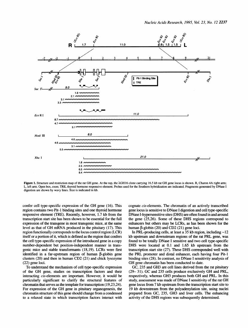
Figure 1. Structure and restriction map of the rat GH gene. At the top, the ?GHl6 clone carrying 16.5 kb rat GH gene locus is shown. R, Charon 4A right arm;L, left arm. Open box, exon; TRE, thyroid hormone responsive element. Probes used for the Southern hybridization are indicated. Fragments generated by DNase Idigestion are shown by wavy lines. Size is indicated in kb.
confer cell type-specific expression of the GH gene (16). Thisregion contains two Pit-I binding sites and one thyroid hormoneresponsive element (TRE). Recently, however, 1.7 kb from thetranscription start site has been shown to be essential for the fullexpression of the transgene in most transgenic mice, at the samelevel as that of GH mRNA produced in the pituitary (17). Thisregion functionally corresponds to the locus control region (LCR)itself or a portion of it, which is defined as the region that confersthe cell type-specific expression of the introduced gene in a copynumnber-dependent but position-independent manner in trans-genic mice and stable transformants (18,19). LCRs were firstidentified in a far-upstream region of human 3-globin geneclusters (20) and then in human CD2 (21) and chick lysozyme(22) gene loci.To understand the mechanism of cell type-specific expression
of the GH gene, studies on transcription factors and theirinteracting cis-elements are important. However, it would beparticularly significant to clarify the structural features ofchromatin that serves as the template for transcription (19,23,24).For expression of the GH gene in pituitary organogenesis, thechromatin structure of this gene should change from a condensedto a relaxed state in which transcription factors interact with
cognate cis-elements. The chromatin of an actively transcribedgene locus is sensitive to DNase I digestion and cell type-specificDNase I-hypersensitive sites (DHS) are often found in and aroundthe gene (25,26). Some of these DHS regions correspond toenhancers but others may be LCRs, as has been shown for thehuman ,-globin (20) and CD2 (21) gene loci.
In PRL-producing cells, at least a 35 kb region, including -12kb upstream and downstream regions of the rat PRL gene, was
found to be totally DNase I sensitive and two cell type-specificDHS were located at 0.1 and 1.65 kb upstream from thetranscription start site (27). These DHS corresponded well withthe PRL promoter and distal enhancer, each having four Pit-1binding sites (28). In contrast, no DNase I sensitivity analysis ofGH gene chromatin has been conducted to date.GC, 235 and GH3 are cell lines derived from the rat pituitary
(29- 31). GC and 235 cells produce exclusively GH and PRL,respectively, whereas GH3 produces both GH and PRL. In thisstudy, assessment was made of DNase I sensitivity of the rat GHgene locus from 7 kb upstream from the transcription start site to19 kb downstream from the polyadenylation site, using nucleiprepared from GC, 235, GH3 and liver cells. The enhancingactivity of the DHS regions was subsequently determined.
0
R 1 1.7
4,0q
11.0
I A J
'4,-
Sac II & 90
Eco R I8.74.7
Hind Il
Xba I
_. _Y -r _1 _ I _. B _
11-Vc
4-e I PR-l Binding Ske0 TRE

2238 Nucleic Acids Research, 1995, Vol. 23, No. 12
A
go ***40 -q0 W,@oqp.
S,0
9,,** '99@@@.::.
Figure 2. DNase I sensitivity of GH gene chromatin analyzed by Sacl orEcoRI digestions. Isolated nuclei were treated with the indicated amounts ofDNase I for 3 min at 20°C. DNAs were purified from nuclei and digested withSacl (A and B) orEcoRl (C and D). The digests (3 jg) were separated by elec-trophoresis on 0.75% agarose gel and blotted onto nitrocellulose filters. (A,C), GC; (B, D), 235. Blots were hybridized with rat GH cDNA (+18 to +424,A and B; +18 to +698, C and D). Restriction fragment size (given in kb) wasdetermined by HindIll-cleaved XDNA. Concentration of DNase I for treat-ment of nuclei (in gig/ml): lane 1, 2; lane 2, 1; lane 3, 0.5; lane 4, 0.25; lane5, 0.13; lane 6, 0.
MATERIALS AND METHODS
Cells
Cells were obtained from the following sources: GC (29) and 235(30), Dr M. Karin (University of California, San Diego, USA);GH3 (31) and HeLa, the Japanese Cancer Research ResourcesBank (Tokyo, Japan). GC and GH3 cells were grown inDulbecco's modified minimal essential medium (D-MEM)/F12containing 5% fetal bovine serum (FBS, JRH Biosciences, USA)and 5% horse senrum (JRH Biosciences). 235 cells were propa-gated in the above medium plus 10 nM P-estradiol (E2) toenhance their growth. HeLa cells were grown in D-MEMcontaining 10o FBS.
Isolation of nuclei
Cells in 12 (GC and GH3) or 30 (235) 9 cm dishes were collectedwith a scraper, washed once with phosphate-buffered saline(PBS) and resuspended in 5 ml Ni buffer (15 mM Tris-HCl, pH7.5,60mM KCI, 15 mM NaCl, 5 mM MgCl2, 0.5 mM D1T, 0.1mM EGTA, 0.3 M sucrose) (32). All subsequent steps werecarried out at 0-4°C. The cell suspension was mixed with 5 mlNI buffer containing 0.4 (GC and GH3) or 0.2% (235) NP-40 bypipetting, incubated on ice for 10 min and centrifuged 15 min at
Figure 3. DNase I sensitivity ofGH gene chromatin analyzed by HindIlI orXbaI digestion. Isolated nuclei were treated with the indicated amounts ofDNase I for 3 min at 20°C. DNAs were purified from nuclei and digested withHindIII (A and B) or XbaI (C and D). The digests (3 jig) were separated byelectrophoresis on 0.75% agarose gel and blotted onto nitrocellulose filters.(A, C), GC; (B, D), 235. All blots were hybridized with rat GH cDNA (+18to +698). Restriction fragment size (given in kb) was determined by HindIII-cleaved .DNA. Concentrations ofDNase I for treatment of nuclei (in jg/ml):lane 1, 2; lane 2, 1; lane 3, 0.5; lane 4, 0.25; lane 5, 0.13; lane 6, 0.
1000 g. The pelleted nuclei were washed once with NI buffer andresuspended in NI buffer (235) or in NI buffer containing 50%glycerol and 2 mM DTT (GC and GH3). A liver from a maleWistar rat was perfused with PBS and cut into small pieces. Tissue(-1 g) was homogenized with 10 ml NI buffer containing 0.4%NP-40 with 10 strokes of a Dounce homogenizer with pestle B,filtered through nylon mesh (#100-200) and centrifuged 15 minat 1000 g. The pelleted nuclei were washed and resuspended inNI buffer. Nuclei (-2 x 10⁸) were obtained from GC, GH3 and235 cells.
Nulease digestion and Southern hybridization
For DNase I (Takara Shuzo, Kyoto, Japan) digestion, 20 ji ofenzyme in NI buffer was mixed in 250 ±1 with 1 x 10⁷ of nucleiand incubated 3 min at 20°C. Reactions were terminated by theaddition of 25 jl stop solution containing 5% SDS and 100mMEDTA. The digests were mixed with 50 jg proteinase K in 90 jiNI buffer and incubated at 37°C overnight. After phenol,phenol/chloroform and chloroform extraction and ethanol preci-pitation, crude DNAs were dissolved inTE (10mM Tris-HCl,pH7.5, 1 mM EDTA). CrudeDNA (-100 jig) was digested overnightwith excess restriction endonuclease (Takara Shuzo), trated 30min with 40 jg/ml of RNase A at 37°C and extracted withphenol/chloroform. Southeem transfer and hybridization werecarried out as previously reported (33). After hybridization at
-eew 40404004wo

Nucleic Acids Research, 1995, Vol. 23, No. 12 2239
65°C overnight, the filter was washed four times (10 min/wash-ing) in 0.3 x SSC and 0.1% SDS at 65°C. Using rat pituitarycDNA as the template (34), a rat GH cDNA (nucleotides 18-698)(35) was amplified by PCR and subcloned into Bluescript. RatGH cDNA fragments, nucleotides 18-684 and 18-424, weremade from the cloned cDNA. The 0.8 kb rat albumin probecontaining the -12 to +113 region of the cDNA (36,37) wasprepared by PCR using male Wistar rat liver DNA and cloned intoBluescript. The probe DNA was labeled by the random primingmethod using [a-32P]dCTP.
A1 2 3 4 5 6
3.1
2.1-*iv* *
L 1IIi~3 3IJTII\319tsLiI.\ 21-LI~~~~~~~~~~~~~~~~~fl:.:
B1 2 3 4 5 6
W. &^ -- 21.0
~ 4 -841)814i:22i+6 ~6 5 1)11
-2t.I)2.-1.8 D)hI
Transfection
The Pica Gene basic vector, PGV-B (5597 bp; Toyo Ink, Japan),and promoter vector, PGV-P (5789 bp, PGV-P plus SV40promoter), possessing the firefly luciferase (Luc) gene, served asreporter genes. The effector plasmid, pRc/RSV (5133 bp; Invi-trogen, USA), containing rat Pit-I cDNA has been described (34).Luc activity was assayed using a Pica Gene Luminescence kit,PKG-100 (Toyo Ink). Reporter genes having rat GH (-318 to +12,GH-P) and PRL (-450 to +36, PRL-P) promoter regions were asdescribed (38). The rat PRL distal enhancer containing the -1800to -1447 region (PRL-E) (39) was made by thePCR method usingliverDNA prepared from male Wistar rat and cloned into the KpnIsite of PRL-P reporter (PRL-P+PRL-E). PRL-E was also clonedinto the KpnI site of the GH-P reporter (GH-P+PRL-E). Using ratGH cDNA, the XGH16 clone carrying 16.5 kb rat GH gene locus(Fig. 1) was isolated from a Spraque-Dawley female rat liverDNAlibrary (Charon 4A vector, Clontech, USA). To determineenhancing activity, reporter plasmids having rat DNA fragmentsderived from a genomic GH locus were constructed as shown inFigures 5-8. Transfections were conducted by the lipofectinmethod (Gibco-BRL) for 16 h as described (38). One dayfollowing medium renewal, luciferase activity was measured aspreviously described (34). A BCA protein assay kit (Pierce) wasused for protein determinations.
RESULTS
DNase I sensitivity ofGH gene chromatin in GC and235 cells
As reported previously (38), Northern hybridizations showed theexclusive production of GH and PRL mRNAs in GH and 235cells, respectively, while GH3 produced both mRNAs. Toexamine the structure of GH gene chromatin which functions asa template in transcription, DNase I-hypersensitive site(s) (DHS)mapping was carried out (Fig. 1). From the transcriptional startsite to the Sacd site located 7 kb upstream that of the rat GH gene,two majorDHS (M-DHS: UIA, -0.35 kb; UHA, -1.4 kb) and twominor DHS (m-DHS: UIB, -0.15 kb; UIIB, -1.6 kb) were found(Fig. 2A and B). UIA, UIB and UIIB were sensitive inGH-producing GC cells, whereas UIIA was sensitive in GH-non-producing but PRL-producing 235 cells. The UIB regioncorresponded to the promoter having two Pit-I binding sites andone TRE.The DNase I sensitivity ofchromatin in the GH gene region and
downstream region from the polyadenylation site was examined(Figs 2 and 3). In the downstream region, twoM-DHS (DIA, +2.7kb; DII, +6.7 kb) were identified by hybridization of EcoRI-digested DNA with the GH cDNA probe after DNase I treatmentof GC nuclei (Fig. 2C). The DIA was confirmed by HindIlI
C
DII1 6.5-
D
4 NP 4 -8.3
-- 38
-- 1.2
\1.0
Figure 4. DNase I sensitivity of GH gene chromatin in GH3, 235 and livercells. Isolated nuclei were treated with indicated amounts ofDNase I for 3 minat 20°C. DNAs were purified from nuclei and digested with Sacl (A), XbaI(B and C) or EcoRI (D). The digests (3 ig) were separated by electrophoresison 0.75% agarose gel and blotted onto nitrocellulose filters. Blot A was hy-bridized with rat GH cDNA (+18 to +424), B and C, with rat GH cDNA (+18to +698) and D, with 0.8 kb albumin DNA. Restriction fragment size (givenin kb) was determined by HindlIl-cleaved XDNA. Lanes (Al, B1), 235; (A2,B2, C, D), liver; (A3-A6, B3-B6), GH3. Concentrations of DNase I for treat-ment of nuclei (in gig/ml): A and B, lanes (1, 4), 2; lane 2, 4; lane 3, 0; lane5, 1; lane6,0.5; C andD, lane 1,4; lane2, 2; lane 3, 1; lane4, 0.5; lane 5,0.25;lane 6,0.
digestion ofGC DNA (Fig. 3A). DIB (m-DHS, +2.1 kb) and DIII(m-DHS, +8.6 kb) sites were detected by XbaI digestion of GCDNA (Fig. 3C). Among DHS, only the DIII was M-DHS inPRL-producing 235 cells (Fig. 3D). The DIII was M-DHS in thenuclei of 235 cells but m-DHS in GC nuclei, as also noted forUIIA and UIIB.
DNase I sensitivity ofGH gene chromatin in GH3 andliver cells
Assessment was made of the DNase I sensitivity of GH genechromatin in GH3 cells in which both GH and PRL are produced(Fig. 4). In the upstream region, hypersensitive sites (UIA, UIB,UIIB) characteristic of GH-producing GC cells, were present inGH3 cells, whereas no UIIA site in 235 cells could be seen (Fig.4A). In the downstream region, two M-DHS (DIA, DII) and twom-DHS (DIB, DIII) observed in GC cells were also detected inGH3 cells (Fig. 4B). In contrast to M-DHS of DIII in 235 cells,it was m-DHS in the GH3 cells. Thus, the DNase I sensitivity ofGH gene chromatin in GH/PRL-producing GH3 cells wasvirtually the same as that in GH-producing GC cells, but clearlydifferent from that in PRL-producing 235 cells.Among DHS in the upstream region of the GH gene, onlyUHA
was slightly hypersensitive in liver cells (Fig. 4A). In the

2240 Nucleic Acids Research, 1995, Vol. 23, No. 12
Ampr
I Pit-i Binding SiteI EREI TRE Luciferase (fold over promoter)
GHWP+PRL-E318-IM0 -1447-318 *12
Gl P+PRL-E I Ion I II
B ; 1.6
BK |
Kn IB
DIB
Sc I
Kn
B Hi -
Hd [
Hd F
Dm22 v
12Hd I * PC
I!d
d
PC P OPCI[jd
Figure 5. Activity of transiently introduced DHS regions of the rat GH gene into GC cells. Reporter plasmids having rat genomic GH locus fragments are shown.Values are means from triplicate determinations (±S.E.M.). GH-P, GH promoter; PRL-E, PRL distal enhancer; ERE, estrogen responsive element; TRE, thyroidhormone responsive element. Fragment size is indicated in kb. Restriction enzyme abbreviations: B H, BamHI; Bi, Bgll; Hd, HindH; Kn, KpnI; P C, PmaCI;Pu, PvuII; Sc, Sacl.
downstream region, the DH site was M-DHS in liver cells (Fig.4B and C). Reported DHS located in an upstream region of thealbumin gene were confirmed in liver cells (Fig. 4D) (40).
Activity of transiently introduced DNase I-hypersensitiveregions of the rat GH gene locus
As shown for the PRL gene, DHS regions often correspond toenhancers (19,24-26). An attempt was thus made to detectenhancing activity in DHS regions of the rat GH gene locus bycombining the regions with the reporter plasmid and transfectingcells. A 0.5 kb fragment in the UII region, a 0.6 kb fragment
including DIB and a 2.2 kb fragment including DHI- showedsignificant orientation-independent enhancer activity followinginsertion of the fragments into the upstream region of the GHpromoter (GH-P) and introduction into GC cells (Fig. 5). Usingthe PRL distal enhancer (PRL-E) as a positive control, the orderof magnitude of enhancing activity was: PRL-E > DIU > UII >DI. The enhancing activity of the DI region was the lowest butreproducible. A 1.4 kb fragment containing the DII site had noenhancer activity when inserted into a different position in thereporter due to difficulty of insertion into the GH-P upstreamregion. Enhancer activity of the UIl and DI regions could be
U'
Bi -4- :::N

Nucleic Acids Research, 1995, Vol. 23, No. 12 2241
Ampr
GCGH-PGH-P+PRL-EPRL-PPRL-P+UIIPRL-P+DIPRL-P+DIII E
HdPRL-P+PRL-ESV-P
SV-P+UIISV-P+DISV-P+DIIISV-P+PRL-E
235GH-PGH-P+UIIGHlP+DIGH-P+DM
HdGH-P+PRL-E
PRL-PPRL-P+UIIPRL-P+DIPRL-P+DII
HdPRL-P+PRL-E
2.2
-318 +12
-1 099 J!"4I I-W
l T1 111.:mJ
n AI Ion
2 n0.6, Bl
se PCI
-1800 -1447rIIN M
L I
2 Sc PC
Hd -1800lf -1447
I P"t-I BSx SitI EREI TRE
-318 +121 11
Il I] ElSc PC22
-1 8m-1447
I Hill
Sc PC
-1 -1?f L111n
Luciferase ( unit/ g protein)
4 ?l l le
I I 1 j 1
Luciferase (unit/mg protein)
qP NoNo o NqpN-b9e*, *~ I I I I I I
- i
I -1 -1 - I l1 I
d;-R'Reee ~
Figure 6. Activity of GH enhancers on GH, PRL and SV40 promoters in GC or 235 cells. Reporter plasmids having rat GH locus enhancers are shown. Valuesshown are means from triplicate determinations (±S.E.M.). GH-P, GH promoter; PRL-P, PRL promoter; PRL-E, PRL distal enhancer; SV-P, SV40 promoter; ERE,estrogen responsive element; TRE, thyroid hormone responsive element. Fragment size is indicated in kb. One light unit of Luc corresponds to 1.4 x iO-17 molLuc. Restriction enzyme abbreviations: Bl, BglII; Hd, HindmI; Kn, KpnI; P C, PmaCI; Pu, PvuH; Sc, SacI.
narrowed to a 0.5-0.6 kb fragment but it was difficult to reducethe size of the active fragment of the 2.2 kb fragment from theDUI region.Three M-DHS (UIA, DIA, DII) in GC cells were located
outside the fragment possessing enhancer activity, whereasm-DHS (UHB, DIB, DIII) in GC cells always mapped inside theenhancer fragments. The UIB site, corresponding to the promoterin the GH-P region, was shown to be m-DHS. Thus, the regions
having enhancing activity inGC cells when combined with GH-P,were not M-DHS but m-DHS in GC cells.
Activity ofGH enhancers on GH, PRL and SV40promoters in GC and 235 cells
Promoter- and cell type-specificities of GH enhancers were
examined (Fig. 6). When GH enhancers were inserted into the
U
I~~~~~~~~~~~~
I
--410. Rdi mm
2.2
-110, Hd I 01iI

2242 Nucleic Acids Research, 1995, Vol. 23, No. 12
Ampr
(-)
SV-P
rPR-l ( 9g 50
1.63.6
3.6
0 0
, 1.6Bl PU 3.6
0.6 1Sc PC 3.6
SV-P+UII
SV-P+DI
SV-P+DIII 01.63.6Hd
SV-P+PRL-E
PRL-P+PRL-E
0-18z-1 A47
1.6
3.6
0
-1 131 .6
3.6
Luciferase ( unit / mg protein )
10 1 5 20 25 30 35_ L L L
-4i
A
k
Figure 7. Effectof Pit- I onGH enhancers in HeLa cells. 0.4 1g reporter and0-3.6 1g effector plasmids were mixed with 8 p1 lipofectin and transfected as described(38). Amount ofDNA used for transfection was kept constant to 4 1ig by the addition of pRc/RSV. Values shown are means from duplicate determinations. SV-P,SV40 promoter; PRL-P, PRL promoter; PRL-E, PRL distal enhancer. Fragment size is indicated in kb. One light unit of the Luc corresponds with 1.4 x 10¹⁷ molLuc. Restriction enzyme abbreviations: Bi, BgflI; Hd, HindIU; Kn, KpnI; P C, PmaCI; Pu, PvuH; Sc, Sacl.
upstream region of the PRL promoter (PRL-P) of reporterconstructs and transfected into GC cells, less stimulation (UII,2.7-fold; DI, 1.5; DM, 0.7) was observed compared with PRL-E(11-fold). The order of stimulation activity (UII > DI > DIU)differed from when they were combined with GH-P. All GHenhancers combined with the SV40 promoter (SV-P) showed<1.4-fold stimulation, compared with PRL-E having 3.4-foldstimulation. Thus, GH enhancers prefer GH-P to PRL-P or SV-P.For GH-P-combined GH enhancers in 235 cells, significant
enhancing activity (UII, 2.3-fold; DI, 2.5; DIII, 2.3) was found,confirming thatGH enhancers function well with GH-P. Since theenhancing activity ofthe above constructs in 235 cells was far lessthan that ofGH-P combined PRL-E, the GH enhancers are shownto work well in GC cells. This was confimned by the introductionof PRL-P-combined GH enhancers into 235 cells.
Effects of Pit-i on GH enhancers in HeLa cells
Characteristics of the three GH enhancers (UII, DI, DIII) werefound to differ from PRL-E when combined with GH-P, PRL-Por SV-P, and then introduced into GC or 235 cells (Fig. 6). Nosignificant enhancing activity of SV-P combined with the GHenhancers could be detected in GC cells, compared with a3.4-fold stimulation of SV-P with PRL-E having four Pit-ibinding sites. It thus follows that Pit-1 does not mediate theenhancer activity, as is also indicated by the observation that theactivity of PRL-E always exceeded that of GH enhancerscombined with PRL-P when introduced into GC or 235 cells, orcombined with GH-P and introduced into 235 cells (Fig. 6). Sincean estrogen responsive element (ERE) was present in PRL-E,there is the possibility that most PRL-E activity is mediated by
2.2--410. Im
Hdm
I
,H

-7 , -2 -1 1 2 3 4 5 6 7 R 9
UII UI
BAAs 4!
DIXi X
DII
DNase I-Hypersensitive Sites mRNAUIIB UIIA UIA UIB DIB DIA DIl DIII GH PRL
2 GC + + + + + + + +j 235 + + +
3! GHs + + + + + + + + +> Liver + +
Enhancing + -? -7Activity + - + + - - +
Functions ENH SWI? LCR? PRO ENH LCR? MAR? SWI?ENH
DIII
+ ,a
I Pit-I Binding SiteTRE
[D Introduced RegionActive Enhancer
PRO: PromoterENH : EnhancerLCR : Locus Control RegionSWI : GH/PRL SwitchMAR: Matrix Attachment Region
Figure 8. Major DHS (M-DHS) and minor DHS (m-DHS) of rat GH gene chromatin and possible functions of regions corresponding to M-DHS and m-DHS.Size is indicated in kb. DNase I-hypersensitive sites: large +, M-DHS; small +, m-DHS. Open box, exon; big arrow (A), M-DHS; small arrow (B), m-DHS; TRE,thyroid hormone responsive element.
E2/estrogen receptor (ER) (4,41-43) rather than by Pit-1.SV-P-combined GH enhancers were consequently introducedinto HeLa cells together with the Pit-I effector plasmid to seewhether Pit-I would stimulate the GH enhancers (Fig. 7). A largestimulation of the positive control, PRL-P+PRL-E, was observedby Pit-1, while Pit-I stimulated SV-P+PRL-E to a certain extentin a Pit-I-dependent manner. In contrast, Pit-1 had no effect onSV-P-combined GH enhancers, thus showing it was not involvedin any of the three GH enhancers.
DISCUSSION
Analysis of the chromatin structure of a particular gene locuswould be essential for understanding the molecular mechanism ofcell type-specific gene expression (19,24). From DNase Isensitivity analysis of the chromatin structure of the rat GH genelocus from 7 kb upstream from the transcription start site to 19 kbdownstream from the polyadenylation site, two and threeM-DHSwere found within 2 kb upstream and 7 kb downstream regions,respectively (Fig. 8). Thus, M-DHS of the rat GH gene locus isdistributed relatively near the GH gene itself. Two m-DHS (UIB,UIIB) in the upstream region and one (DIB) in the downstreamside were found associated with M-DHS and thus a total of fiveM-DHS and three m-DHS were mapped. Five (UIIB, UIA, UIB,DIB, DIA) including two (UIA, DIA) M-DHS were specific forGH-producing cells.DNA fragments, including UIIA-UIIB, DIB and DIII sites,
were found to express enhancer activity that functions well withthe GH promoter in GC cells. Like UHIB and DIB, DNase I-
hypersensitive sites in enhancer fragments were m-DHS but notM-DHS in GC cells. Although the DIII site was m-DHS in GCcells, it was M-DHS in 235 cells, possibly due to the lowresolution of mapping, so that the DNase I sensitive sitecorresponding to enhancer in GC cells would appear to bem-DHS (DUB?) and differ from M-DHS (DIUA?) in 235 cells.It follows from these findings that all GH enhancers correspondto m-DHS in GH-producing cells. Since GH gene expression canbe affected by hormones and factors such as thyroid hormone,glucocorticoid, GRF, somatostatin and activin, examination ofchanges in the pattern of DNase I-hypersensitivity followingthese hormone treatments would be worthwhile.
Transient transfection studies show that as much as 235 bp(BglII site) of the region upstream to the transcriptional start siteis sufficient for cell type-specific expression of the rat GH gene(14). Three enhancers corresponding to m-DHS have beenidentified in this study. Since significant enhancer activity ofUIIB and DIB regions could not be detected until the fragmentshad been reduced to -0.5-0.6 kb, it may possibly have escapednotice. Since the order of magnitude of enhancer activity was notalways constant (Figs 5 and 6) when enhancers were combinedwith GH-P, PRL-P or SV-P and introduced into GC or 235 cells,it follows that they may differ from each other, although variousfactors may affect the enhancer locus since enhancer fragmentsize was relatively long in this study. Although cis-elementsmediating enhancer activity remain to be determined, owing inpart to absence of sequence data on DIB and DIII regions, someenhancer activity of the 0.5 kb BglII-PvuII fragment including
NucleicAcids Research, 1995, Vol. 23, No. 12 2243
21I - a %J I d. -F -T j v I 0 7 4. IgrA -#11 A-OL
i.
--VW cC.Y v
-p- 0 LV CY..4 1.6 2.4Bgl 11 Sac I Hind HI
0.5 0.6

2244 Nucleic Acids Research, 1995, Vol. 23, No. 12
UIIB site may be mediated by T3R since its binding site wasfound in this region (44).Two M-DHS, UIA and DIA located close to the GH gene that
were not enhancers, were found specific in GH-producing cells.These DHS regions may thus each have locus control region(LCR) activity in cell type-specific expression of the GH gene.The UIA region may correspond to a region having LCR-likeactivity identified in the 0.3-1.7 kb upstream region ofthe ratGHgene (17). Most PRL-producing cells derive from GH/PRL-pro-ducing cells (5-9) and thus, expression of the GH gene should beshut off in proceeding from GH/PRL-production to PRL-produc-tion only. Since UIIA and possibly DII(A?) were specific toPRL-producing cells and situated outside putative LCRs of theGH gene, UHA and DEI regions may serve as switch regions toturn off GH gene expression during differentiation from GH/PRL-producing cells to PRL-producing cells. Functions of theDII region appear difficult to determine. Since this region wasDNase I-hypersensitive inGC cells and also in liver cells, and notan enhancer, it may have structural functions in chromatin suchas the matrix attachment region (MAR) (45) or scaffold bindingregion (46). This site is DNase I-hypersensitive in GH-producingcells but insensitive in 235 cells. The sensitivity may thus berequired forGH gene expression but not after switching. Throughthe use of transgenic mice or stable transformants, it will bepossible to confirm whether DNA regions having M-DHS orm-DHS have the predicted functions.
ACKNOWLEDGEMENTS
We thank N. B. Hecht for editing, N. Matsuura for hisencouragement and M. Karin and the Japanese Cancer ResearchResources Bank for providing the cells. This work was supportedby a grant from the Fisheries Agency, Ministry of Education,Culture, and Sciences of Japan (to M.O.), and the Foundation ofGrowth Science (to K.K.).
REFERENCES1 Miller, W. L. and Eberiardt, N. L. (1983) Endocr Rev., 4, 97-130.2 Nicoll, C. S., Mayer, G. L. and Russel, S. M. (1986) Endocr Rev., 7,
169-203.3 Dolle, P., Castrillo, J.-L., Theill, L. E., Deerinck, T., Ellisman, M. and
Karin, M. (1990) Cell, 60,809-820.4 Simmons, D. M., Voss, J. W., Ingaham, H. A., Holloway, J. M., Broide, R.
S., Rosenfeld, M. G. and Swanson, L. W. (1990) Genes Dev., 4, 695-711.5 Hoeffer, J. P., Boockfor, F. R. and Frawley, L. L. (1985) Endocrinology,
117, 187-195.6 Chatelain, A., Dupuoy, J. P. and Dubois, M. P. (1979) Cell Tissue Res.,
196,409-427.7 Watanabe, Y. D. and Daikoku, S. (1979) Dev. Biol., 68, 559-567.8 Behringer, R. R., Mathews, L. S., Palmiter, R. D. and Brinster, R. L.
(1988) Genes Dev., 2,453-461.9 Borrelli, E., Heyman, R. A., Arias, C., Sawchenko, P. E. and Evans, R. M.
(1989) Nature, 339,538-541.10 Ingraham, H. A., Albert, V. R, Chen, R., Crenshaw, E. B., HI., Elsholtz, H.
P., He, X., Kapiloff, M. S., Mangalam, H. J., Swanson, L. W., Treacy, M.N. and Rosenfeld, M. G. (1990) Annu. Rev. Physiol., 52, 773-791.
11 Theill, L. E. and Karin, M. (1993) Endocr. Rev., 14, 670-689.12 Bodner, M., Castrillo, J.-L., Theill, L. E., Deerinck, T., Ellisman, M. and
Karin, M. (1988) Cell, 55, 505-518.13 Ingraham, H. A., Chen, R., Mangalam, H. J., Elsholtz, H. P., Flynn, S. E.,
Lin, C. R., Simmons, D. M., Swanson, L. and Rosenfeld, M. G. (1988)Cell, 55,519-529.
14 Nelson, C., Crenshaw HII, E. B., Franco, R., Lira, S. A., Albert, V. R.,Evans, R. M. and Rosenfeld, M. G. (1986) Nature, 322, 557-562.
15 Lipkin, S. M., Naar, A. M., Kalla, K. A., Sack, R. A. and Rosenfeld, M. G.(1993) Genes Dev., 7, 1674-1687.
16 Lira, S. A., Crenshaw III, E. B., Glass, C. K., Swanson, L. W. andRosenfeld, M. G. (1988) Proc. Natl. Acad. Sci. USA, 85,4755-4759.
17 Lira, S. A., Kalla, K. A., Glass, C. K., Drolet, D. W. and Rosenfeld, M. G.(1993) Mo. Endocrinol., 7, 694-701.
18 Dillon, N. and Grosveld, F. (1993) Trends Genet., 9, 134-137.19 Felsenfeld, G. (1992) Nature, 355, 219-224.20 Grosveld, F., van Assendelft, G. B., Greaves, D. R. and Kollias, G. (1987)
CeU, 51,975-985.21 Greaves, D. R., Wilson, F. D., Lang, G. and Kioussis, D. (1989) Cell, 56,
979-986.22 Bonifer, C., Vidal, M., Grosveld, F. and Sippel, A. E. (1990) EMBO J., 9,
2843-2848.23 Komberg, R. D. and Lorch, Y. (1992) Annu. Rev. Cell Biol., 8, 563-587.24 Paranjape, S. M., Kamakara, R. T. and Kadonaga, J. T. (1994) Annu. Rev.
Biochem., 63, 265-297.25 Gross, D. S. and Garrard, W. T. (1988) Annu. Rev. Biochem., 57, 159-197.26 Elgin, S. C. R. (1988) J. Biol. Chem., 263, 19259-19262.27 Durrin, L. K., Weber, J. L. and Gorski, J. (1984) J. Biol. Chem., 259,
7086-7093.28 Mangalam, H. J., Albert, V. R., Ingraham, H. A., Kapiloff, M., Wilson, L.,
Nelson, C., Elsholtz, H. and Rosenfeld, M. G. (1989) Genes Dev., 3,946-958.
29 Bancroft, F. C. (1973) Endocrinology, 92, 1014-1021.30 Reymond, M. J., Nansel, D.D., Burrows, G.H., Neaves, W.B., Porter, J.C.
(1984) Acta Endocrinol., 106, 459-470.31 Tashjian, A. H., Jr., Yasumura, Y, Levine, L., Sato, G., Parker, P. (1968)
Endocrinology, 82, 342-352.32 Wu, C. (1989) Methods Enzymol., 170, 269-289.33 Takayama, Y, Rand-Weaver, M., Kawauchi, H. and Ono, M. (1991) Mol.
Endocrinol., 5, 778-786.34 Ono, M., Hangai, T., Kaneko, T., Sato, Y., Ihara, S. and Kawauchi, H.
(1994)Mol.Endocrinol.,8, 109-115.35 Seeburg, P. H., Shine, J., Martial, J. A., Baxter, J. D. and Goodman, H. M.
(1977) Nature, 270, 486-494.36 Sargent, T. D., Yang, M. and Bonner, J. J. (1981) Proc. Natl. Acad. Sci.
USA, 78, 243-246.37 Sargent, T. D., Jagodzinski, L. L., Yang, M. and Bonner, J. J. (1981) Mol.
Cell. Biol., 1, 871-833.38 Ono, M., Mochizuki, E., Mori, Y, Aizawa, A. and Harigai, T. (1995)
Gene, 153, 267-271.39 Kladde, M. P., D'Cunha, J. and Gorski, J. (1993) J. Mol. Biol., 229,
344-367.40 Babiss, L. E., Bennett, A., Friedmann, J. M. and Darnell, J., J.E. (1986)
Proc. Natl. Acad. Sci. USA, 83, 6504-6508.41 Day, R. N., Koike, S., Sakai, M. and Maurer, R. A. (1990) Mol.
Endocrinol., 4, 1964-1971.42 Maurer, R. A. and Notides, A. C. (1987) Mol. Cell. Biol., 7, 4247-4254.43 Waterman, M. L., Adler, S., Nelson, C., Greene, G. L., Evans, R. M. and
Rosenfeld, M. G. (1987) Mol. Endocrinol., 2, 14-21.44 Lavin, T. N., Baxter, J. D. and Horita, S. (1988) J. Biol. Chem., 263,
9418-9426.45 Cockerill, P. N. and Garrard, W. T. (1986) Cell, 44, 273-282.46 Gasser, S. M. and Laemmli, U. K. (1986) Cell, 46,521-530.