Embed Size (px)
Citation preview

DNA PACKING IN VIRUS AND ANIMAL CELL NUCLEI
Raghuram.A (ME04B043)
Victor Prem Sagar.B(ME04B063) Nishanth.F(ME04B075)

1. Virus: An Introduction A virus can be considered as a container which transports a piece of genetic material a genome (either RNA or DNA). The virus attacks or infects a host cell by releasing its genome inside the cell 1.1 Structure of VIRUS
DNA is packed in the head very tightly. The head is icosahedral in structure. It has 2,3-5 rotational symmetry. A complete virus particle, known as a virion, is little more than a gene transporter, consisting at the most basic level of nucleic acid surrounded by a protective coat of protein called a capsid. A capsid is composed of proteins encoded by the viral genome and its shape serves as the basis for morphological distinction. Virally coded protein units called protomers will self-assemble to form the capsid, requiring no input from the virus genome - however, a few viruses code for proteins, which assist the construction of their capsid.

1.2 Evolution of structure in a Bacteriophage
In most cases, the linear virus genome when stretched out in solution is at least an order of magnitude longer than the diameter of the capsid. Folding the genome in order to stuff it into such a confined space is quite a feat of topology, but is compounded by repulsion by the cumulative negative electrostatic charges on the phosphate groups of the nucleotide backbone resulting in the genome resisting being crammed into a small space. Viruses overcome this difficulty by packaging along with the genome a number of positively charged molecules in order to counteract this negative charge repulsion. These include:
• small positively charged ions (Na, Mg, K, etc) • polyamines • various nucleic acid-binding proteins
The packing of DNA in the viral head is a complicated process and is studied extensively.

2. DNA-Deoxyribonucleic Acid 2.1 Introduction DNA is a polymer. The monomer units of DNA are nucleotides, and the polymer is known as a "polynucleotide." There are four different types of nucleotides found in DNA, differing only in the nitrogenous base. The four nucleotides are given one-letter abbreviations as shorthand for the four bases.
• A is for adenine • G is for guanine • C is for cytosine • T is for thymine
DNA is a normally double stranded macromolecule. Two polynucleotide chains, held together by weak thermodynamic forces, form a DNA molecule
Features of the DNA Double Helix
• Two DNA strands form a helical spiral, winding around a helix axis in a right-handed spiral
• The two polynucleotide chains run in opposite directions • The sugar-phosphate backbones of the two DNA strands wind around the helix
axis like the railing of a spiral staircase • The bases of the individual nucleotides are on the inside of the helix, stacked on
top of each other like the steps of a spiral staircase

3 DNA packing in Virus
3.1 Introduction From a simplistic view point, a virus can be considered as a container which transports a piece of genetic material—a genome (either RNA or DNA). The virus attacks or infects a host cell by releasing its genome inside the cell. The machinery of the host cell then processes the genetic information of the virus and is thereby ‘‘hijacked’’, and tricked into producing all of the components for new progeny viruses. For most viruses, once assembly of the protein shell or capsid of a new virus is complete, the newly replicated genome is threaded inside, segment-by-segment. This packaging process can be a remarkable feat of confinement. Many bacteriophages (viruses which attack bacteria cells) in particular are able to package their genomes extremely tightly. For instance, whereas the length of the T4 bacteriophage genome is of the order of 54 lm, the diameter of the head of the T4 virus is a mere 100 nm. Therefore the packaging of the T4 genome entails a 540:1 linear compression ratio. Simply scaling these dimensions to more a tangible size, this is similar to packing 400 feet of quarter-inch electrical extension cord into a (nine-inch-diameter) basketball!.
EM Image of a T4 and phi29 bacteriophage ( middile one is phi29) It is 50nm wide
The force generated by the bacteriophage ǿ29 packaging motor has been directly measured using optical tweezers, which showed a continuous buildup of the internal pressure as packaging proceeds. The measured stall force of 57 pN makes the f29 packaging motor one of the most powerful protein motors known to date. Similarly, the pressurization of packaged phage-l has been confirmed by using osmotic pressure as a resistive force against DNA ejection, causing the ejection to cease and leaving partially filled capsid particles . The internal pressure within these packaged viruses is directly related to the free-energy cost of forcing the DNA into a small cavity and provides the energy for conversion to work in the injection of the DNA into the host cell. 3.2 Inverse Spool Confirmation Cryo-electron microscopy and image processing have provided observational evidence supporting an axisymmetric packing motif known as the inverse spool conformation. Cryo-electron microscopy observations of also revealed that the duplex DNA genome of the T4 bacteriophage forms a highly condensed series of concentric layers, spaced about 2:36 nm apart, that follow the general contour of the inner wall of the protein capsid. By electron microscopy with image reconstruction, the conformation of the packaged genome of phage T4 appears to adopt a spool- like conformation with several layers of order, consistent with the coaxial spool model, where the spools are ordered parallel to each other and perpendicular to the axis of the tail.
Cross Sections of Empty and Full Phi 29 Phages (Cryo EM Reconstructions by Y. Tao et. al.) Capsid is about 50 nm wide while DNA inside is about 6.5 microns long!!

The DNA in the inner core region buckled out of the spooling plane, the capsid is able to accommodate more DNA at a lower density without suffering a high cost in bending energy. The chain is packed in an inverse-spool fashion initially, wrapping in a helical layer around its outer layer of confinement. However, the inverse-spool process was not maintained after this initial layer was formed, with the inner core filling in a disordered fashion. The chain is extruded from the bottom of the cylindrical core, located near the center of the icosahedron. The most recently extruded portion of the chain is directed toward the lower vertex of the icosahedron. The potential of the confining walls in this lower region of the capsid is symmetric about the packing axis, causing the most recently extruded portion of chain to form a small-radius circular loop perpendicular to the packing axis. Subsequent loops are forced underneath previously formed loops, which consequently rise upwards along the packing axis. The loops are driven up also in search of a wider cross-sectional radius into which
they can expand, available in the region away from the poles of the icosahedron. Cartoon of the structure of DNA packing
DNA strand Icosahedral strcture
Spool like Confirmation

3.3 Mechanics Involved in the formation A number of new experimental insights into the way DNA in viruses is packaged and ejected have been garnered. For example, the structure of both the portal motor as well as an example of the membrane puncturing device that leads to the delivery of the viral genome have been determined. At lower resolution, results of cryo-electron microscopy experiments have revealed the structure of certain viruses at various stages during self-assembly and the ordered arrangements of DNA in concentric circles within viral capsids. These insights have recently been complemented by single-molecule experiments in which the force exerted by the portal motor is measured during the process of viral packing itself . The problem of DNA packing is intriguing not only on the grounds of sheer geometric crowding, but also because of the recognition that the regions within which DNA is packaged (such as in a viral capsid) have linear dimensions that are comparable to the persistence length of the DNA, resulting in a steep elastic energy cost to be paid to effect such packing. 3.3.1 The Energetics of Viral Packing The energetics of viral packing is characterized by a number of different factors including: (i) the entropic-spring effect that causes the DNA in solution to adopt a more spread-out configuration than that in the viral capsid, (ii) the energetics of elastic bending that results from inducing curvature in the DNA on a scale that is smaller than the persistence length of p 50 nm, and (iii) those factors related to the presence of charge both on the DNA itself and in the surrounding solution. As shown by Riemer and Bloomfield ,the entropic contribution is smaller by a factor of 10 or more relative to the bending energies and those mediated by the charges on the DNA and the surrounding solution, and hence we make no further reference to it. As a result, just like in earlier work, we examine the interplay of elastic and interaction forces, though we neglect surface terms originating from DNA–capsid interactions. We present the formulation of Director field theory. 3.3.2 Director Field theory The objective of the theory is the formulation of a continuum theory of viral DNA packaging based on a director field representation of the encapsidated DNA.The defining property of the director field is that its point values give the local direction and density of the DNA.Thus, the director field supplies a complete description of the DNA conformation, and its determination is the central goal of the theory.The continuity of the DNA strand requires the director field to be divergence-free and tangent to the capsid.The energy of the DNA is defined as a functional of the director field which accounts for bending, torsion, and for electrostatic interactions through a density-dependent cohesive energy.

3.3.2 a) Principles The operative principle which determines the DNA conformation is assumed to be energy minimization. This principle, together with the complex structure of the energy functional, suggests the use of direct methods of the calculus of variations for the characterization of energy-minimizing conformations .In particular, we bypass the use of the Euler–Lagrange—or equilibrium—equations of the energy functional and seek to directly formulate constructions yielding low-energy conformations of the DNA.For axisymmetric capsids, a simple construction known as the inverse spool confirmation. We regard the inverse spool as a ‘baseline’ DNA conformation, and subsequent constructions are aimed at successively lowering the energy from that baseline. In particular, we show that torsionless toroidal solenoids, consisting of planar coils contained on meridional planes and wrapped around a spool core, and fine mixtures of the solenoid and spool phase, beat the inverse spool construction. 3.3.2 b)Conclusions from Director field Theory: Torsionless toroidal solenoids, consisting of planar coils contained within meridional planes and wrapped around a spool core, and fine mixtures of the solenoid and spool phase, beat the inverse spool construction.. 3.4 FLASH FIELD MODEL Aim of this model is to explain the steps of packing of DNA in virus Many molecular motors move unidirectionally along a DNA strand powered by nucleotide hydrolysis. These motors are multimeric ATPases with more than one hydrolysis site. This model explains how these motors generate the requisite force to process along their DNA track. This novel mechanism for force generation is based on a fluctuating electrostatic field driven by nucleotide hydrolysis. We apply the principle to explain the motion of certain DNA helicases and the portal protein, the motor that bacteriophages use to pump the genome into their capsids. The motor can reverse its direction without reversing the polarity of its electrostatic field, that is, without major structural modifications of the protein. The amazing facts involved in the packing process are published. (News paper release) (http://www.berkeley.edu/news/media/releases/2001/10/18_motor.html
The tight packing in the virus is achieved by one of the most powerful molecular motors ever observed, stronger than the motors that move our muscles or the nanoscale molecular motors that duplicate DNA or transcribe it into RNA. The motor the researchers studied is part of the bacteriophage ø29 (phi-29), a virus that is the scourge of the common soil bacterium Bacillus subtilis.

The first piece of the virus made is the empty capsid with a protein complex, called the portal motor, at the mouth. This motor grabs hold of the viral genome, a double strand of DNA, and pushes it into the capsid to complete assembly of the virus.
The biophysicists suspected that the motor generates a strong force since it
compacts DNA nearly 6,000 times its normal volume. To achieve this, the motor has to overcome DNA's resistance to bending, the electrostatic forces of repulsion encountered when pushing charged atoms close together, and the forces of entropy that make anything resist being constrained in a tight space.
A powerful molecular motor (yellow) translocates the twisted strands of DNA (right) of the Bacillus subtilis bacteriophage ø29 into a protein capsid. By using optical tweezers to pull on the DNA while it is being packed, UC Berkeley and University of Minnesota researchers have measured the force generated by the motor and the packing pressure. Flashing Field Model The model is based on the following central assumption: Binding of ATP to a hydrolysis site induces a conformational change that exposes a pair of negative and positive charge regions near the inner surface of the channel. The charged regions are not of equal size, and they are oriented at an angle to the circumferential meridian. We will assume that there is one charge pair region per nucleotide hydrolysis site, and that the axes of the charge pair regions are tilted with respect to the axis of the hole. The helicase binds three nucleotides at a time, and so we infer it has three hydrolysis sites however, the portal protein has perhaps 12 or 13 hydrolysis sites, located in two tiers. As we will see, the pattern of hydrolysis affects the performance of the motor (analogous to the firing sequence of a car engine). Therefore, if each hydrolysis site controls the expression of a single charge pair then, for more than a few hydrolysis sites, the number of firing sequences becomes too large to compute.

Qualitatively, the model works like this. The negatively charged phosphates spaced along the backbone of the DNA strand interact sequentially with the field of the charge pair regions as they "flash" on and off with the binding and hydrolysis of ATP. Each charge pair field gives the closest phosphate(s) an "electrostatic push" in the direction of the charge pair axis. The net effect of the charge pairs flashing on and off creates a sustained torsional and axial thirst. It turns out that the order in which the fields flash is not too important; even random flashing will drive the rotation. Note that the motor is an electrostatic machine that transduces strain fluctuations generated by nucleotide hydrolysis into a fluctuating electric field. Biased diffusion plays no role in torque generation; Brownian motion serves only as a "lubricant" to ensure the rotor does not hang up in local energy minima. Fig. 2a & 2b illustrates the model geometry and the operating principle.
Fig 1. This figure shows ATP getting hydrolyzed in hydrolysis sites

Conclusion: The DNA is packed in an Inverse Spool confirmation with DNA in the inner core region buckled out of the spooling plane, the capsid is able to accommodate more DNA at a lower density without suffering a high cost in bending energy.
Fig 2a) This picture shows the angles at which fields are generated
The figure demonstrates the fields which are present around the charges. The white colored part is positive charge and black colored part is negative charge. It shows the positive charge is double the negative charge.

4. DNA PACKAGING IN ANIMAL CELL NUCLEI
4.1 Introduction:
Cell is been an exicting thing since ages. We are concerned with the animal cell in particular its Nucleus where the genetic material is stored. Its proven fact that DNA is the one that carries hereditary information. The image illustrated down gives an idea about the location of the nucleus, which is an Animal Cell, also called as Eukaryotic Cell.
4.2 A Brief Glance Of The Nucleus : The nucleus is a highly specialized organelle that serves as the information and administrative center of the cell. This organelle has two major functions.
o It stores the cell's hereditary material, or DNA, and o It coordinates the cell's activities, which include intermediary metabolism,
growth, protein synthesis, and reproduction (cell division).
The spherical nucleus occupies about 10 percent of a cell's volume, making it the cell's most prominent feature.A double-layered membrane, the nuclear envelope, separates contents of the nucleus from the cellular cytoplasm.

The envelope is riddled with holes called nuclear pores that allow specific types and sizes of molecules to pass back and forth between the nucleus and the cytoplasm. It is also attached to a network of tubules, called the endoplasmic reticulum, where protein synthesis occurs. These tubules extend throughout the cell and manufacture the biochemical products that a particular cell type is genetically coded to produce
The image briefly illustrates about the nucleus with the labeled parts
4.3 A Glance at the DNA :
• Deoxyribonucleic acid (DNA) is a nucleic acid that contains the genetic instructions for the biological development of a cellular form of life or a virus. All known cellular life and some viruses have DNAs. DNA is a long polymer of nucleotides (a polynucleotide) that encodes the sequence of amino acid residues in proteins, using the genetic code: each amino acid is represented by three consecutive nucleotides (a triplet code).
• DNA is responsible for the genetic propagation of most inherited traits. In humans, these traits range from hair color to disease susceptibility. The genetic information encoded by an organism's DNA is called its genome. During cell division, DNA is replicated, and during reproduction is transmitted to offspring. The offspring's genome is a combination of the genomes of its parents. Lineage studies can be done because mitochondrial DNA only comes from the mother, and the Y chromosome only comes from the father

5. Hierarchies Of Compaction
The DNA is compacted in a series of sequential steps from a single strand to the Higher level of compaction
5.1 Packing Ratio :
The length of DNA divided by the length into which it is packaged is Packing Ratio
The degree to which DNA is condensed is expressed as its packing ratio. The length of DNA in the nucleus is far greater than the size of the compartment in which it is contained.
Example :
The shortest human chromosome contains 4.6 x 10⁷ bp of DNA (about 10 times the genome size of E. coli). This is equivalent to 14,000 µm of extended DNA. In its most condensed state during mitosis, the chromosome is about 2 µm long. This gives a packing ratio of 7000 (14,000/2).
5.2 Explanation : To achieve the overall packing ratio, DNA is not packaged directly into final
structure of chromatin. Instead, it contains several hierarchies of organization. The first level of packing is achieved by the winding of DNA around a protein core to produce a "bead-like" structure called a nucleosome.
This electron micrograph shows chromatin from the nucleus of a chicken red blood cell (birds, unlike most mammals, retain the nucleus in their mature red blood cells). The arrows point to the nucleosomes. You can see why the arrangement of nucleosomes has been likened to "beads on a string

This gives a packing ratio of about 6. This structure is invariant in both the euchromatin and heterochromatin of all chromosomes. The second level of packing is the coiling of beads in a helical structure called the 30 nm fiber that is found in both interphase chromatin and mitotic chromosomes.
This structure increases the packing ratio to about 40. The final packaging occurs when the fiber is organized in loops, scaffolds and domains that give a final packing ratio of about 1000 in interphase chromosomes and about 10,000 in mitotic chromosomes.
5.3 Evident On the basis of Salt Concentration :
The DNA compaction is evident from the electron microscopes and various other methods have proved this compaction to the extent of Nucleosomes.
The DNA molecule of a chromosome is folded and folded again in such a way that it is convenient to think of chromosomes as having several hierarchical levels of organization, each responsible for a particular degree of shortening of the enormously long strand. First Level : Assembly of DNA and histones can be considered the first level- namely, a sevenfold reduction in the length of DNA and the formation of a beaded flexible fiber 11 nm wide, roughly five times the width of free DNA. The structure of chromatin varies with the concentration of salts, and the 11 nm fibre is present only when the salt concentration is very low. If the salt concentration is increased slightly, the fibre becomes shortened somewhat by forming a zigzag arrangement of closely spaced beads between which the linking DNA is no longer visible in electron micrographs. Second Level : If the salt concentration is further increased to that present in living cells, a second level of compaction occurs- namely, the organization of the 11 nm nucleosome fiber into a shorter, thicker fiber with an average diameter ranging from 300 to 350 A, called the 30-nm fiber. In forming this structure , the 11 nm fiber apparently coils in a somewhat irregular left-handed super helix or solenoidal supercoil with six nucleosomes per turn. It is believed that most intracellular chromatin has the solenoidal supercoiled configuration. Final Level : The final level of organization is that in which the 30-nm fiber condenses into a chromatid of the compact metaphase chromosome. Little is known about this process other than that it seems to proceed in stages. In electron micrographs of isolated metaphase chromosomes from which histones have been removed, the partly unfolded DNA has the form of an enormous number of loops that seem to extend from a central core, or scaffold, composed of non histone chromosomal proteins. Electron microscopic studies of chromosome condensation in mitosis and meiosis suggests that the scaffold extends along the chromatid and that the 30-nm fiber becomes arranged into a helix of loops radiating from the scaffold. Details are not known about the additional folding that is required of the fiber in each loop to produce the fully condensed metaphase chromosome.

The genetic significance of the compaction of DNA and protein into chromatin and ultimately into the chromosome is that it greatly facilitates the movement of the genetic material during nuclear division. Relative to a fully extended DNA molecule, the length of a metaphase chromosome is reduced by a factor of approximately 104 because of chromosome condensation, the chromosomes would become so entangled that there would be many more abnormalities in the distribution of genetic material into daughter cells 5.3 FORMATION OF THE NUCLEOSOME: The electron micrograph of chromatin in the fig illustrated shows a long, nub –studded fibers bursting from the nucleus of a chick red blood cell. The chromatin fibers resemble beads on a string, with the beads having a diameter of about 1nm and the string having a diameter of about 2 nm. The 2 nm string is DNA. The chromatin has beads. Each bead is a nucleosome containing roughly 160 bp of DNA wrapped around a core composed of eight histones two each of H2A, H2B, H3, H4.The 160 bp of DNA wrap twice around this core octamer .An additional 40bp form linker dna, which contains one nucleosome with the next Histone H1 from the chromatin, some dna unwinds from each nucleosome but thenucleosomes do not fall apart; about 140 bp remain wrapped around each core

5.4 Models Of Higher-Level Packaging seek to Explain the
Extreme Compaction of Chromosomes at Mitosis
Several models been proposed by researchers. We shall deal with two of them. Formation of the 30 nm Fiber Through Super coiling : One model of additional compaction beyond nucleosomal winding purposes that the 10nm nucleosomal chromatin supercoils into a 30 nm super helix, achieving a further six fold chromatin condensation. Support for this model comes in part from electron microscope images of 30 nm that contain about six nucleosomes per turn. Whereas the 10 nm fiber looks three beads wide. Removal of some H1 from a 30nm causes it to unwind to 10nm.Adding back the H1 reinstates the 30nm fiber. Although electron microscopes can see the 30nm fiber.They do not know exactly how it forms.Higher level of compaction is even less understood. The electro micrograph is shown where 10nm(left) and 30nm(right) fibers are shown.
Mechanism Status What it accomplishes Nucleosome confirmed by crystal structure condenses naked DNA 7-fold to a
100 Å fiber Super coiling Hypothetical model( the 30nm
fiber predicted by the model has been seen in the electron microscope)
cause additional 6-fold compaction, achieving a 40-50 fold condensation relative to the naked eye
Radial Loops- Scaffolds Hypothetical model (preliminary experimental support exists for this model)
Through progressive compaction of 30 nm fiber condenses DNA to rod like mitotic chromosome that is 10000 times more compact than naked DNA

The Radial Loop-Scaffold Model The Radial Loop-Scaffold Model Seeks to Explain Compaction of the 30 nm fiber This model proposes that certain nonhistone proteins , including topoisomerase 2, bind to chromatin every 60-100kb and tether the supercoiled nucleosome studded 30 nm fiber into structural loops. Evidence that non-histone proteins fasten these loops come from chemical manipulations in which the removal of histones does not cause chromatin to unfold;d completely. Other nonhistone proteins may inturn gather the loops into daisylike rosettes; and additional nonhistone proteins may then compress the rosette centers into a compact bundle. A range of nonhistones thus forms the condensation scaffold. This proposal of looping and gathering is known as the Radial Loop-Scaffold Model of compaction. Visualisation : To visualize how it achieves condensation , imagine a long piece of string . to shorten it, you knot it at intervals to form loops separated by straight streches;the knots are at the base of each loop.To shorten the string still further you clip together set of knots. Finally you pin together all the clips. The Radial Loop-Scaffold Model of chromosome packaging offers a simple explanation of progressive chromosome compaction from interphase to metaphase chromosomes. At interphase the nucleosome-studded chromatin forms many structural loops, which are anchored together in rosettes in some areas. This initial looping and gathering compresses the genetic material sufficiently to fit into the nucleus , where it appears as a mass of tangled string. As the chromosomes enter the prophase of mitosis, looping and gathering increase and bundling through protein cross-ties begins. By metaphase , height of looping, the gathering and bundling achieves 250 fold compaction of the roughly 40 fold compacted 30 nm fiber, giving rise to the highly condensed , rod like shapes we refer to as mitotic chromosomes.
Is it Evident? Several pieces of biochemical and micrographic evidence support that radial loop-scaffold model. For example, metaphase chromosomes from which experimenters have extracted all the histones still maintain their familiar X-like shapes. Moreover, electron micrographs of whole-mounted mitotic chromosomes show loops of chromatin at the periphery of the chromosomes. And recent analysis of DNA indicate that special, irregularly spaced repetitive base sequences associate with nonhistone proteins to define the chromatin loops. These stretches of DNA are known as Scaffold-associated region, Or SARs. Found at the base of the chromatin loops, SARs are most likely the sites at which the DNA is anchored to the condensation scaffold. Despite bits and pieces of experimental evidence, studies that directly confirm or reject the radial loop-scaffold model have not yet been completed. Thus the loop and scaffold concept of higher level chromatin packaging remains a hypothesis. The hypothetical status of this higher-level compaction model contrasts sharply with nucleosomes, which are entities that investigators have isolated, crystallized and are being analyzed.

References: 1. Hud, N. V., and K. H. Downing. 2001. Cryoelectron microscopy of lambda phage DNA condensates in vitreous ice: the fine structure of DNA toroids. Proc. Natl. Acad. Sci. USA. 98:14925–14930
2. Hud, N. V., K. H. Downing, and R. Balhorn. 1995. A constant radius of curvature model for the organization of DNA in toroidal condensates. Proc. Natl. Acad. Sci. USA. 92:3581–3585.
3. Kindt, J., S. Tzlil, A. Ben-Shaul, and W. M. Gelbart. 2001. DNA packaging and ejection forces in bacteriophage. Proc. Natl. Acad. Sci. USA. 98:13671–13674
4. Purohit, P. K., J. Kondev, and R. Phillips. 2003. Mechanics of DNA packaging in viruses. Proc. Natl. Acad. Sci. USA. 100:3173–3178
5. Odijk, T. 1998. Hexagonally packed DNA within bacteriophage T7 stabilized by curvature stress. Biophys. J. 75:1223–1227.
6. Tzlil, S., J. T. Kindt, W. M. Gelbart, and A. Ben-Shaul. 2003. Forces and pressures in DNA packaging and release from viral capsids. Biophys. J. 84:1616–1627
7. Spakowitz, A. J., and Z. G. Wang. 2005. DNA packaging in bacteriophage: is twist important? Biophys. J. 88:3912–3923
8.Langevin Dynamics Simulations of Genome Packing in Bacteriophage Biophys. J., July 1, 2006; 91(1): 25 - 41
9. Robert Olby; "The Path to The Double Helix: Discovery of DNA"; first published in October 1974 by MacMillan, with foreword by Francis Crick; ISBN 046681173; the definitive DNA textbook, revised in 1994, with a 9 page postscript.
10. Handwerger, Korie E., Joseph G. Gall (Janurary 2006). "Subnuclear organelles: new insights into form and function".
11.Three-dimensional director-field predictions of viral DNA packing arrangements W. S. Klug _ M. T. Feldmann _ M. Ortiz
5 April 2004 / Accepted: 8 July 2004 / Published online: 22 September 2004. 12. DNA Packaging in Bacteriophage: Is Twist Important? Andrew James Spakowitz and Zhen-Gang Wang Division of Chemistry and Chemical Engineering, California Institute of Technology, Pasadena, California 91125 13. A director-field model of DNA packaging in viral capsids W.S.Klug, M.Ortiz ∗ Engineering and Applied Science Division, California Institute of Technology, Pasadena, CA 91125, USA 10 December 2002; received in revised form 14 April 2003 14. Langevin Dynamics Simulations of Genome Packing in Bacteriophage Christopher Forrey and M. Muthukumar 15. Lodish H, Berk A, Matsudaira P, Kaiser CA, Krieger M, Scott MP, Zipursky SL, Darnell J. (2004). Molecular Cell Biology. WH Freeman: New York, NY.


The inverse spool confirmation depicted inside an icosahedral virus Mechanics Involved in the Structure formation.
The packaging process involves a delicate balance of forces which are both mechanical and electrostatic in nature. The packing dimensions turn out to be exactly of the scale at which DNA can be considered a mechanically stiff rod. However, this rod has a very large negative total charge to it, such that strong repulsive electrostatic forces are experienced when the DNA is densely packed in the head of a virus.
Impressive advances in structural biology and techniques of single-biomolecule
experimentation have produced, in recent years, a number of observations about the mechanics of DNA as it is packaged into viruses.
Single-molecule experiments measuring the stiffness properties of DNA
and the electrostatic interactions of condensed DNA have enabled the formulation of quantitative, coarse-grained theories of packing mechanics.

DNA is a highly charged polymer, with one negative charge for every 0.17 nm of its length .Under typical ‘‘physiological’’ conditions, sections of DNA experience
repulsion when brought close to each other. However, with the addition of sufficient numbers of polyvalent cations, the hydration forces can be attractive, condensing DNA into toroidal bundles. Experimental proofs for the Inverse Spool Confirmation
Single molecule experiments measuring the stiffness properties of DNA and the electrostatic interaction of condensed DNA have enabled to predict the packing mechanics
buckled out of the spooling plane, the capsid is able to accommodate more DNA at a lower density without suffering a high cost in bending energy. Hence the Structure The DNA is packed in an Inverse Spool confirmation with DNA in the inner core region buckled out of the spooling plane, the capsid is able to accommodate more DNA at a lower density without suffering a high cost in bending energy .

DNA PACKING IN ANIMAL CELL
The length of DNA in the nucleus is far greater than the size of the compartment in which it is contained.
The degree to which DNA is condensed is expressed as its packing ratio.
This condensation is carried on as shown:
Packing ratio
The length of DNA divided by the length into which it is packaged
EXAMPLE
For example, the shortest human chromosome contains 4.6 x 10⁷ bp of DNA (about 10 times the genome size of E. coli). This is equivalent to 14,000 µm of extended DNA. In its most condensed state during mitosis, the chromosome is about 2 µm long. This gives a packing ratio of 7000 (14,000/2).
Explanation
To achieve the overall packing ratio, DNA is not packaged directly into final structure of chromatin. Instead, it contains several hierarchies of organization. The first level of packing is achieved by the winding of DNA around a protein core to produce a "bead-like" structure called a nucleosome.
This electron micrograph shows chromatin from the nucleus of a chicken red blood cell (birds, unlike most mammals, retain the nucleus in their mature red blood cells). The arrows point to the nucleosomes. You can see why the arrangement of nucleosomes has been likened to "beads on a string
This gives a packing ratio of about 6. This structure is invariant in both the euchromatin and heterochromatin of all chromosomes. The second level of
packing is the coiling of beads in a helical structure called the 30 nm fiber that is found in both interphase chromatin and mitotic chromosomes.
This structure increases the packing ratio to about 40. The final packaging occurs when the fiber is organized in loops, scaffolds and domains that give a final packing ratio of about 1000 in interphase chromosomes and about 10,000 in mitotic chromosomes.
When chromosomes are stained with dyes, they appear to have alternating lightly and darkly stained regions. The lightly-stained regions are euchromatin and contain single-copy, genetically-active DNA. The darkly-stained regions are heterochromatin and contain repetitive sequences that are genetically inactive.
The formation of nucleosomes helps somewhat, but not nearly enough, to make the DNA sufficiently compact to fit in the nucleus. In order to fit 46 DNA molecules (in humans), totaling over 2 meters in length, into a nucleus that may be only 10 µm across requires more extensive folding and compaction.
• Interactions between the exposed "tails" of the core histones causes nucleosomes to associate into a compact fiber 30 nm in diameter.
• These fibers are then folded into more complex structures whose precise configuration is uncertain and which probably changes with the level of activity of the genes in the region.
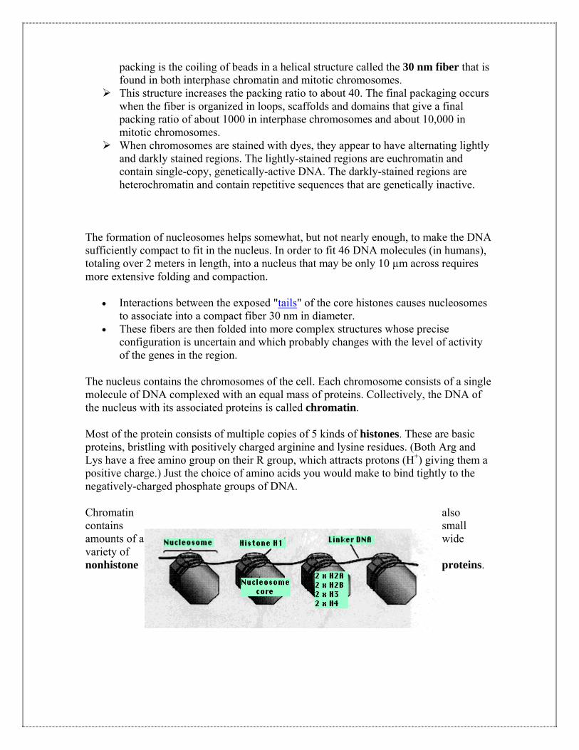
The nucleus contains the chromosomes of the cell. Each chromosome consists of a single molecule of DNA complexed with an equal mass of proteins. Collectively, the DNA of the nucleus with its associated proteins is called chromatin.
Most of the protein consists of multiple copies of 5 kinds of histones. These are basic proteins, bristling with positively charged arginine and lysine residues. (Both Arg and Lys have a free amino group on their R group, which attracts protons (H+) giving them a positive charge.) Just the choice of amino acids you would make to bind tightly to the negatively-charged phosphate groups of DNA.
Chromatin also contains small amounts of a wide variety of nonhistone proteins.

References: 1. Hud, N. V., and K. H. Downing. 2001. Cryoelectron microscopy of lambda phage DNA condensates in vitreous ice: the fine structure of DNA toroids. Proc. Natl. Acad. Sci. USA. 98:14925–14930
2. Hud, N. V., K. H. Downing, and R. Balhorn. 1995. A constant radius of curvature model for the organization of DNA in toroidal condensates. Proc. Natl. Acad. Sci. USA. 92:3581–3585.
3. Kindt, J., S. Tzlil, A. Ben-Shaul, and W. M. Gelbart. 2001. DNA packaging and ejection forces in bacteriophage. Proc. Natl. Acad. Sci. USA. 98:13671–13674
4. Purohit, P. K., J. Kondev, and R. Phillips. 2003. Mechanics of DNA packaging in viruses. Proc. Natl. Acad. Sci. USA. 100:3173–3178
5. Odijk, T. 1998. Hexagonally packed DNA within bacteriophage T7 stabilized by curvature stress. Biophys. J. 75:1223–1227.
6. Tzlil, S., J. T. Kindt, W. M. Gelbart, and A. Ben-Shaul. 2003. Forces and pressures in DNA packaging and release from viral capsids. Biophys. J. 84:1616–1627
7. Spakowitz, A. J., and Z. G. Wang. 2005. DNA packaging in bacteriophage: is twist important? Biophys. J. 88:3912–3923
8.Langevin Dynamics Simulations of Genome Packing in Bacteriophage Biophys. J., July 1, 2006; 91(1): 25 - 41