Embed Size (px)
Citation preview
IN THIS ISSUE
DNA Methylation, Sample Preparation & Antibodies2 5-Hydroxymethylcytosine: A New Analysis
in DNA Methylation
3 NEW: Biotin-based Enrichment of5-Hydroxymethylcytosine
4 NEW: Enzymes for the Study of 5-Hydroxymethylcytosine
5 NEW: MeDIP Enrichment of 5-Methylcytosine or 5-Hydroxymethylcytosine
6 NEW: ChIP qPCR Primers and Controls Ensure Your Experiments Work Every Time
7 NEW: Multi-Sample Sonicator, Probe Sonicator and Cooled Sonication Platform
8 DNMT Assay to Analyze Changes in CpG Methylation
8 NEW: Dounce Homogenizer to Assist in Cell and Chromatin Preparations
9 NEW: Protease Inhibitor Cocktail for Better Sample Preparation
9 Nuclear Extraction Kit for Nuclear, Cytoplasmic or Whole-cell Lysates
10 Antibodies to Stem Cell Regulators to Further your Stem Cell Research
10 Highly Validated Antibodies and ELISAs for the Study of Nuclear Receptors
11 Antibodies and Reagents for High Resolution Confocal STED Microscopy
High Resolution STED Microscopy Products (page 11)
Confocal
STED
2 North America 877 222 9543 Europe +32 (0)2 653 0001 Japan +81 (0)3 5225 3638 www.activemotif.com
5 - H y d r o x y m e t h y l c y t o s i n e D N A M e t h y l a t i o n
5-Hydroxymethylcytosine: A New Analysis in DNA Methylation
the fi rst company to provide a 5-hydroxymethylcytosine antibody that could be used for the detec-tion and immunoprecipitation of 5-hmC DNA. We have since expanded our product offering to include enzymes capable of distinguishing between 5-mC and 5-hmC residues, and affi nity en-richment kits to selectively isolate 5-hydroxymethylcytosine DNA.
Additional product details are available within this newsletter. To learn more about all of the 5-hmC products available, please give us a call or visit us on our website at www.activemotif.com/hmc.
In mammals and other verte-brates, DNA methylation usu-ally occurs at the C5 position of cytosines (5-mC), mostly within CpG dinucleotides. Normally, CpG islands, short CG-rich regions, are unmethylated; in cases where methylation does occur, the as-sociated gene is silenced. Aberrant methylation of these CpG regions is often associated with disease.
In 2009, Kriaucionis and Heintz as well as Tahiliani et al. discovered that a novel form of DNA methy-lation, 5-hydroxymethylcytosine (5-hmC), exists in mammals, and is elevated in neurons and embryonic stem cells. 5-Hydroxymethylcy-tosine results from the enzymatic conversion of 5-methylcytosine into 5-hydroxymethylcytosine by the TET family of cytosine oxy-genases (Figure 1). To date, little is understood about the functional relevance of 5-hmC in the mam-malian genome.
One of the diffi culties in studying 5-hmC is the fact that many of the traditional techniques employed
to study DNA methylation, such as bisulfi te conversion or certain methylation-sensitive restric-tion enzymes, do not distinguish between 5-mC and 5-hmC resi-dues. Methyl CpG binding protein enrichment methods only serve to selectively bind 5-mC DNA methylation, but cannot be used to enrich for 5-hmC. In an effort to better analyze the location and function of 5-hydroxymethylcyto-sine, new tools and techniques will need to be employed.
Active Motif is pleased to offer antibodies, assays and enzymes for the direct analysis of 5-hy-droxymethylcytosine. We were
Figure 1: Schematic of the conversion of 5-methylcytosine (5-mC) to 5-hydroxymethylcytosine (5-hmC).5-methylcytosine is converted to 5-hydroxymethylcytosine by the TET family of cytosine oxygenases.
Save 10% on reagents to study 5-HydroxymethylcytosineFor a limited time, get 10% off select products for 5-hmC.
For complete details and a list of eligible products, please visit www.activemotif.com/promo.
N
N
NH2OH
H
5-methylcytosine 5-hydroxymethylcytosine
ON
N
TET1, TET2, TET3
NH2
HO
3www.activemotif.com Japan +81 (0)3 5225 3638 Europe +32 (0)2 653 0001 North America 877 222 9543
O c t o b e r 2 0 1 1 • v o l u m e 1 2 • n u m b e r 2
To better interrogate the functional relevance of 5-hydroxymethylcytosine (5-hmC) DNA methylation, Active Motif
has developed an assay kit that enables detection and affi nity enrichment of DNA fragments containing the
5-hydroxymethylcytosine residue. The Hydroxymethyl Collector™ kit utilizes a β-glucosyltransferase enzyme to
transfer a modifi ed glucose moiety to 5-hydroxymethylcytosine residues in double-stranded DNA. This modifi ed
glucose is then used to chemically attach a biotin conjugate for capture and enrichment with magnetic streptavidin
beads. The enriched DNA can then be analyzed by PCR, microarray or sequencing to better understand the role of
5-hydroxymethylcytosine in mammalian genomes.
5 - H y d r o x y m e t h y l c y t o s i n e E n r i c h m e n t
NEW: Biotin-based Enrichment of 5-Hydroxymethylcytosine
Product Format Catalog No. Price ($US)
Hydroxymethyl Collector™ 25 rxns 55013 375
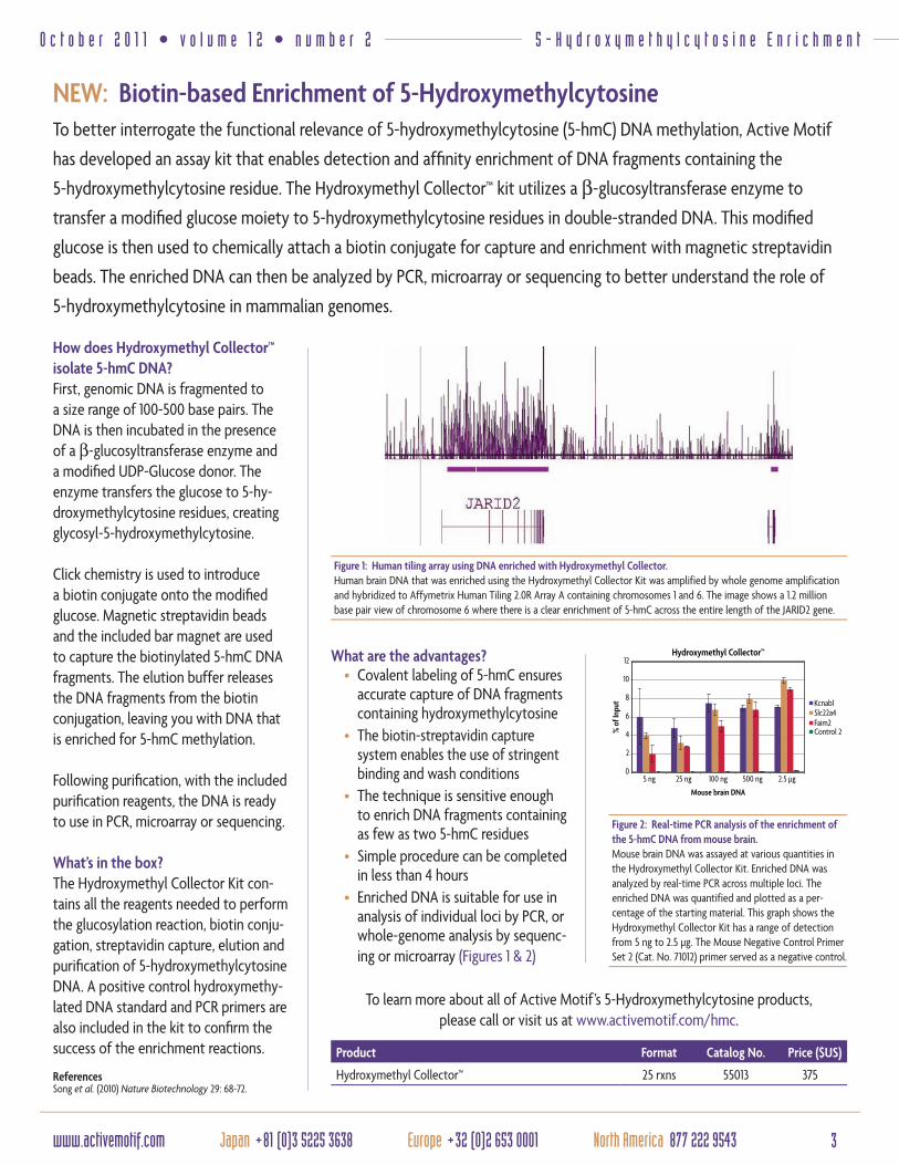
Figure 1: Human tiling array using DNA enriched with Hydroxymethyl Collector.Human brain DNA that was enriched using the Hydroxymethyl Collector Kit was amplified by whole genome amplification and hybridized to Affymetrix Human Tiling 2.0R Array A containing chromosomes 1 and 6. The image shows a 1.2 million base pair view of chromosome 6 where there is a clear enrichment of 5-hmC across the entire length of the JARID2 gene.
To learn more about all of Active Motif’s 5-Hydroxymethylcytosine products, please call or visit us at www.activemotif.com/hmc.
How does Hydroxymethyl Collector™ isolate 5-hmC DNA?First, genomic DNA is fragmented to a size range of 100-500 base pairs. The DNA is then incubated in the presence of a β-glucosyltransferase enzyme and a modifi ed UDP-Glucose donor. The enzyme transfers the glucose to 5-hy-droxymethylcytosine residues, creating glycosyl-5-hydroxymethylcytosine.
Click chemistry is used to introduce a biotin conjugate onto the modifi ed glucose. Magnetic streptavidin beads and the included bar magnet are used to capture the biotinylated 5-hmC DNA fragments. The elution buffer releases the DNA fragments from the biotin conjugation, leaving you with DNA that is enriched for 5-hmC methylation.
Following purifi cation, with the included purifi cation reagents, the DNA is ready to use in PCR, microarray or sequencing.
What’s in the box?The Hydroxymethyl Collector Kit con-tains all the reagents needed to perform the glucosylation reaction, biotin conju-gation, streptavidin capture, elution and purifi cation of 5-hydroxymethylcytosine DNA. A positive control hydroxymethy-lated DNA standard and PCR primers are also included in the kit to confi rm the success of the enrichment reactions.
ReferencesSong et al. (2010) Nature Biotechnology 29: 68-72.
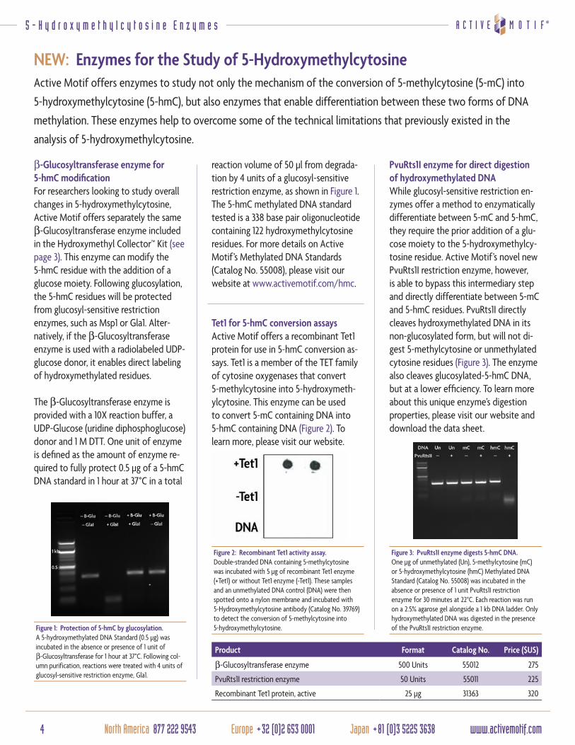
Figure 2: Real-time PCR analysis of the enrichment of the 5-hmC DNA from mouse brain.Mouse brain DNA was assayed at various quantities in the Hydroxymethyl Collector Kit. Enriched DNA was analyzed by real-time PCR across multiple loci. The enriched DNA was quantified and plotted as a per-centage of the starting material. This graph shows the Hydroxymethyl Collector Kit has a range of detection from 5 ng to 2.5 µg. The Mouse Negative Control Primer Set 2 (Cat. No. 71012) primer served as a negative control.
What are the advantages?• Covalent labeling of 5-hmC ensures
accurate capture of DNA fragments containing hydroxymethylcytosine
• The biotin-streptavidin capture system enables the use of stringent binding and wash conditions
• The technique is sensitive enough to enrich DNA fragments containing as few as two 5-hmC residues
• Simple procedure can be completed in less than 4 hours
• Enriched DNA is suitable for use in analysis of individual loci by PCR, or whole-genome analysis by sequenc-ing or microarray (Figures 1 & 2)
4 North America 877 222 9543 Europe +32 (0)2 653 0001 Japan +81 (0)3 5225 3638 www.activemotif.com
5 - H y d r o x y m e t h y l c y t o s i n e E n z y m e s
Active Motif offers enzymes to study not only the mechanism of the conversion of 5-methylcytosine (5-mC) into
5-hydroxymethylcytosine (5-hmC), but also enzymes that enable differentiation between these two forms of DNA
methylation. These enzymes help to overcome some of the technical limitations that previously existed in the
analysis of 5-hydroxymethylcytosine.
NEW: Enzymes for the Study of 5-Hydroxymethylcytosine
Product Format Catalog No. Price ($US)
β-Glucosyltransferase enzyme 500 Units 55012 275
PvuRts1I restriction enzyme 50 Units 55011 225
Recombinant Tet1 protein, active 25 µg 31363 320
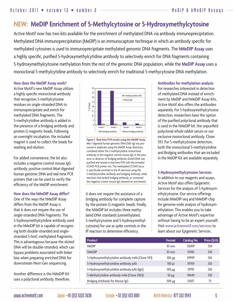
Figure 3: PvuRts1I enzyme digests 5-hmC DNA.One µg of unmethylated (Un), 5-methylcytosine (mC) or 5-hydroxymethylcytosine (hmC) Methylated DNA Standard (Catalog No. 55008) was incubated in the absence or presence of 1 unit PvuRts1I restriction enzyme for 30 minutes at 22°C. Each reaction was run on a 2.5% agarose gel alongside a 1 kb DNA ladder. Only hydroxymethylated DNA was digested in the presence of the PvuRts1I restriction enzyme.
PvuRts1I enzyme for direct digestion of hydroxymethylated DNAWhile glucosyl-sensitive restriction en-zymes offer a method to enzymatically differentiate between 5-mC and 5-hmC, they require the prior addition of a glu-cose moiety to the 5-hydroxymethylcy-tosine residue. Active Motif’s novel new PvuRts1I restriction enzyme, however, is able to bypass this intermediary step and directly differentiate between 5-mC and 5-hmC residues. PvuRts1I directly cleaves hydroxymethylated DNA in its non-glucosylated form, but will not di-gest 5-methylcytosine or unmethylated cytosine residues (Figure 3). The enzyme also cleaves glucosylated-5-hmC DNA, but at a lower effi ciency. To learn more about this unique enzyme’s digestion properties, please visit our website and download the data sheet.
Tet1 for 5-hmC conversion assaysActive Motif offers a recombinant Tet1 protein for use in 5-hmC conversion as-says. Tet1 is a member of the TET family of cytosine oxygenases that convert 5-methylcytosine into 5-hydroxymeth-ylcytosine. This enzyme can be used to convert 5-mC containing DNA into 5-hmC containing DNA (Figure 2). To learn more, please visit our website.
Figure 1: Protection of 5-hmC by glucosylation.A 5-hydroxymethylated DNA Standard (0.5 µg) was incubated in the absence or presence of 1 unit of β-Glucosyltransferase for 1 hour at 37°C. Following col-umn purification, reactions were treated with 4 units of glucosyl-sensitive restriction enzyme, Gla1.
β-Glucosyltransferase enzyme for 5-hmC modifi cationFor researchers looking to study overall changes in 5-hydroxymethylcytosine, Active Motif offers separately the same β-Glucosyltransferase enzyme included in the Hydroxymethyl Collector™ Kit (see page 3). This enzyme can modify the 5-hmC residue with the addition of a glucose moiety. Following glucosylation, the 5-hmC residues will be protected from glucosyl-sensitive restriction enzymes, such as Msp1 or Gla1. Alter-natively, if the β-Glucosyltransferase enzyme is used with a radiolabeled UDP-glucose donor, it enables direct labeling of hydroxymethylated residues.
The β-Glucosyltransferase enzyme is provided with a 10X reaction buffer, a UDP-Glucose (uridine diphosphoglucose) donor and 1 M DTT. One unit of enzyme is defi ned as the amount of enzyme re-quired to fully protect 0.5 µg of a 5-hmC DNA standard in 1 hour at 37°C in a total
Figure 2: Recombinant Tet1 activity assay.Double-stranded DNA containing 5-methylcytosine was incubated with 5 µg of recombinant Tet1 enzyme (+Tet1) or without Tet1 enzyme (-Tet1). These samples and an unmethylated DNA control (DNA) were then spotted onto a nylon membrane and incubated with 5-Hydroxymethylcytosine antibody (Catalog No. 39769) to detect the conversion of 5-methylcytosine into 5-hydroxymethylcytosine.
reaction volume of 50 µl from degrada-tion by 4 units of a glucosyl-sensitive restriction enzyme, as shown in Figure 1.The 5-hmC methylated DNA standard tested is a 338 base pair oligonucleotide containing 122 hydroxymethylcytosine residues. For more details on Active Motif’s Methylated DNA Standards (Catalog No. 55008), please visit our website at www.activemotif.com/hmc.
5www.activemotif.com Japan +81 (0)3 5225 3638 Europe +32 (0)2 653 0001 North America 877 222 9543
O c t o b e r 2 0 1 1 • v o l u m e 1 2 • n u m b e r 2
NEW: MeDIP Enrichment of 5-Methylcytosine or 5-HydroxymethylcytosineActive Motif now has two kits available for the enrichment of methylated DNA via antibody immunoprecipitation.
Methylated DNA immunoprecipitation (MeDIP) is an immunocapture technique in which an antibody specifi c for
methylated cytosines is used to immunoprecipitate methylated genomic DNA fragments. The hMeDIP Assay uses
a highly specifi c, purifi ed 5-hydroxymethylcytidine antibody to selectively enrich for DNA fragments containing
5-hydroxymethylcytosine methylation from the rest of the genomic DNA population, while the MeDIP Assay uses a
monoclonal 5-methylcytidine antibody to selectively enrich for traditional 5-methycytosine DNA methylation.
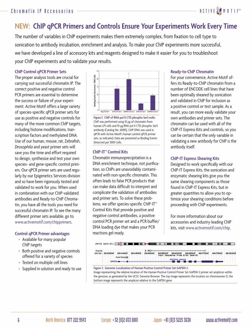
Figure 1: Real time PCR results using the MeDIP Assay.Mse I digested human genomic DNA (500 ng) was pro-cessed in duplicate using the MeDIP Assay. Reactions contained either the 5-methylcytidine monoclonal antibody or the negative control mouse IgG in the pres-ence or absence of bridging antibody. Eluted DNA was purified and tested in real time PCR with the included ZC3H13 PCR primer mix. The methylated ZC3H13 locus is specifically enriched in the IP reactions using the 5-methylcytodine antibody and bridging antibody, while reactions that lacked bridging antibody, or contained the negative control mouse IgG showed no enrichment.
How does the MeDIP Assay work?Active Motif’s new MeDIP Assay utilizes a highly specifi c monoclonal antibody that recognizes 5-methylcytosine residues on single-stranded DNA to immunoprecipitate and enrich for methylated DNA fragments. The 5-methylcytidine antibody is added in the presence of a bridging antibody and protein G magnetic beads. Following an overnight incubation, the included magnet is used to collect the beads for washing and elution.
For added convenience, the kit also includes a negative control mouse IgG antibody, positive control MseI digested human genomic DNA and real time PCR primers that can be used to verify the effi ciency of the MeDIP enrichment.
M e D I P & h M e D I P A s s a y s
5-Hydroxymethylcytosine ServicesIn addition to our reagents and assays, Active Motif also offers Epigenetic Services for the analysis of 5-hydroxym-ethylcytosine. Our service offerings include hMeDIP-seq and hMeDIP-chip for genome-wide analysis of hydroxym-ethylation. This enables you to take advantage of Active Motif’s expertise without having to be an expert yourself.Visit www.activemotif.com/services to learn about our Epigenetic Services.
Product Format Catalog No. Price ($US)
MeDIP 10 rxns 55009 350
hMeDIP 10 rxns 55010 350
5-Hydroxymethylcytidine antibody mAb (Clone 59.1) 100 µg 39999 330
5-Hydroxymethylcytidine antibody pAb 100 µl 39769 330
5-Hydroxymethylcytidine antibody pAb (IgG) 100 µg 39791 330
5-Methylcytidine antibody mAb (Clone 33D3) 50 µg 39649 330
Bridging Antibody for Mouse IgG 500 µg 53017 75
How does the hMeDIP Assay differ?One of the ways the hMeDIP Assay differs from the MeDIP Assay is that it does not require the use of single-stranded DNA fragments. The 5-hydroxymethylcytidine antibody used in the hMeDIP kit is capable of recogniz-ing both double-stranded and single-stranded 5-hmC methylated fragments. This is advantageous because the eluted DNA will be double-stranded, which can bypass problems associated with linker bias when preparing enriched DNA for downstream Next-Gen sequencing.
Another difference is the hMeDIP Kit uses a polyclonal antibody, therefore,
Antibodies for methylation analysisFor researches interested in detection of methylated DNA instead of enrich-ment by MeDIP and hMeDIP Assay Kits, Active Motif also offers the antibodies separately. For 5-hydroxymethylcytosine detection, researchers have the option of the purifi ed polyclonal antibody that is used in the hMeDIP kit, the unpurifi ed polyclonal whole rabbit serum or our exclusive monoclonal antibody, Clone 59.1. For 5-methylcytosine detection, both the monoclonal 5-methylcytidine and Bridging antibodies that are included in the MeDIP Kit are available separately.
it does not require the assistance of a bridging antibody for complete capture by the protein G magnetic beads. Finally, the hMeDIP kit includes three methy-lated DNA standards (unmethylated, 5-methylcytosine and 5-hydroxymethyl-cytosine) for use as spike controls in the IP reaction to determine effi ciency.
6 North America 877 222 9543 Europe +32 (0)2 653 0001 Japan +81 (0)3 5225 3638 www.activemotif.com
ChIP Control qPCR Primer SetsThe proper analysis tools are crucial for carrying out successful chromatin IP. The correct positive and negative control PCR primers are essential to determine the success or failure of your experi-ment. Active Motif offers a large variety of species-specifi c qPCR primer sets for use as positive and negative controls for many of the more common ChIP targets, including histone modifi cations, tran-scription factors and methylated DNA. Use of our human, mouse, rat, Zebrafi sh, Drosophila and yeast primer sets will save you the time and effort required to design, synthesize and test your own species- and gene-specifi c control prim-ers. Our qPCR primer sets are used regu-larly by our Epigenetics Services division and so have been rigorously tested and validated to work for you. When used in combination with our ChIP-validated antibodies and Ready-to-ChIP Chroma-tin, you have all the tools you need for successful chromatin IP. To see the many different primer sets available, go to www.activemotif.com/chipprimers.
Control qPCR Primer advantages• Available for many popular
ChIP targets• Both positive and negative controls
offered for a variety of species• Tested on multiple cell lines• Supplied in solution and ready to use
The number of variables in ChIP experiments makes them extremely complex, from fi xation to cell type to
sonication to antibody incubation, enrichment and analysis. To make your ChIP experiments more successful,
we have developed a line of accessory kits and reagents designed to make it easier for you to troubleshoot
your ChIP experiments and to validate your results.
NEW: ChIP qPCR Primers and Controls Ensure Your Experiments Work Every Time
ChIP-IT™ Control KitsChromatin immunoprecipitation is a DNA enrichment technique, not purifi ca-tion, so ChIPs are unavoidably contami-nated with non-specifi c chromatin. This often leads to false PCR products that can make data diffi cult to interpret and complicate the validation of antibodies and primer sets. To solve these prob-lems, we offer species-specifi c ChIP-IT™ Control Kits that provide positive and negative control antibodies, a positive control PCR primer set and a PCR buffer/DNA loading dye that makes your PCR reactions gel-ready.
Ready-to-ChIP ChromatinFor your convenience, Active Motif of-fers its Ready-to-ChIP chromatin from a number of ENCODE cell lines that have been optimally sheared by sonication and validated in ChIP for inclusion as a positive control or test sample. As a result, you can more easily validate your own antibodies and primer sets. The chromatin can be used with all of the ChIP-IT Express Kits and controls, so you can be certain that the only variable in validating a new antibody for ChIP is the antibody itself.
ChIP-IT Express Shearing KitsDesigned to work specifi cally with our ChIP-IT Express Kits, the sonication and enzymatic shearing kits give you the same shearing components as those found in ChIP-IT Express Kits, but in greater quantities to allow you to op-timize your shearing conditions before proceeding with ChIP experiments.
For more information about our accessories and industry-leading ChIP kits, visit www.activemotif.com/chip.
Figure 2: Genomic Localization of Human Positive Control Primer Set GAPDH-2.Image representing the relative location of the Human Positive Control Primer Set GAPDH-2 primer set amplicon within the genome, as generated by the UCSC Genome Browser. The top image represents the location on chromosome 12, the bottom image represents the amplicon relative to the GAPDH gene.
Figure 1: ChIP of RNA pol II CTD phospho Ser5 mAb.ChIP was performed using 10 µg of chromatin from human LP1 cells and 10 µg RNA pol II CTD phospho Ser5 antibody (Catalog No. 61085). ChIP DNA was used in qPCR with Active Motif’s human control qPCR primer sets, as indicated. Data are presented as Binding Events Detected per 1000 Cells.
C h r o m a t i n I P A c c e s s o r i e s

7www.activemotif.com Japan +81 (0)3 5225 3638 Europe +32 (0)2 653 0001 North America 877 222 9543
O c t o b e r 2 0 1 1 • v o l u m e 1 2 • n u m b e r 2 S o n i c a t i o n E q u i p m e n t
Programmable generators – why not choose complete control?Both the EpiShear™ Multi-Sample Sonicator and the EpiShear™ Probe Sonicator are controlled by microproces-sor units that offer both programmable and manual operation. Each has a keypad and digital display that make it easy to program the amplitude and duration of On and Off pulse cycles. Pulse intensity to be set from 20-100%, enabling you to choose exact parameters for your cell type. The digital display shows the total elapsed time, and provides real-time energy monitoring of both wattage and joules. These features make it easier to obtain more reproducible results when you perform subsequent sonications.
Process up to 8 samples at onceThe Multi-Sample Sonicator is a high-in-tensity cup horn sonicator. Its 750 watts of power enable continuous operation to shear even the most problematic samples, like T cells. The unit can process up to eight 1.5 ml vial samples at once, and comes with a sound enclosure and a thermoelectric chiller (Figure 1). For more complete information, please visit us at www.activemotif.com/cuphorn.
Sonicate large or small samplesWhile the EpiShear™ Probe Sonicator is a compact, economical unit (Figure 2), it is still a fully programmable unit with all the features that will help ensure your reproducible results. It is supplied with a 1/8" microtip probe that enables you to shear one 500 µl to 15 ml sample at a time. Other size probes are available that expand its range from 200 µl to 50 ml. For more complete information, please visit www.activemotif.com/probe.
Sonication is frequently used to prepare sheared chromatin for ChIP, as well as for standard cell disruption, DNA/RNA
shearing and other homogenization applications. Active Motif is pleased to introduce its EpiShear™ line of sonication
products, which will save you time and effort while ensuring that you obtain more reproducible results.
NEW: Multi-Sample Sonicator, Probe Sonicator and Cooled Sonication Platform
Make any probe sonicator easier to useThe EpiShear Cooled Sonication Platform, affectionately known as “Big Jack”, greatly increases reproducibility by enabling you to precisely position the depth of the probe in your sample every
time. It can be used in a sound enclosure or on a base. Coolers that fi t microfuge, 15 ml and 50 ml tubes keep your sample cold during sonication, so you don’t have shuttle it to and from an ice bucket. See www.activemotif.com/platform.
Figure 1: The EpiShear Multi-Sample Sonicator / Chiller.The EpiShear Multi-Sample Sonicator / Chiller includes a powerful 750-watt generator with a keypad and digital display that make it easy to program and monitor sonication. The unit’s cup horn sonicator is housed in a compact sound enclosure to reduce sonication noise. The cup horn can process up to 8 samples simultaneously, which rotate continuously to ensure all samples are processed equally. It is mounted in an acrylic bath that is filled with water. The bath and enclosure are plumbed with quick-connect hoses that attach to a thermoelectric chiller, which keeps the samples at 2-4°C during sonication.
Figure 2: The EpiShear Probe Sonicator and the EpiShear Cooled Sonication Platform.The EpiShear Probe Sonicator is a compact 120-watt unit that can process small or large samples. The EpiShear Cooled Sonication Platform, sold separately, precisely positions the probe every sonication, greatly enhancing sample-to-sample reproducibility. The tube coolers, available in 3 sizes, eliminate the need to rest the sample in an ice bath between pulses.
8 North America 877 222 9543 Europe +32 (0)2 653 0001 Japan +81 (0)3 5225 3638 www.activemotif.com
C p G M e t h y l a t i o n A n a l y s i s a n d S a m p l e P r e p a r a t i o n
NEW: Dounce Homogenizer to Assist in Cell and Chromatin Preparations
DNMT Assay to Analyze Changes in CpG MethylationDNA methylation usually occurs at the fi fth carbon of cytosine, within the context of a CpG dinucleotide. Modifi ca-
tions at these sequences by DNA methyltransferase enzymes (DNMTs) can have profound effects on transcription.
Active Motif’s DNMT Activity / Inhibition Assay is a fast, user-friendly assay to simplify the measurement of DNA
methyltransferase activity or the effects of inhibitor compounds without the need for radioisotopes.
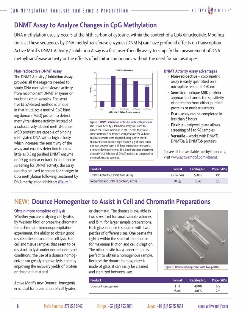
Non-radioactive DNMT AssayThe DNMT Activity / Inhibition Assay provides all the reagents needed to study DNA methyltransferase activity from recombinant DNMT enzymes or nuclear extract samples. The sensi-tive ELISA-based method is unique in that it utilizes a methyl-CpG bind-ing domain (MBD) protein to detect methyltransferase activity, instead of a radioactively labeled methyl-donor. MBD proteins are capable of binding methylated DNA with a high affi nity, which increases the sensitivity of the assay and enables detection from as little as 0.5 ng purifi ed DNMT enzyme or 0.5 µg nuclear extract. In addition to screening for DNMT activity, the assay can also be used to screen for changes in CpG methylation following treatment by DNA methylation inhibitors (Figure 1).
Figure 1: DNMT inhibition of MCF-7 cells with procaine.The DNMT Activity / Inhibition Assay was used to screen for DNMT inhibition in MCF-7 cells that were either untreated or treated with procaine for 96 hours. Nuclear extracts were prepared using Active Motif’s Nuclear Extract Kit (see page 9) and 5 µg of each condi-tion was assayed with a 1.5 hour incubation time and a 3 minute developing time. The 5 mM procaine treatment showed 43% inhibition of DNMT activity as compared to the mock treated samples.
Product Format Catalog No. Price ($US)
DNMT Activity / Inhibition Assay 1 x 96 rxns 55006 495
Recombinant DNMT1 protein, active 10 µg 31335 320
Obtain more complete cell lysisWhether you are analyzing cell lysates by Western blot, or preparing chromatin for a chromatin immunoprecipitation experiment, the ability to obtain good results relies on accurate cell lysis. For cell and tissue samples that seem to be resistant to lysis under normal detergent conditions, the use of a dounce homog-enizer can greatly improve lysis, thereby improving the recovery yields of protein or chromatin material.
Active Motif’s new Dounce Homogeniz-er is ideal for preparation of cell lysates
or chromatin. The dounce is available in two sizes, 1 ml for small sample volumes and 15 ml for larger sample preparations. Each glass dounce is supplied with two pestles of different sizes. One pestle fi ts tightly within the shaft of the dounce for maximum friction and cell disruption. The other pestle has a looser fi t and is perfect to obtain a homogenous sample. Because the dounce homogenizer is made of glass, it can easily be cleaned and sterilized between uses.
DNMT Activity Assay advantages• Non-radioactive – colorimetric
assay is easily quantifi ed on a microplate reader at 450 nm
• Sensitive – unique MBD protein approach enhances the sensitivity of detection from either purifi ed proteins or nuclear extracts
• Fast – assay can be completed in less than 3 hours
• Flexible – stripwell plate allows screening of 1 to 96 samples
• Versatile – works with DNMT1, DNMT3a & DNMT3b proteins
To see all the available methylation kits, visit www.activemotif.com/dnamt.
Product Format Catalog No. Price ($US)
Dounce Homogenizer 1 ml15 ml
4040140415
175225
Figure 1: Dounce homogenizer with two pestles.
9www.activemotif.com Japan +81 (0)3 5225 3638 Europe +32 (0)2 653 0001 North America 877 222 9543
O c t o b e r 2 0 1 1 • v o l u m e 1 2 • n u m b e r 2
Nuclear Extraction Kit for Nuclear, Cytoplasmic or Whole-cell Lysates
S a m p l e P r e p a r a t i o n
NEW: Protease Inhibitor Cocktail for Better Sample Preparation
Product Format Catalog No. Price ($US)
Nuclear Extract Kit 100 rxns400 rxns
4001040410
205585
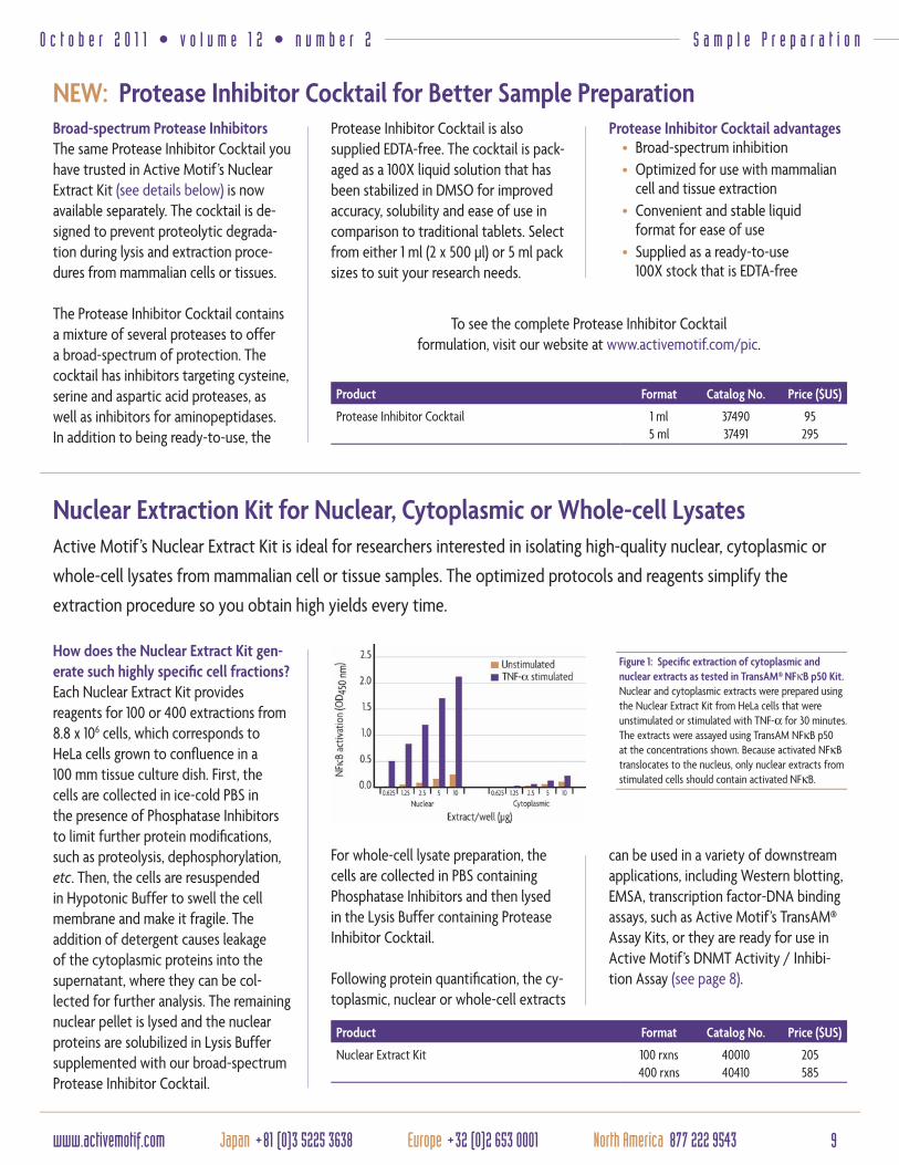
Figure 1: Specifi c extraction of cytoplasmic and nuclear extracts as tested in TransAM® NFκB p50 Kit.Nuclear and cytoplasmic extracts were prepared using the Nuclear Extract Kit from HeLa cells that were unstimulated or stimulated with TNF-α for 30 minutes. The extracts were assayed using TransAM NFκB p50 at the concentrations shown. Because activated NFκB translocates to the nucleus, only nuclear extracts from stimulated cells should contain activated NFκB.
How does the Nuclear Extract Kit gen-erate such highly specifi c cell fractions?Each Nuclear Extract Kit provides reagents for 100 or 400 extractions from 8.8 x 10⁶ cells, which corresponds to HeLa cells grown to confl uence in a 100 mm tissue culture dish. First, the cells are collected in ice-cold PBS in the presence of Phosphatase Inhibitors to limit further protein modifi cations, such as proteolysis, dephosphorylation, etc. Then, the cells are resuspended in Hypotonic Buffer to swell the cell membrane and make it fragile. The addition of detergent causes leakage of the cytoplasmic proteins into the supernatant, where they can be col-lected for further analysis. The remaining nuclear pellet is lysed and the nuclear proteins are solubilized in Lysis Buffer supplemented with our broad-spectrum Protease Inhibitor Cocktail.
Broad-spectrum Protease InhibitorsThe same Protease Inhibitor Cocktail you have trusted in Active Motif’s Nuclear Extract Kit (see details below) is now available separately. The cocktail is de-signed to prevent proteolytic degrada-tion during lysis and extraction proce-dures from mammalian cells or tissues.
The Protease Inhibitor Cocktail contains a mixture of several proteases to offer a broad-spectrum of protection. The cocktail has inhibitors targeting cysteine, serine and aspartic acid proteases, as well as inhibitors for aminopeptidases. In addition to being ready-to-use, the
Protease Inhibitor Cocktail is also supplied EDTA-free. The cocktail is pack-aged as a 100X liquid solution that has been stabilized in DMSO for improved accuracy, solubility and ease of use in comparison to traditional tablets. Select from either 1 ml (2 x 500 µl) or 5 ml pack sizes to suit your research needs.
Protease Inhibitor Cocktail advantages• Broad-spectrum inhibition• Optimized for use with mammalian
cell and tissue extraction• Convenient and stable liquid
format for ease of use• Supplied as a ready-to-use
100X stock that is EDTA-free
Product Format Catalog No. Price ($US)
Protease Inhibitor Cocktail 1 ml5 ml
3749037491
95295
To see the complete Protease Inhibitor Cocktail formulation, visit our website at www.activemotif.com/pic.
Active Motif’s Nuclear Extract Kit is ideal for researchers interested in isolating high-quality nuclear, cytoplasmic or
whole-cell lysates from mammalian cell or tissue samples. The optimized protocols and reagents simplify the
extraction procedure so you obtain high yields every time.
For whole-cell lysate preparation, the cells are collected in PBS containing Phosphatase Inhibitors and then lysed in the Lysis Buffer containing Protease Inhibitor Cocktail.
Following protein quantifi cation, the cy-toplasmic, nuclear or whole-cell extracts
can be used in a variety of downstream applications, including Western blotting, EMSA, transcription factor-DNA binding assays, such as Active Motif’s TransAM® Assay Kits, or they are ready for use in Active Motif’s DNMT Activity / Inhibi-tion Assay (see page 8).

10 North America 877 222 9543 Europe +32 (0)2 653 0001 Japan +81 (0)3 5225 3638 www.activemotif.com
S t e m C e l l & N u c l e a r R e c e p t o r A n t i b o d i e s
The biology of stem cells involves a wide variety of proteins found in
the nucleus that are required to maintain cell pluripotency (the ability
to differentiate into any cell type) and self-renewal. Many of these
proteins are associated with chromatin and serve to keep large regions
of the genome in an open, accessible confi guration. In fact, chromatin
biology and epigenetics are integral components of stem cell biology.
To assist you in your study of stem cells and the role of chromatin
regulation, Active Motif offers a wide range of antibodies to
important stem cell proteins, including:
• Transcription factors
• Chromatin modifying proteins
• Histones and histone modifi cations
• Polycomb proteins
For more information on our antibodies to regulators of stem cell function, visit www.activemotif.com/stemcellabs.
Antibodies to Stem Cell Regulators to Further your Stem Cell Research
Highly Validated Antibodies and ELISAs for the Study of Nuclear Receptors
Figure 1: Immunofl uorescence staining of Progesterone receptor.Mouse hypothalamic section stained with Progesterone receptor antibody (Catalog No. 61023).
Nuclear receptors are important regula-tors of endocrine activity, mediating responses to changing hormone levels. Most nuclear receptors bind steroid hormone ligands. But, there are some (orphan receptors) that do not, or for which the ligand has not yet been identi-fi ed. Because nuclear receptors can bind DNA directly and regulate gene expres-sion, they are also transcription factors, many of which recruit co-activators and co-repressors that possess histone-modifying activity. Active Motif offers a number of highly validated antibodies for the study of nuclear receptors and steroid hormone activity.
For more information on Active Motif’s antibodies to nuclear receptors, visit www.activemotif.com/nrabs.
Figure 1: Ring1B staining in U2OS cells.Human U2OS cells were stained with Ring1B monoclonal antibody (Catalog No. 39663) and visualized by indirect immunofluorescence.
Active Motif also offers sandwich ELISAs that enable you to quantitatively mea-sure the levels of the Androgen, Estrogen
and Progesterone nuclear receptors. For more complete information, please visit www.activemotif.com/nrelisa.
Figure 2: Androgen receptor analyzed by chromatin IP.ChIP was performed using 30 µg of VCAP60 cell chro-matin and 10 µl of Androgen receptor antibody (Catalog No. 39781). ChIP DNA was used in qPCR with the Human Negative Control Primer Set 1 (Catalog No. 71001) and primer pairs specific for the KLK3 and FKBP5 genes. Data are presented as Binding Events Detected per 1000 Cells.
11www.activemotif.com Japan +81 (0)3 5225 3638 Europe +32 (0)2 653 0001 North America 877 222 9543
O c t o b e r 2 0 1 1 • v o l u m e 1 2 • n u m b e r 2 H i g h R e s o l u t i o n M i c r o s c o p y
One of these novel techniques is STED (STimulated Emission Depletion), which can achieve up to 12-fold higher resolu-tion than classical confocal microscopy. Active Motif is proud to help scientists take advantage of this powerful technol-ogy by providing IF-tested primary anti-bodies, and high-quality conjugated sec-ondaries & sample preparation reagents that are certifi ed by Leica Microsystems for use with its STED microscopes. For more complete information on STED, please visit www.activemotif.com/sted.
Optimized sample preparation for STED and classical confocal microscopyProper sample preparation is among the most signifi cant factors for obtaining high-quality images in both STED and classical confocal microscopy. To help ensure that you consistently achieve the best results possible, Active Motif’s scientists developed the Chromeo™ STED
Immunofl uorescence System and the MAX Stain™ Immunofl uorescence Tools. These sample preparation reagents were designed to increase the intensity and specifi city of your fl uorescent labeling.
Customized conjugation protocols ensure the highest quality secondariesThe quality of any fl uorescent second-ary antibody depends on the spectral properties of the dye, the quality of the secondary antibody, and the dye-to-protein ratio & purity of the conjugate. To control all variables possible, Active Motif produces all its conjugates using protocols that have been optimized for each dye/antibody combination. This ensures the quality of the conjugates, and makes the fl uorescent secondaries brighter while lowering the fl uorescent background. For more information, visit www.activemotif.com/secondary.
The resolution attainable in fl uorescent microscopy is limited by the diffraction of light, which was shown by Ernst
Abbe in 1873. The Abbe Limit restricts the ability of the observer to visually resolve objects that are separated by
less than ~200 nm. Recently, several super-resolution techniques have been developed to overcome this limitation,
providing the ability to image structures as small as 20 nm. These improvements in microscopy enable scientists to
decipher the nanostructure of the cell in details that could not be visualized before.
Antibodies and Reagents for High Resolution Confocal STED Microscopy
Product Format Catalog No. Price ($US)
Chromeo™ 488 Goat anti-Mouse IgG 1 mg 15031 135
Chromeo™ 488 Goat anti-Rabbit IgG 1 mg 15041 135
Chromeo™ 505 Goat anti-Mouse IgG 1 mg 15030 135
Chromeo™ 505 Goat anti-Rabbit IgG 1 mg 15040 135
Chromeo™ 494 Goat anti-Mouse IgG 1 mg 15032 135
Chromeo™ 494 Goat anti-Rabbit IgG 1 mg 15042 135
ATTO 647N (STED) Goat anti-Mouse IgG 250 µl 15038 260
ATTO 647N (STED) Goat anti-Rabbit IgG 250 µl 15048 260
ATTO 655 (STED) Goat anti-Mouse IgG 250 µl 15039 260
ATTO 655 (STED) Goat anti-Rabbit IgG 250 µl 15049 260
Chromeo™ STED Immunofl uorescence System 1 kit 15260 230
MAXpack™ Immunostaining Media Kit 1 kit 15251 295
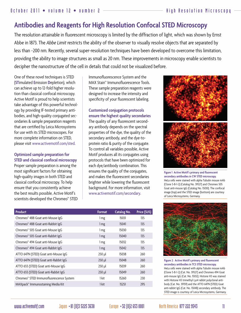
Figure 2: Active Motif’s primary and fl uorescent secondary antibodies in TCS STED microscopy.HeLa cells were stained with alpha Tubulin mouse mAb (Clone 5-B-1-2) (Cat. No. 39527) and Chromeo 494 Goat anti-mouse IgG (Cat. No. 15032). Histone H3 was stained with Histone H3 trimethyl Lys4 rabbit polyclonal anti-body (Cat. No. 39159) and the ATTO 647N (STED) Goat anti-rabbit IgG (Cat. No. 15048) secondary antibody. The STED image is courtesy of Leica Microsystems, Germany.
Figure 1 Active Motif’s primary and fl uorescent secondary antibodies in CW STED microscopy.HeLa cells were stained with alpha Tubulin mouse mAb (Clone 5-B-1-2) (Catalog No. 39527) and Chromeo 505 Goat anti-mouse IgG (Catalog No. 15030). The confocal image (top) and the STED image (bottom) are courtesy of Leica Microsystems, Germany.
Enabling Epigenetics Research
Superior Antibodies & Kits for EPIGENETICS RESEARCH
Visit us at www.activemotif.com
Active Motif develops validated antibodies and assays that enable the discovery and characterization of key epigenetic processes.
Our products are developed in-house and supported by scientists with expertise in chromatin biology. For a complete product listing please visit www.activemotif.com.
CHROMATIN ANALYSISChIP kits and ChIP-validated antibodies
HISTONE MODIFICATIONAntibodies & ELISAs, Arrays, HAT/HDAC assays, Histone purification and Recombinant Histones
DNA METHYLATION Methylated DNA enrichment, bisulfite conversion, DNMT assays, whole genome amplification, hMeDIP, MeDIP, 5-hmC & 5-mC antibodies