Embed Size (px)
Citation preview

DNA & Genetics
DNA Replication Errors and Mutations
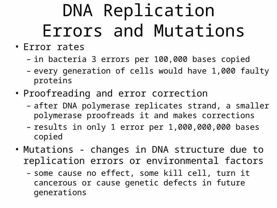
• Error rates– in bacteria 3 errors per 100,000 bases copied
– every generation of cells would have 1,000 faulty proteins
• Proofreading and error correction– after DNA polymerase replicates strand, a smaller polymerase
proofreads it and makes corrections
– results in only 1 error per 1,000,000,000 bases copied
• Mutations - changes in DNA structure due to replication errors or environmental factors– some cause no effect, some kill cell, turn it cancerous or cause
genetic defects in future generations

DNA Function
• Serves as code for protein synthesis, cell replication and reproduction
• Gene - sequence of DNA nucleotides that codes for one polypeptide
• Genome - all the genes of one person

Translation of mRNA Flow Chart

DNA & Peptide Formation
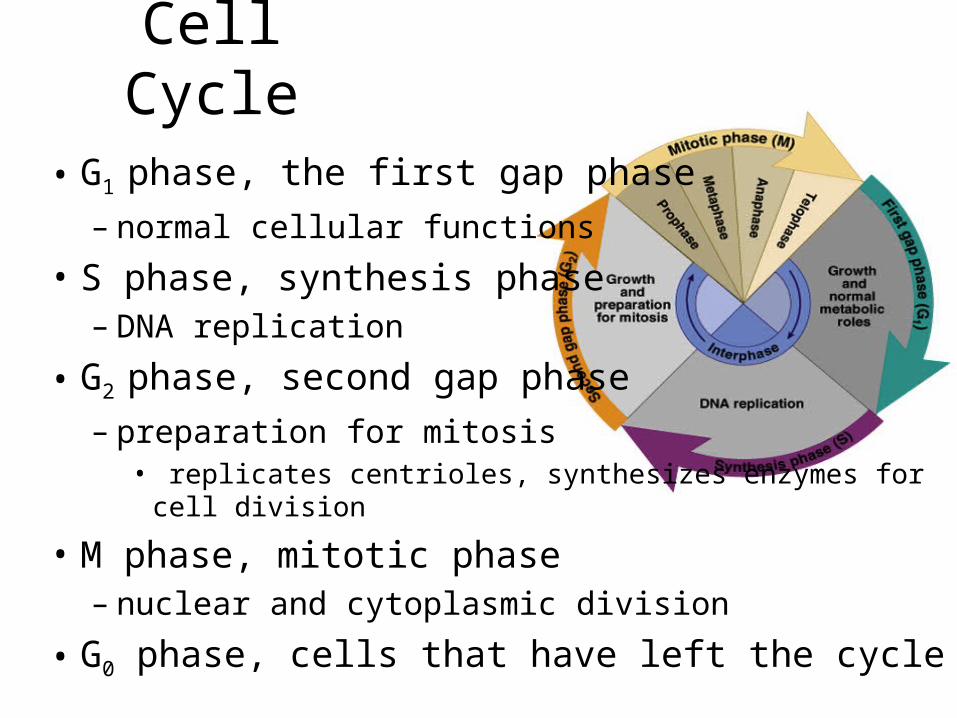
Cell Cycle
• G1 phase, the first gap phase
– normal cellular functions
• S phase, synthesis phase– DNA replication
• G2 phase, second gap phase
– preparation for mitosis• replicates centrioles, synthesizes enzymes for cell division
• M phase, mitotic phase– nuclear and cytoplasmic division
• G0 phase, cells that have left the cycle

Functions of Mitosis
• Embryonic development • Tissue growth• Replacement of old and dead cells• Repair of injured tissues
Mitosis: Prophase
• Chromatin supercoils into chromosomes
• Nuclear envelope disintegrates
• Centrioles sprout microtubules, mitotic spindle
• Centrioles move to poles
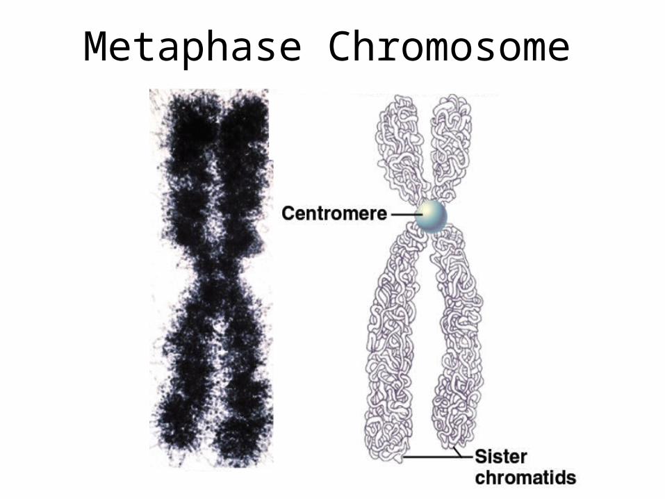
Metaphase Chromosome
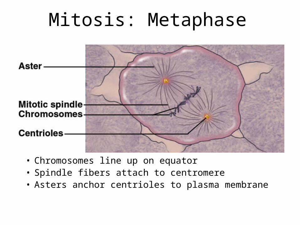
Mitosis: Metaphase
• Chromosomes line up on equator• Spindle fibers attach to centromere• Asters anchor centrioles to plasma membrane
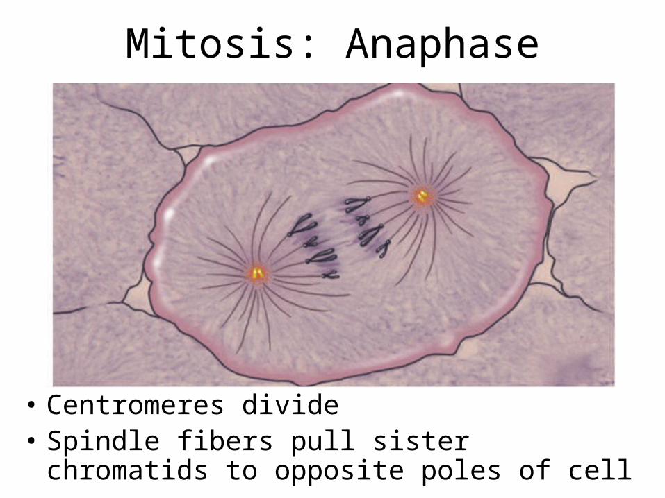
Mitosis: Anaphase
• Centromeres divide• Spindle fibers pull sister chromatids to opposite
poles of cell

Mitosis: Telophase
• Chromatin uncoils
• Nuclear envelopes form
• Mitotic spindle breaks down

Cytokinesis
• Division of cytoplasm
• Myosin pulls on actin in the membrane skeleton
• Causes crease around cell equator called cleavage furrow
• Cell pinches in two

Timing of Cell DivisionCells divide when:
• Cells large enough
• DNA replicated
• Adequate supply of nutrients
• Growth factor stimulation
• Open space in tissue
Cells stop dividing when:
• Loss of growth factors or nutrients
• Contact inhibition

Cancer
• Tumours– abnormal growth, when cells multiply faster than they
die– oncology is the study of tumors
• Benign– connective tissue capsule, grow slowly, stays local – potentially lethal by compression of vital tissues
• Malignant– unencapsulated, fast growing, metastatic (causes 90%
of cancer deaths)

Causes of Cancer
• Carcinogens - estimates of 60 - 70% of cancers from environmental agents – chemical
• cigarettes, food preservatives
– radiation• UV radiation, particles, rays, particles
– viruses• type 2 herpes simplex - uterus, hepatitis B - liver

Mutagens• Trigger gene mutations
– cell may die, be destroyed by immune system or produce a tumor
Defenses against mutagens• Scavenger cells
– remove them
• Peroxisomes – neutralize nitrites, free radicals and oxidizing agents
• Nuclear enzymes– repair DNA
• Tumor necrosis factor (TNF) destroys tumors

Cancer Genes• Oncogenes
– mutated form of proto-oncogenes – sis oncogene causes excessive production of growth
factors• stimulate neovascularization of tumor
– ras oncogene codes for abnormal growth factor receptors
• sends constant divide signal to cell
• Tumor suppressor genes– inhibit development of cancer– damage to one or both removes control of cell division

Effects of Malignancies
• Displaces normal tissue, function deteriorates– rapid cell growth of immature nonfunctional cells
– metastatic cells different tissue origin
• Block vital passageways– respiratory or vascular
• Diverts nutrients from other tissues– tumors have high metabolic rates
– causes weakness, fatigue, emaciation, infection
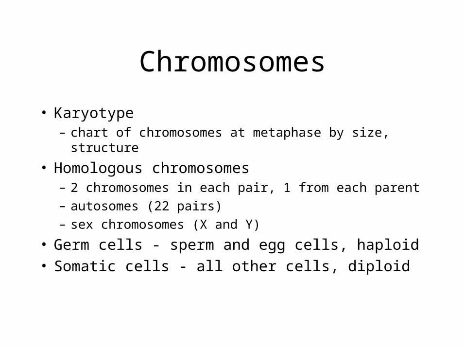
Chromosomes
• Karyotype– chart of chromosomes at metaphase by size, structure
• Homologous chromosomes– 2 chromosomes in each pair, 1 from each parent
– autosomes (22 pairs)
– sex chromosomes (X and Y)
• Germ cells - sperm and egg cells, haploid• Somatic cells - all other cells, diploid

Genes and Alleles• Gene loci
– location of gene on chromosome
• Alleles– two homologous chromosomes have same gene at
same locus, may be different forms of gene
• Dominant allele– produces normal, functional protein
• Recessive allele– when both alleles are recessive produces abnormal
protein or no protein

Genetics of Earlobes• Genotype
– alleles for a trait (DD)
• Phenotype– trait that results
• Dominant allele (D)– expressed with DD or Dd– Dd parent ‘carrier’ of
recessive gene
• Recessive allele (d)– expressed with dd only
Punnett square
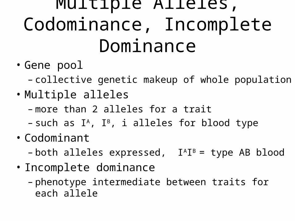
Multiple Alleles, Codominance, Incomplete Dominance
• Gene pool– collective genetic makeup of whole population
• Multiple alleles– more than 2 alleles for a trait– such as IA, IB, i alleles for blood type
• Codominant– both alleles expressed, IAIB = type AB blood
• Incomplete dominance– phenotype intermediate between traits for each allele
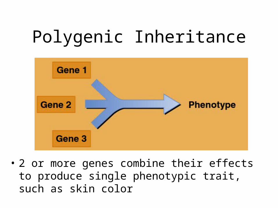
Polygenic Inheritance
• 2 or more genes combine their effects to produce single phenotypic trait, such as skin color
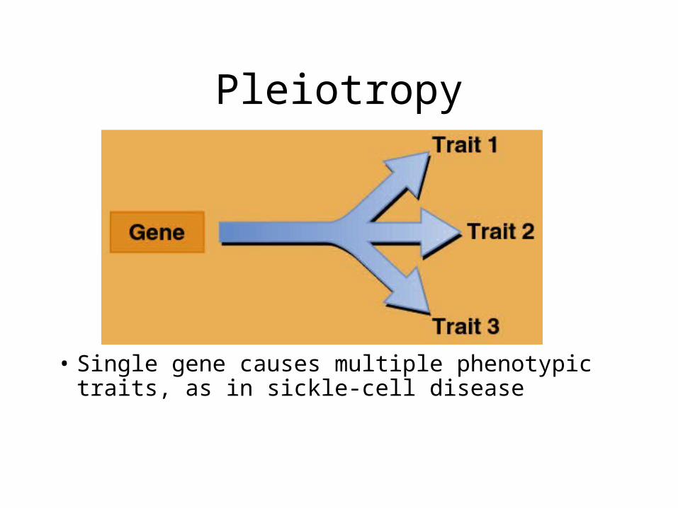
Pleiotropy
• Single gene causes multiple phenotypic traits, as in sickle-cell disease
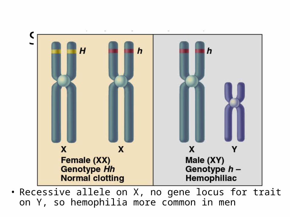
Sex-Linked Inheritance
• Recessive allele on X, no gene locus for trait on Y, so hemophilia more common in men

Penetrance and Environmental Effects
• Penetrance– % of
population to express predicted phenotype
• Role of environment– brown eye
colour requires phenylalanine from diet to produce melanin, the eye pigment

Alleles at the Population Level
• Dominance and recessiveness of allele do not determine frequency in a population
• Some recessive alleles, blood type O, are the most common
• Some dominant alleles, polydactyly, are rare
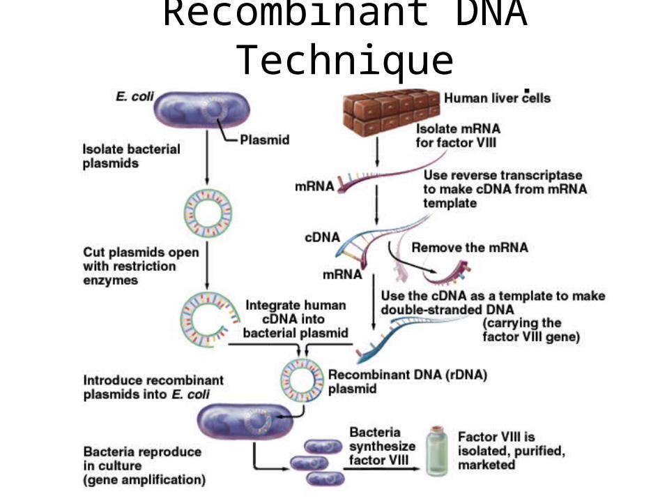
Recombinant DNA Technique


![[PPT]DNA Replication - Rothology - homereplication.pptx · Web viewDNA Replication copyright cmassengale Replication Facts DNA has to be copied before a cell divides DNA is copied](https://img.dokumen.tips/doc/110x75/5aa6232f7f8b9a7c1a8e5563/pptdna-replication-rothology-home-replicationpptxweb-viewdna-replication.jpg)