Embed Size (px)
Citation preview

Accepted Manuscript
Title: DNA and bone structure preservation in medievalhuman skeletons
Author: Yvette M. Coulson-Thomas Andrew L. NortonVivien J. Coulson-Thomas Rinaldo Florencio-Silva Nadir AliSamir Elmrghni Cristiane D. Gil Gisela R.S. Sasso Ronald A.Dixon Helena B. Nader
PII: S0379-0738(15)00151-6DOI: http://dx.doi.org/doi:10.1016/j.forsciint.2015.04.005Reference: FSI 7961
To appear in: FSI
Received date: 13-9-2014Revised date: 29-1-2015Accepted date: 3-4-2015
Please cite this article as: Y.M. Coulson-Thomas, A.L. Norton, V.J. Coulson-Thomas,R. Florencio-Silva, N. Ali, S. Elmrghni, C.D. Gil, G.R.S. Sasso, R.A. Dixon, H.B.Nader, DNA and bone structure preservation in medieval human skeletons, ForensicScience International (2015), http://dx.doi.org/10.1016/j.forsciint.2015.04.005
This is a PDF file of an unedited manuscript that has been accepted for publication.As a service to our customers we are providing this early version of the manuscript.The manuscript will undergo copyediting, typesetting, and review of the resulting proofbefore it is published in its final form. Please note that during the production processerrors may be discovered which could affect the content, and all legal disclaimers thatapply to the journal pertain.

Page 1 of 41
Accep
ted
Man
uscr
ipt
DNA and bone structure preservation in medieval human skeletons
Yvette M. Coulson-Thomas*a,b, Andrew L. Nortonc, Vivien J. Coulson-Thomasa,d,
Rinaldo Florencio-Silvae, Nadir Alib, Samir Elmrghnib, Cristiane D. Gile, Gisela R. S.
Sassoe, Ronald A. Dixonb and Helena B. Nadera
aDepartment of Biochemistry, Universidade Federal de São Paulo, Rua Três de Maio 100,
São Paulo, 04044-020, Brazil; bSchool of Life Sciences, University of Lincoln, Brayford
Pool, Lincoln, LN6 7TS, UK; cDurham University, Woodland Road, DH7 9RH, Durham,
UK; d Current address: John van Geest Centre for Brain Repair, University of Cambridge,
Forvie Site, Robinson Way, Cambridge CB2 0PY, UK; eDepartment of Morphology and
Genetics, Universidade Federal de São Paulo, Rua Botucatu 740, São Paulo, 04023-900,
Brazil
*Corresponding author: Y M Coulson-Thomas; email: [email protected]; tel.:
+55 11 5576 4438 or +44 (0)7753 224005; fax: +55 11 55736407; address: Departamento
de Bioquímica, Disciplina de Biologia Molecular, Universidade Federal de São Paulo,
INFAR, Rua Três de Maio 100, São Paulo, 04044-020, Brazil

Page 2 of 41
Accep
ted
Man
uscr
ipt
Abstract
Morphological and ultrastructural data from archaeological human bones are scarce,
particularly data that have been correlated with information on the preservation of
molecules such as DNA. Here we examine the bone structure of macroscopically well-
preserved medieval human skeletons by transmission electron microscopy and
immunohistochemistry, and the quantity and quality of DNA extracted from these
skeletons. DNA technology has been increasingly used for analyzing physical evidence in
archaeological forensics; however, the isolation of ancient DNA is difficult since it is
highly degraded, extraction yields are low and the co-extraction of PCR inhibitors is a
problem. We adapted and optimized a method that is frequently used for isolating DNA
from modern samples, Chelex® 100 (Bio-Rad) extraction, for isolating DNA from
archaeological human bones and teeth. The isolated DNA was analysed by real-time PCR
using primers targeting the sex determining region on the Y chromosome (SRY) and STR
typing using the AmpFlSTR® Identifiler PCR Amplification kit. Our results clearly show
the preservation of bone matrix in medieval bones and the presence of intact osteocytes
with well preserved encapsulated nuclei. In addition, we show how effective Chelex®
100 is for isolating ancient DNA from archaeological bones and teeth. This optimized
method is suitable for STR typing using kits aimed specifically at degraded and difficult
DNA templates since amplicons of up to 250 bp were successfully amplified.
Keywords: ancient DNA; archaeological bone; archaeological teeth; Chelex; DNA
typing; electron microscopy

Page 3 of 41
Accep
ted
Man
uscr
ipt
1 Introduction
Literature on archaeological human bone morphology and ultrastructure is scarce,
particularly data correlating bone structure with DNA preservation. The survival of
ancient DNA is of particular interest since DNA technology has been increasingly used in
archaeological forensics, for example, for the identification of diseases [1, 2],
identification of migration patterns [3, 4] and patrilineage determination [5].
The isolation of amplifiable DNA does not always reflect bone taphonomy and,
occasionally, DNA can be isolated from bones that appear to be morphologically
degraded [6]. However, analysis at a microscopic level can provide an indication as to the
preservation of amplifiable DNA; the isolation of amplifiable DNA has been correlated
with integrity of the microscopic structure, particularly lamellae [7]. In addition, more
compact appearance of bone in scanning electron micrographs is one of the factors that
indicates that amplifiable DNA can be isolated [8]. Other factors include high collagen
content, low racemization values of aspartic acid, leucine and glutamic acid, low infrared
splitting factor and small size of crystallite [8].
The long-term persistence of DNA in skeletons has been associated with the fact that it
can bind to hydroxyapatite, the main mineral component of bone tissue, which makes it
more resistant to decay and less susceptible to degradation by enzymes [9]. In addition,
DNA has been shown to bind to collagen forming a DNA-collagen complex [10], and can
be extracted from archaeological bone powder in both the hydroxyapatite and collagen
fractions [11]. The differential survival of DNA in ancient specimens is generally

Page 4 of 41
Accep
ted
Man
uscr
ipt
unpredictable but depends on both the burial environment and how well they were
retrieved and preserved.
DNA extraction from archaeological bones and teeth is a challenge and new analytical
processes, particularly DNA extraction methods, are required. Methods chosen for
extracting ancient DNA must focus on preserving the integrity of the remaining DNA
during extraction, whilst ensuring sufficient yield and that it is free of PCR inhibitors [12].
Similar methods can be used for forensic samples and archaeological samples since they
share similar characteristics [13]. In forensics, one frequently used method for isolating
DNA is extraction using Chelex® 100 (Bio-Rad). The efficiency of the Chelex® 100
extraction method has been described for DNA extraction from various modern forensic
samples, including blood, semen, buccal swabs and hair [14]. Previous research has
described a protocol using Chelex® 100 for extracting DNA from fresh bone samples
[15]. We have adapted and optimized the extraction protocol using Chelex® 100 for
isolating ancient DNA from archaeological bones and teeth with the intention of
preserving the integrity of the DNA, and ensuring the yield is sufficient and the product is
free of PCR inhibitors. Subsequent autosomal STR DNA typing using the AmpFℓSTR®
Identifiler® PCR Amplification Kit gave us an indication as to how well-preserved the
isolated DNA was. Our results demonstrate that Chelex® 100 is effective for isolating
ancient DNA from archaeological bones and teeth.
It is also important to know how well the bones have been preserved at a microscopic
level since this has been shown to reflect the integrity of the isolated DNA [7, 8]. This

Page 5 of 41
Accep
ted
Man
uscr
ipt
study therefore looked to provide information on the bone structure of medieval human
bones. The morphology and ultrastructure of the archaeological human bones was
analyzed by transmission electron microscopy and immunohistochemistry. The data were
then correlated with the preservation of amplifiable DNA. Our results clearly show the
preservation of bone matrix in medieval bones and the presence of apparently intact
osteocytes.
2 Material and methods
2.1 Samples
The medieval skeletons used in this study were curated at the University of Lincoln, UK.
Sixty-eight partial or complete skeletal remains were excavated in the last decade from a
site on the south side of Monk’s Road, Lincoln. The site was determined to be the
extramural graveyard of the defunct parish of St Peter at Welles (ad fontem). The age of
the site was estimated to between 1150 AD and 1400 AD. The appropriate Coroners
licence was obtained to excavate the skeletons. Various types of bone were chosen to be
analysed; femur (adult, adolescent and juvenile), humerus (adult) and ulna (adult and
adolescent). Two archaeological teeth were also analysed. The study of these
archaeological human skeletons was approved by the University of Lincoln ethics
committee in the UK and CEP/UNIFESP in Brazil (CAAE: 07934412.2.0000.5505).
2.2 Transmission Electron Microscopy (TEM)
Transverse bone sections (75 m thick) were obtained using a Leica SP1600 Saw
Microtome (Leica Biosystems, Nussloch, Germany). The bone sections were immersed

Page 6 of 41
Accep
ted
Man
uscr
ipt
in cacodylate-buffered 1% osmium tetroxide at pH 7.2 for 2 hours. Subsequently, the
samples were washed in distilled water and immersed in 2% aqueous uranyl acetate for 2
hours. After washing, the bone slices were dehydrated in graded concentrations of
ethanol, treated with propylene oxide and then embedded in Araldite. Semi-thin sections
(600-800 nm) were obtained using an ultramicrotome (Leica UCT) in order to ascertain
the presence of material, and then ultra-thin sections (70-85 nm) were collected onto
grids and stained in alcoholic 2% uranyl acetate and in lead citrate solution, and
examined using a ZEISS EM900 electron microscope (Department of Morphology and
Genetics, UNIFESP).
2.3 Imunohistochemistry
Transverse bone sections (60 m thick) were obtained using a Leica SP1600 Saw
Microtome (Leica Biosystems, Nussloch, Germany), which can slice hard materials such
as bone without any previous treatment that could lead to changes in bone composition
and structure. The bone slices were hydrated in PBS buffer for 1 hour at 4°C and then
fixed in 2% buffered paraformaldehyde for 30 min followed by washing in PBS and
antigen recovery (10 min incubation in 10 mM sodium citrate pH 6 at 100°C). The bone
slices were then washed, incubated in 10% hydrogen peroxide for 30 min, washed, and
unspecific protein binding sites were blocked with 5% fetal bovine serum (FBS). The
slices were then incubated with rabbit anti-osteocalcin (30044, Santa Cruz, Santa Cruz,
CA) and mouse monoclonal anti-collagen III (Millipore MAB3392) overnight at 4°C.
Bone sections were washed and then processed using the Universal Dako LSAB®+ Kit,
Peroxidase (LSAB+ Kit, HRP) and Dako Liquid DAB+ Substrate Chromogen System
Page 7 of 41
Accep
ted
Man
uscr
ipt
(Dako, Glostrup, Denmark). Finally, bone sections were incubated sequentially in 30%
ethanol, 50% ethanol, 70% ethanol, 90% ethanol, 100% ethanol, ethanol:xylol (1:1),
xylol, and then mounted on glass slides in Permount (Thermo Fisher Scientific Inc.,
Waltham, MA) and sealed with nail polish. Negative control immunostainings were
performed with omission of the primary antibodies, in the presence of FBS, overnight at
4°C.
2.4 DNA extraction
2.4.1 Optimized Chelex® 100 DNA extraction method
The bones and teeth were initially cleaned in water and allowed to dry. In addition, bone
samples were mechanically cleaned with sandpaper. Subsequently, the bone and tooth
samples were exposed to sodium hypochlorite for 15 minutes at room temperature [16].
Bone powder and tooth powder were collected using a drill (Draper) with drill bits that
had been previously exposed to sodium hypochlorite and autoclaved. The bone powder
and tooth powder were stored at -20ºC until they were processed.
Bone or tooth powder (100 mg) was suspended in 1 ml 10% Chelex® 100 (Bio-Rad), and
the protocol described by Willard et al. [15] was adapted and optimised for the
archaeological samples in order to improve DNA yield and integrity. Essential
modifications to the method were that the initial incubation period was increased to 3
hours at 56ºC and the boiling period to 20 minutes. The resulting solution (containing
DNA) was subjected to phenol/chloroform purification (to remove PCR inhibitors) using
conventional methods (adding equal volume of phenol:chloroform:isoamyl alcohol

Page 8 of 41
Accep
ted
Man
uscr
ipt
(25:24:1), centrifuging at 15,000 g for 5 minutes and collecting the aqueous layer)
followed by isopropanol precipitation [17]. Sodium acetate (500 mM final concentration)
was added to the samples prior to the isopropanol. The recovered DNA was stored at -
20ºC. A Chelex® 100 extraction reagent blank control was processed at the same time as
the samples. Positive modern DNA controls were processed in a different location and at
a different time to the ancient DNA samples, as discussed below.
2.4.2 Digestion buffer DNA extraction method
Bone powder was obtained as described above and DNA was isolated using the digestion
buffer DNA extraction method as described by Foran et al.[18], which is ideal for
recovering residual DNA. Briefly, 100 mg bone powder was suspended in 1 ml digestion
buffer (20 mM Tris, 50 mM EDTA, 0.1% SDS, pH 7.5) and 15 l of proteinase K (20
mg/ml) was added. The samples were incubated overnight at room temperature and then
at 56°C for 3 hours. This initial incubation step was slightly modified since it was
observed that this resulted in better DNA yield and integrity. The supernatant (containing
DNA) was subjected to phenol:chloroform:isoamyl alcohol purification and
isopropanol/sodium acetate precipitation, as described above. The isolated DNA was
stored at -20ºC. No more than two DNA extractions were carried out at the same time,
and a digestion buffer extraction reagent blank control was processed at the same time as
the samples.
2.4.3 DNA extraction from control samples

Page 9 of 41
Accep
ted
Man
uscr
ipt
DNA was isolated from cheek cells from a volunteer (male) and the investigator carrying
out the STR (Short Tandem Repeat) typing assays (female) using a standard teaching
protocol [19], and stored at -20ºC. The female DNA was used in the STR-typing assays
as a quality control and the male DNA in the real-time PCR amplification assays as a
positive control. DNA extraction from these samples was carried out in a different
location on a different day to the extraction of DNA from the archaeological samples.
2.5 DNA quantification
Isolated DNA was quantified using a NanoDrop ND-1000 spectrophotometer
(Nanodrop Technologies, Wilmington, DE, USA).
2.6 Autosomal STR DNA typing
The quality of the ancient DNA recovered was evaluated using the AmpFℓSTR®
Identifiler® PCR Amplification Kit (Applied Biosystems, Warrington, UK) according to
the manufacturer’s protocol. This kit includes the following markers: D8S1179, D3S1358,
TH01, D19S433, vWA, TPOX, D5S818, D21S11, D7S820, CSF1PO, D13S317,
D16S539, D2S1338, D18S51, FGA and the gender identification locus Amelogenin. A
control DNA sample, extracted from cheek cells from the investigator carrying out the
STR typing, was also typed for quality control. The PCR products were analysed using an
Applied Biosystems 310 Genetic Analyser (Applied Biosystems) and the profiles
determined using the GeneMapper® ID V3.2 Software (Applied Biosystems). Alleles
were assigned according to the International Society of Forensic Genetics (ISFG)
guidelines for forensic STR [20].

Page 10 of 41
Accep
ted
Man
uscr
ipt
2.7 Real-time PCR
The sex of archaeological human bone and teeth samples (adult, adolescent and juvenile
femur; adult humerus; adult and adolescent ulna; and adult teeth) was identified by
amplification of the sex determining region on the Y chromosome (SRY) [21, 22]. A
sequence of 93-bp was amplified using the primers described by Esteve Codina et al.
(without the initial GTTT sequence) [23]. DNA extracted from male cheek cells was
amplified as a positive control.
The DNA was amplified in a total volume of 20 l comprising DNA (40 ng), primers (3
M), Syber Green PCR Master Mix (Applied Biosystems) in MicroAmp Optical 96-
Well Reaction Plates (Applied Biosystems). Real-time PCR amplifications were carried
out in a 7500 Real-Time PCR system (Applied Biosystems) and the thermal cycling
conditions were: 95°C for 10 min, 40 cycles of 95°C for 15 sec, 58°C for 1 min and 72°C
for 30 sec. Amplified-product specificity was analysed by means of dissociation curves.
2.8 Quality Control
The laboratory has previously passed a quality assurance exercise (YHRD) test on 20
May 2010 [24].
3 Results
3.1 Electron microscopy

Page 11 of 41
Accep
ted
Man
uscr
ipt
Transmission Electron Microscopy was carried out in archaeological bone samples.
Collagen fibers were observed compounding an apparently well-organized bone matrix,
demonstrating how well preserved the bones are (arrows in Figure 1 B and C).
Interestingly, an apparently intact osteocyte was observed inside a lacuna, surrounded by
bone matrix (Figure 1 A, B and C). In figure 1 B and C, an electron opaque cytoplasmic
portion of the osteocyte can be seen, with its cytoplasmic process traversing the interior
of a canaliculus. Apparently degenerated osteocytes were also observed (Figure 1 D and
E). In Figure 1D, the osteocyte is inside a large lacuna presenting disperse and flocculent
material (asterisks). This material can be seen in more detail in figure 1F, and seems to be
composed of collagen fibers and other molecules such as proteoglycans that resulted from
some form of degradation of the peri-lacuna bone matrix material, which is shown in
figure 1D as an electron opaque region surrounding the lacuna (arrows in figure 1D).

Page 12 of 41
Accep
ted
Man
uscr
ipt
Figure 1 Electron micrographs of adolescent femur slices. A shows an apparently intact
osteocyte (Ot) inside a lacuna (La), surrounded by bone matrix (BM). B and C represent
amplified regions of A, showing an electron opaque cytoplasmic portion of the osteocyte
(Ot), with its cytoplasmic process (P) traversing the interior of a canaliculus (C), and
collagen fibers (black arrows). D and E show apparently degenerative osteocytes (Ot). In
D the osteocyte is inside a large lacuna (La) presenting disperse and flocculent material
(asterisks), which is seen in more detail in image F. This material seems to result from
degradation of the peri-lacuna bone matrix material, the electron opaque region
surrounding the lacuna (La) (arrows).
3.2 Imunohistochemistry
We analyzed the location of osteocalcin and collagen III in archaeological human bone
slices. Due to the potential fragility and already compromised structure of the
archaeological bones, care was taken to obtain bone slices with no previous
decalcification. Osteocalcin and collagen III were immunostained using rabbit anti-
osteocalcin (30044, Santa Cruz, Santa Cruz, CA) and mouse monoclonal anti-collagen III
(Millipore MAB3392), respectively. A similar type of immunostaining pattern was
observed for collagen III and the non-collagenous protein osteocalcin; evenly distributed
throughout the bone matrix (Figure 2). The osteons are well preserved and remnants of
the lacuno-canalicular network can also be observed (asterisk in figure 2).

Page 13 of 41
Accep
ted
Man
uscr
ipt
Figure 2 Immunolocalization of osteocalcin and collagen III in archaeological human
bone slices. Archaeological human femur slices (60 m thick) were labeled for
osteocalcin and collagen III, and developed using DAB (brown). Remnants of the lacuno-
canalicular network can be observed (asterisk). Scale bar: 200 μm.
3.3 Optimisation of the DNA extraction method
In the present study we optimised a method for extracting ancient DNA from
archaeological bones and teeth using Chelex® 100 (Bio-Rad). The established method
for fresh bone [15] was adapted and optimised for archaeological bones by incubating
bone powder in a 10% rather than 5% Chelex® 100 suspension, and for an extended
incubation period of three hours (under vertical rotation) rather than 30 minutes to ensure
that sufficient DNA was released from the bone powder. Quantification of DNA isolated
with incubation periods of one, three and five hours showed that the three- and five-hour
periods resulted in a similar amount of DNA but more than the one-hour period. The
subsequent step of boiling the DNA sample with Chelex® 100 was increased from the
established 10 minutes to 20 minutes. We included the steps of phenol:chloroform
purification and isopropanol precipitation following the Chelex® 100 extraction to ensure

Page 14 of 41
Accep
ted
Man
uscr
ipt
any PCR inhibitors were removed and to finally concentrate the DNA. The ancient DNA
recovered using this method was suitable for STR typing and real-time PCR
amplification as described below.
3.4 DNA quality
The quality of the ancient DNA was evaluated by STR typing using the AmpFlSTR®
Identifiler PCR Amplification kit (Applied Biosystems). Ancient DNA extracted from
archaeological human bones using Chelex® 100 followed by phenol:chloroform:isoamyl
alcohol purification and isopropanol/sodium acetate precipitation was successfully
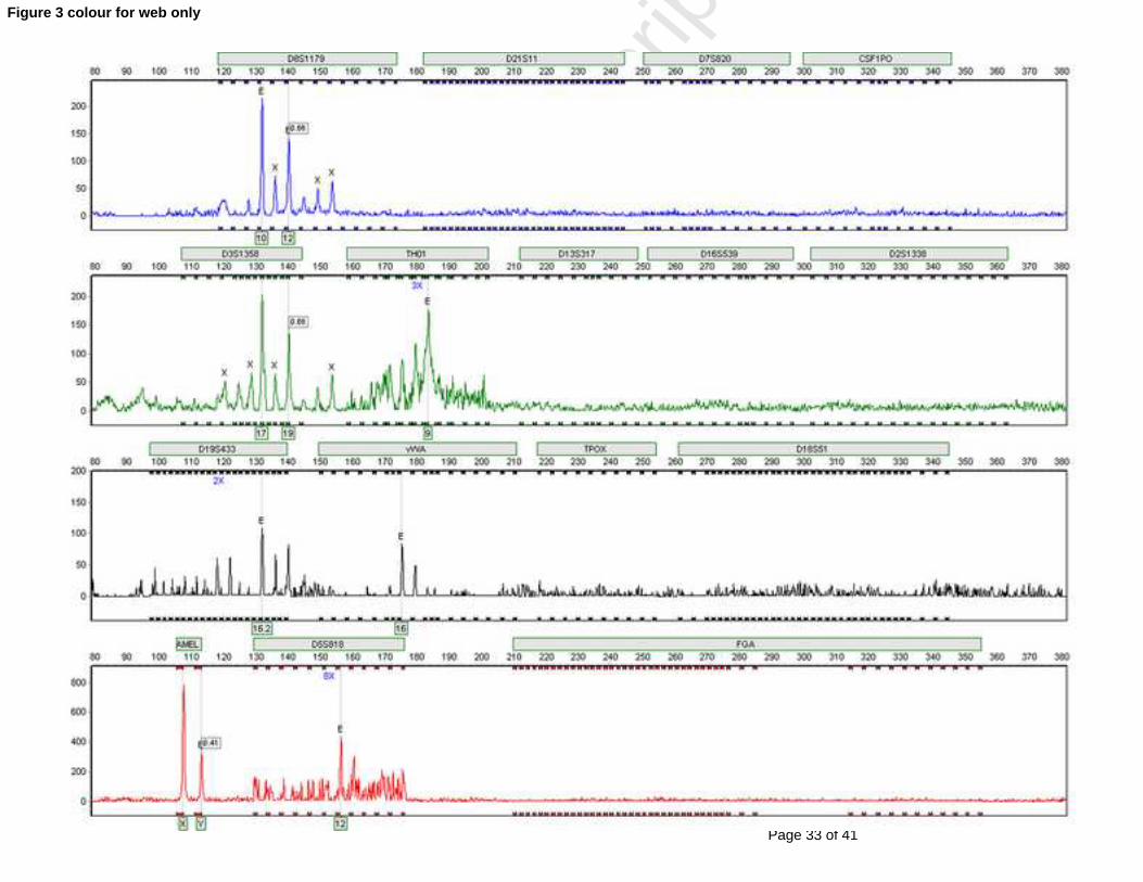
amplified for STR markers producing amplicons of up to 250 bp (Figure 3). The
electropherogram peaks were informative and a partial profile was obtained. DNA
recovered from archaeological skeletons using this optimized protocol would be suitable
for STR typing using kits aimed specifically at degraded and difficult DNA templates,
such as Applied Biosystems’ AmpFℓSTR® MiniFiler™ PCR Amplification kit.

Page 15 of 41
Accep
ted
Man
uscr
ipt
Figure 3 AmpFℓSTR® Identifiler® electropherogram for DNA extracted from
archaeological human adult femur using Chelex® 100 (Bio-Rad) followed by
phenol/chloroform purification and isopropanol precipitation.
Ancient DNA isolated from archaeological human bones using only Chelex® 100
(without phenol:chloroform:isoamyl alcohol purification and isopropanol/sodium acetate
precipitation) resulted in unsuccessful ancient DNA amplification (Results not shown).
This could be due to isolated DNA that is not concentrated enough for STR typing and/or
the presence of PCR inhibitors.

Page 16 of 41
Accep
ted
Man
uscr
ipt
DNA extracted from cheek cells from the investigator carrying out the STR typing was
also STR typed for quality control purposes and the electropherogram obtained reveals a
profile different to that obtained for the archaeological human bone samples (Figure 4).
Figure 4 mpFℓSTR® Identifiler® electropherogram for DNA extracted from modern
cheek cells.
3.5 PCR amplification for gender identification
DNA was extracted from a further eight archaeological human bone samples (adult,
adolescent and juvenile femur; adult humerus (two bones); adult radius; and adult and
Page 17 of 41
Accep
ted
Man
uscr
ipt
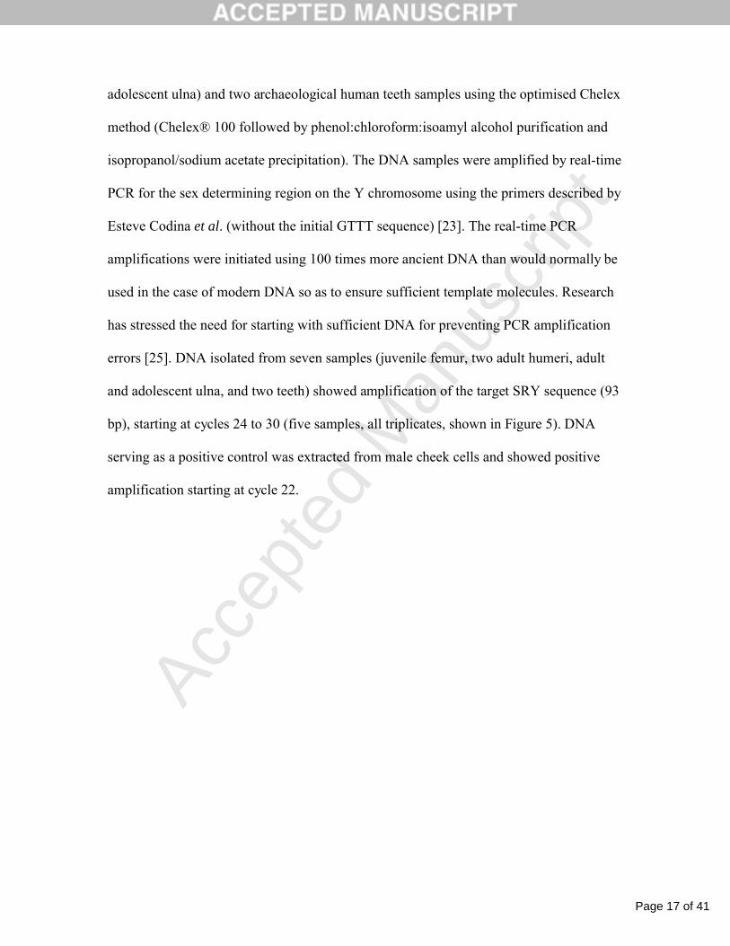
adolescent ulna) and two archaeological human teeth samples using the optimised Chelex
method (Chelex® 100 followed by phenol:chloroform:isoamyl alcohol purification and
isopropanol/sodium acetate precipitation). The DNA samples were amplified by real-time
PCR for the sex determining region on the Y chromosome using the primers described by
Esteve Codina et al. (without the initial GTTT sequence) [23]. The real-time PCR
amplifications were initiated using 100 times more ancient DNA than would normally be
used in the case of modern DNA so as to ensure sufficient template molecules. Research
has stressed the need for starting with sufficient DNA for preventing PCR amplification
errors [25]. DNA isolated from seven samples (juvenile femur, two adult humeri, adult
and adolescent ulna, and two teeth) showed amplification of the target SRY sequence (93
bp), starting at cycles 24 to 30 (five samples, all triplicates, shown in Figure 5). DNA
serving as a positive control was extracted from male cheek cells and showed positive
amplification starting at cycle 22.
Page 18 of 41
Accep
ted
Man
uscr
ipt
Figure 5 Real-time PCR amplification of the SRY region of DNA extracted from
archaeological human bones (A) and teeth (B) using the optimised Chelex 100 method.
DNA extracted from male cheek cells was used as the positive control. C: control; JF:
juvenile femur; AdU: adolescent ulna; AU: adult ulna; T1: tooth one; T2: tooth two.
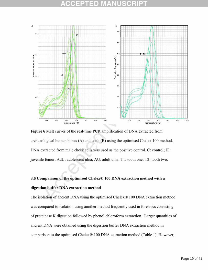
Amplified-product specificity was analysed by means of dissociation curves and only one
peak was observed per sample indicating that only one product had been amplified
(Figure 6).
Page 19 of 41
Accep
ted
Man
uscr
ipt
Figure 6 Melt curves of the real-time PCR amplification of DNA extracted from
archaeological human bones (A) and teeth (B) using the optimised Chelex 100 method.
DNA extracted from male cheek cells was used as the positive control. C: control; JF:
juvenile femur; AdU: adolescent ulna; AU: adult ulna; T1: tooth one; T2: tooth two.
3.6 Comparison of the optimised Chelex® 100 DNA extraction method with a
digestion buffer DNA extraction method
The isolation of ancient DNA using the optimised Chelex® 100 DNA extraction method
was compared to isolation using another method frequently used in forensics consisting
of proteinase K digestion followed by phenol:chloroform extraction. Larger quantities of
ancient DNA were obtained using the digestion buffer DNA extraction method in
comparison to the optimised Chelex® 100 DNA extraction method (Table 1). However,

Page 20 of 41
Accep
ted
Man
uscr
ipt
on the basis of the quality of the isolated ancient DNA, the optimised Chelex® 100 DNA
extraction method proved to be a more efficient method since longer amplicons were
obtained when STR typing using the AmpFlSTR® Identifiler PCR Amplification kit.
Table 1 Ancient DNA isolated from archaeological human bone powder
Sample DNA extraction methodTotal DNA
(ng)
Chelex® 100 1204
Chelex® 100 + phenol:chloroform:isoamyl alcohol 1577Archaeological
human adult femurDigestion buffer + phenol:chloroform:isoamyl alcohol 6132
Chelex® 100 1417
Chelex® 100 + phenol:chloroform:isoamyl alcohol 2721
Archaeological
human adult
humerus Digestion buffer + phenol:chloroform:isoamyl alcohol 3293
Modern cheek cells Chelex® 100 2971
Chelex® 100
extraction reagent
blank control
Chelex® 100 + phenol:chloroform:isoamyl alcohol 0
Digestion buffer
extraction reagent
blank control
Digestion buffer + phenol:chloroform:isoamyl alcohol 0
As mentioned above, ancient DNA extracted from archaeological human bones using the
optimised Chelex® 100 method produced amplicons of up to 250 bp (Figure 3), whereas
ancient DNA isolated using the digestion buffer method provided electropherograms
without informative peaks, due to artefacts such as additional peaks or elevated stutters,
or due to the absence of peaks (Figure 7).

Page 21 of 41
Accep
ted
Man
uscr
ipt
Figure 7 AmpFℓSTR® Identifiler® electropherogram for DNA extracted from
archaeological human adult femur using digestion buffer followed by phenol/chloroform
purification and isopropanol precipitation.
4 Discussion
DNA recovered from ancient materials does not always produce PCR amplification
products due to DNA template damage [26], and the extraction method used is of critical
importance for isolating amplifiable DNA. An effective method for isolating DNA
frequently used in forensics is extraction using Chelex® 100 (Bio-Rad), so we optimized
this method for isolating ancient DNA from archaeological bones and teeth, seeking to

Page 22 of 41
Accep
ted
Man
uscr
ipt
preserve the integrity of the DNA, and ensure sufficient yield and purity for real-time
PCR amplification and STR typing. This method proved to be a more efficient method
for isolating ancient DNA than another method frequently used in forensics consisting of
proteinase K digestion followed by phenol:chloroform extraction, which can result in
extracts that inhibit Taq polymerase [25].
Since boiling forensic DNA samples with Chelex® 100 has been shown to be critical for
protecting the DNA from degradation [14], we increased this incubation period in
Chelex® 100 for archaeological bone and tooth samples to ensure the quality of the DNA.
In order to remove PCR inhibitors from the archaeological samples, steps of
phenol:chloroform purification and isopropanol precipitation were included, which have
been shown to remove PCR inhibitors from ancient bone extracts [17]. These steps also
served to concentrate the DNA ensuring sufficient quantity for PCR amplification.
Chelex® 100 extraction is a very effective method for isolating DNA from forensic bone
samples; it has, for example, been used for successfully isolating amplifiable DNA from
bones that have been heated during a prolonged period [27]. Our results clearly show that
Chelex® 100 is also effective for isolating ancient DNA from archaeological bones and
teeth, and the isolated DNA is suitable for STR typing and real-time PCR amplification.
Other methods have been compared and optimized for extracting ancient DNA from
archaeological skeletons; however, the PCR products analyzed were in the range of 100
bp and the amplification of larger products was not evaluated [28]. We were able to
verify what amplicon size range could be obtained for ancient DNA isolated from

Page 23 of 41
Accep
ted
Man
uscr
ipt
archaeological bones and teeth using the optimized Chelex® 100 extraction method by
carrying out STR typing using the AmpFlSTR® Identifiler PCR Amplification kit.
Amplicons of up to 250 bp were obtained, providing a partial STR profile using this kit.
This size would be ideal for STR typing using kits aimed specifically at degraded and
difficult DNA templates, such as the AmpFLSTR® MiniFilerTM PCR Amplification Kit
(Applied Biosystems®).
Relatively well-preserved DNA has been identified within the protective environment of
intergrown crystal aggregates within fossil bones [29]. Furthermore, the preservation of
DNA in archaeological bones has been correlated with various factors including nearly
perfect micromorphology, with only small areas of localized demineralization [30],
lamellae integrity [7], a more compact appearance of bone in scanning electron
micrographs and high collagen content [8]. We therefore analyzed the medieval human
bone samples by transmission electron microscopy, which is a powerful tool that has
been used to study the ultrastructural characteristics of both archaeological and fossilized
bones [31, 32]. Our findings show intact osteons and an apparently well-organized bone
matrix, containing molecules such as collagen and osteocalcin, in medieval human bones,
demonstrating how well preserved the bones are.
Interestingly, in the transmission electron microscopy analysis we observed an apparently
intact osteocyte inside a lacuna, surrounded by bone matrix. Usually, only the lacuno-
canalicular network is observed in archaeological bones, such as in the
immunohistochemistry images in the present study. Mineralized osteocytes have been

Page 24 of 41
Accep
ted
Man
uscr
ipt
previously described in fossilized bone dating to the Cretaceous and Jurassic periods [32,
33] and 5 million years BP [31]. These cells have been shown to mineralize in vivo, as a
form of in vivo death, and are a potential source of preserved DNA [31].
5 Conclusions
In conclusion, this paper provides morphological and ultrastructural information on
medieval human bones, and describes an optimized method using Chelex® 100 for
isolating ancient DNA from archaeological bones and teeth. The isolated DNA can be
used for sex determination and DNA profiling, and this optimised Chelex 100 method is
efficient, simple and reproducible.
References
[1] Dixon R, Roberts CA. Modern and ancient scourges: the application of ancient DNA to the analysis of tuberculosis and leprosy from archaeologically derived human remains. Anc biomol. 2001;3:181-93.[2] Taylor GM, Young DB, Mays SA. Genotypic analysis of the earliest known prehistoric case of tuberculosis in Britain. J Clin Microbiol. 2005;43:2236-40.[3] Forster P. Ice Ages and the mitochondrial DNA chronology of human dispersals: a review. Philos Trans R Soc Lond B Biol Sci. 2004;359:255-64; discussion 64.[4] Richards MB, Macaulay VA, Bandelt HJ, Sykes BC. Phylogeography of mitochondrial DNA in western Europe. Ann Hum Genet. 1998;62:241-60.[5] King TE, Bowden GR, Balaresque PL, Adams SM, Shanks ME, Jobling MA. Thomas Jefferson's Y chromosome belongs to a rare European lineage. Am J Phys Anthropol. 2007;132:584-9.[6] Dixon R. The experimental degradation of archaeological human bone by anaerobic bacteria and the implications for recovery of ancient DNA. Proceedings of the 9th International Conference on Ancient DNA and Associated Biomolecules. Pompeii2009.[7] Marinho AN, Miranda NC, Braz V, Ribeiro-Dos-Santos AK, de Souza SM. Paleogenetic and taphonomic analysis of human bones from Moa, Beirada, and Ze Espinho Sambaquis, Rio de Janeiro, Brazil. Mem Inst Oswaldo Cruz. 2006;101 Suppl 2:15-23.[8] Sosa C, Vispe E, Nunez C, Baeta M, Casalod Y, Bolea M, et al. Association between ancient bone preservation and dna yield: a multidisciplinary approach. Am J Phys Anthropol. 2013;151:102-9.

Page 25 of 41
Accep
ted
Man
uscr
ipt
[9] Brundin M, Figdor D, Sundqvist G, Sjogren U. DNA binding to hydroxyapatite: a potential mechanism for preservation of microbial DNA. J Endod. 2013;39:211-6.[10] Mrevlishvili GM, Svintradze DV. DNA as a matrix of collagen fibrils. Int J Biol Macromol. 2005;36:324-6.[11] Campos PF, Craig OE, Turner-Walker G, Peacock E, Willerslev E, Gilbert MT. DNA in ancient bone - where is it located and how should we extract it? Ann Anat. 2012;194:7-16.[12] Budowle B, Bieber FR, Eisenberg AJ. Forensic aspects of mass disasters: strategic considerations for DNA-based human identification. Leg Med (Tokyo). 2005;7:230-43.[13] Capelli C, Tschentscher F. Protocols for ancient DNA typing. Methods Mol Biol. 2005;297:265-78.[14] Walsh PS, Metzger DA, Higuchi R. Chelex 100 as a medium for simple extraction of DNA for PCR-based typing from forensic material. Biotechniques. 1991;10:506-13.[15] Willard JM, Lee DA, Holland MM. Recovery of DNA for PCR amplification from blood and forensic samples using a chelating resin. Methods Mol Biol. 1998;98:9-18.[16] Kemp BM, Smith DG. Use of bleach to eliminate contaminating DNA from the surface of bones and teeth. Forensic Sci Int. 2005;154:53-61.[17] Hanni C, Brousseau T, Laudet V, Stehelin D. Isopropanol precipitation removes PCR inhibitors from ancient bone extracts. Nucleic Acids Res. 1995;23:881-2.[18] Foran DR, Gehring ME, Stallworth SE. The recovery and analysis of mitochondrial DNA from exploded pipe bombs. J Forensic Sci. 2009;54:90-4.[19] Fankhauser DB. ISOLATION OF BUCCAL CELL DNA. Batavia: University of Cincinnati Clermont College; 2009.[20] Olaisen B, Bar W, Brinkmann B, Budowle B, Carracedo A, Gill P, et al. DNA recommendations 1997 of the International Society for Forensic Genetics. Vox Sang. 1998;74:61-3.[21] George R, Sriram G, Saraswathi T, Sivapathasundharam B. Isolation of epithelial cells from acrylic removable dentures and gender identification by amplification of SRY gene using real time PCR. J Forensic Dent Sci. 2010;2:32-6.[22] Kastelic V, Budowle B, Drobnic K. Validation of SRY marker for forensic casework analysis. J Forensic Sci. 2009;54:551-5.[23] Esteve Codina A, Niederstatter H, Parson W. "GenderPlex" a PCR multiplex for reliable gender determination of degraded human DNA samples and complex gender constellations. Int J Legal Med. 2009;123:459-64.[24] Elmrghni S, Coulson-Thomas YM, Kaddura M, Dixon RA, Williams DR. Population genetic data for 17 Y STR markers from Benghazi (East Libya). Forensic Sci Int Genet. 2012;6:224-7.[25] Handt O, Krings M, Ward RH, Paabo S. The retrieval of ancient human DNA sequences. Am J Hum Genet. 1996;59:368-76.[26] Hoss M, Jaruga P, Zastawny TH, Dizdaroglu M, Paabo S. DNA damage and DNA sequence retrieval from ancient tissues. Nucleic Acids Res. 1996;24:1304-7.[27] Sivolap Y, Krivda G, Kozhuhova N, Chebotar S, Benecke M. A homicide in the Ukraine: DNA-based identification of a boiled, skeletonized, and varnished human skull, and of bone fragments found in a fireplace. Am J Forensic Med Pathol. 2001;22:412-4.[28] Rohland N, Hofreiter M. Comparison and optimization of ancient DNA extraction. Biotechniques. 2007;42:343-52.

Page 26 of 41
Accep
ted
Man
uscr
ipt
[29] Salamon M, Tuross N, Arensburg B, Weiner S. Relatively well preserved DNA is present in the crystal aggregates of fossil bones. Proc Natl Acad Sci U S A. 2005;102:13783-8.[30] Hagelberg E, Bell LS, Allen T, Boyde A, Jones SJ, Clegg JB. Analysis of ancient bone DNA: techniques and applications. Philos Trans R Soc Lond B Biol Sci. 1991;333:399-407.[31] Bell LS, Kayser M, Jones C. The mineralized osteocyte: a living fossil. Am J Phys Anthropol. 2008;137:449-56.[32] Schweitzer MH, Zheng W, Cleland TP, Bern M. Molecular analyses of dinosaur osteocytes support the presence of endogenous molecules. Bone. 2013;52:414-23.[33] Schweitzer MH, Wittmeyer JL, Horner JR. Soft tissue and cellular preservation in vertebrate skeletal elements from the Cretaceous to the present. Proc Biol Sci. 2007;274:183-97.

Page 27 of 41
Accep
ted
Man
uscr
ipt
Table 1 Ancient DNA isolated from archaeological human bone powder
Sample DNA extraction methodTotal DNA
(ng)
Chelex® 100 1204
Chelex® 100 + phenol:chloroform:isoamyl alcohol 1577Archaeological
human adult femurDigestion buffer + phenol:chloroform:isoamyl alcohol 6132
Chelex® 100 1417
Chelex® 100 + phenol:chloroform:isoamyl alcohol 2721
Archaeological
human adult
humerus Digestion buffer + phenol:chloroform:isoamyl alcohol 3293
Modern cheek cells Chelex® 100 2971
Chelex® 100
extraction reagent
blank control
Chelex® 100 + phenol:chloroform:isoamyl alcohol 0
Digestion buffer
extraction reagent
blank control
Digestion buffer + phenol:chloroform:isoamyl alcohol 0

Page 28 of 41
Accep
ted
Man
uscr
ipt
Electron microscopy reveals the extracellular matrix is preserved in medieval bones
Medieval bone extracellular matrix proteins may be analyzed by immunohistochemistry
An efficient method is provided for isolating ancient DNA for forensic analysis
This adapted Chelex®100 method isolates high yields of archaeological skeleton DNA
Ancient DNA isolated using Chelex®100 is suitable for STR typing and real-time PCR

Page 29 of 41
Accep
ted
Man
uscr
ipt
Figure 1

Page 30 of 41
Accep
ted
Man
uscr
ipt
Figure 2 black and white for printing

Page 31 of 41
Accep
ted
Man
uscr
ipt
Figure 2 colour for web only

Page 32 of 41
Accep
ted
Man
uscr
ipt
Figure 3 black and white for printing
Page 33 of 41
Accep
ted
Man
uscr
ipt
Figure 3 colour for web only

Page 34 of 41
Accep
ted
Man
uscr
ipt
Figure 4 black and white for printing

Page 35 of 41
Accep
ted
Man
uscr
ipt
Figure 4 colour for web only

Page 36 of 41
Accep
ted
Man
uscr
ipt
Figure 5 black and white for printing

Page 37 of 41
Accep
ted
Man
uscr
ipt
Figure 5 colour for web only

Page 38 of 41
Accep
ted
Man
uscr
ipt
Figure 6 black and white for printing

Page 39 of 41
Accep
ted
Man
uscr
ipt
Figure 6 colour for web only

Page 40 of 41
Accep
ted
Man
uscr
ipt
Figure 7 black and white for printing

Page 41 of 41
Accep
ted
Man
uscr
ipt
Figure 7 colour for web only