Embed Size (px)
Citation preview


P R O S T H E T I C S R E S E A R C H B O A R D
Division of Engineering and Industrial Research
and the Division of Medical Sciences
NATIONAL ACADEMY OF SCIENCES-NATIONAL RESEARCH COUNCIL
F. S. Strong, Jr., Chairman
Carl E. Badgley, Vice-Chairman
Ward Darley
Chester C. Haddan
Paul E. Klopsteg
Paul B. Magnuson
Robert R. McMath
C. Leslie Mitchell
Simon Ramo
Howard A. Rusk
Augustus Thorndike
Tracy S. Voorhees
C O N S U L T A N T
Robert S. Allen
E X E C U T I V E D I R E C T O R
Harold W. Glattly
E X E C U T I V E S E C R E T A R Y
Robert E. Benjamin
S T A F F E D I T O R
Bryson Fleer

Artificial Limbs
VOL. 4 SPRING 1 9 5 7 NO. 1
CONTENTS
GETTING DOWN TO CASES
Charles O. Bechtol 1
SOME EXPERIENCE WITH PROSTHETIC PROBLEMS OF UPPER-EXTREMITY AMPUTEES
Marvin S. Gottlieb, Robert L Mazet, Jr., Craig L. Taylor, and Marian P. Winston 4
SOME EXPERIENCE WITH PROSTHETIC PROBLEMS OF ABOVE-KNEE AMPUTEES
Charles W. Radcliffe, Norman C. Johnson, and James Foort 41
THE MANAGEMENT OF THE NONFUNCTIONAL HAND—RECONSTRUCTION VS. PROSTHESIS
Sterling Bunnell 76
TECHNICAL NOTES FROM THE ARTIFICIAL LIMB PROGRAM. 1 0 3
ABSTRACTS OF CURRENT LITERATURE 1 0 5
DIGEST OF MAJOR ACTIVITIES OF THE ARTIFICIAL LIMB PROGRAM 1 0 8
While this issue of ARTIFICIAL LIMBS, unavoidably much delayed, was still in press, word was received of the death of Dr. Sterling Bunnell, of a heart attack, at his home in San Francisco, on August 20, 1957. On behalf of the Prosthetics Research Board, ARTIFICIAL LIMBS expresses deepest regret at the passing of its distinguished contributor.
P R O S T H E T I C S R E S E A R C H B O A R D
NATIONAL ACADEMY OF SCIENCES—NATIONAL RESEARCH COUNCIL
2101 Constitution Ave. Washington 25, D. C.

Artificial Limbs is a publication of the Prosthetics Research Board, National Academy of
Sciences—National Research Council, issued twice a year, in the spring and in the autumn,
in partial fulfillment of Veterans Administration Contract VAm-21223. Copyright 1957 by
the National Academy of Sciences—National Research Council. Quoting and reprinting are
freely permitted, providing appropriate credit is given. The opinions expressed by con
tributors are their own and are not necessarily those of the Prosthetics Research Board.
Library of Congress Catalog Card No. 55-7710.
Editorial Board: Herbert Elftman, College of Physicians and Surgeons, Columbia University,
New York City; Eugene F. Murphy, Prosthetic and Sensory Aids Service, Veterans Adminis
tration, New York City.

Getting Down to Cases
CHARLES O. BECHTOL, M.D.1
IT is the common teaching of all experience that even the most carefully planned activities seldom follow the course originally laid out for them. Man tends to play himself through life by ear, as it were, in a series of false starts and fortunate recoveries. In all fields of endeavor, therefore, hindsight is more often than not the quality which, in the long run, keeps people going in the general direction of progress. That such is the way things are is perhaps nowhere more patent than in the evolution of the Artificial Limb Program.
When, for example, in 1945, the Committee on Prosthetic Devices (now the Prosthetics Research Board) set out to improve the lot of the amputee population, it chose for itself the seemingly obvious, if also apparently simple, goal— the design and development of new and improved artificial-limb components. Because of the more or less widely held misconception, even among amputees themselves, that improved devices alone might well raise the level of the art of limb prosthetics to that existing in other fields of science and invention, the Committee established, through arrangements for contract research, a far-flung program with principal emphasis on the fundamental investigation of human locomotion, on time-and-motion studies of the human arm and hand, and on what might by some be called professional gadgeteering.
After a few years of organized effort on the part of engineers and prosthetists, with the consequent development of new and supposedly improved models and techniques, and after the application of experimental prostheses to amputees for initial tests of the new equipment, it became perfectly clear that, if genuine improvement in amputee service were to be had, something more would be needed. In retrospect came realization of the circumstance that no single design of prosthesis is ever apt to be superior for all amputees of a given type and, conversely, that every amputee presents in one way or another a special problem not amenable to mass treatment. Put in engineering language, the difficulty was seen to lie in the fact that dealing with the rehabilitation of
1 Associate Professor of Surgery and Chief of the Division of Orthopedic Surgery, Yale University; Orthopedic Consultant, Veterans Administration Hospital, West Haven, Conn.; formerly Assistant Clinical Professor of Orthopedic Surgery, University of California, and Western Area Consultant for Orthopedic and Prosthetic Appliance Clinic Teams, Veterans Administration; member, Committee on Prosthetics Research and Development, PRB, NRC.

amputees means dealing with a "nonstandard product," the human being. He comes in all sizes, shapes, and conditions. And his reaction to any given selection of equipment is almost always grossly influenced by his individual personal needs and characteristics—physical and mental—as well as by his activity requirements. Since most of the new devices and new methods were largely untried at the clinical level, there existed no valid criteria either for determining when components had been prescribed and fitted to best advantage in the individual case or for assessing the degree of utilization achieved by a given wearer. In the absence of demonstrable proof of successful application on a relatively broad scale, the limb industry was understandably reluctant to adopt the new ways and means with any ostensible enthusiasm. But at the beginning of the Artificial Limb Program in 1945 no one was in a position to predict such eventualities.
Lacking, in brief, was the experience necessary for the construction of a general set of principles of amputee management. In recognition of this state of affairs, and in view of the especially challenging problems prevailing in the upper extremity, there was established in mid-1950, in the Department of Engineering at the University of California at Los Angeles, the so-called "Case Study Program," with the purpose of investigating the application of prostheses to a wide variety of amputee types and of developing effective methods for evaluation of amputee service, not only with regard to the quality and applicability of the mechanical equipment but also with concern for the effect of training and of occupational, educational, recreational, and other personal factors on the final success of prescription and fitting. Intended to bridge the gap between fundamental work in the laboratory and practice in the field, and with excellent industry participation, the work continued until 1953. Analysis of the data thus accumulated continued until late in 1956.
So fruitful was the case-study work in upper extremities at UCLA that in the spring of 1953 there was organized at the University of California at Berkeley a similar investigation into the problems of the leg amputee, especially the above-knee case, a matter that had already been the subject of fundamental research and engineering design at that institution since the beginning of the Artificial Limb Program eight years earlier. Again with the wholehearted cooperation of the limb industry, the so-called "Clinical Study" in lower extremities has, like the UCLA Case Study, now garnered much valuable information on which to base some general principles.
Because the experience gained at UCLA and at Berkeley represents the most reliable data available on what now constitutes good practice in limb prosthetics, the bulk of this issue of ARTIFICIAL LIMBS is devoted to a presentation of selected case histories, predominantly the histories of typical problem cases as contrasted with cases that responded readily and well to routine fitting. The balance is given over to a discussion, by one of the world's best-known leaders in hand surgery, of the possibilities for surgical reconstruction of damaged hands and of the application of prostheses for the partial hand, an

area which offers, if anything, even more highly specialized individual cases and which therefore has not yet been the subject of any major investigation within the Artificial Limb Program. Bunnell's contribution fills admirably what would otherwise be a noticeable gap in the coverage.
As regards the broad implications of the case material, it is worth observing how many and diverse are the ways in which the problem of amputee rehabilitation must be attacked and how wide is the variety of skills necessarily brought to bear. In pursuit of clinical work it was found essential to enlist the participation of numerous specialists, each with his own particular interests and abilities. Functioning together, these people not only aided materially several hundred cooperating amputee subjects but at the same time contributed to their own self-development and hence to the growth of techniques suitable for widespread dissemination to practicing clinic teams. Thus, in a larger sense, they laid the basis for the nationwide program of prosthetics education now so well under way. Because, in turn, the education program resulted in a vast increase in the number of available clinic teams, amputees in the United States are today reaping benefits that could scarcely have been visualized seven or eight years ago. Here then, in the results of the case studies, lies the key to continued advancement in the mastery of limb prosthetics.
Some Experience with Prosthetic Problems of Upper-Extremity Amputees
MARVIN S. GOTTLIEB, M.A.1
ROBERT L. MAZET, JR., M.D.
CRAIG L. TAYLOR, Ph.D.,3 AND
MARIAN P. WINSTON, B.A.4
THE history of the upper-extremity prosthetics program up to 1954 has been outlined in a previous article in this journal (7). From 1950 to the present, the upper-extremity research group established in the Department of Engineering, University of California at Los Angeles, has processed some 300 arm amputees: 72 during the Case Study Program (3), an overlapping 250 during the 12 schools at the Prosthetics Training Center (1), a small group of adult research amputees, and 104 children seen at the Child Amputee Prosthetics Project (4) prior to July 1, 1956. From the adult cases we have selected 23 of special interest to summarize in this article.
First presented are five cases that responded well to standard methods, the purpose being to
1 Formerly Junior Research Engineer, Engineering Artificial Limbs Project, Department of Engineering, University of California (Los Angeles).
2 Clinical Professor of Orthopedic Surgery, University of California Medical School (Los Angeles); Chief of the Orthopedic Service, Wadsworth Veterans Hospital; member, Committee on Prosthetics Research and Development, PRB, NRC; Past-President, American Board for Certification of the Prosthetic and Orthopedic Appliance Industry, Inc.
3 Professor of Engineering and Physiology, University of California (Los Angeles); Project Leader, Engineering Artificial Limbs Project, Department of Engineering, University of California (Los Angeles); member, Committee on Prosthetics Research and Development, PRB, NRC.
4 Editor, Engineering Artificial Limbs Project, Department of Engineering, University of California (Los Angeles); formerly Editor, Prosthetics Education Project, UCLA Medical Center,
establish a baseline for comparison with the problem cases. Cases aided by the development of special equipment and by training in its use are grouped in one section because of the interrelationship between fitting, correct equipment, and amputee training. Under the heading of special equipment come the prototypes of several devices now standard in the armamentarium and also some modifications that remain unique to the individual wearer.5
Cases aided by medical and biomechanical treatment are grouped together, again because of the interrelationship involved.
Although some three fourths of all arm amputees encountered in the program have become consistent users of functional prostheses, we have chosen to present unsolved problems in nearly half of the case histories given here. The reason, obviously, is to draw attention to the areas of need. Apart from some unilateral wrist-disarticulation and long-below-elbow amputees who operate easily and efficiently without prostheses (whom we do not consider to be problem cases), arm amputees who have the opportunity to be fitted properly, but who fail to use their prostheses, most often fall into one of three classes:
1. Women of limited strength who object to the weight of forearm and terminal device.
2. Persons with severe biomechanical limitations, such as forequarter amputees.
3. Individuals suffering from disabling pain,
5 Since these case histories are drawn from the UCLA experience, the devices presented as solving problems are those designed by this particular project. We

SOME ARM CASES
Just to show that arm amputees are no exception to the general orneriness of mankind, the closing section covers cases presenting unsolved psychosocial problems.
It will be clear that several of the case histories might have been classified under some of the other headings. For example, in view of the drastic effects that the patient's postampu-tation decrease in earnings had on his family life, Case 9, discussed from the viewpoint of special equipment, could as reasonably have been classified under psychosocial problems. Case 13, discussed under biomechanical treatment, represents also an achievement in equipment modification. And so forth.
The expression "man-machine combination" is a well-worn phrase in contemporary bio-technical research. In limb prosthetics, one might say, there is a "man-equipment-training combination" in which the man may be modified by medicine, by surgery, by physical or occupational therapy, by developments in the psychosocial realm, or by training in control and use of the prosthesis. The equipment must be compatible with all these and may have to be modified by redesign or special fitting to overcome the man's biomechanical limitations. Training may be either of negligible importance, as in Case 12, or crucial, as in Cases 7 and 11. I ts usual importance tends to be somewhere between the two extremes.
Finally, it may be noted that the standards, procedures, and techniques employed in fitting, fabrication, and training are all described in detail in the Manual of Upper Extremity Prosthetics, 2nd Edition (8). Similarly, all materials and most of the components mentioned are listed in the Manual, together with sources and characteristics. Of the components not otherwise referenced directly, all have already been described in previous issues of ARTIFICIAL LIMBS, in the collaboration by
Klopsteg, Wilson, et al. (5), in manufacturers' catalogs, or in the general literature of the field. A number of the special components are described in recent reports of the Engineering Artificial Limbs Project at UCLA.
were in no position to present the stories behind valuable components which emerged from other laboratories and limbshops.
CASES RESPONDING W E L L TO STANDARD
METHODS
CASE 1, FOREQUARTER
History
Case 1, male, a 30-year-old medical photographer, was first seen in the Case Study in February 1951, eight years postoperative. His left forequarter amputation, in which the left scapula and two thirds of the clavicle had been removed, followed injury in wartime Naval service. The Navy had provided him with a Navy-Fitch (2) arm (double-coupled-flexion type with wooden forearm, leather socket, catgut cords, and double chest-strap harness) but had not trained him to use it. Because of socket discomfort, he had worn no prosthesis for the preceding five years and was unable to operate his Navy-Fitch arm at all for testing purposes. He was able to fulfill all his functional needs satisfactorily with one hand, did not believe that any functional prosthesis for his level of amputation was available, and sought only a cosmetic replacement.
Examination and Evaluation
The patient was 6 ft. 4 in. tall, weighed 195 lb., was well muscled, and had good posture considering the extent of his loss (Fig. 1). The operative scar on the left shoulder girdle was well healed and not tender, but the area of the axilla was hypersensitive to touch. The subject was able to move the end of the remaining third of the clavicle only very slightly in flexion-extension but was judged to have a good range of motion in elevation-depression.
Treatment
The patient's unusually good conformation enabled him to be fitted with a modified shoul-der-disarticulation prosthesis rather than with the usual forequarter type. Accordingly, a sectional type of shoulder prosthesis was prescribed, with emphasis on the cosmetic shaping of the shoulder cap. It included (Fig. 2) a chest-strap harness with four attachment points on the shoulder cap, an opposite-shoulder loop for dual control of terminal-device operation and forearm flexion, and nudge control of the elbow lock since the patient had no desire for an actively operated

GOTTLIEB, MAZET, TAYLOR, AND WINSTON
Fig. 1. Case 1. Patient as seen on referral.
Fig. 2. Case 1. Prosthesis provided at UCLA. The unusually good physical conformation and range of motion of this forequarter amputee enabled him to be fitted successfully with a modified shoulder disarticulation type of prosthesis rather than with the full forequarter socket. There was more functional regain than usual considering the patient's level of amputation. Compare with Cases 15 and 16.

SOME ARM CASES
elbow. The nudge control failed mechanically several times, a circumstance which led to a satisfactory redesign. Originally provided with a Dorrance hook, the patient later requested and received an APRL hand and hook. The pressure-control feature of the APRL hook proved "invaluable" in his darkroom work.
Training in use of the prosthesis was aided by the patient's wife, who was an occupational therapist. After training, the amputee passed nine out of ten activity tests and was judged to perform with extreme smoothness and remarkable ease and dexterity considering his level of amputation. When followed up a year later, the subject reported that he wore his prosthesis during most of his waking hours, sometimes as much as 120 hours a week, using the hand for most of his picture-taking and public-contact work and the hook in developing negatives and making prints.
Summary
In this case, better results were obtained than might reasonably have been expected. A unilateral forequarter amputee, the patient was interested only in a cosmetic replacement, did not seek functional regain, and did not believe that it was possible. Yet by proper fitting, followed by good training, he became an excellent prosthesis user.
CASE 2, WRIST DISARTICULATION
History
Case 2, male, a 38-year-old machine operator and assembler of tools and outdoor furniture, was first seen by the Case Study in June 1952, seven years after amputation. His left hand had been lost by a shrapnel injury to the wrist while he was serving in a Polish-French tank combat crew in Berlin. He had been fitted with a plastic socket with interchangeable Dorrance No. 8 hook and Becker wooden hand but had not worn the prosthesis for the preceding five months because the socket was broken. Prior to the breakdown, the patient had used the wooden hand 10 hours a day.
Examination and Evaluation
Examination showed a screwdriver-shaped stump with the styloids intact (Fig. 3). Physical condition was good, although forearm
rotation was somewhat limited. The amputee had never received any physical therapy or prosthetic training.
Treatment
There is available no wrist cap that matches the elliptical cross-section of the human wrist, and the wrist-disarticulation socket must therefore be faired out to meet the round wrist caps used. In this case, an attempt was made to develop a manually operated wrist unit of elliptical cross-section using rubber O-rings to supply the friction necessary for resistance to rotation. But the resulting appearance was not satisfactory, the added length (1.3 in.) was too great, and frictional characteristics were not as desired. Rather than devote the time and effort necessary to redesigning the unit, the practical solution was adopted of using a Sierra Model C wrist cap instead and fairing out the socket accordingly (Fig. 4). Use of the Model C wrist cap decreased the length by half an inch and improved the functional characteristics.
In accordance with the patient's desire, he was supplied with an APRL hook. He preferred it because of the selective prehension and "better mechanism" and because he felt that exposed rubber bands, as in the Dorrance models, would accumulate grease in his work. But the hook required weekly servicing because of dirt accumulation, and when the patient ripped the stud off he requested a Dorrance No. 5 hook instead. After experience with the Dorrance hook, however, he reported that it tended to scratch the furniture he polished on the job. At the patient's insistence, an auxiliary prosthesis was constructed for use with the old Becker hand, which he considered ideal for the polishing operation. The patient's one remaining objection to his prosthetic equipment was that, with his limited pronation-supination, the hook could not be positioned fast enough, but the length of his stump contraindicated use of a step-up rotation prosthesis. At last report, the patient was wearing a prosthesis 10 hours a day, 70 hours a week.
Summary
Case 2 was a relatively uncomplicated case that responded well to standard methods of

GOTTLIEB, MAZET, TAYLOR, AND WINSTON
Fig 3 Case 2. Patient as seen on referral.
Fig. 4. Case 2. Prosthesis provided at UCLA. Because of required weekly cleaning and relative breakability in heavy work, the APRL hook shown here was later given up in favor of a Dorrance No. 5.

SOME ARM CASES
fitting and prescription. This particular case points up the unavailability of certain desirable equipment for the wrist-disarticulation amputee and the importance of considering all the occupational requirements in prescribing a terminal device.
CASE 3, MEDIUM BELOW-ELBOW
History
Case 3, male, a 48-year-old butcher specializing in breaking and boning fore-quarters of beef, was first seen in the Case Study in July 1951, nine months after amputation of his left arm below the elbow and one month after prosthetic fitting. He wore his new prosthesis at work but not otherwise, and he complained of stump soreness and pressure, a shoulder saddle that tended to slip under load, and awkward placement of the thumb of the Dorrance No. 1 hook. He had received no training in the use of his prosthesis.
Examination and Evaluation
Examination showed a screwdriver-shaped stump, 7.8 in. from epicondyle to tip, exceptionally finn and well muscled, with the radius approximately half an inch longer than the ulna (Fig. 5). The forearm flexors were markedly hypertrophied, and forearm flexion was limited to 120 deg.
Treatment
Because of the patient's heavy work, a heavy-duty short-below-elbow type of prosthesis was prescribed (Fig. 6). The amputee specified modification in harness which called for replacing the leather shoulder saddle by one of washable webbing. In view of the patient's desire for selective prehension force, an APRL hook was prescribed experimentally, but it was badly damaged in the course of the patient's work and was therefore replaced by a Dorrance No. 1 hook. An F-M disconnect was tried. But after the patient's hard use broke the gear teeth of the disconnect three times, a threaded type of disconnect was prescribed instead. The first three sockets fabricated proved unsatisfactory—the first because it interfered with circulation, the next two
because of rubbing against the distal end of the radius and the ulna when the patient rotated his forearm. The fourth socket proved satisfactory, but the cables continued to fray with use and had to be replaced every few weeks.
Summary
This case emphasizes the importance of rugged equipment for heavy work in the manual trades and the shortcomings in this respect of many available components. The amputee made a contribution to limb prosthetics in initiating the washable webbing shoulder saddle. His experience with cable wear and frequent replacement indicates the problem which has since been very largely solved by swaged fittings and by the nylon cable-housing liner.
CASE 4, BELOW-ELBOW BICEPS CINEPLASTY
History
Case 4, male, a husky 18-year-old student, first entered the Case Study in December 1951, six years after a right below-elbow amputation that followed an explosion in a chemistry experiment in his home. About six months after the accident, he had been fitted with a laced leather socket and wooden hand, but he abandoned the device because he continued to break the fingers in the course of surf-casting and other outdoor activities. About a year later, the patient obtained his second prosthesis, with a David work hook, and wore it daily until it became inoperable. He had received no prosthetic training.
Examination and Evaluation
The stump was 83 percent of forearm length, screwdriver-shaped, and well muscled. The patient had a complete range of motion except for forearm rotation, which was limited to 30 deg. of pronation, no supination.
Treatment
Classified as a long-below-elbow type, the amputee was fitted with the standard prosthesis for his level of amputation, with an APRL hand and APRL hook. Operation of the voluntary-closing device was learned readily, and the patient was judged an excellent user.

GOTTLIEB, MAZET, TAYLOR, AND WINSTON
Fig. 5. Case 3. Patient as seen on referral,
Fig. 6. Case 3. Heavy-duty prosthesis as prescribed for reason of occupation.

SOME ARM CASES
In the trainer's judgment, the wearer's performance of test activities was as good as that of a normal person.
Having heard of the increased range of motion and the freedom from shoulder harness made possible by the cineplastic procedure, the amputee returned to the clinic three months later as a candidate for biceps cineplasty under the experimental program. The operation was prescribed, and the biceps muscle tunnel was constructed in July 1952 without postoperative complications (Fig. 7). Six weeks after surgery, the patient returned to the clinic, where his below-elbow biceps-cineplasty prosthesis was completed (Fig. 8).
After fitting and training, the patient was tested, and his performance was found to be nearly as good as it had been with the harness-controlled prosthesis. At that time, he experienced pain when the load on the tunnel reached 15 lb., but when this problem was overcome he proved to have a tunnel that could develop 105 lb. of pull when under 1 lb. of initial tension and 120 lb. under 10 lb. of initial tension. Two or three years later, the amputee modified his epicondyle clip by cutting it down in size and padding it deeply with foam rubber. Vinyl plastic was tried as a covering material, but the patient proved sensitive to it and went back to leather.
After almost five years, this patient was wearing his prosthesis with APRL hook all of his waking hours. He had no interest in a hand and would not consider a voluntary-opening hook, although he complained of the relative susceptibility of the APRL device to breakage. After several years' experience, he no longer broke his hooks, but the rubber linings wore off the hook fingers and required replacement every few months.
Summary
This case is an example of successful application of the below-elbow biceps cineplasty. Although the amputee was an excellent user of a satisfactory harness-operated prosthesis, he thought the increased range of motion and freedom from shoulder harness worth the surgery. This case also shows the amputee's insistence on using his preferred terminal
device, even for activities for which he knew it was unsuitable.
CASE 5 , A B O V E - E L B O W / H U M E R A L - N E C K COM
B I N A T I O N WITH BILATERAL PECTORAL CINEPLASTY
History
Case 5, male, a 31-year-old Air Force fighter pilot and former ail-American football player, entered the project in November 1950 on special leave from a military hospital. He had been under medical treatment since 1947, when the fire that followed a jet crash-landing severely burned his head, the left side of his body, and both arms, resulting in bilateral arm amputation. Both pectoral muscles had been tunneled. The patient had been fitted with Navy-Fitch double-coupled-flexion arms, the cineplastic tunnels being used for prehension control (6). He complained of poor socket fit, restrictive harnessing, rotation of the sockets on the stumps, and the absence of an elbow lock and expressed a desire to learn to perform essential services for himself independently. Except for a six-month program of exercise to strengthen the muscle tunnels, he had never received any training in connection with his amputations.
Examination and Evaluation
Examination showed a right above-elbow stump and a left humeral-neck amputation, the two sides having the same pattern of scarring over the deltoid and the anterior and posteromedial aspects. There was limitation of humeral motion on the right side and no motion at all on the left. Exercises were prescribed. The patient appeared to be in excellent general condition, physically and psychologically. The right tunnel had a maximum excursion of 3 in. and a maximum force of 51 lb., the left 2.75 in. and 56 lb.
Treatment
To overcome the rotation of the sockets when the pectoral tunnels were contracted, to enable the amputee to don his prostheses independently, and to avoid the restriction of motion involved in force transmission through

GOTTLIEB, MAZET, TAYLOR, AND WINSTON
Fig. 7. Case 4. Patient after construction of biceps muscle tunnel.
Fig. 8. Case 4. Patient wearing cineplastic prosthesis. Tunnel could develop 120 lb. of pull under 10 lb. of initial tension.

SOME ARM CASES
bilateral pectoral cineplasty, the right side (above-elbow) was fitted and harnessed without use of the pectoral tunnel. The tunnel pin on the left side (humeral-neck) was modified in an effort to improve efficiency of the power-transmission system and to make it possible for the amputee to insert the pin either by means of the opposite prosthesis or by means of the mouth.
Forearm flexion and prehension control were of the standard, harness-operated dual type powered on the right side by humeral flexion and on the left by scapular abduction (Fig. 9), elbow lock on the left being operated by the left pectoral tunnel. After about three hours of
Fig. 9. Case 5. Prostheses provided at UCLA. Use of the pectoral tunnel for elbow lock on the left side was later given up.
training in the control and use of his new prostheses, the amputee was judged proficient.
The unused right pectoral tunnel was removed surgically, and about three years later the patient gave up use of the other tunnel but continued to use the prescribed arms without modification. He had had new prostheses made in 1953 but used them only for gardening and similar activities because he considered the upper portion of the right arm too long. In February 1957, more than six years after fitting, he was still wearing the prescribed arms and the same harness, although he had worn out four Northrop-Sierra two-load hooks and had been interchanging the two Northrop Model C elbows throughout the six years whenever service was required. He used the right prosthesis for most functions, with occasional help from the left. The patient did not bother with his own buttons or cutting his meat for himself, but he was active in the insurance business, took up hunting, and reported: "I write, drive, just like anyone else—only thing, I ain't as pretty."
Summary
One of many cases in which pectoral tunnels did not work out as planned, this bilateral arm amputee was made independent through standard prosthetic fitting and training. He modified his bilateral prosthetic control system to emphasize unilateral function.
CASES AIDED BY SPECIAL EQUIPMENT AND
TRAINING
CASE 6, SHOULDER DISARTICULATION
History
Case 6, male, a 23-year-old office worker and preamputation bakery-truck driver-salesman, entered the clinic in September 1952, five months postoperative. His right arm had been disarticulated at the shoulder (Fig. 10) because of a malignant tumor.
Examination and Evaluation
Examination showed no medical contraindications to prosthetic fitting. Exercises to in-

Fig. 10. Case 6. Patient as seen on referral.
Fig. 11. Case 6. Pioneer fitting of a shoulder disarticulation, including prototype of the UCLA manually controlled, friction-type shoulder joint The amputee refused to give up the prosthesis even when bodily changes due to illness made it irritating.

SOME ARM CASES
crease the range of motion of the shoulder girdle were prescribed.
Treatment
At first, a standard, sectional type of shoulder-disarticulation prosthesis was prescribed and fitted, with dual control for forearm flexion and prehension and with nudge control of the elbow lock, a Dorrance No. 555 hook being used to keep weight to a minimum (Fig. 11). Later the patient was given a Northrop-Sierra two-load hook to evaluate; he adopted it enthusiastically.
Since the amputee experienced difficulty in putting on a shirt or coat, he asked for a movable shoulder joint which would allow him to flex his prosthesis in the parasagittal plane. Designed to his satisfaction, this device proved to be the prototype of the UCLA manually controlled, friction-type shoulder joint. At the patient's suggestion also, the nudge control was redesigned to cut down its protrusion and prevent clothing from catching in it. A month later, the subject reported that he wore his prosthesis 12 to 15 hours a day, that it was adequate for the needs of daily living, but that he would prefer a cosmetic hand of some kind for social occasions.
In May 1955, the patient underwent surgery for removal of a large metastatic tumor mass in the right lung, and beginning in September 1956 he received x-ray therapy for an inoperable lesion of the left lung. Loss of weight and atrophy of the shoulder girdle impaired the fit of the prosthesis, but the subject rejected medical advice that he wear only a shoulder cap to decrease the weight. He continued to wear the prosthesis until irritation of the bony prominences of clavicle and scapula necessitated prescription of a new soft-socket liner in February 1957. At that time he was in good general health and working regularly.
Summary
This pioneer fitting of a shoulder-disarticulation case resulted in devices now standard in the armamentarium. The satisfaction gained by the patient from his prosthesis is indicated by the fact that he insisted on wearing it even when bodily changes made it irritating physically.
CASE 7, BILATERAL SHOULDER DISARTICULATION
History
Case 7, male, a 63-year-old bridge and building-construction foreman with bilateral shoulder disarticulations (Fig. 12), entered the clinic in November 1953, three months after the amputation of his right arm because of osteomyelitis. The left arm had been amputated 15 years earlier as an ultimate aftereffect of trauma in 1923. The patient had never worn a prosthesis. In addition to independence in self-care, he particularly needed to be able to sign his name—the one manual function required in his job.
Examination and Evaluation
Examination showed a well-healed scar in the left shoulder region but on the right some postoperative edema, encrustation, and weeping. Shoulder motion was limited, and strength was poor.
Treatment
After an interruption due to an unrelated operation (splenectomy), the amputee was fitted at the Prosthetics Training Center bilaterally and also unilaterally with a right shoulder-disarticulation prosthesis. A year later, in 1955, he reported that he wore either the bilateral set or the unilateral prosthesis all his waking hours, usually the unilateral prosthesis, which had greater force and excursion and did not present the problem of interaction of controls. But he used this prosthesis only for picking up and carrying light objects and for nonprehension activities, such as pushing, pulling, striking, and hooking.
In May and June of 1955, the patient spent seven days at the Prosthetics Laboratory for alterations, experimentation, and training. His shoulder turntable was modified by addition of a Belleville washer in order to maintain constant friction, and nylon cable-housing liners were installed. Several experimental modifications of the elbow unit were tried in an attempt to secure smooth, reliable operation, but the final solution consisted of generous lubrication of the cable with paraffin, plus replacement of the housing by another long enough to allow an in-line entry of the cable into the locking unit.

GOTTLIEB, MAZET, TAYLOR, AND WINSTON
Fig. 12. Case 7. Patient as seen on referral.
The amputee's difficulties with the other components of his prosthesis resulted from lack of understanding of the mode of function, and he was therefore given intensive training. Patterns of activity feasible for this particular patient were worked out, and practice was supervised. Under this guidance, he learned to eat "all shapes and consistencies of food" with a fork, to write legibly, to unzip and zip his trousers (with a 3-in. elkhide thong attached to the zipper pull) for independent urination, to put on and take off a shirt or coat, to turn book and magazine pages, and to perform other activities. The therapist devised special equipment for his use, including
a stand for his electric shaver and a simple trouser belt with a D-ring buckle that he could tighten or loosen with one prosthesis.
In January 1956, it was found that the patient had not been employing these techniques at home because it upset his wife to see him struggle and she preferred to do things for him. In March 1956, he was fitted with a unilateral prosthesis employing the UCLA manually controlled, friction-type shoulder joint, modified arm-rotation turntable, nylon cable-housing liners, and a cable-excursion multiplier (Fig. 13). He was the first of the amputees fitted with this system. Two months later, he wrote that he had leveled a building lot by hand and prepared it for planting, performed household chores, and worked in an office answering the phone, writing down messages, and checking workmen in and out with equipment. In December 1956, the amputee wrote, in his own shaky but legible penmanship, to report the prolonged illness of his wife, during which he had taken care of himself after years of dependence.
Fig. 13. Case 7. Successful unilateral fitting of the bilateral shoulder-disarticulation case

S O M E A R M CASES
Summary
This complex case has been given in some detail because it highlights several different aspects of the problem of the severely handicapped amputee. The interrelationship of equipment and training is pointed up. When the patient was unable to operate his components, the solution resided in modification of some, realignment in one case, and better training in use of the others. The effect of oversolicitous family members in keeping the handicapped person dependent is shown. Given usable prosthetic equipment and training, this elderly bilateral shoulder-disarticula-tion amputee was able to operate independently when his wife was no longer able to help him. The case meets one of the prevailing standards of rehabilitation—gainful employment at an appropriate task.
CASE 8, VERY SHORT BELOW-ELBOW
History
Case 8, male, a 32-year-old clerk, was first seen in the Case Study in November 1950. His very short below-elbow amputation had resulted from machine-gun fire during service as an Army rifleman in France in September 1944. Except for the insertion of the biceps, the forearm musculature had been lost. Several unsuccessful efforts at prosthetic fitting—unsuccessful because of the limited stump motion—had convinced him that he would have to undergo reamputation above the elbow in order to be fitted with a useful prosthesis. He came to the Case Study as a last resort before reamputation.
Examination and Evaluation
Examination revealed a 3.8-in. below-elbow stump. A bony block in the elbow limited forearm motion to between 150 and 165 deg. of extension.
Treatment
A very short-below-elbow split-socket prosthesis was prescribed, with an above-elbow type of dual control for forearm flexion and prehension and with a special device which enabled the 15-deg. of stump motion to operate the elbow lock (Fig. 14). This was the proto-
Fig. 14. Case 8. Amputee wi th very short (3.8-in.) below-elbow amputa t ion fitted wi th the s tump-actuated elbow lock. Reamputa t ion previously considered, was avoided.

GOTTLIEB MAZET, TAYLOR, AND WINSTON
type of the stump-actuated elbow lock now-standard in the armamentarium.
Although the patient rated the prosthesis as excellent, he felt that more practice was needed in learning to operate the elbow lock with his stump and was found not to be wearing the prosthesis as many hours a week as he had reported. Three years later, however, he was wearing the limb constantly.
Summary
In this clear-cut case, the design of a special device to meet a special situation solved the amputee's problem. The patient was saved from reamputation by the development of a device that is now standard. The history suggests, however, that the solution would have been still more successful, in terms of prosthesis use, had the amputee received more training and perhaps psychological counseling.
CASK 9, VERY SHORT BELOW-ELBOW WITH
BICEPS CIXEPLASTY
History
Case 9, male, age 40, was seen as an industry-counseling case in October 1951, two and a half years after an amputation which resulted from an industrial accident while he was working as an elevator and control-system installer. On the patient's return to work, after nearly two years' disability, the elevator company had transferred him to office work at slightly more than half his former salary. On the reduced income, he had been forced to give up his home. his wife suffered a nervous breakdown, and the two children had to live with relatives during a long period of readjustment. He had been provided in 1949 with a cosmetic arm and "Realastic" hand but had never had a functional prosthesis.
Examination and Evaluation
Examination showed a left very short below-elbow stump, badly scarred, with flexion limited to 90 deg. by a bony block in the elbow. Shoulder motion also was limited.
Treatment
The amputee was given a short-below-elbow prosthesis with an APRL hand and with the
Fig. 15. Case 9. Patient after construction of biceps muscle tunnel.
forearm set in 20 deg. of initial flexion. Five months later he reported himself satisfied with this limb and, although he said he was wearing it 12 hours every day, he desired a step-up hinge to increase forearm flexion. In September 1953, a split-socket prosthesis with variable-ratio step-up hinge was fitted, with both hook and hand as terminal devices. The new prosthesis increased the patient's maximum forearm flexion to 120 deg., and he was judged as being "very adept" with both hand and hook. After acquiring a functional prosthesis, the amputee was able to return to his skilled trade with another employer, although he had to start as an elevator-mechanic's helper.

SOME ARM CASKS
Fig 16 Case 9 Patient fitted with UCLA below-elbow biceps-cineplasty system using split socket and the 1.5-ratio step-up elbow hinge.
Learning that still greater functional regain (ability to operate the prosthesis above shoulder level) was possible with biceps-cineplasty control, the patient had his left biceps muscle tunneled in August 1954 as an experimental subject in the below-elbow biceps-cineplasly program (Fig. 15). Shortly after the surgerv, he was fitted with a below-elbow biceps-cineplasty prosthesis with split socket, variable-ratio step-up hinge, and UCLA control system. In March 1956, an experimental prosthesis was fabricated for him using the new UCLA 1.5-ratio step-up elbow hinge (Fig. 16). With this limb he was able to lift 11 lb., nearly twice his previous maximum. It should be remembered that in this case slump flexion was not aided by the biceps because the biceps tendon had, of course, been severed The 1.5-ratio hinge gave 5 deg. more forearm flexion than did the variable-ratio hinge. Although this increase in forearm flexion was of no importance to the patient, who had fell that the variable-ratio hinge gave all the forearm flexion he needed in his left arm, he greatly appreciated the ease and smoothness of action of the 1.5-ratio hinge. By 1957 he had advanced to the position of elevator inspector.
Summary
This case highlights the contribution of new devices to the welfare of the amputee with a very short below-elbow stump.. It also points up the socioeconomic value of a functional prosthesis in the manual trades. When this amputee was prevented from working at his highest level of skill, severe dislocation was experienced by an entire family. Fitting of a suitable prosthesis enabled him to return to gainful employment.
CASK 10, CONGENTINAL BELOW-ELBOW.
History
Case 10, female, a 37-year-old teaching nun, entered the clinic in January 1955. A congenital left below-elbow amputee, she had worn cosmetic arms since the age of four She was
wearing a cosmetic appliance 6 hours a day, 5 days a week, but desired more prosthetic function. Her particular desire was to be able

GOTTLIEB, MAZET, TAYLOR, AND WINSTON
to hold an open book while writing at the blackboard.
Examination and Evaluation
The patient was of slight build (Fig. 17). Stump length was on the borderline of the very short below-elbow type (3 in. below the epi-condyles). Forearm flexion was limited to 90 deg., and strength was also limited. There was pain on pressure at the tip of the stump and along the anterior surface; x-rays showed two bony spurs on the anterior surface of the ulna.
Treatment
The patient was first fitted with a short-below-elbow prosthesis with Hosmer PC-100 hinges, flexion range being sacrificed for simplicity. Three months later, another prosthesis was made, with outside-locking elbow hinges as commonly used with the elbow-disarticula-tion type of prosthesis. For greater gripping surface, the Dorrance No. 555 hook was replaced by a Dorrance No. 5X. To help relieve pressure on the stump during forearm flexion, the therapist suggested use of humeral abduction, and the patient found this technique made many activities more comfortable and less awkward.
For further relief from pressure, a polyure-thane foam socket liner was made the following July. The seam coincided with a bony prominence, however, so that a new liner was necessary. At the same time, the socket was cut out to free the medial epicondyle.
When nylon cable-housing liner was installed in February 1956, the patient reported that:, although it afforded great mechanical advantage, it deprived her of the "vibration feedback" on which she had previously relied for information as to her cable tension and amount of hook opening. The final modification (Fig. 18) was made in July 1956, when a chest strap was added to the harness to prevent it from slipping off the shoulder when the arm was raised in upward and backward motions. Over the period covered, the patient tried several hooks, alternating between her needs for greater gripping surface and for lighter weight. Her final choice was the Dorrance No. 5XA. In February 1957 she was provided with
Fig 17. Case 10 Patient as seen on referral.
three interchangeable socket liners for purposes of cleanliness.
This patient's desire to pass out papers to her classes was met by the technique of holding the stack of papers upright with the right hand and picking off copies with the hook.
Summarv
This case indicates the experimental approach that must be adopted to meet the needs of an amputee with special physical limitations. It also suggests the use of the custom-fitted soft-socket liner when the amputee's stump configuration is too complex and painful to be made comfortable in the conventional plastic socket. The outside-locking elbow hinge provided the needed stability for this short-below-elbow amputee with limited strength.

SOME ARM CASES
Fig 18 Case 10 Present prosthesis.
CASE 1 1 , SHORT A B O V E - E L B O W / H U M E R A L -
NECK COMBINATION
History
Case 11, female, a 35-year-old health educator and graduate dentist, entered the program in March 1953, 11 years after amputation. With right short above-elbow and left humeral-neck stumps, she had lost her arms as a result of electrical burns in a sailing accident. Before her marriage, she was self-supporting as a teacher and lecturer. After marriage, she was an active housewife and mother of two small sons. She had been fitted with bilateral prostheses of modern type in 1947. Her second and third prostheses were for the above-elbow side only, and the third, fitted in November 1952, was the first to incorporate an elbow lock. The family moved from Michigan to Los Angeles so that the patient could enter the UCLA program. They remained for two and a half years, during which various combinations of prosthetic equipment were tried.
Examination and Evaluation
Examination showed a right stump extending 5.3 in. below the acromion, a left stump 3 in. below the acromion (Fig. 19). The patient was tall and broad-shouldered, with excellent mobility of the shoulder girdles. The right stump required shrinkage, however, and in September 1954 the subject underwent surgery for excision of a neuroma, a spur, and a bursa. Simultaneously, excess fat and skin were trimmed off. About six months later, a fibular bone graft into the left humeral head was performed, but the stump thus produced was not functional, it projected at an awkward angle, and it proved sensitive to socket pressure.
Treatment
Before the bone graft, the amputee was fitted bilaterally (Fig. 20). She was trained to use each arm effectively, but because of interaction of controls she had great difficulty in coordinated activities and she found that the left arm was in the way in many functions. She was

GOTTLIEB, MAZET, TAYLOR, AND WINSTON
much better without cross-controlling, but she stated that bilateral fitting was worth some sacrifices for the sake of body balance and prevention of spasm of the neck and back muscles.
Fig. 20. Case 11. Patient as fitted bilaterally.
taught to drive an automobile (for the first time) using the driving ring, obtained her driver's license, and from that time continued to drive for herself and to take her turn at the wheel on long trips. She prepared the family meals and washed the dishes but did not feed herself because of limited forearm ilexion. Later, with the addition of a wrist-flexion unit and with intensive training, she learned to use a fork effectively but found it an activity too fatiguing for everyday use.
In June 1955, before the grafted slump was ready for fitting, the patient was fitted with a right prosthesis, with only a shoulder reaction cap on the left side (Fig. 21). Function was
Fig. 21. Case 11. Patient as fitted unilaterally with opposite-shoulder reaction cap. Properly aligned unilateral prosthesis gave body balance without counterweigh ting.

SOME ARM CASES
The disadvantages mentioned were found to be due to subtle misalignment of the single arm and were corrected by fabrication of a unilateral prosthesis correctly aligned.
In a final attempt to achieve successful bilateral fitting, the patient suggested a perineal strap. This change in harnessing, tried in January 1956, succeeded in separating the control motions but at the cost of limiting motion and preventing the wearer from putting on her prostheses independently. After this, the subject concluded that unilateral fitting without perineal harnessing gave her the maximum of function, especially with the aluminum Dorrance 5XA hook and a slightly shortened forearm. Several months after the family moved away, the amputee sent word that her final prosthesis was the lightest and most comfortable of all and reported that she fed herself quite nicely with the swivel "spork" (combination of spoon and fork).
Summary
The maximum comfort and function attained by this bilateral high-level amputee was obtained with unilateral equipment. Even body balance was restored by careful alignment without further counterweighting of the opposite side. Intensive training, plus high motivation on the amputee's part, resulted in regain of many functions and the learning of some new ones (e.g., driving a car). The attempt to lengthen the humeral-neck stump by a bone graft, while successful from a surgical viewpoint, was of no prosthetic value because of the angle of the resulting stump.
CASES AIDED BY MEDICAL AND
BIOMECHANICAL TREATMENT
CASE 12 , SHOULDER DISARTICULATION WITH
WEAK PECTORAL TUNNEL
History
Case 12, male, a 22-year-old beekeeper, entered the program as an industry-counseling case in February 1952, 18 months after the loss of his right arm in a mortar barrage during the Korean War. The small cineplastic pectoral tunnel that had been constructed was intended to operate the elbow lock of the
shoulder-disarticulation prosthesis with which he had been fitted. But when the patient was seen at UCLA, he was operating the elbow lock manually with the opposite hand because the tunnel pin excoriated his muscle tunnel and also because operation of the elbow required more excursion than he could produce (because of stretching of the nylon control cord).
Examination and Evaluation
Examination showed the pectoral tunnel to be unusually narrow and superficially placed (Fig. 22). The maximum force developed during testing was 8 lb., less than one sixth the force normally available from a pectoral tunnel. Although the two shoulders were at the same height, the patient had developed a thoracic curve with compensating lumbar curve.
Treatment
Prescribed physical therapy included posture instruction and practice, exercises to develop the left arm and right shoulder girdle, and DeLorme progressive pulley exercises for the muscle tunnel. After 20 half hours of super-
Fig. 22. Case 12. Patient as seen on referral.

GOTTLIEB, MAZET, TAYLOR, AND WINSTON
vised practice and eight hours of massage and irradiation, the maximum force available from the pectoral tunnel had more than doubled to 19 lb., still about a third of the normal amount
but more than enough to operate the prescribed elbow lock. The tremor which had been evident on contraction had disappeared.
A question-mark muscle pin was prescribed to overcome the rubbing and pressure-pain experienced with the straight muscle pin, and an adjustment turnbuckle was included. A larger shoulder cap (with circular cut-out for the muscle tunnel) provided stability, and the modern cable transmission system lessened friction and increased efficiency (Fig. 23). Instead of the hinge joint which had allowed the patient to abduct his prosthesis by bending his body to the right, the prescribed prosthesis included the new UCLA manually controlled friction-type shoulder joint,
flex the humeral
Fig. 23. Case 12. Present prosthesis. By physical therapy and suitable adaptation of equipment, a weak, superficial pectoral tunnel was reclaimed for elbow-lock operation
which allowed him to
section. Training results cannot be reported be
cause the subject left for his home state as soon as his new prosthesis was checked out. The physical therapist, however, reported that the patient was "quite adept without instruction."
Summary
This amputee represents a case of a surgically inadequate pectoral tunnel which, by
physical therapy and proper adaptation of equipment, was reclaimed for elbow-lock operation.
CASK 1 3 , FEMALE CONGENITAL BELOW-ELBOW
WITH WEAK BICEPS TUNNEL
History
Case 13, a 25-year-old office worker first seen in March 1951, is the only female cineplasty case in the UCLA experience. A congenital left below-elbow amputee, she had been fitted with her first prosthesis in October 1949 after biceps cineplasty and had never received any training. The patient reported that since graduation from high school she had been employed in secretarial work, bookkeeping, filing, sorting, operating "Mimeograph," running an "Addressograph," manning a PBX switchboard, and typing and that her amputation had not affected her earning power. She stated that her cineplastic Huffner prosthesis with magnesium forearm and metal hand was too heavy, fitted poorly, rubbed at the elbow joint, and caused damage to clothing. The tunnel pin was observed to slip to one side during operation, and the prosthesis rotated accordingly so as to require readjustment every 15 minutes.
Examination and Evaluation
Examination showed a firm stump with a full range of forearm flexion. Curvature of the bones limited extension of the forearm to about 150 deg. The muscle tunnel showed a usable excursion of approximately 2.5 in. and a rest-length force of 13 lb.
Treatment
Resistive exercises were prescribed to be performed at home, and tunnel exercise pins of increasing diameter up to 1/3 in. were given successively. Work on the prescribed prosthesis was started during the fifth week of exercise. Although there was a temporary gain of 1 in., tunnel excursion did not increase permanently as a result of exercise, but the force more than doubled to approximately 30 lb. While this value is markedly less than normal biceps-cineplasty tunnel force in a

SOME ARM CASES
male amputee, lack of comparative data on female cases prevents a judgment as to whether this relative weakness of the biceps is normal for the patient's sex.
In any event, the tunnel was not adequate to operate the desired terminal device, the APRL hand. Accordingly the mechanical advantage of the lexer system of an APRL hand was doubled, thereby reducing the force requirements by one half but doubling the excursion requirements. The problem of slipping of the tunnel pin was eliminated by the development of the UCLA equalizing yoke, which also increased the available force by maintaining the tunnel ina slightly prestretched position (now the standard procedure). The
new prosthesis (Fig. 24) enabled the patient to obtain 5 lb. of prehension force at 1 in. of opening, as contrasted to the 1 lb she was able to obtain with her old equipment
Unfortunately, family reasons required the patient's return to Chicago immediately after checkout, without any training. During the next two years she wore the prosthesis little. After two years, referral to Dr. Clinton L. Compere in Chicago resulted in the fitting of a new prosthesis, with proper training in its use, after which the amputee became a satisfied and consistent user When followed up three vears later, she continued to express satisfaction with her prosthesis and recommended cineplasty to other female amputees.
Fig 24. Case 1.3, Patient with new prosthesis Physical therapy, modification of equipment, and special training made useful an otherwise surgically inadequate biceps tunnel.

GOTTLIEB, MAZET, TAYLOR, AND WINSTON
Summary
This case points up the interrelationship between considerations of surgery, physical therapy, engineering, and training. An essentially inadequate muscle tunnel (a surgical problem) was rendered useful by exercise, special individual modifications of equipment, and development of components which benefit all below-elbow biceps-cineplasty amputees. The results of physical therapy and engineering design were negated by lack of prosthetic training. When training became available, the amputee was changed from a virtual non-wearer to an enthusiastic user.
CASE 14 , SHOULDER DISARTICULATION
History
Case 14, male, a 27-year-old purchasing liaison representative with a paralyzed right arm, first appeared at the project in June 1952. A brachial plexus traction injury six years earlier had resulted in loss of arm control and virtual loss of forearm control.
Examination and Evaluation
A few intrinsic muscles remained in the hand, the forearm could be flexed very slightly, and a low level of sensation remained, but all the major arm and scapular musculature had atrophied. The patient was exceedingly anxious to have the flail arm removed so that he could wear a functional prosthesis. He said that the flail arm was useless and in the way. He was experiencing marital difficulties during this period, and the clinic psychologist suspected that the desire for amputation might be an emo-
Fig. 25. Case 14. Patient as seen one month after voluntary disarticulation of a flail arm against clinic advice.
tional reaction to the home situation. The clinic strongly recommended against amputation until functional bracing had been tried. It prescribed such bracing. But this advice was not followed, and the arm was amputated.
Treatment
In August 1952, the patient reappeared at the clinic, a month postoperative, for fitting as a shoulder-disarticulation amputee (Fig. 25). He was instructed in how to correct posture and was given shoulder exercises to do. Fitting and training in the use of a standard shoulder-disarticulation prosthesis resulted in excellent use (Fig. 26). The amputee continued to serve the schools of the Prosthetics Training Center and the UCLA research program as an amputee subject, was considered an excellent user of his prosthesis, and stated three years after amputation that he had never regretted his decision. As far as the staff can judge, his emotional difficulties appear to have been resolved by the amputation and successful prosthetic fitting.

SOME \RM CASKS
Summary
It is difficult to prescribe the removal of an extremity that retains some sensation and some function, with a view toward replacing it with a mechanism. This patient knew what he wanted, obtained it against the advice of the clinic, and is apparently well satisfied with the results.
CASES PRESENTING UNSOLVED PROBLEMS OF
BIOMECHANICAL LIMITATION
The chief unsolved problem of biomechanical limitation in upper-extremity prosthetics is the case of the forequarter (interscapulo-thoracic) amputee, whose entire scapula and clavicle have been removed. In the UCLA experience to date, there has been no congenital forequarter amputee and only one caused by injury.6 All the rest had undergone amputation because of malignancies. With the possible exception of one traumatic child case, which is still in question, within the knowledge of the staff no true forequarter amputee has become a successful user of a prosthesis.
6 Case 1, although classified as a forequarter, is excluded from this discussion because he retained most of the clavicle, which had a good range of motion.
In forequarter cases, any functional regain is achieved at the cost of great effort because so little excursion is available by way of body control motions and because so much area must be covered by the socket for stability—virtually the entire thorax and back to the mid-line on the side of amputation plus a curved lobe that hooks around the neck onto the opposite shoulder. So far, none of our forequarter cases have considered the effort and discomfort worthwhile. Their attitudes may be influenced by a conscious or unconscious fear of stirring up malignancies, for the mortality rate among these cases has been high.
CASE. 1 5 , FOREQUARTER
Case 15, a 30-year-old housewife, entered the project in June 1955, seven months after amputation for a recurrence of rhabdomyosarcoma. She was intelligent and anxious to cooperate. After a three-month period of training and practice in use of the prescribed prosthesis, she doubted whether the functional regain was worth the effort and discomfort. Later, word of her death reached the clinic.
CASE 16 , FOREQUARTER
Case 16, a 31-year-old housewife, was seen in July 1955, about four months after amputa
tion for a malignant synovial tumor. After prescription, fitting. and instruction, she was unable to operate the prosthesis enough to check it out for mechanical functioning. Because she was able to manage adequately with one hand all of her activities except sewing and knitting, and because she found the prosthesis hot, heavy, uncomfortable, and difficult to operate, she withdrew from the program and was referred to a maker of cosmetic restorations.
Fig 26 Case 14. Successful shoulder disarticulation prosthesis.

CASE 17 , CONGENITAL QUADRILATERAL
History
Case 17, male, a 27-year-old congenital quadrilateral amputee 29 in. tall, entered the clinic early in 1951. Born without legs (bilateral hip disarticulations), he managed locomotion at home by hopping on his pelvic musculature. Away from home he was dependent on others for transportation; he could maneuver his wheelchair into the street but not across curbs. On the right was a below-elbow stump, while the left stump was above-elbow (.Fig. 27).
The patient operated a 24-hour telephone-answering service at home with the help of his wife and one part-time employee. He often worked the switchboard for eight hours without relief, writing down messages by means of a pencil inserted in a leather band worn on the below-elbow stump. He also ran a baby-sitting agency and from time to time recruited and managed telephone sales crews for special sales campaigns. His regular working day was 10 hours. His businesses were growing, but he felt handicapped by his inability to visit prospective clients. He had been fitted with
Fig. 27 Case 17. Upper extremities of patient as seen on referral.
Fig 28. Case 17 Prostheses provided Right prosthesis is cut out to accommodate characteristics of the below-elbow stump.
artificial arms at the age of 21, but he found them in the way for the quick motions necessary in his work. Except for a wooden ladder used to reach chairs, toilets, and so on, he took care of all his vocational, avocational, and personal-hygiene activities without the use of prostheses or special facilities.
Examination and Evaluation
Examination showed the above-elbow stump to be limited lo 70 deg. of abduction, 95 deg. of flexion, and 5 deg. of extension, with no rotation at all. The arm on the below-elbow side was limited to 80 deg. of abduction, 120 deg. of llexion, 15 deg. of extension, and 10 deg. of rotation, the elbow being fused at approximately 90 deg. The patient had never had physical therapy, and none was prescribed because his strength was satisfactory and it was felt that, in view of the fact that he was a congenital amputee, the muscles could not be stretched without severe pain.
Treatment
The new prostheses fitted to the patient (Fig. 28) were evaluated by him and shown by
GOTTLIEB, MAZET, TAYLOR, AND WINSTON

SOME ARM CASES
test to be excellent in relation to his old pair. But 20 hours of training led to the conclusion that interference with old habit patterns was insurmountable, especially because the subject wore the prostheses only six hours a week and was too busy to practice. At no time did his performance of test activities with the prostheses approach his performance with bare stumps. But he found the limbs useful for social occasions. His evaluation remained the same after a year of wearing the prostheses six hours a week.
One benefit the patient received from his participation in the UCLA program was the design of a special pigeonhole device which served his filing needs far better than did the notebook system he had been employing. A specially designed prosthesis holder enabled him to put his arms on without help.
Summary
In the case of an amputee who combines severe limitations (by ordinary standards) with well-established habit patterns that enable him to function quickly and efficiently without prostheses, training in the use of prostheses may be futile. This amputee, who in his vocation operated far better without prostheses than with, nevertheless appreciated prostheses for wear on social occasions.
CASES PRESENTING UNSOLVED MEDICAL
PROBLEMS
CASE 18 , FOREQUARTER
Case 18, a 68-year-old housewife, was seen in November 1953, seven months after her right forequarter amputation. The medical report obtained from her physician indicated that she had undergone a simple mastectomy of the right breast in October 1944, x-ray-therapy of the axillary areas in 1945 and 1947, and a left radical mastectomy for metastasis to the contralateral breast and axilla in 1950. Paralysis of the right arm had developed in 1952, and forequarter amputation was performed in March 1953.
In view of the advanced age and history of malignancy, the clinic agreed that a functional prosthesis was contraindicated. A soft cosmetic shoulder cap was prescribed to meet the
amputee's need for body balance and symmetrical appearance.
CASE 19 , SHOULDER DISARTICULATION/SHORT
ABOVE-ELBOW COMBINATIONS
History
Case 19, male, a 60-year-old railroad pensioner, entered the clinic in November 1951. Ten years earlier, he had been run over by a boxcar. Shoulder disarticulation of the right arm, amputation of the left arm about 3 in. below the acromion, and application of a tibial graft to the above-elbow stump had followed (Fig. 29). The stumps proved too sensitive to
Fig. 29 Case 19 Patient as seen on referral.

GOTTLIEB, MAZET, TAYLOR, AND WINSTON
be fitted with prostheses, and the patient had been unemployed ever since, living on his railroad pension and dependent on others for his daily needs. Throughout that time, he had had intense sensation of phantom hands, with the "fingers" painfully pinched together and somewhat overlapped.
Examination and Evaluation
In May 1952, the patient underwent with good results a partial resection of the pectoralis major tendon for the purpose of lengthening the above-elbow stump. At the same time, three supposed neuromata, which turned out to be tender masses of scar tissue, were removed from the most sensitive areas. The operation was of some help, but the pain remained in the scar areas and in the distal 3 in. of the anterior aspect of the bone graft and prevented the amputee from sleeping and from wearing the prosthesis prescribed and fitted to him.
Later in 1952, the patient was hospitalized for two weeks at the Pain Clinic at the University of California Medical Center in San Francisco. Under relatively mild sedation of phenobarbital and Seconal, he slept well and required only a few grains of codeine. Indefinite continuation of the mild sedation was recommended. The phantom pain disappeared after injections of sodium amytal, but the tender areas of the stump were not eliminated. Efocaine was ineffective, and treatment with a strong vibrator was not well tolerated. The intraspinous injection of sodium chloride solution as a counterirritant caused the trigger points to disappear only temporarily. Finally, in view of the patient's improved frame of mind, it was decided that minor pressure, such as would be exerted by the prosthesis, might be tolerated.
Treatment
Although pairs of prostheses of modern design were prescribed and fitted to the patient during the schools at the Prosthetics Training Center, his stump pain remained an unsolved problem. In April 1956, when the subject was 65 years of age, intensive research began on the case. The decision was made to fit the shoulder-
disarticulation side only and to make a reaction-cap socket for the above-elbow side rather than to make further attempts at bilateral fitting. Sectional plates were modified to form the UCLA manually controlled, friction-type shoulder joint and skewed 20 deg. to the sagittal plane so as to enable passive flexion and abduction of the humeral segment. The arm-rotation turntable was modified by addition of a Belleville washer for finer adjustment of tension, and a cable-excursion multiplier was added. The use of nylon cable-housing liners, which had been adopted as standard procedure at UCLA, greatly decreased cable friction and increased smoothness.
Mechanically, the prosthesis enabled the patient to perform simple grooming and eating manipulations for himself. But pain under the left reaction cap intensified with the use of the prosthesis. Investigation showed that this problem was due partly to inadequate training. In addition to left shoulder flexion to stabilize the reaction cap, the amputee was employing flexion of the above-elbow stump. Although training in the correct motion was given, it was not expected that the patient would overcome his faulty habit patterns, and a mechanical solution was sought.
After several unsuccessful trials, a reaction cap was made from a wrap taken with the humeral segment snug against the body but with the distal end of the stump projecting slightly (Fig. 30). This expedient transferred the undesirable pressure to the anterior portion of the stump. To alleviate the pressure there, a cutout was made and margined with foam-rubber padding.
Staff evaluation was that, while the mechanical results were very good, the potential functional regain would be somewhat limited by the patient's outlook and by his habits of dependence. It should be mentioned, perhaps, that this amputee supplemented his meager pension by earnings in part-time employment at a men's club. With his prosthesis he carried a specially built tray for holding several drinks.
Summary
Here was a very complicated case in which intense phantom pain of 11 years' standing was eliminated but in which stump pain per-

SOME ARM CASES
Fig. 30. Case 19. Patient as fitted unilaterally with specially designed opposite-shoulder reaction cap.
sisted. Mechanical problems were solved by the UCLA unilateral equipment for bilateral shoulder cases, but the amputee's habits and motivations limited full prosthetic effectiveness. At least this patient was enabled to earn some money for the first time in 15 years.
CASE 2 0 , SHOULDER DISARTICULATION
History
Case 20, male, a 26-year-old Polish-born Israeli plumber, well driller, and, after amputation, clerk, entered the project in August 1951. During the Arab-Israeli War of 1948, when a jeep in which he was riding struck a land mine, he had suffered a crush injury to the left arm, which resulted in shoulder disarticulation. Afterward, the patient experienced intense and continuing phantom pain in the missing hand, in the distal third of the phantom forearm, and occasionally in the entire phantom arm. Usually the phantom hand was localized in the normal position, but sometimes it was perceived as telescoped to the phantom elbow.
Paravertebral punctures had been employed, but the relief lasted only until the anesthetic
wore off. Sympathectomy of the thoracic chain had no effect, nor did eight electric-shock treatments administered by a psychiatrist. The patient was then sent by the Israeli Government to California for treatment.
Examination and Evaluation
Examination showed marked scoliosis (the left shoulder carried 1.5 in. higher than the right), an extreme anterior protrusion of the thorax, and lateral curvature of the spine (Fig. 31). The patient had never received physical therapy, and the left shoulder girdle was atrophied.
Treatment
Exercises to correct scoliosis and to increase range of motion were prescribed, the Sayre head sling was used to stretch tight neck musculature, and self-corrective mirror instruction in posture was given. When last seen in May 1952, the subject was still performing his exercises, and his posture and shoulder mobility had improved markedly.
Case 20 was fitted with a standard shoulder-disarticulation prosthesis (Fig. 32), which he

GOTTLIEB, MAZET, TAYLOR, AND WINSTON
Fig. 31. Case 20. Patient as seen on referral.
valued highly and which he wore all of his waking hours despite the discomfort of a perineal strap, which, because of unhealed operative wounds, he preferred to an opposite-shoulder loop. But his phantom pain continued to be disabling. Two stellate-ganglion blocks were attempted but failed. In October 1951, a neuroma of the left brachial plexus was removed, and a marked fibrotic scalenus anticus muscle was cut and allowed to retract. The patient was pain-free for 10 days during the next month, but thereafter the pain returned with even greater intensity. In December 1951, therefore, he was referred to the Pain Clinic at the University of California Medical School in San Francisco.
On examination, the staff of the Pain Clinic found a strip of complete anesthesia below the left clavicle (thought to be related to the scalenectomy) and generally poor sensation on the left side of the entire body, with reduction of urinary and sexual function. These deficiencies were gradually eliminated during the weeks of the patient's treatment at the Pain
Clinic. But no relief whatever of the phantom pain was obtained by counterirritant injection of sodium chloride into the intraspinous ligaments, by injection of sodium amytal into the trigger point in the neck, by vibration treatment, or by intravenous injection of ponto-caine. The amputee was enabled to sleep, however, by the use of phenobarbital, plus almost daily intravenous injections of 10-percent sodium amytal to the point of slight drowsiness. The latter did not eliminate the pain but seemed to relax the phantom hand and lower the pain to tolerable levels. On the clinic's recommendation, these injections were continued, but within a few weeks the patient proved refractory to the sodium amytal. When he left Los Angeles in May 1952, he was resigned to living with his phantom pain and hoped only to keep busy enough to keep his mind from it.
Summary
This case was a success prosthetically but a complete failure from the standpoint of re-

SOME ARM CASES
Fig. 32 Case 20 Standard shoulder-disarticulation prosthesis supplied to patient,
lieving the amputee's phantom pain. Neurosurgery, drug therapy, and psychiatry were equally fruitless; the first resulted only in the additional pain of multiple operative wounds.
CASKS PRESENTING UNSOLVED PSYCHOSOCIAL
PROBLEMS7
CASE 21, VERY SHORT BELOW-ELBOW
History
Case 21, male, age 52, entered the clinic in October 1951, five years after the explosion of an enemy mine resulted in the very short below-elbow amputation of his right arm. A revision had been performed five months after the amputation. Before his wartime service as a captain and major, the patient had worked for a railroad for 20 years, his civilian occupation
7 Two of the three problem cases included in this section are clear-cut. That Case 21 is placed in the category of psychosocial problems represents a judgment on the part of the staff and of officials of the Veterans Administration; from Case 21's viewpoint, his problem related to inadequate fitting and alignment.
being given as trainmaster. Since his amputation, he had been unemployed much of the time, living on rental income and Federal pension benefits.
While in an Army hospital in 1946, the patient had been fitted with a modern prosthesis with polycentric hinges. He was wearing it five years later and at that time stated that he wore it 12 hours a day. But he was not satisfied with the limb. During the four years between 1947 and the lime of the patient's appearance at the clinic, the VA paid for three additional prostheses and also for an extensive series of modifications. Finally, in January 1951, convinced that the amputee was not wearing any prosthesis regularly, and under pressure from him for a satisfactory prosthesis, the VA representative referred him to the UCLA Case Study.
Examination and Evaluation
Examination showed a stump 3-5/8 in. long measured from the medial epicondyle to the end (Fig. 33), the distal area of the stump being

GOTTLIEB, MAZET, TAYLOR, AND WINSTON
Fig. 33. Case 21. Patient as seen on referral.
sensitive to pressure. The amputee had received physical and occupational therapy and prosthesis-use training, all of which he evaluated as excellent. Strength and range of motion were good, and no exercises were prescribed.
Treatment
After the patient's first UCLA prosthesis (Fig. 34) had been fitted, revised several times, and worn for a short period, and after the amputee had complained of the same pressure pain as before, a special study of his forearm flexion was made. Thereafter the clinic prescribed a prosthesis with flexible insert hinges, thus sacrificing flexion step-up in order to provide a comfortable fit. To obtain a useful range of flexion, the socket was so formed and the hinges so aligned as to place the forearm in 20 deg. of initial flexion. After wearing the second arm a short time, the amputee rejected it with the complaint that the outer wall of the socket was bent laterally about 15 deg. from the normal plane of flexion, thus preventing
him from using it in driving a car. He complained also that the prosthesis lacked a stop to prevent him from hurting his stump on full extension. The staff was unable to relate these complaints to any objective measurements, and no stump soreness or discoloration was found.
Investigation of the patient's Army and VA records revealed no personality disturbance that might explain a hypercritical attitude toward prostheses. The UCLA staff psychologist examined all of the amputee's previous prostheses (which, except for the first, were in nearly new condition) and obtained the patient's relative ranking of each. It was found that the amputee's rankings were consistently related to the degree of misalignment between the epicondylar axis and the elbow axis of the prosthesis.
When, in 1952, the prosthesis last prescribed was fitted, the relationship of the prosthetic elbow center to the epicondylar axis was measured as a function of forearm flexion, and the

SOME ARM CASES
Fig. 34. Case 21 First prosthesis provided at UCLA, using split socket and variable-ratio step-up hinges to increase forearm flexion Because the patient complained of pressure pain upon flexion, the step-up hinges were later abandoned in favor of flexible insert hinges.

GOTTLIEB, MAZET, TAYLOR, AND WINSTON
greatest discrepancy was found to be 1 in. with the forearm fully flexed. It was explained that this degree of misalignment was within the unavoidable error of the best techniques then available. As before, the prosthesis passed all checkout tests, was taken home, and returned with little evidence of wear. The amputee complained of the same pressure pain as before. Since the staff's resources had been exhausted, the case was closed. The staff psychologist was of the opinion that the patient was unconsciously rejecting a satisfactory prosthesis to retain a disabled state that absolved him from the necessity of working at a lower level of prestige and authority than characterized his preamputation history as safety engineer, trainmaster, and field officer.
Summary
Case 21 was a frustrating case for everybody concerned. It raised many questions and provided no answers.
CASE 2 2 , BELOW-ELBOW WITH BICEPS CINE
PLASTY
History
Case 22, male, a 30-year-old unemployed right below-elbow amputee, appeared before the cooperating VA hospital clinic in October 1954 requesting a cineplasty operation, although he had never had personal contact with any cineplasty case. His amputation three years earlier had resulted from an automobile accident, and there had been a reamputation six weeks later. The patient had never had a prosthesis and stated that he could not get a job without one. His previous employment record was poor.
Examination and Evaluation
Examination showed a man 6 ft. 2-1/2 in. tall, weighing 155 lb., with a normal range of motion and no conditions requiring medical or physical therapy (Fig. 35).
Treatment
The patient was referred to the Prosthetics Training Center to observe cineplasty wearers. There he served as an amputee subject, was fitted satisfactorily with a conventional below-
Fig. 35. Case 22. Patient as seen on referral,
elbow prosthesis (Fig. 36), and impressed the staff favorably by his cooperative attitude. He returned to the VA hospital with even greater enthusiasm for cineplasty, and with some misgivings a biceps tunnel was prescribed and constructed in November 1954. Postoperative convalescence was uneventful but was marked by a multitude of vague complaints with no assignable physical foundation, a demand for attention, and unwillingness to leave the hospital until forced to do so. The amputee returned to the next prosthetics course, where a cineplasty prosthesis was fabricated about seven weeks postoperative. During training, it became evident that his attention span was poor; disassociation of elbow flexion from biceps contraction was slow, and he was an inept student.
About three months after his operation, while in the laboratory, the subject induced an episode of hyperventilation during which he seemed to be choking. He was removed by ambulance to the Los Angeles County General

SOME ARM CASES
Hospital, where a tracheotomy was performed, but he signed out on discovering that he was scheduled for a laryngoscopy. On his return, he informed all the laboratory staff that his tracheotomy was necessitated by cancer of the larynx. Thereafter he delighted in wheezing through his tracheotomy tube on every possible occasion until the tube was removed.
It had previously been noted that the patient delighted in wearing short-sleeved shirts and exposing his muscle tunnel to everyone with whom he came in contact. He also revealed himself as an inveterate fabricator, and psychiatric consultation disclosed him to be a dependent and insecure individual. About two months after the hyperventilation episode, he was admitted to the hospital with chest pain and unexplained fever. The hyperventilation was noted again in the hospital. His "fever" was explained when he was observed putting the thermometer on the radiator. Upon discharge, the patient disappeared.
Fig. 36. Case 22. Conventional below-elbow prosthesis first fitted to patient.
Summary
The results of prosthetic fitting, which were in the main successful, were largely negated in this case by the extreme maladjustment of the amputee. Again the principle of careful selection in a cineplasty program was emphatically illustrated.
CASK 2 3 , BILATERAL BELOW-ELBOW
History
Case 23, male, an unemployed 31-year-old bilateral below-elbow amputee, was referred by the California State Department of Rehabilitation in October 1951. He had lost his hands in August 1949 in a punch-press accident while learning to be a tool and die maker. He gave his previous work as coilspring winder and crane operator. He had been fitted with below-elbow rotation prostheses (APRL-Sierra) on both sides but with no wrist-flexion device. He reported that he wore his prostheses 15 hours a day but that he found them inadequate for all but the simplest personal tasks and could not return to the trade he had been learning. He was anxious to dress himself, eat independently, drive a car, and so on.
Examination and Evaluation
Examination showed no postural abnormalities. The patient was well muscled and had a good range of motion. His right stump was 84 percent of estimated forearm length, his left 73 percent (Fig. 37).
Treatment
Although the length of the left forearm placed the patient in the medium-below-elbow class, long-below-elbow prostheses were prescribed for both arms because both retained forearm rotation (160 deg. in the right, 110 deg. in the left). Wrist-flexion units and Dorrance No. 5 hooks with rubber-lined fingers were prescribed for both prostheses (Fig. 38). In mechanical tests, the new prostheses and the original pair made approximately the same scores.
Although the amputee had had no prosthetic-use training and was inadequate in the use of his original prostheses, after about four hours of training in the use of the prescribed

GOTTLIEB, MAZET, TAYLOR, AND WINSTON
Fig, 37 Case 23 Patient as seen on referral.
prostheses he was judged proficient. His level of performance with either side was regarded by the trainer as excellent.
Because the patient desired independence, much practice was given in opening doors and similar activities. When training was completed in October 1951, the subject stated that he felt independent and that he was going to move out of his parents' home and seek employment. After two weeks, he reported that he was totally independent and required no help in his everyday activities. He gave much of the credit to the wrist-flexion units, with which he accomplished many activities formerly impossible for him.
The day after the patient's discharge from the project in November, his picture was in the local newspapers under such headlines as NAB HANDLESS BANDIT IN MARKET ROBBERY. The stories revealed that he had had a brief notoriety as the "Paper-Bag Bandit" in 1945, when a series of seven bank robberies in four months netted him approximately S10,000 and a 5-year-to-life term at Folsom Prison. There he
had lost his hands in a license-plate pressing machine. He had been on parole when referred to the clinic. To the humiliation of the UCLA amputee trainer, the subject was captured in the market parking lot as he struggled to open the door of the stolen stale vehicle he was using as his getaway car. The clinic staff which had discharged the patient with new prostheses one day earlier was surprised also to read his statement that he had turned to robbery because he ''needed money fast to replace a broken pull wire and a couple of rubber tips."
Summary
Does rehabilitation mean returning the patient to his former occupational status?
CONCLUSION
From the case histories given here, certain facts emerge. A primary feature is the individual nature of the problem, in which rules are only general guides. The amount of functional regain cannot always be predicted. Compare, for example, the results obtained with

SOME ARM CASES
Fig. 38. Case 23. Bilateral prostheses as fitted at UCLA.
three of the forequarler amputees (Cases 1, 15, and 16). Even in the abbreviated histories here, and far more in the actual case records, it is clear that the fitting of an arm amputee is a custom job usually involving a certain amount of experimentation and successive approximation before satisfaction is achieved.
It is now obvious that by far the majority of arm amputees can be satisfactorily and usefully lilted with prostheses. The exceptions, as of this writing, are those amputees with long arm stumps who have so much residual function that they may not feel the need for mechanical assistance and, at the other extreme, amputees who are so handicapped that it is difficult to provide enough stability and body control motions. During the course of the UCLA study thus far, the titling of the shoulder amputee was raised from a marginal to a truly worthwhile procedure, as was the fitting of the bilateral high-level amputee. The forequarler amputee remains, in most cases, an unsolved problem.
At (his lime, it appears that unilateral fitting
of the bilateral high-level amputee (shoulder and very shorl above-elbow types) provides greater function than does bilateral fitting. A bilateral shoulder amputee can achieve considerable independence if equipped with the UCLA manually controlled, friction-type shoulder joint, cable-excursion multiplier, arm-rotation turntable modified for constant tension by addition of a Belleville washer, and swaged cable fittings with nylon cable-housing liner. The latter two apply to all arm amputees.
Some cases of phantom pain are refractory to every therapeutic measure Yet painful pressure-sensitive areas on the stump may often be dealt with by careful fitting techniques. In general, below-elbow. biceps-cine-plasty cases were successful while other types involving cineplasty were not. The stories behind the development of now-standard armamentarium components are drawn from the UCLA experience, and such background is therefore necessarily given only for UCLA-developed items and not for the valuable developments of other agencies such as Northrop

GOTTLIEB, MAZET, TAYLOR, AND WINSTON
Aircraft, the Army Prosthetics Research Laboratory, and the commercial limb industry.
ACKNOWLEDGMENTS The authors wish to express their thanks
both to the amputees whose records are presented here and also to the past and present members of the Engineering Artificial Limbs Project whose notations are found in the case files, particularly Tonnes Dennison, Jerry Leavy, Hyman Jampol, Gilbert M. Motis, Lester Carlyle, William R. Santschi, Harry E. Campbell, Jeannine F. Dennis, and William H. Henderson.
LITERATURE CITED 1. Artificial Limbs, passim. 2. Canty, Thomas J., Amputations and recent develop
ments in artificial limbs, Armed Forces Med. J., 3: 1147 (19S2).
3. Gottlieb, M. S., Final report of the UCLA upper extremity amputee case study, Department of Engineering, University of California (Los Angeles), in preparation 1957.
4. Henderson, William H., Artificial arms for child amputees—fabrication and fitting developments to July 1, 1956, Department of Engineering and School of Medicine, University of California (Los Angeles), October 1, 1956.
5. Klopsteg, Paul E., Philip D. Wilson, et al., Human limbs and their substitutes, McGraw-Hill, New York, 1954.
6. Navy Prosthetic Research Laboratory, U. S. Naval Hospital, Oakland, Calif., Interim Progress Report, Research Project NM 007 084.26, Cine-plastic above-elbow prosthesis, 1 November 1954.
7. Taylor, Craig L., The objectives of the upper-extremity prosthetics program, Artificial Limbs, January 1954. p. 4.
8. University of California (Los Angeles), Department of Engineering, Manual of upper extremity prosthetics, 2nd ed., W. R. Santschi and Marian P. Winston, eds., in press 1957.

Some Experience with Prosthetic Problems of Above-Knee Amputees
CHARLES W. RADCLIFFE, M.S., M.E.,1
NORMAN C. JOHNSON, M.D.,2 AND JAMES FOORT, M.A.Sc.3
F O R almost a dozen years the University of California has been active in prosthetics research. On the recommendation of the then Committee on Prosthetic Devices (now the Prosthetics Research Board) of the National Research Council, there was established in 1945, with the cosponsorship of the School of Medicine in San Francisco and the College of Engineering in Berkeley, the Prosthetic Devices Research Project (now the Lower-Extremity Amputee Research Project), a program designed primarily for the purpose of conducting studies in several areas of importance to leg amputees, especially fundamental studies of the processes of human locomotion. Supported on a continuing basis with funds supplied by the Veterans Administration, the work has from the beginning been under the supervision of Howard D. Eberhart, Professor of Civil Engineering, and Verne T. Inman, Professor of Orthopedic Surgery.
In the course of fundamental research, the need for experimental devices required the activation of an engineering design group, and consequently a small staff of design engineers,
1 Associate Professor of Engineering Design, University of California, Berkeley; member, Committee on Prosthetics Research and Development, PRB, NRC.
2 Orthopedic Consultant, Lower-Extremity Amputee Research Project, University of California, Beikeley.
3 Assistant Research Engineer, Lower-Extremity Amputee Research Project, University of California, Berkeley; formerly with Prosthetic Services Centre, Canadian Department of Veterans Affairs, Sunnybrook Hospital, Toronto.
draftsmen, and technicians has been active since 1948. This group, working with the fundamental study groups, research prosthe-tists, and amputee subjects, has designed improved prosthetic devices, developed mechanical aids to fitting and alignment, and assisted in the application of well-known principles of engineering mechanics to the problems of fitting and aligning lower-extremity prostheses.
As correlation of the results of the various fundamental study groups progressed, and as the engineering design group developed improved devices, it became increasingly apparent that, in order to make their results useful to the members of the medical profession and to prosthetists serving amputees, a program of amputee application was indicated. Accordingly, there was organized in the spring of 1953 a Clinical Study aimed at providing increased opportunity for application of research results to the solution of typical prosthetic problems of leg amputees. The work in fundamental research had studied the "man"; the Clinical Study was needed to consider the 'man-machine combination." Its objectives
were to evaluate current prosthetic practice and to develop improved procedures where needed, to establish basic principles of fit and alignment for all levels of lower-extremity amputation, to evaluate medical and prosthetic factors in the rehabilitation of amputees, and to develop methods for evaluation of lower-extremity amputees and their prostheses.
An immediate outgrowth of the Clinical Study was an increasing awareness of the need for additional research directed toward the solution of the medical problems of the amputee. At the present time, the Medical

RADCLIFFE, JOHNSON, AND FOORT
Division of the Lower-Extremity Amputee Research Project, located at the Medical Center in San Francisco, includes groups active in the fields of stump dermatology, amputation surgery, skeletal changes, energy, neuroanatomy, psychology, and the physiology of pain. The Clinical Study provides an opportunity for the solution of the prosthetic problems associated with the medical studies and also of the purely prosthetics research problems connected with better materials and improved techniques of fitting. To date, most of the experience has been had with above-knee amputees, as here reported, although more than 100 patients, presenting all levels of lower-extremity amputation, are currently under study.
PROCEDURES
Each amputee processed through the Clinical Study has certain unique problems, and each must therefore be considered on an individual basis. Initially, it was thought that it would be possible to process amputees in certain rather loosely defined groupings—such as ''short-stump above-knee," "long-stump below-knee," and so on. But this procedure has not been found practical since each amputee is referred to the study as his particular problem arises. Largely because of the attendant requirements of time, travel, and inconvenience, it is difficult to induce an amputee to become a research subject when he considers his prosthesis to be comfortable and well fitted. The cases reported here have almost without exception been referred to the Clinical Study as "problem cases" and have had chronic difficulties upon referral. The sample does not, therefore, necessarily indicate a typical cross-section of the amputee population. The prosthetic problems of the group as a whole, however, constitute what we believe to be a rather common group of problems facing above-knee amputees.
Each amputee referred to the Clinical Study is given a preliminary examination for the purpose of obtaining information as to the nature of his problems, if any. The preliminary examination includes:
1. An interview with an amputee specialist (i.e., a trainer). The amputee specialist obtains a brief pros
thetic history, explains the research program to the amputee, and records personal data.
2. Medical examination by an orthopedic surgeon. The orthopedic surgeon obtains a brief medical history and endeavors to classify the major complaints of the amputee.
3. Prosthetic evaluation by staff prosthetists and other specialists. A group consisting of three or more people examines the amputee's stump, his prosthesis, and his performance in order to analyze the fit, alignment, and functional behavior of the amputee with his prosthesis.
The results of each of these examinations are recorded in the form of a written memorandum report. Upon completion of the reports, a group discussion is held for the purpose of making recommendations as to the further handling of the case. For example, it may appear on preliminary examination that a particular amputee has a severe skin infection of unknown origin. In such a case, the recommendation might be to refer the patient to the Skin Study Group at the Medical Center in San Francisco before considering any work directed toward improving fit and alignment.
Certain cases considered to be of interest to the research staff as a whole are referred to the Amputee Conference held at the Medical Center, San Francisco, on a regularly scheduled weekly basis. Amputees may be referred to the Amputee Conference by medical study groups as well as by the Clinical Study Group. Attendance at the conference is limited to University of California staff members, and not more than three amputees are presented for discussion at any one session. The Amputee Conference provides an opportunity for presentation of the results of the preliminary examination and, thereafter, a general group discussion. At this time a general plan of treatment, including broad research objectives, is formulated.
If an amputee is accepted by the Clinical Study as a case of research interest, a more complete medical examination is required. Cases referred to the Clinical Study from the medical study groups or the Amputee Conference have usually been examined at the Medical Center prior to referral. The complete medical examination includes routine clinical tests, plus x-rays of the stump and pelvis.
Before any actual treatment is undertaken, a plan of approach is worked out by a team

SOME ABOVE-KNEE CASES
consisting of an orthopedic surgeon, a pros-thetist, an engineer, and an amputee specialist. The team discusses research objectives in detail, writes a prescription for one or more phases of prosthetic treatment, and lays out an estimated schedule. A report is then written summarizing the discussion and recommendations, and the team meets periodically, as necessary, to review progress and to make further recommendations. Each phase of the treatment of the amputee is reported in a memorandum which becomes a written record of progress. Permanent records embrace medical records, including x-rays; evaluation records, including evaluation forms and 16-mm. motion pictures (100 ft. per evaluation); black-and-white still photographs; 35-mm. color transparencies; and memorandum reports on plans and progress.
GENERAL PRINCIPLES OF ABOVE-KNEE
PROSTHETICS
As already noted, one of the major objectives of the Clinical Study was to provide the means for additional amputee trials of certain principles of fitting and alignment which had been evolved during several years of fundamental research, evaluation of current practices, and amputee trials but which had been developed with a limited number of amputee subjects. The technique of fitting the suction-socket prosthesis to an above-knee amputee has been reported by the University of California in a series of publications (1,3,4,5,7,8,9,10) which have been revised periodically as new knowledge and techniques became available. The latest article (6) stressed the interdependence of the shaping and fitting of the socket and the biomechanics of alignment of the prosthesis. A rational basis for planning and fitting the above-knee prosthesis was presented. All of the patients reported upon in the present paper were fitted in accordance with these principles. It is therefore well to offer here a brief summary of the more important considerations.
The prosthetist is undoubtedly the person on the prosthetics team with the heaviest responsibilities. His skill with his hands is largely responsible for the eventual rehabilitation of the amputee. But in carrying out his assignment of providing the amputee with a
satisfactory prosthesis, he is faced with something of a dilemma in the establishment of an order of procedure. In order of importance, he must provide the amputee with, first, comfort; second, function; and third, appearance. It can be argued that he should approach the solution of these problems in reverse order if optimum results are to be achieved. Actually, there are two separate and distinct phases of equal importance in the fitting of a leg prosthesis—the planning phase and the construction phase. It is during the planning phase that the objectives listed above should be considered in the reverse order. One of the principal reasons for failure to achieve optimum results in the fitting of a suction-socket above-knee leg is lack of appreciation of, and hence failure to formulate, a working plan before beginning the construction of the prosthesis.
In order properly to plan the fitting and alignment of a prosthesis, the clinic team must have in mind a rational sequence which will eventually result in a satisfactory fitting for the amputee. The order of the sequence is necessarily dictated by the type of problem to be solved at a particular stage. Let us consider, for example, the case of a typical leg amputee. During the medical and prosthetic examination by members of the clinic team, a careful analysis is made of the patient's potential as a wearer of a prosthesis. This analysis includes classification as to stump type, stump length, activity level, habit patterns, and special medical factors. It dictates in general terms the type of alignment to be incorporated in the amputee's prosthesis (Fig. 1).
The alignment of the prosthesis will in large measure establish the gait pattern of the amputee, assuming of course that he has been trained to use his prosthesis in a manner consistent with its alignment. A leg amputee can walk efficiently with a symmetrical, narrow-based gait only if his prosthesis has been planned and constructed to achieve such a gait pattern. The type of alignment also affects the manner of fitting the socket. An amputee walking with a narrow base may require a distribution of contact forces between stump and socket entirely different from that of an amputee walking with a wide base (i.e., abducted gait).

Fig 1. Variations in alignment to accommodate stumps of different functional lengths. With the short stump, the slow or hesitant walker, having limited use of the hip abductors and extensors, needs considerable alignment stability. The moderate walker, with stump of medium functional length, has average use of the hip abductors and extensors. Alignment for the long stump is for an active walker having good use of the hip abductors and extensors. These figures serve as a guide to typical features of alignment once the amputee has been classified After Radcliffe (6).

SOME ABOVE-KNEE CASES
The distribution of stump-socket contact forces is determined by the functions the socket must perform, the major functions of a typical above-knee suction socket being as follows:
1. Suspension of the leg in the swing phase of walking. This requirement dictates that an airtight seal be maintained between stump and socket, especially in the proximal third of the stump.
2. Vertical support of body weight in the stance phase. The only efficient areas of an above-knee stump for weight-bearing are the ischial tuberosity and the gluteus maximus. Attempts to use for weight-bearing in a suction socket the attachments of the adductor musculature in the perineal area have been unsuccessful Almost without exception this procedure leads either to painful pressure on the pubic ramus or to skin irritation where there exists a definite roll of adductor musculature over the medial brim of the socket.
3. Stabilization of the ischial tuberosity on the posterior brim (ischial seat) of the socket. Failure to provide stabilization of the tuberosity will allow the pelvis to slide forward and down into the socket, a circumstance which causes chafing and irritation of the skin under the ischial tuberosity and, in addition, is a major source of crotch discomfort.
4. Provision of effective stump reaction points for utilization of hip musculature on the side of the amputation. Any attempt to use the hip musculature either for control of the torso above the hip joints or for control of knee-joint movements below the hip joint will require that the stump transmit a moment, or torque. For lateral stabilization of the torso, there is required a pair of mediolateral reaction forces equal in magnitude but opposite in direction—one acting on the lateral side of the stump, concentrated in the lower third, and a second acting horizontally against the medial side of the stump in the upper third. During those times when the stump acts to maintain knee stability by active stump extension, the reaction points are against the posterodistal and the anteroproximal areas of the stump.
On the basis of these functional requirements, the quadrilateral shape of suction socket shown in Figure 2 has been developed. It not only conforms to the anatomical skeleton and musculature but also provides the four functions already listed—suspension, support, ischial stabilization, and torque reaction points.
Thus far the objectives of appearance and function have been accounted for. It has been
Fig. 2. Quadrilateral shape of suction socket, as developed at the University of California, showing anatomical features of an above-knee stump in weight-bearing. Cross-section 1/2 in. below ischial level. After Radcliffe (6).

RADCLIFFE, JOHNSON, AND FOORT
stated that appearance is determined by proper alignment and use of the prosthesis and that function is dictated by proper alignment accompanied by a rational design of socket to provide the necessary accommodation of stump-socket forces. These concepts can be restated in the following two principles:
1. Gait and alignment establish a definite pattern of stump-socket forces.
2. The force pattern, in combination with anatomical proportions, dictates a rational design of socket of a generally quadrilateral shape.
The third objective is to provide a completely comfortable socket which will be consistent with the functional requirements and yet allow the amputee to use his prosthesis for long periods. Comfort is achieved by application of three additional principles:
1. Relative motion or rubbing between stump and socket should be held to a minimum.
2. Stump-socket contact forces can never be eliminated. Contact forces can be tolerated most comfortably if distributed over a large skin area.
3. Where a contact force must be transmitted in an area of the stump involving both soft and firm tissues, a uniform distribution of the contact pressure is accomplished by a proportionately greater distortion of the softer tissues.
Application of the three principles relating to comfort have resulted in four features of socket shape at the brim that are of particular importance:
1. The anteroposterior dimension of the socket must be determined with considerable accuracy from skeletal measurements. Any error in this dimension will be reflected in improper placement of the ischial tuberosity on the posterior brim of the socket.
2. To ensure distribution of vertical support over the entire posterior brim (i.e., to achieve ischial-gluteal weight-bearing), a rather flat posterior contour with a flare in the gluteal area is required.
3. An anterior wall extending into the inguinal area (the high front), when used with the proper anatomical dimension, is extremely efficient in stabilization of the ischium on the ischial seat.
4. A definite protuberance into Scarpa's triangle (the adductor area extending downward into the socket), accompanied by a channel to fit the belly of the rectus femoris, is necessary to ensure a uniform pressure distribution and an airtight seal across the anterior brim of the socket.
The following cases have been selected as illustrative of typical problem cases and as
being informative to others engaged in the rehabilitation of above-knee amputees. Treatment was not completed in all cases because considerable improvement over the previous condition sometimes caused the individual to believe the optimum had been reached and to be reluctant to devote additional time. The cases are in general indicative of the kind of results that can be obtained under the team approach to the problem of amputee rehabilitation.
SOME ABOVE-KNEE CASES
CASE 1, LOWER THIRD OF THIGH
History
Case 1, a male, was 32 years of age, measured 5 ft. 8-1/2 in., and weighed 190 lb. His left leg had been amputated above the knee in October 1944 as the result of a wound. He was employed as a civil engineer. For two years after the amputation, he received intermittent physical therapy and exercise before being fitted with a conventional prosthesis with pelvic belt. The patient's second and third prostheses were similar. His fourth prosthesis, also a pelvic-belt leg, was worn with fair results for 18 months. It was then converted to suction suspension in an unsuccessful attempt to increase comfort. The fifth prosthesis, also suspended by suction, was worn for a year with continuous stump trouble before the amputee was finally hospitalized.
The patient was referred to the clinical-study program in November 1953 following hospitalization for severe edema precipitated by his suction-socket prosthesis. Treatment consisted of remaining off the prosthesis during and immediately following hospitalization.
Examination and Evaluation
The stump was 12 in. long, with limited tolerance to weight-bearing on the end. Subcutaneous tissue was light and musculature soft, with some bunching of the hamstrings and slight atrophy at the distal end. X-ray showed a healed but laterally displaced fracture of the distal 5-1/2 in. of the femur. The end of the femur was slightly rounded, was closed with new bone growth, and had a small medial spur. Approximately half an inch of muscle padding
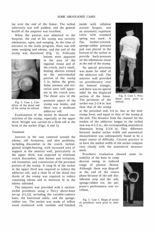
SOME ABOVE-KNEE CASES
lay over the end of the femur. The ischial tuberosity was well padded, and the general health of the amputee was excellent.
When the patient was admitted to the hospital, the end of his stump was severely edematous, open, and weeping. At the time of entrance to the study program, there was still some weeping and edema, and the end of the stump was discolored (Fig. 3). Follicular
lesions were apparent in the area of the inguinal crease and of the crotch, and a small, healing abscess existed on the anteromedial portion of the stump 5 in. below the groin. Some rawness and irritation were still apparent in the crotch area. The distal area of the
Fig. 3. Case 1. Condition of the distal end of the stump on referral.
posterior aspect of the stump was tender, and there was a moderate adductor roll.
Examination of the socket fit showed constriction of the stump, especially in the upper third. Weight was carried on a flesh roll at the brim of the socket (Figs. 4 and 5).
Treatment
Interest in the case centered around the edema, roll formation, and skin problems, including discomfort in the crotch. Ischial-gluteal weight-bearing, with increased area of support at the anterior wall, particularly in the upper third, was expected to eliminate crotch discomfort, skin lesions and irritation, roll formation, and constriction of the proximal portion of the stump. A snug fit of the socket in the upper third was required to reduce the adductor roll, and a close fit of the distal two thirds of the stump was required to reduce remaining edema and to maintain fit as the edema subsided.
The amputee was provided with a suction-socket prosthesis using a Navy above-knee set-up (11,12), including the variable-cadence knee, the functional ankle, and the sponge-rubber toe. The socket was made of willow wood reinforced with rawhide and finished
Fig. 4. Case 1. Prosthesis worn prior to referral.
inside with cellulose acetate lacquer, and an automatic expulsion valve with standard spring was used. A flat, leather-covered, sponge-rubber pressure pad was placed in the bottom of the socket to provide back-pressure on the edematous tissue at the end of the stump.
No special provision was made for relief of the adductor roll. The anterior wall provided no protuberance over the femoral triangle, and there was no special relief for the displaced section of the femur. The perimeter of the socket was 2-1/4 in. less than that of the stump at the proximal end, 3/4 in. less at the mid-stump level, and equal to that of the stump at the end. The distance from the channel for the tendon of the adductor longus to the ischial seat was 4-1/2 in., the corresponding anatomical dimension being 3-3/4 in. This difference between medial socket width and anatomical measurement was subsequently found to be a major source of difficulty. Current practice is to have the medial width of the socket compare very closely with the anatomical measurement.
Prosthetic evaluation showed some instability of the knee in ramp descent owing to reduced range of plantar flexion. Although there was drop-off at the end of the stance phase because of the soft dor-siflexion stop and the soft, sponge-rubber toe, the amputee's performance was excellent.
Fig. 5. Case 1. Shape of socket of prosthesis worn prior to referral.

RADCLIFFE, JOHNSON, AND FOORT
During the final fitting, the end of the stump turned red, but a sponge-rubber pad placed in the bottom of the socket improved stump color markedly within two hours. One week after delivery of the prosthesis, the edema was reduced; three weeks after, there was no edema; nine months after fitting, some edema was evident at the distal end of the stump.
Evaluation indicated that the ischial tuberosity was sliding anteriorly off the ischial seat so that the stump was settling deeper into the socket, with increased constriction at the proximal end. Several factors were involved. The stump had shrunk, and the anteroposterior dimension of the socket, especially in the medial third, which had been too great initially, had been increased in an unsuccessful attempt to relieve discomfort in the inguinal crease and in the weight-bearing area of the stump. The edema was confined to the areas of the stump which extruded into the valve recess and into the gap between the pad and the socket walls. The valve recess was lowered, the pad was refitted so that more weight was carried on the end of the stump, and the space between the pad and socket walls was eliminated. The edema cleared up.
Roll formation over the anterior brim of the socket was eliminated through extension of the anterior wall of the socket above the level of the ischial seat. The adductor roll was completely contained within the socket. Tightness of fit in the upper third was a source of minor discomfort immediately, but this problem decreased with reduction of the roll, which was complete within six months (Fig. 6).
Follicular lesions in the area of the crotch and the inguinal crease quickly cleared up with reduced forces from the socket brim and elimination of roll formation over the brim. One year after treatment began, the discoloration at the end of the stump was markedly decreased. Irritation of the skin over the posterior brim was a persistent problem directly related to decreased effectiveness of ischial-gluteal weight-bearing and wedging of the posterior aspect of the stump against the inside edge of the posterior brim of the socket. Attempts to increase weight-bearing on the distal end of the stump showed that the amputee preferred
ischial-gluteal weight-bearing because of discomfort experienced on the stump end with prolonged support of body weight.
Reduced support on the ischial tuberosity followed stump and socket changes and caused discomfort on the ramus. The medial brim of the socket was lowered to provide relief. This expedient was partially successful, but the stump sank deeper into the socket after wear, and ramus discomfort has recurred.
Summary
The problems of edema, roll formation, skin lesions, and discomfort in the crotch were studied. Edema was originally caused by a tight fit of the stump with constriction of the proximal end. Definite ischial-gluteal weight-bearing, with increased area of anterior support, was the primary factor in clearing up the edema. A pressure pad under the end of the stump helped to reduce the edema. Stump and socket changes which allowed the ischial tuberosity to slide into the socket, with wedging of the stump proximally, caused edema to recur. Improved fit of the pressure pad, with increased end-bearing, cleared up the edema. The adductor roll was brought about by weight-bearing in the crotch on the tight socket. Ischial-gluteal support, adduction of the femur in the socket with relaxation of the adductors, and extension of the medial brim to the level of the ischial seat, without provision of a relief pocket, eliminated the adductor roll. Discomfort due to tightness of fit for adductor- • roll reduction decreased as the roll reduced. The high anterior wall eliminated roll formation over the anterior brim of the socket. Skin lesions and irritation were caused by high force concentrations on the stump. Ischial-
Fig. 6. Case 1. Present prosthesis.
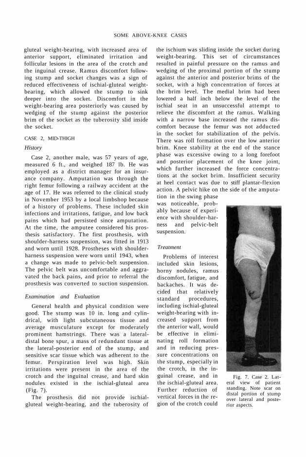
SOME ABOVE-KNEE CASES
gluteal weight-bearing, with increased area of anterior support, eliminated irritation and follicular lesions in the area of the crotch and the inguinal crease. Ramus discomfort following stump and socket changes was a sign of reduced effectiveness of ischial-gluteal weight-bearing, which allowed the stump to sink deeper into the socket. Discomfort in the weight-bearing area posteriorly was caused by wedging of the stump against the posterior brim of the socket as the tuberosity slid inside the socket.
CASE 2, MID-THIGH
History
Case 2, another male, was 57 years of age, measured 6 ft., and weighed 187 lb. He was employed as a district manager for an insurance company. Amputation was through the right femur following a railway accident at the age of 17. He was referred to the clinical study in November 1953 by a local limbshop because of a history of problems. These included skin infections and irritations, fatigue, and low back pains which had persisted since amputation. At the time, the amputee considered his prosthesis satisfactory. The first prosthesis, with shoulder-harness suspension, was fitted in 1913 and worn until 1928. Prostheses with shoulder-harness suspension were worn until 1943, when a change was made to pelvic-belt suspension. The pelvic belt was uncomfortable and aggravated the back pains, and prior to referral the prosthesis was converted to suction suspension.
Examination and Evaluation
General health and physical condition were good. The stump was 10 in. long and cylindrical, with light subcutaneous tissue and average musculature except for moderately prominent hamstrings. There was a lateral-distal bone spur, a mass of redundant tissue at the lateral-posterior end of the stump, and sensitive scar tissue which was adherent to the femur. Perspiration level was high. Skin irritations were present in the area of the crotch and the inguinal crease, and hard skin nodules existed in the ischial-gluteal area (Fig. 7).
The prosthesis did not provide ischial-gluteal weight-bearing, and the tuberosity of
the ischium was sliding inside the socket during weight-bearing. This set of circumstances resulted in painful pressure on the ramus and wedging of the proximal portion of the stump against the anterior and posterior brims of the socket, with a high concentration of forces at the brim level. The medial brim had been lowered a half inch below the level of the ischial seat in an unsuccessful at tempt to relieve the discomfort at the ramus. Walking with a narrow base increased the ramus discomfort because the femur was not adducted in the socket for stabilization of the pelvis. There was roll formation over the low anterior brim. Knee stability at the end of the stance phase was excessive owing to a long forefoot and posterior placement of the knee joint, which further increased the force concentrations at the socket brim. Insufficient security at heel contact was due to stiff plantar-flexion action. A pelvic hike on the side of the amputation in the swing phase was noticeable, probably because of experience with shoulder-harness and pelvic-belt suspension.
Treatment
Problems of interest included skin lesions, horny nodules, ramus discomfort, fatigue, and backaches. It was decided that relatively standard procedures, including ischial-gluteal weight-bearing with increased support from the anterior wall, would be effective in eliminating roll formation and in reducing pressure concentrations on the stump, especially in the crotch, in the inguinal crease, and in the ischial-gluteal area. Further reduction of vertical forces in the region of the crotch could
Fig. 7. Case 2. Lateral view of patient standing. Note scar on distal portion of stump over lateral and posterior aspects.

RADCLIFFE, JOHNSON, AND FOORT
be achieved by adduction of the femur in the socket, thus eliminating pelvic drop in the stance phase.
The amputee was provided with a suction-socket prosthesis which included a single-axis constant-friction knee, a plantar-dorsiflexion ankle, and a foot with single toe-break. Segments of the prosthesis were willow wood reinforced with rawhide. The socket interior was finished with cellulose acetate lacquer, and use was made of an automatic expulsion valve with standard spring.
Since the ischial tuberosity was not adapted to weight-bearing, the gluteal channel was held shallow to increase gluteal support. In addition, this arrangement offered increased sitting comfort by allowing a thinner posterior wall. Definite hamstring relief was provided by channeling the posteromedial apex of the socket (Fig. 8). The medial brim was approximately 1/4 in. lower than the posterior brim to provide clearance for the ramus. The medial socket width was 4-3/4 in. as compared to an anatomical measurement of 3-3/4 in., a difference subsequently found to be a major source of difficulty. As already mentioned, current practice is to have the medial width of the socket compare very closely with the anatomical measurement. The anterior wall was extended 2 in. above the level of the ischial seat and was relieved slightly over the femoral triangle. But this idea, which was tried for fear that pressure in the femoral triangle would interfere with circulation, has since been abandoned in favor of a definite protuberance into this area. Such a shape gives considerable distributed anterior support.
The socket was placed well forward on the knee block to allow initial flexion of the femur in the socket for increased voluntary control,
reduced energy requirements, and decreased lordosis of the lumbar portion of the spine. Prosthetic evaluation indicated that there was
Fig. 8. Case 2. Socket shape of prosthesis worn at present.
excessive stability at the end of the stance phase owing to a long forefoot and the posterior location of the knee axis, the long forefoot having been dictated by the large foot size. In the swing phase, there was some whip, which was not removed during alignment trials on the adjustable leg (3,4,5), but the gait was markedly improved on the new prosthesis. There was no ramus discomfort, no irritation, and no roll formation in the crotch or inguinal crease. The ischial tuberosity was close to the inside edge of the socket, so that the medial wall had to be lowered to prevent ramus discomfort.
After stump shrinkage, ramus discomfort recurred. The medial brim was lowered, but this measure provided only temporary relief as the stump settled deeper into the socket. Skin irritations from the anterior brim were reduced but persisted, since wedging occurred owing to inefficient ischial weight-bearing. Force concentration at the anterior brim was reduced somewhat by extension of the brim 2 in. above the level of the ischial seat. Undercut of the anterior wall over the femoral triangle reduced the effective area of anterior support.
Skin lesions in the crotch cleared up initially but recurred with failure of ischial weight-bearing. Formation of horny nodules in the weight-bearing areas was unchanged because poor ischial support allowed the tuberosity to move in and out of the socket over the inside edge of the posterior wall, thus creating abrasive and wedging action. Excessive perspiration was considered a factor both in the formation of horny nodules and in stump irritation because of the deteriorating effect it had on the inside finish of the socket.
Although the anterior and posterior brims were rolled and adjusted periodically to reduce discomfort from skin irritations in the inguinal crease and from nodules in the weight-bearing areas, this expedient provided only temporary relief, since the forces involved were either unchanged or increased. Reduced alignment stability, with increased flexion of the stump in the socket, did not relieve the backache. Activity level was not noticeably changed, and fatigue also remained unchanged.
In the course of treatment, redundant tissue at the lateral-distal portion of the stump was a

SOME ABOVE-KNEE CASES
problem in fitting because of the sensitivity of the adherent scar tissue. A large pocket was provided to give relief. Doing so reduced the effective length of the femur available for stabilization of the pelvis.
Summary
Failure of ischial-gluteal weight-bearing resulted in ramus discomfort and skin lesions. Lowering the medial brim provided only temporary relief, since the stump settled further into the socket. With recurrence of vertical pressure in the crotch, skin lesions were again a problem. The need for effective anterior stabilization to maintain the ischial tuberosity on the ischial seal was definitely indicated.
The high anterior wall eliminated roll formation and reduced skin infections in the inguinal crease. Undercut of the anterior wall over the femoral triangle reduced the anterior support area and increased the force concentration al the brim. Modifications of the anterior wall and of the posterior brim reduced discomfort temporarily only, since the force pattern was unchanged.
Placement of the prosthetic toe-break at the shoe crease provided excessive knee stability at the end of the stance phase. This result suggested that the conventional location of the toe-break was too far forward.
CASE 3, MID-THIGH
History
Case 3 was another male, age 51, height 6 ft., weight 180 lb. He lost his left leg above the knee at the age of 17 after an injury sustained in a baseball game. Since his original surgery, he had had no further revision. For the first five years after amputation, he used crutches without a prosthesis. He had since worn three prostheses during his 34 years as an amputee. The first leg had a shoulder-harness suspension. The leg worn upon his acceptance as a research patient had been converted in 1952 from an aluminum socket, pelvic-belt leg to a wooden suction-socket prosthesis a year and nine months previously. He was employed as an expediter in a shipyard, and the nature of his employment was such as to involve consider
able standing and moving about over short distances. His chief complaint was concerned with persistent edema of the stump since conversion to suction suspension, and this was the reason for his referral to the clinical-study program by a local limbshop in November 1952. The patient complained of occasional phantom pain but had no persistent local pain.
Examination and Evaluation
The patient was in good general health, with normal reflexes and above-average musculature. The stump was cylindrical, with 6-1/2 in. of femur below the perineum and 2-1/2 in. of soft tissue over the end of the femur (Fig. 9). Stump musculature was not abnormally prominent, and subcutaneous tissue was light. On contraction of the muscles, the redundant tissue pulled upward without bunching. A large scar, adherent to the distal end of the femur, extended 6 in. up the lateral side of the stump (Fig. 10). There was severe, nonpitting edema in the redundant tissue. The skin in the edematous area was without hair, distended, discolored, and scaly, with an orange-peel
Fig. 9- Case 3. X-ray of stump, mediolateral view.

RADCLIFFE, JOHNSON, AND FOORT
texture. Small cysts and horny nodules were evident in this region as well as in the inguinal area and in the crotch. The patient said that these cysts frequently enlarged and broke down, producing a pinkish-yellow discharge.
X-ray revealed the usual finding that there was lessened bone density on the amputated side and that the femur tapered and curved medially toward the distal end, which appeared to be closed. Comparison of socket and stump perimeters showed the socket smaller than the stump by 1-1/4 to 2 in. at corresponding levels. The distance from the tendon of the adductor longus to the ischial tuberosity was 3-1/2 in., as compared to the corresponding socket measurement of 5 in.
The suction socket the patient was wearing, although of the ischial-bearing type, did not achieve ischial bearing. The anteroposterior dimension was too large, the mediolateral dimension too small (Fig. 11). The socket was too tight, especially in the distal half, and the proximal end of the stump was constricted because of a wedging action precipitated by failure to establish ischial-gluteal bearing. Weight was borne on the medial brim of the socket, which was 3/8 in. below the level of the ischial seat and generously flared. There was a small adductor roll, and the anterior brim of the socket was level with the posterior brim, with some roll formation in the area of the inguinal crease. The anterior wall was undercut, a feature that caused localized high pressure on the stump at the anterior brim. The patient was well adapted to the use of the prosthesis, although a number of undesirable characteristics of gait were apparent, including a 7-in. walking base, considerable sidesway, and exaggerated arm swing on the side of the amputation.
Treatment
This patient's chief problem was the severe edema. It was felt that this disorder, as well as the skin lesions, could probably be controlled adequately by proper fit and alignment. The question of prime interest to the study group was whether or not suction suspension was the cause of the edema in this case.
Fig. 10. Case 3. Lateral view of stump. Note large, adherent scar.
The amputee was provided with a suction-socket prosthesis with conventional components, including a single-axis constant-friction knee, a plantar-dorsiflexion ankle, and a foot with a single toe-break. Segments were made of wood and reinforced with rawhide. An automatic expulsion valve with a strong spring was used to increase positive pressure in the socket during the stance phase.
The socket perimeters were 1-1/2 in. less than corresponding stump dimensions in the top third and equal to stump dimensions below that. The distance from the tendon of the adductor longus to the ischial tuberosity was 3-1/2 in., and the corresponding socket dimension was 4 in. The anterior wall was relieved to avoid pressure in the area contacting the femoral triangle (Fig. 12), and a flat sponge-rubber pad covered with soft leather was placed in the bottom of the socket to provide back-pressure on the edematous tissue.
After the patient had worn the prosthesis for six weeks, the edema in the redundant tissue had decreased markedly. The improvement was maintained over a nine-month period, although at no time was the condition completely eliminated. About the ninth month, there was a sudden increase in the amount of edema. Three factors seemed to be involved. There was increased activity. A weaker valve spring had been installed to reduce loss of suction. And there had been stump shrinkage, as indicated by the experience of ramus discomfort. The thickness of the control pad was

SOME ABOVE KNEE CASES
increased, but doing so did not alter the condition. Xext, a stronger valve spring was provided to increase the positive pressure in the stance phase, and there was then a marked and immediate improvement in the edematous condition of the stump. To provide increased ischial weight-bearing by reducing the anteroposterior dimensions of the socket, liners were added in the area of the socket contacting the femoral triangle. Although ischial weight-bearing was improved, as evidenced by the elimination of ramus discomfort, there was no change in the edema.
The decision was then made to provide the amputee with a socket that would make total contact with the stump end, thus exerting greater back-pressure on the edematous tissue. After a four-day trial period, the patient found
Fig. 11, Case 3. Socket shape of prosthesis worn on referral.
Fig 12 Case 3 Socket shape, present prosthesis
that accumulated perspiration irritated the stump acutely, and the socket had to be discarded. At present the amputee continues to wear the first prosthesis provided and still has moderate edema.
Skin infections initially present in the crotch area were cleared with provision of ischial-gluteal weight-bearing, but with stump shrinkage the condition recurred because of decreased effectiveness of such weight-bearing. Provision of liners over the area of the socket contacting the femoral triangle increased the effectiveness of ischial-gluteal weight-bearing and reduced the skin problems. Throughout treatment, there was irritation on the weight-bearing area of the stump, especially around the ischial tuberosity. Provision of a section of nylon stocking, fastened to the outside of the socket and draped interiorly over the weight-bearing area, improved comfort considerably by reducing shear between the skin and the socket. The

RADCLIFFE, JOHNSON, AND FOORT
skin irritations were due primarily to excessive anteroposterior socket dimensions, especially along the medial wall. This situation allowed the tuberosity to slip into the socket and the entire stump to settle deeper, with consequent wedging of the stump against the posterior brim and the anterior wall, thus creating a high force concentration on the ischial tuberosity. A pressure pad was found very helpful in controlling edema when other elements of the fit were satisfactory.
Minor skin irritations resulted from deterioration of the inside finish of the socket, but refinishing the socket cleared them.
Summary
Although treatment was never completely successful in eliminating this patient's edema, function and comfort were markedly improved, and the course of his prosthetic treatment served to demonstrate several principles. Provision of ischial-gluteal weight-bearing eliminated ramus discomfort, reduced edema, and cleared skin infections anteriorly and medially where the stump contacted the socket brim. The posterior brim caused irritation of the stump when ischial-bearing was indefinite, with the tuberosity near the inside edge of the socket, or when the radius of curvature over the inside edge was too small, or when the ischial area was not conditioned for weight-bearing. Use of liners to decrease the anteroposterior dimension increased comfort. When there was stump shrinkage and decreased ischial support, edema increased, and a pressure pad alone was not successful in controlling it. Use of a stronger valve spring, to increase the positive pressure, decreased edema. In spite of the failure to control the edema completely, the patient was able to perform at a high level of activity.
CASE 6, LOWER THIRD OF THIGH
History
Case 6, male, was 56 years old, stood 6 ft. 2 in. tall, and weighed 142 lb. He lost his right leg above the knee as a result of a motor-coach accident when he was 47. The patient's first prosthesis, fitted six months after amputation,
used shoulder-harness suspension. It was worn for two years. The next prosthesis provided pelvic-belt suspension. It was being worn when he entered the Clinical Study in November 1953 (Fig. 13). Complaints included tightness of the socket, discomfort due to abrasion of the hip by the belt on the side of the amputation, and irritation in the distal-lateral area of the stump. The prosthesis was in a state of general disrepair.
Examination and Evaluation
General health was good, and activity level was high both at home and at work. The stump was conical, with light subcutaneous tissue and very light musculature, muscular atrophy having been brought about by stump inactivity in the walking cycle. Tissue over the end of the stump was very thin. The lateral-distal portion of the stump was scarred, and the ischial tuberosity was small, sharp, and lightly padded. Scars in the crotch area indicated periodic folliculitis and boil formation, and there was local pain posterodistally.
A number of points were of interest in this case. They included a heavy adductor roll due to abducted gait and the plug fit; inexperience with suction suspension and ischial-gluteal weight-bearing; gait faults, including the abducted gait and pelvic hike on the side of the amputation; and the history of boils and folliculitis in the crotch due to weight-bearing in that area (Figs 14, 15, and 16).
Treatment
The amputee was provided with a suction-
Fig 13 Case 6 Original prosthesis. Weight carried on adductor roll

SOME ABOVE-KNEE CASES
Fig. 14. Case 6. Re- Fig. 15. Case 6. Ad-laxed position of stump ductor roll prior to treat-prior to treatment. ment.
socket prosthesis which included a single-axis knee with constant-friction swing-phase control, a plantar-dorsiflexion ankle, and a foot with a single toe-break. Segments were willow wood reinforced with rawhide (Figs. 17 and 18).
Since the gluteus maximus was atrophied, the extensor channel and gluteal flare were fitted closely. No relief was provided for the heavy adductor roll, which was drawn completely into the socket as a part of the process of reduction. The socket perimeters at the brim of the socket were 3 in. less than stump dimensions. Two inches below the level of the ischial seat, socket perimeters were approximately half an inch less than stump dimensions. At the lower levels, socket and stump perimeters were identical. The distance from the ischial seat to the channel for the tendon of the adductor longus was 3-3/4 in., the corresponding anatomical measurement being 3 in.
Adduction of the femur in the socket relaxed the adductors and permitted inclusion of the roll in the socket with less difficulty (Fig. 19). Initially a safety belt was provided to increase
the amputee's sense of security, since he had a fear of losing the leg.
For the first three months of treatment the prosthesis was worn three hours a day. During the next six months use of the prosthesis was increased to all day, the extended period of adaptation to use of the prosthesis being due to discomfort at the ischial tuberosity. After nine months there was marked increase in comfort, a circumstance which induced the amputee to discard the cane he had theretofore used regularly. A soft pad over the ischial seat reduced discomfort but was discontinued after two weeks in the expectation that adaptation would be accelerated.
Tight fit of the proximal third of the stump for reduction of the adductor roll resulted in edema in the distal portion of the stump. But
Fig. 16. Case 6. Triangular shape of original socket.

RADCLIFFE, JOHXSOX, AND FOORT
when the socket perimeters in the upper third were increased to reduce constriction, the edema cleared up. Two weeks later the adductor roll had shrunk, and there was loss of suction. A new socket was made and modified with liners at intervals for a period of a month as shrinkage continued. By this time, the perimeter of the stump at the perineum had been reduced 2-1/2 in., so that a new socket was required. The dimension of this socket from the ischial seat to the channel for the tendon of the adductor longus was reduced by half an inch, and a protuberance was provided over the area contacting the femoral triangle.
Edema recurred after six months, and examination of fit showed considerable development of the hamstring muscles. Accordingly, the socket was opened at the posterior wall
starting 2 in. below the ischial-seat level; the edema cleared up.
Further development of stump musculature resulted in edema at the end of the stump during the ninth month of treatment. Increased hamstring relief was provided, a stronger valve spring was installed, and a sponge-rubber pad was placed in the bottom of the socket to increase back-pressure on the end of the stump. Again the edema cleared up.
Training was provided for a period of one hour a day for six weeks. Gait was excellent under observation, although there was some reversion to old habits when the amputee was not under supervision. Pelvic hike was particularly per-
Fig. 18. Case 6. Shape of socket of new prosthesis
sistent. Those habits which were dependent on fit and alignment, including abducted gait, were gradually eliminated.
Summary
Problems studied included stump changes, particularly at the large adductor roll, adaptation to suction suspension, adaptation to ischial weight-bearing, and gait faults. Boils and folliculitis did not recur during the process of treatment.
Reduction of the large adductor roll formed in five years of weight-bearing in the crotch with abducted gait required six months of treatment. During this period there was some edema owing to constriction of the proximal third of the stump. Edema was reduced by increasing the perimeters of the socket in this region. Edema resulted again owing to constriction following hamstring hypertrophy.
Fig. 17. Case 6. New prosthesis, with ischial-gluteal weight-bearing.

SOME ABOVE-KNEE CASES
Relief for this development, the use of a stiffer valve spring for increased positive pressure in the stance phase, and a sponge-rubber pad in the bottom of the socket cleared up the edema.
There was periodic loss of suction following stump shrinkage. The light subcutaneous tissue could be distorted very little. As a result, slight stump changes led to loss of suction.
Initially some lateral instability and reduced control of the prosthesis, probably resulting from weakness of the gluteus medius, was experienced. With adaptation to suction suspension, there was increased stability and control as the gluteus medius became stronger. Adaptation was completed within the nine months required to stabilize the stump. The ischial tuberosity took more than nine months to condition for weight-bearing, chiefly because of the lack of previous experience, the light padding over the tuberosity, and the especially sharp configuration of the bone.
CASE 8, MID-THIGH
History
Case 8, another male, was 42 years old, measured 6 ft., and weighed 175 lb. His right leg was amputated above the knee in December 1949 after a shotgun wound received in a hunting accident approximately a year previously. He had had only one prosthesis since amputation and was wearing it at the time he was accepted by the Clinical Study in January 1954. Although the prosthesis provided suction suspension, the components were conventional. The patient was dissatisfied with the prosthesis primarily on the basis of poor fit, but he felt that the alignment could be improved and that such improvement might give him more comfort and better function. He also complained of needlelike phantom pains in the ball or sole of the "foot," with persistent tingling.
Examination and Evaluation
General physical examination was normal. The stump was conical (Fig. 20), approximately 9 in. of femur remained below the perineum, and about an inch of tissue covered the end of the femur. Musculature of the stump was firm, but there was retracted muscle on
the lateral side about 2-1/2 in. from the tip of the femur. There was little subcutaneous fat. On the posterior aspect of the stump at the distal end was an inverted T-shaped scar, and the distal end of the femur was sensitive to pressure. X-ray showed a medioposterior spur arising from the end of the femur, curving upward, and tapering. There was edema and brown discoloration at the end of the stump (Fig. 21), and small follicular lesions were evident in the areas contacting the anterior and medial brims of the socket.
The prosthesis had a wooden socket reinforced with rawhide, a single-axis knee with constant friction for swing-phase control, an ankle providing plantar-dorsiflexion action, and a foot with a single toe-break 5 in. anterior
Fig 19. Case 6. Condition of subject one year after application of new prosthesis. Note reduced adductor roll.

RADCLIFFE, JOHXSOX, AND FOORT
Fig 20. Case 8 Stump molded by tight fit.
to the ankle axis. Weigh t was carried through a roll of flesh at the brim of thesocket (Fig. 22), and the amputee walked with a wide-based gait owing to crotch discomfort and out-set of the foot. Knee stability was excessive because of the long forefoot and the posterior position of the knee axis, which fell approximately 1 in. posterior to the tro-chanter-ankle reference
line. The prosthesis was short, but this detail was not too apparent since the ischial tuberosity was 1-1/2 in. above the posterior brim of the socket. Because of insufficient knee friction and excessive kicker action, there was heavy impact at the end of the swing phase, and there
Fig. 21. Case 8. Socket shape of original prosthesis. Note edema and brown discoloration at the end of the stump.
was whip during the swing phase, probably owing to muscle activity within the socket and to the vigorous stump action required to break the prosthetic knee at the end of the stance phase. Rotation at heel contact was due to excessive stiffness of the plantar-flexion bumper.
Problems of interest to the Clinical Study included the edema encountered with use of suction suspension, skin infections, the adductor roll, the time and circumstances involved in conditioning the amputee to ischial-gluteal w e i g h t - b e a r i n g , and gait training.
Treatment
Reduction of the edema required reduced constriction of the proximal third of the stump through provision of ischial-gluteal weight-bearing, extension of the anterior wall above the level of the ischial seat, and close fit of the distal two thirds of the stump. Reduction of pressure on the proximal end of the stump from the superior brim of the socket was required to clear up skin infections. At the same time, snug fit, with adduction of the femur in the socket to relax the adductors, was required
for reduction of the adductor roll. Improved fit and alignment, with training, were planned to correct gait faults.
The amputee was provided with a suction-socket prosthesis which included a single-axis knee with constant-friction swing-phase control, a plantar-dorsiflexion ankle, and a foot with a single toe-break. Segments were of wood,
Fig. 22. Case 8. Original prosthesis. Plug fit, with roll formation over the socket brim.

SOME ABOVE-KNEE CASES
reinforced with plastic laminate. The extensor channel was held shallow and flared minimally at the brim to increase gluteal weight-bearing, since the amputee was not accustomed to ischial weight-bearing. Xo relief was provided for the adductor roll. The anterior wall of the socket was slightly relieved over the area contacting the femoral triangle (Fig. 23). The toe-break was cut 5 in. anterior to the ankle axis so as to coincide with the normal break of the shoe. To increase knee stability in the initial phase of the fitting, the knee axis was placed 3/4 in. behind the trochanter-ankle reference line. Socket perimeters were 1-1/4 in. under stump perimeters at the proximal end and equal to stump perimeters at the level of the distal two thirds. The distance between the ischial tuberosity and the adductor longus tendon was 3-3/4 in., the corresponding socket dimension being 4-1/2 in.
Evaluation following delivery of the prosthesis indicated that knee stability was excessive owing to the long forefoot and the posterior position of the knee axis. Training was required to improve balance and cadence
Fig. 23. Case 8 Socket shape, new prosthesis
Fig. 24 Case 8. New prosthesis. Note elimination of roll formation over the brim of the socket.
symmetry and to overcome the vaulting as well as to reduce the width of the walking base. The ischial tuberosity was on the seat, and there was no ramus contact with the medial brim of the socket. The adductor roll was contained in the socket. Roll formation over the anterior brim of the socket was eliminated (Fig. 24).
Initially there was some edema at the distal end of the stump owing to constriction proximally. As the flesh roll reduced, constriction and edema decreased, and finally the edema cleared up.
After an illness which caused the patient to lose considerable weight, the stump settled deeper into the socket as the ischial tuberosity slipped inside. This circumstance allowed the ramus to contact the medial brim and caused the anterior and posterior brims to constrict the stump. But because the somewhat conical shape of both stump and socket maintained the snug fit over the entire stump as the latter settled down into the socket, and because, consequently, the pressure differential between the proximal and distal portions of the stump was not increased sufficiently, edema did not recur. Nevertheless, ramus discomfort decreased activity on the prosthesis. The problem was eliminated with provision of a new socket.
Follicular lesions cleared up with effective ischial-gluteal weight-bearing but recurred when ischial support was decreased following

RADCLIFFE, JOHNSON, AND FOORT
loss of weight. Provision of a new socket with ischial-gluteal weight-bearing again cleared up the skin condition. With the first socket, poor stabilization of the ischial tuberosity on the seat contributed to skin irritation and to the formation of horny nodules in the weight-bearing area. Comfort was greatly improved by reduced anteroposterior dimensions, with improved anterior support by provision of a protuberance on the anterior wall over the area contacting the femoral triangle.
Gait training improved walking habits but focused attention on deficiencies of fit by forcing the amputee to walk according to a preconceived pattern rather than one that provided maximum comfort. Sixteen months after training was complete, evaluation indicated that, because of discomfort from loss of fit, gait was somewhat worse than before training. Since in any case the amputee adapted his gait pattern to provide maximum comfort, training was of doubtful value as compared with good prosthetic treatment. With the first prosthesis, excessive knee stability detracted from naturalness of gait, and this condition also was a factor in causing the discomfort in the ischial-gluteal area and at the end of the femur anteriorly, where the stump showed the results of the force required to break the knee. Subsequent fit and alignment corrected these problems and greatly improved comfort. The length of the forefoot was reduced to approximately 3-1/2 in. to decrease knee stability, and the knee axis was placed on the ankle-trochanter reference line.
Summary
The patient's edema on the prosthesis worn at the time of referral was apparently caused by constriction of the stump in the socket, especially in the proximal third. A contributing factor was weight-bearing on the adductor roll over the medial brim. Provision of ischial-gluteal weight-bearing, with wide distribution of the pressure on the anterior aspect of the stump, had a number of consequences. The edema disappeared with the reduction of the adductor and anterior rolls and recurred only when fit and ischial support were lost with loss of weight from illness. Skin irritations in the crotch, along the gluteal fold, and around the
ischial tuberosity were cleared up by reduction of shearing forces when positive support was provided. Reduction of alignment stability by shortening the toe-break length and by moving the knee axis forward cleared up the skin irritation on the anterodistal aspect of the stump by reducing the force required to break the knee at toe-off. Training appeared to have far less effect on symmetry of gait than did fit and alignment. When the patient was able to walk symmetrically with comfort, he did so. When last seen, the amputee reported a general increase in comfort and a corresponding increase in his level of activity.
CASE 9, BILATERAL ABOVE-KNEE, UPPER THIRD
OF THIGHS
History
Case 9, male, 27 years of age, weight 100 lb., underwent amputation at the age of eight as a result of crushing injuries to both legs sustained in a truck accident. He had had six pairs of legs since his amputation, the first pair having been fitted four months after surgery, without preliminary conditioning iherapy or exercise. That pair, employing shoulder-harness suspension, was worn for four years. Between that time and 1948, he had had three sets of legs, all employing pelvic-belt suspension and using conventional components. In 1948 he was fitted at the University of California with suction suspension. The prostheses were worn for two years and then discarded because of disrepair. New suction sockets, provided in 1950, were worn for two years. These were uncomfortable owing to tightness of fit. In 1953 a local limbshop fitted the patient with the suction-socket prostheses he was wearing when referred to the Clinical Study in September 1953. The complaints included skin irritation with folliculitis, boils, and abrasion on areas of the stump contacting the socket brim; crotch discomfort; and edema in the ends of the stumps. Although working at essentially a sedentary occupation, he did a great deal of walking around his office.
Examination and Evaluation
There were no significant physical findings except as pertaining to the amputations. This

SOME ABOVE-KNEE CASES
young man was well nourished, healthy, and of average intelligence. The stumps were almost identical. They were approximately 6 in. long, measured from the perineum, and cylindrical, with approximately 2 in. of tissue over the distal ends of the femurs (Fig. 25). Hygiene of the stumps and prostheses was poor, perspira-ation level high. There were boils, folliculitis, and abrasions on the stumps and the crotch areas, with boils and folliculitis in the inguinal creases. Both stumps had heavy, nonpitting edema and petechiae at the distal ends. The stumps were held in 28 deg. of abduction, but ranges of motion and muscle power were normal and equal. X-ray showed medial curvatures of both femurs distally. The medullary cavities appeared to be closed, and there were no sensitive areas.
Fig. 25. Case 9. Stumps relaxed.
The prostheses (Figs. 26, 27, and 28) had rectangular suction sockets on single-axis knees with constant-friction swing-phase control, plantar-dorsiflexion ankles, and wooden feet with single toe-breaks. Segments of the prostheses were made of willow reinforced with rawhide. No auxiliary suspension or control straps were used. Although the sockets were intended to provide ischial-gluteal weight-bearing, the ischial tuberosities were down inside the sockets so that weight was carried on the medial brims, which had been lowered in an unsuccessful attempt to provide relief, with severe wedging of the stumps against the anterior and posterior brims. This situation was a cause of irritation and infection of the stumps in the areas contacting the medial and posterior brims of the sockets and promoted edema by restriction of circulation. Excessive alignment stability due to posterior placement of the knee axes increased forces on the posterior aspects of the stumps as the amputee attempted to break the knees to initiate swing phase.
The patient walked with a wide-based gait, at least partially because of the abducted position of his stumps. He customarily used a cane but was able to walk without it. Because the amount of friction in the swing-phase-control units was adjusted to provide minimum resistance to rotation, there was impact at the end of the swing phase. Rotation at heel contact was due to excessive stiffness of the heel bumper of the left prosthesis. Torso and pelvic list were due to shortness of the right prosthesis.
Treatment
The objectives of treatment in this instance were:
1. Elimination of crotch discomfort and skin problems by providing definite ischial-gluteal weight-bearing ;
2. Elimination of irritation and follicular lesions in the inguinal areas by reducing force concentrations in these areas through use of high anterior walls and definite ischial-gluteal weight-bearing;
3. Reduced wedging of the stumps proximally through provision of definite ischial-gluteal weight-bearing and high anterior walls for increased area of support;

RADCLIFFE, JOHNSON, AND FOORT
4. Close fit oi the slumps along their entire lengths, with decreased wedging of the stumps proximally, for reduction of the edema;
5. Reduction in energy consumption by providing increased voluntary control with flexion of the stumps in the sockets and reduced alignment stability; and
6. Study of the effect of narrow- and wide-base alignment on lateral stability, within the limits imposed by the abducted positions of the stumps.
In March 1954, the patient was provided with two suction-socket prostheses (Fig. 29). A light webbing belt was furnished to aid suspension. Single-axis constant-friction knees, plantar-dorsiflexion ankles, and wooden feet with rocker toe-breaks and foam crepe shoe-sole material in the toes were used. The prostheses were reinforced with rawhide, the inside surfaces of the sockets were finished with cellulose acetate lacquer, and automatic expulsion valves with standard springs were used. For knee stability, the reference line joining the ankle axis to the point of contact of the greater trochanter passed 1 in. ahead of the knee axis on both prostheses. The ankles were provided with stiff plantar-flexion bumpers to increase anteroposterior stability.
Within approximately a month from the time of fitting the initial prostheses, there was substantial improvement in comfort and skin problems in the crotch areas. The medial brims were not appreciably lower than the
Fig. 26. Case 9. Socket shape of prostheses worn at time of referral, medial walls nearest patient's hands.
posterior brims. Skin problems in the inguinal creases were relieved by ischial-gluteal weight-bearing, by high anterior walls, and by provision of a protuberance over the region of the femoral triangle (Figs. 30 and 31).
Provision of ischial-gluteal weight-bearing and increased anterior support resulted in reduced wedging of the stumps proximally, and a close fit of the stumps over their entire length produced a prompt and marked reduction in edema. Irritation in the weight-bearing area was a persistent problem in the early stages of fitting. At one point, the amputee had the sockets modified in a commercial limbshop in an attempt to relieve this discomfort. But these changes increased the anteroposterior
Fig. 27. Case 9. Prostheses worn at time of referral, medial view.

SOME ABOVE-KNEE CASES
adjusted to he could walk with adequate control and stability when using stiff plantar-flexion bumpers, with the ischial seats well behind the projected lines through the ankle and knee axes, and with initial flexion of the stumps for voluntary control (Fig. 31). Because of long-established habits of abduction, it was necessary to provide wide-base a-lignment of the second pair of prostheses. At the time of the final evaluation, the stumps were in excellent condition.
Fig 28. Case 9. Pros-theses worn at time of referral. posterior view.
dimensions of the sockets medially in the upper third. The tuberosities slipped into the sockets, and edema recurred. New | sockets were fitted to re-establish ischial support. Irritation and discomfort in the area of the tuberosities disappeared after approximately two months of conditioning.
I s c h i a l - g l u t e a l weight-bearing raised the stumps in the sockets and decreased voluntary control, but after four months the patient became
this change. It was found that
Fig. 30. Case 9. Socket shape, present prostheses. Ischial seats are at bottom center.
Summary
The patient's problems of edema and skin irritation in the areas of the crotch, the inguinal creases, and the gluteal folds responded well to the standard principles of fitting. Irritation in the weight-bearing area was a temporary problem which cleared up with tissue conditioning. A wide walking base was required in this case for lateral stability. The stiff plantar-flexion bumpers provided anteroposterior stability both standing and walking. Placement of the sockets well forward on the knees provided adequate security.
Fig. 29. Case 9. Present prostheses. Fig. 31. Case 9. Present prostheses, medial view.

RADCLIFFE, JOHNSON, AND FOORT
CASE 10, VERY SHORT ABOVE-KNEE
History
Case 10, male, was 51 years of age, weighed 150 lb., and was 5 ft. 7 in. tall. Amputation was through the left femur in the upper third. The original amputation had been carried out in December 1952 as a result of arteriosclerosis, and the stump had been revised in August 1953.
This patient was referred by a local limb-shop that was in the process of fitting him with a pelvic-belt prosthesis converted from a suction socket because of failure to maintain suction. He was pessimistic about the use of suction suspension and was unwilling to attempt it except for the benefit of the research group. Because of pressure in the groin, insecurity at the knee, toe-scuffing in the swing phase, and stump withdrawal when sitting, he was dissatisfied with the leg being fitted by the local shop.
Examination and Evaluation
Physiologically, the patient appeared older than his age, but he was alert and cooperative. The abdomen was severely scarred from surgical incisions for appendectomy and double sympathectomy, and scars also extended from the distal end of the stump up the antero-medial aspect to mid-groin. The medial scars were deeply adhered to underlying tissue. Subcutaneous fat was moderate and musculature firm, with prominent adductors and gluteus maximus. There were no sensitive areas. Skin was normal. The femur extended 1-1/2 in. below the perineum and 7 in. below the great trochanter (Figs. 32 and 33). A spur extended upward on the medial side, and the stump showed some abduction contracture.
This patient was of interest primarily because of the very short stump. Also of interest was the patient's inexperience, which offered an opportunity to study problems of adaptation and stump changes. Experience in prosthetic treatment of cases with circulatory impairment was also desired.
Treatment
The patient was provided with a suction-socket prosthesis. At first a single-axis knee
with constant-friction swing-phase control, a p l an t a r -do r s i f l ex ion ankle, and a foot with single toe-break were used. Segments were of wood, reinforced with plastic laminate. Later the knee was changed to a friction-stabilized type. The final socket was made of plastic laminate, and a SACH foot (solid ankle, cushioned heel) was used instead of the conventional foot.
Two sockets were fitted within the first two months. With the second socket, all requirements for the successful application of suction suspension had been met, but, because of obscuring factors related to the amputee's a t t i t u d e , this condition was not altogether understood at the time. A remolding process had brought about elongation of the stump, a feature which made suction easier to maintain.
Successful application of suction suspension depended upon undercutting the posterior, medial, and anterior walls of the socket below the ischial-seat level, maintaining the lateral wall above the level of the ischial seat, and holding the fit close in the proximal part of the stump. Because of the undercut medial wall, bunching of the adductors did not break the suction. Suspension aids, valuable in providing increased sense of security in the initial phase of treatment, were unnecessary once the amputee was adapted to the use o'f his prosthesis. Because of the limited amount of femur available, and also because of the abducted position of the stump, no attempt was made to adduct the femur in the socket. Flexion of the stump in the socket was designed
Fig. 32. Case 10. Lateral view of the stump in the hanging position.

SOME ABOVE-KNEE CASES
to meet the natural requirements of the stump and spine rather than to provide voluntary control of the knee, since stump power was limited and undercutting of the anterior and posterior walls of the socket reduced the effectiveness of the stump in controlling the knee (Fig. 34).
Initially the patient was provided with a conventional single-axis knee with adequate knee stability. Fear of falling and buckling of the prosthetic knee due to weakness of the normal leg, inexperience, and other factors, however, led to use of a friction-stabilized knee. But aligning the friction-stabilized knee in accordance with the rules for the single-axis conventional knee resulted in excess stability at the end of the stance phase. Accordingly.
Fig. 33. Case 10. Stump in 90 deg. of flexion.
Fig. 34. Case 10. Rectangular socket provided for patient.
Fig. 35. Case 10. New prosthesis, posterior view.
the knee was later aligned to provide decreased alignment stability with greater reliance on the friction mechanism.
Although narrow-based gait was not anticipated in the alignment of the limb, the amputee was able to walk with a 4- to 6-in. base. The prosthesis was made about 1-1/2 in. shorter than the normal limb (Fig. 35) because the amputee found that the shorter prosthesis permitted better control. Before prosthetic treatment started, the patient had back pains,
and it was anticipated that the shorter leg might lead to recurrence. As a matter of fact, he had some recurrence of the back pains early in the fitting when a webbing belt was tried as a supplement to suction suspension. But this problem disappeared when use of the belt was discontinued.
Stump changes during the study were minimal. In the first two and a half months, stump shrinkage occurred, but the stump remained stable throughout the following two years of observation. After the patient had worn the first suction-socket prosthesis a short time, the tissue of the stump started to extend, so that by the time the stump had stabilized there was an increase from 1-1/2 in. to 3 in. of tissue available below the perineum for effecting a suction seal. At no time was there more than a reddening of the stump in the weight-bearing area, and, as the tissue became conditioned, the skin became dark and tough. There was no edema.
Summary
The problem in this case was to attempt application of suction suspension to a very

RADCLIFFE, JOHNSON, AND FOORT
short, heavy, above-knee stump. Suction proved to be a practical means of suspension. All walls were concave, and relief was provided for bunching adductors to prevent the stump from being forced away from the socket in the medial apexes with consequent failure of the suction seal. For increased control and reduced effort, the amputee preferred the prosthesis approximately 1-1/2 in. shorter than the normal leg. There were no back pains. The stump shrank slightly during the first two months, and there was elongation of the stump, particularly on the medial side. Because of insufficient knowledge for adequate prosthetic treatment of the patient, and because of the poor adjustment of the patient to his amputation and physical condition, rehabilitation was a lengthy process. Once the patient was successfully treated, no changes in fit and alignment were required over a two-year period.
CASE 2 4 , LOWER THIRD OF THIGH
History
Case 24, female, was 41 years of age, stood 5 ft. 2 in. tall, and weighed 126 lb. She had undergone amputation at the level of the lower third of the left femur. There had been a congenital lymphangioma involving the tissues of the left leg from the knee down. Infection developed in the soft tissues over the anterior portion of the tibia, and subsequently there was an osteomyelitis of the tibia. Later a mass, which was diagnosed as carcinoma, appeared in the ankle. Amputation was performed in May 1954.
The amputee had had only one prosthesis since amputation. It consisted of conventional components and a molded leather socket laced anteriorly (Fig. 36). When she entered the study program in January 1955, complaints included skin irritation and infection, with discomfort in the crotch, discomfort and restriction from the corset used to suspend the prosthesis, right-sided backache, and excessive wear of hosiery.
Examination and Evaluation
Normally a very active person, the patient had a rather low activity level owing to
limitations imposed by her prosthesis. General physical condition was good, except for very flabby abdominal musculature, but the patient experienced phantom s e n s a t i o n s a s though the "leg" were "falling asleep." Phantom pains in the form of c ramps in the ' 'calf" and shooting pains on the medial side of the "ankle" also were present. They lasted only a few seconds and were less frequent since she had been fitted with a prosthesis.
The stump was cylindrical and 9 in. long measured from the perineum, including approximately 2 in. of redundant tissue over the end of the femur. Subcutaneous tissue was heavy but f i rm. Musculature was of
average strength, with no group particularly prominent. There was no significant edema or skin problem in the distal end of the stump, but follicular lesions existed in the crotch, and areas of irritation were present on the torso from pinching and bruising by the corset stays.
The prosthesis provided weight-bearing on a flesh roll around the brim of the socket, in the crotch, and against the side walls of the socket (Figs. 37 and 38). A pressure pad in the bottom of the socket did not provide appreciable support, and the patient walked with a wide base and with torso and pelvic list owing to excessive length of the prosthesis, wide-base alignment, and discomfort in the crotch. Stride length, cadence, and arm attitude were unsymmetrical because of excessive stability of the prosthetic knee.
This amputee presented several problems of interest to the study group. They included
Fig. 36. Case 24. Prosthesis worn on referral. Note lateral displacement of the socket on the stump.

SOME ABOVE-KNEE CASES
skin infections, the relationship between redundant tissue at the end of the stump and edema with suction suspension, factors involved in changeover from corset to suction suspension, factors involved in changeover to i s c h i a l - g l u t e a l weight-bearing, stump changes, the cosmetic problems of a female amputee, and use of the SACH foot with high-heeled shoes.
Fig. 37. Case 24. Socket shape at the brim, prosthesis worn on referral.
Treatment
In September 1955, the patient was provided with a suction-socket prosthesis consisting of a single-axis constant-friction knee, a SACH foot made for a high-heeled shoe, and wooden segments reinforced with plastic laminate. An automatic expulsion valve with standard spring was used, and the inside surface of the socket was finished with phenolic resin varnish. A polyvinyl chloride-acetate cosmetic cover was provided for the shank. Since the amputee was unaccustomed to ischial weight-bearing, and also in order to increase sitting comfort, the gluteal flare and the extensor channel were fitted closely to provide increased gluteal support. The lateral wall was closed in over the stump above the ischial-seat level, and the anterolateral apex was closed to reduce conspicuousness of the brim of the socket under the clothing.
This socket was worn on the adjustable leg (4) for approximately a month and was then installed in a finished prosthesis. After a week, the redundant tissue at the end of the stump was moderately edematous. The only known difference between the prosthesis with the adjustable leg and the finished prosthesis was that the latter had five coats of "Platon" varnish on the inside of the socket. There were three possibilities related to the finish of the socket interior. The stump worked down into the socket through weight-bearing, with increased effect of muscle activity on negative
pressure and reduced positive pressure in the stance phase owing to reduced excursion of the stump in the socket. There was adherence of the stump to the walls of the socket and, hence, reduced massaging action at the distal end. Vacuum seal was improved so that negative pressure was maintained, especially during sitting.
The edema cleared up after provision of a pressure pad in the bottom of the socket and after atrophy of the stump, which reduced constriction proximally. In addition there was, as a result of aging and lubrication of the surface finish by body oils, decreased adherence of the stump to the socket. Addition of liners to compensate for shrinkage did not cause edema with this socket.
A second prosthesis was supplied in December 1955, the socket of this limb being fitted snugly in anticipation of further shrinkage of the stump. Six weeks after delivery of the prosthesis, examination showed edema at the end of the stump. A pressure pad was provided, but there was no reduction in the edema during the next two months. Pad thickness was then increased. When the amputee was examined next, six weeks later, the pad had been discarded owing to ineffectiveness in controlling the edema. Edema was reduced, and the stump had atrophied further. This development had reduced constriction of the proximal
Fig. 38. Case 24. Lateral view of the stump in the hanging position.

RADCLIFFE, JOHNSON, AND FOORT
portion of the stump without reducing the effectiveness of ischial support, thus indicating that edema was caused by constriction of the proximal area and that the pressure pad was ineffective in reducing it so long as constriction persisted proximally. Six weeks later there was no edema, and the socket was looser. Liners were installed over the anterior and posterior walls to decrease perimeters of the socket in the upper third by one inch. Edema did not recur. Socket perimeters proximally were reduced another inch two months later as a result of stump atrophy. There was no edema before or after addition of the liners, and at no time was there failure of ischial support.
The patient adapted very well to the changeover from corset to suction suspension and appreciated the comfort and freedom that resulted. Control of the prosthesis was excellent. Adaptation to ischial-gluteal weight-bearing was immediate, and skin lesions and discomfort in the crotch cleared up quickly. The shallow gluteal flare and extensor channel, with relief for hamstring attachments at the ischial tuberosity, provided sitting comfort. As the stump atrophied, proper weight-bearing was maintained effectively by the addition of liners to the anterior and posterior walls of the socket as necessary (Fig. 39).
Stump atrophy due to heavy subcutaneous tissue was a problem, and the stump had not stabilized at the end of a year. It was found that initial fitting of the socket snugly, in anticipation of shrinkage, was satisfactory practice and did not produce a serious amount of edema. Such edema as was produced subsided as stump atrophy proceeded. As further
shrinkage occurred, liners were added without complications.
Closing the lateral wall over the stump above the ischial-seat level and curving the anterolateral apex im-
Fig. 39. Case 24. Socket shape, new prosthesis. Note liners added for shrinkage adjustment.
Fig 40. Case 24. Anterior view of new prosthesis with cosmetic covering.
proved appearance of the prosthesis under clothing. The SACH foot provided a good cosmetic junction between the shank and the foot and permitted use of high-heeled shoes. The polyvinyl acetate cosmetic covers used were sufficiently durable (Fig. 40) and were acceptable in appearance until they become discolored. Staining was ob jec t ionab le wi th in about six months.
Although the cosmetic effect of the SACH foot was appreciated by the amputee, there was objection to the decreased plantar-flexion action and to the damage this caused to spike-heeled shoes. As a result, the patient requested a foot with an articulated ankle. It was found that the decreased plantar-flexion action was a problem down ramps only.
Summary
In spite of the excessive amount of redundant tissue at the distal end of the stump, it was possible to use successfully suction suspension embodying the principles and techniques previously outlined. Problems of edema, skin changes, and loss of suction that occurred during fitting and wearing of the prosthesis were successfully treated by controlling the fit and alignment. Proper fit and alignment were instrumental in promoting stump reduction to a more firm and functional state and in eliminating skin lesions and discomfort in the crotch due to the heavy subcutaneous tissue and steady stump reduction. With a properly fitted suction-socket prosthesis, the patient was able to assume a more satisfactory level of activity without discomfort, and it was possible without difficulty to adapt this type of prosthesis to the require-

SOME ABOVE-KNEE CASES
ments of cosmetic appearance. Foot and ankle function were suitable for use with high-heeled shoes, but frequent examinations and modifications to socket fit were required to maintain comfort.
CASE 28, LOWER THIRD OF THIGH
History
Case 28, male, age 62, height 5 ft. 11 in., weight 121 lb., underwent amputation in the lower third of the left femur in June 1955 following circulatory failure. When he entered the Clinical Study in August 1955, he was wearing a plaster socket on a peg leg with shoulder-harness suspension. He disliked the peg leg because of its appearance and because of discomfort in the crotch. Activity level postoperatively was very much less than prior to amputation and was a matter of great concern to the amputee, who had just retired to a small farm.
Examination and Evaluation
The amputee's physiological age was in advance of his chronological age. The stump was 11 in. long and cylindrical, with light subcutaneous tissue, average musculature considering the age of the patient, and full range of motion at the hip (Fig. 41). The end of the femur was adequately covered and tolerated considerable pressure. At the end of the stump there was persistent, mild pain not related to use of the temporary prosthesis, and there were diminishing shooting phantom pains. X-ray showed a spur on the lateral-distal end of the femur. Postoperative edema was slight.
The plaster socket on the pylon leg had been furnished to aid in reducing the stump. As shrinkage proceeded, the number of stump
socks used had been increased to adjust for it. Considerable stump shrinkage had occurred, a circumstance which, despite the added stump socks, allowed the stump to drop into the
Fig. 41. Case 28. Stump in abduction, slack tissue on medial side.
socket. Severe crotch discomfort was present, since ischial weight-bearing was not used. The amputee walked with a circumducting gait, stride length on the prosthesis was shorter than that on the normal side, there was rotation around the pylon in stance phase, gait was abducted, and one cane was used.
Of interest to the research group were the effects of the temporary plaster socket and the peg leg, stump changes, the rate of rehabilitation of the amputee, and evaluation of suction suspension on an elderly amputee with circulatory deficiency.
Treatment
The patient was provided with a suction-socket prosthesis which included a willow socket, a variable-cadence friction-controlled knee, a willow shank, and a SACH foot (Figs. 42 and 43). Wooden segments were reinforced with plastic laminate, and the socket was finished inside with a phenolic varnish. The extensor channel was shallow, and there was minimal gluteal flare to ensure as much gluteal support as possible (Fig. 44). The anterior wall protruded over the area contacting the femoral triangle starting at the anterior brim and extending downward to a point one third the distance down into the socket. Evaluation of the finished prosthesis indicated that the forefoot was too long and that there was insufficient initial flexion of the stump in the socket.
At first the amputee found it difficult to don the prosthesis, partly because of his age and partly because of the snugness of fit, which was intended to aid in stump reduction. The difficulty decreased as the patient became more accustomed to the limb and as snugness was reduced with stump shrinkage. Because of the patient's age and physical condition, it was necessary to maintain a comparatively low activity level during fitting and training, a matter which resulted in minimum discomfort or abrasion of the skin from impact between the stump and socket. The single-axis knee offered sufficient stability under normal circumstances, but the amputee felt insecure at such activities as gardening. As a result, he was provided with a friction-stabilized knee, and alignment stability was reduced. Initial

RADCLIFFE, JOHNSON, AND FOORT
Fig. 42. Case 28. Lateral view of new prosthesis. Note amount of initial flexion of the stump, as indicated by the angle between the shank and thigh sections- Note also extension of the gluteus maxi-mus over the brim of the socket, indicating weight-bearing on the tendinous hamstring attachments.
indications were that the friction-stabilized knee was an advantage, especially at heel contact. More supervision during fitting and training was required than is usually the case with younger amputees. Suction suspension offered good control, but had the patient been weaker this method of suspension might not have been practical because of the difficulty of putting the leg on.
Gait evaluation showed two faults. One, circumduction of the prosthesis, was probably carried over from the peg leg. The other, which consisted of stepping from the prosthesis to the normal leg as soon as the resistance of the prosthetic forefoot was felt, may also have been related to the use of the pylon. Training reduced but did not eliminate these characteristics of nonsymmetrical gait.
There was consistent stump shrinkage over the first six months of treatment. The initially snug fit of the socket was of limited significance since shrinkage was extensive. Addition of liners as the stump shrank was a successful means of maintaining fit, although it eventually affected alignment. Most extensive shrinkage occurred in the proximal third of the stump. Postoperative edema was not a significant factor, nor was stump hypertrophy. To avoid excessive enlargement of the socket during adjustments, it was necessary to apply a shrinker bandage before prosthetic treatment was started.
There were no fitting problems related to the limited time lapse between amputation
and prosthetic treatment. Adaptation was rapid, and the cane used with the plaster pylon was discarded before delivery of the permanent prosthesis. Use of the prosthesis as part of the reduction treatment introduced the difficulty of frequent examinations and adjustments but was less troublesome and more effective than applying the shrinker, especially at the proximal end of the stump, which the amputee found difficult to wrap properly.
At no time were there skin or circulatory problems with suction suspension, but initially there was moderate discoloration at the distal end of the stump owing to the snugness of fit at the proximal end. Loss of suction, a matter related to the lightness of the subcutaneous tissue, was a frequent problem. Only a small amount of stump shrinkage produced loss of suction, since the amount of tissue distortion possible prior to shrinkage was limited, which is to say that stump fit had to be maintained close to optimal at all times.
Some end-bearing was provided on a pad of foam crepe shoe-sole material in the bottom of the socket. When end-bearing was increased periodically as the stump dropped deeper into the socket with stump shrinkage, the stump end became sensitive and even painful.
Summary
Problems studied with this amputee included those involving his age, his experience on the peg leg, stump changes, rate of rehabilitation, use of suction suspension where there had been circulatory impairment, and training. Prosthetics treatment was more time-
Fig 43. Case 28. Posterior view of new prosthesis providing ischial-gluteal weight-bearing and narrow-based alignment. Knee is friction-stabilized.

SOME ABOVE-KNEE CASES
Fig. 44. Case 28. View of the crotch area of the stump and of the socket brim of the new prosthesis. Note the distance between the level of the ischial tuberosity and the very prominent tendon of the adductor longus. Note also the corresponding socket dimension.
consuming, but stump discomfort was not a problem since activity level was low. At first the patient found the leg hard to put on, but this problem was overcome with practice. Fitting and training schedules were less strenuous than would be followed with a younger amputee. Use of the friction-stabilized knee increased the amputee's confidence, and voluntary control was thus improved because it was possible to provide more flexion of the stump in the socket. Use of the temporary prosthesis with plaster socket and peg-leg attachment introduced gait problems which could not be eliminated entirely, but the plaster socket was effective as a means of reducing postoperative edema. There was, for example, almost no postoperative edema when treatment was started. It was therefore not a problem. Suction suspension caused no circulatory difficulties,
CASE 37, VERY SHORT ABOVE-KNEE
History
Case 37, male, age 39, height 5 ft. 11 in., weight 180 lb., underwent amputation through the right femur, 1-1/2 in. below the perineum, as a result of an injury sustained in World War I I . At various times, but without success, attempts had been made to fit him with above-knee prostheses, including suction (Fig. 45) and belt suspension. These circumstances led to a proposal by the referring agency to
have the patient fitted as a hip-disarticulation case using the Canadian type of prosthesis (2). A plaster-cast check socket had been fitted as a preliminary, with apparent success.
Examination and Evaluation
The stump, though short, was powerful, and the end of the femur was covered by approximately 1 in. of muscle padding. Subcutaneous tissue was fairly heavy. The ischial tuberosity was broad and well padded, but there was pressure-sensitive scar tissue on the lateral side of the stump and in the crotch (Figs. 46, 47,
and 48). The distal end of the stump tolerated considerable pressure but, because of a trigger point on the anterodistal aspect, it was unsatisfactory for end-bearing. Abduction and flexion contracture of the hip was typical of an above-knee amputee with a short stump. Because of the habit of extending the knee when crutch-walking, a practice which had reduced extensor control at the knee, the normal knee buckled occasionally under load in the flexed position. A triple arthrodesis of the normal ankle was an additional complication.
Treatment
The problem presented was how to fit an amputee who was on the borderline between the above-knee case and the hip disartic-ulation. Were the patient fitted as a hip disarticulation, there would be loss of stump function, and, since the amputee lived in a hot-summer climate, the socket would present a particularly acute heat problem. Joint placement with the hip-disarticulation prosthesis would also be a problem, since, when the stump was flexed, it extended 2
Fig. 45. Case 37. Unsuccessful suction socket worn on referral.

RADCLIFFE, JOHNSON, AND FOORT
Fig. 46. Case 37. Posterior view of stump. Note scar on posterolateral apex.
in. ahead of the usual anterior contour for a hip-disarticulation amputee.
An above-knee type of prosthesis offered the advantage of preserving stump function, particularly where suction suspension could be used effectively, since with the use of suction there is reduced excursion and piston action of the stump in the socket. At the same time, the shortness of the stump, with the reduced area for effecting a suction seal, and the large volume change in the stump between the relaxed and the tensed states, accompanied by prominent bunching of the adductors, could cause difficulty in achieving a reliable suction seal. Moreover, the possibility existed that the close fit required could cause discomfort to the sensitive scar tissue in the crotch.
It was decided to treat this patient simultaneously as an above-knee and as a hip-disarticulation amputee, first to check the possibilities of using suction suspension on such a short stump and, second, to check the Canadian hip-disarticulation prosthesis as a method of treating very short above-knee stumps. The amputee was successfully fitted with a plaster hip-disarticulation check socket and walked for two hours with a peg leg attached. The socket constructed from this check socket would have provided sufficient clearance for installation of the hip joint, but the hip-disarticulation fitting was discontinued
at this stage because of success achieved with suction suspension.
Within five days of the beginning of treatment the subject walked successfully on the adjustable leg using suction suspension. The first socket failed from loss of suction through the posterolateral apex because the socket had been enlarged in this area in an at tempt to compensate for the action of the gluteus maxi-mus, which tended to force the stump away from the socket. The second socket, modified in view of lessons learned from the first, held suction and was comfortable (Fig. 49). It was provided in a finished prosthesis. A short-above-knee pelvic harness, as designed at the University of California (Fig. 50), was added to assist in swing-phase control and to maintain the prosthesis on the stump in the sitting position.
Components incorporated into the finished prosthesis included a SACH foot, a wooden shank reinforced with plastic laminate, a friction-stabilized knee, and a wooden socket providing definite ischial-gluteal weight-bearing. The tuberosity was located on the posterior edge of the seat, so that considerable weight was carried on the tendinous hamstring
Fig 47. Case 37. Anterior view of stump. Note scars in crotch and on lateral side of anterior aspect.

SOME ABOVE KNEE CASES
Fig. 48. Case 37. Lateral view- of stump in maximum flexion.
attachments, and the gluteal flare was fitted close to provide some gluteal support and to ensure a suction seal. The medial brim was held level with the posterior brim, while the anterior brim extended 2-1/2 in. above the ischial-seat level. The socket protuberance into the area of the femoral triangle did not extend below the level of the ischial seat, and the lateral brim was held at the same level as the anterior brim. To aid in effecting a suction seal, the perimeter of the socket was enlarged below the brim level. The concavity of the anterior wall started 1 in. above the level of the ischial seat and extended downward, while the concavity of the lateral wall was above the ischial-seat level. Concavity of the medial wall below the brim provided room for the prominent adductors, so that suction was not lost when the stump was tensed. At the brim, the socket perimeter was approximately 3 in. less than the stump perimeter. Alignment stability was reduced, inasmuch as a friction-stabilized knee was provided.
The short-stump harness was successful in preventing loss of suction during sitting, the leg adhered firmly with tensing of stump musculature when the amputee walked, and there was no ramus discomfort. The scar in the crotch proved to be no problem in fitting
and was not a source of discomfort. Except for a mild discomfort in sitting, which resulted in stretching of the skin, especially while sitting in soft seats, there was no discomfort from the posterior brim of the socket. Gait was satisfactory using one cane.
Examination one month after treatment. was begun showed little change in the stump since the initial examination; it had simply elongated about 1/2 in. on the medial side. The ischial tuberosity tolerated weight-bearing without difficulty, and the skin over that area was somewhat toughened. No edema, skin abrasion, or skin infection was evident. Six weeks after treatment started the amputee used the leg all day with one cane, but at a low activity level. Buckling of the normal knee was reduced as a problem when use of the knee increased with improved physical condition.
Five months after treatment started the amputee wore the prosthesis from rising to retiring, and the stump was in excellent condition. Activity level was average, considering the level of amputation. One cane was used, although the patient was able to walk without it. There was some brownish discoloration in the weight-bearing area. Stump shrinkage was not noticeable, and there was no further elongation of the stump. The tuberosity was firmly supported on the ischial seat, and there was no crotch discomfort, although there was slight skin irritation in the crotch due to roughness of the socket finish. The amputee
Fig. 49. Case 37 Successful suction socket.
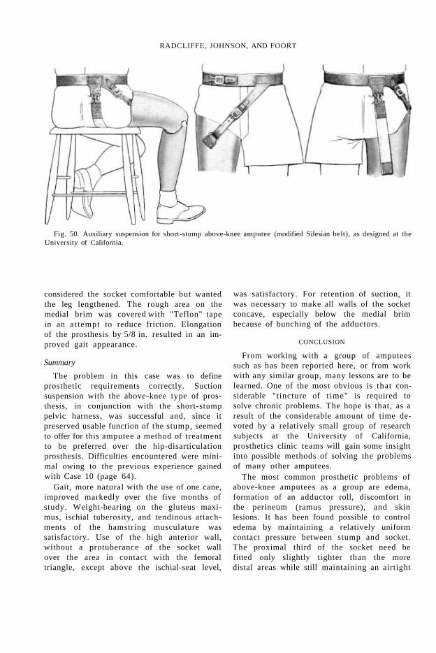
RADCLIFFE, JOHNSON, AND FOORT
Fig. 50. Auxiliary suspension for short-stump above-knee amputee (modified Silesian belt), as designed at the University of California.
considered the socket comfortable but wanted the leg lengthened. The rough area on the medial brim was covered with "Teflon" tape in an at tempt to reduce friction. Elongation of the prosthesis by 5/8 in. resulted in an improved gait appearance.
Summary
The problem in this case was to define prosthetic requirements correctly. Suction suspension with the above-knee type of prosthesis, in conjunction with the short-stump pelvic harness, was successful and, since it preserved usable function of the stump, seemed to offer for this amputee a method of treatment to be preferred over the hip-disarticulation prosthesis. Difficulties encountered were minimal owing to the previous experience gained with Case 10 (page 64).
Gait, more natural with the use of one cane, improved markedly over the five months of study. Weight-bearing on the gluteus maxi-mus, ischial tuberosity, and tendinous attachments of the hamstring musculature was satisfactory. Use of the high anterior wall, without a protuberance of the socket wall over the area in contact with the femoral triangle, except above the ischial-seat level,
was satisfactory. For retention of suction, it was necessary to make all walls of the socket concave, especially below the medial brim because of bunching of the adductors.
CONCLUSION
From working with a group of amputees such as has been reported here, or from work with any similar group, many lessons are to be learned. One of the most obvious is that considerable "tincture of t ime" is required to solve chronic problems. The hope is that, as a result of the considerable amount of time devoted by a relatively small group of research subjects at the University of California, prosthetics clinic teams will gain some insight into possible methods of solving the problems of many other amputees.
The most common prosthetic problems of above-knee amputees as a group are edema, formation of an adductor roll, discomfort in the perineum (ramus pressure), and skin lesions. It has been found possible to control edema by maintaining a relatively uniform contact pressure between stump and socket. The proximal third of the socket need be fitted only slightly tighter than the more distal areas while still maintaining an airtight

SOME ABOVE-KNEE CASES
seal. In the case of an edematous stump, it is important to recognize the necessity for skillful application of socket liners to maintain a functional socket fit as the edema is reduced. Such liners are usually applied along the anterior and lateral walls only.
The adductor roll which typically occurs with plug fit requires considerable time before the change can be made to an efficient, well-fitting suction socket. Almost without exception, such a condition requires a second socket to complete the fitting.
Ramus pressure is a thing of the past. Use of the higher anterior brim and the proper anteroposterior dimension from Scarpa's triangle to the posterior brim will eliminate completely this most troublesome complaint of above-knee amputees. No longer need the above-knee amputee suffer in silence because he just naturally expects his leg to be uncomfortable in this area.
Provision of an efficient supporting surface along the posterior brim of the socket and of a proper fitting of the lateral wall of the socket to provide femur stabilization will relieve common areas of skin irritation, such as at the anterior brim, at the ischial seat, at the medial brim, and at the lateral-distal end of the stump. The most common sources of skin difficulties are poor stump hygiene or rubbing and abrasion. Abrasion can be minimized by a functional fitting of the socket.
ACKNOWLEDGMENTS
The group that worked on the patients presented in this article comprises in fact the whole staff of the Lower-Extremity Amputee Research Project. Even more than amputee treatment must be, this study has been the product of many minds and many labors. Particular mention should be made of the staff members who were especially concerned in the study of the patients here reported. Dr. Henry E. Loon, Coordinator of the Medical Division, served as medical consultant to the group through much of the work. Jack C. Bates, Prosthetic Specialist, served in the vital function of trainer, in addition to making all arrangements with patients. And Jim C. McKennon and William H. Hoskinson served
as prosthetists and faced the task of interpreting the plans of the research group as fully as possible to the patients in the fitting room.
LITERATURE CITED
1. Eberhart, Howard D., and Jim C. McKennon, Suction-socket suspension of the above-knee prosthesis, Chapter 20 in Klopsteg and Wilson's Human limbs and their substitutes, McGraw-Hill, New York, 1954.
2. Foort, J., and C. W. Radcliffe, The Canadian type hip disarticulation prosthesis, University of California (Berkeley), Prosthetic Devices Research Project, [Report to the] Prosthetics Research Board, National Research Council, March 1956.
3. Radcliffe, C. W., Use of the adjustable knee and alignment jig for the alignment of above knee prostheses, University of California (Berkeley), Prosthetic Devices Research Project, Report to the Advisory Committee on Artificial Limbs, National Research Council, August 1951.
4. Radcliffe, Charles W., Mechanical aids for alignment of lower-extremity prostheses, Artificial Limbs, May 1954.
5. Radcliffe, Charles W., Alignment of the above-knee artificial leg, Chapter 21 in Klopsteg and Wilson's Human limbs and their substitutes, McGraw-Hill, New York, 1954.
6. Radcliffe, Charles W., Functional considerations in the fitting of above-knee prostheses, Artificial Limbs, January 1955.
7. University of California (Berkeley), Prosthetic Devices Research Project, [Report to the] Committee on Artificial Limbs, National Research Council, The suction socket above-knee artificial leg, revised edition, April 1948.
8. University of California (Berkeley), Prosthetic Devices Research Project, [Report to the] Advisory Committee on Artificial Limbs, National Research Council, The suction socket above-knee artificial leg, 3rd edition, April 1949.
9. University of California (Berkeley), Prosthetic Devices Research Project, [Report to the] Advisory Committee on Artificial Limbs, Functional considerations in fitting and alignment of the suction socket prosthesis, March 1952.
10. University of California (Berkeley), Prosthetic Devices Research Project, [Report to the] Advisory Committee on Artificial Limbs, National Research Council, Functional considerations in the fitting and alignment of the suction socket above-knee prosthesis, 2nd edition, August 1953.
11. Wagner, Edmond M., Contributions of the lower-extremity prosthetics program, Artificial Limbs, May 1954.
12. Wagner, Edmond M., and John G. Catranis, New developments in lower-extremity prostheses, Chapter 17 in Klopsteg and Wilson's Human limbs and their substitutes, McGraw-Hill, New York, 1954. See especially pp. 497 and 547.

The Management of the Nonfunctional Hand—Reconstruction vs. Prosthesis
STERLING BUNNELL, M.D.1
AN THE course of routine practice, the orthopedic surgeon is frequently confronted with the task of dealing with hands that are damaged by trauma or disease or that are otherwise nonfunctional owing to any of a variety of causes. In all such cases, he is called upon to decide whether or not to undertake amputation of parts of the hand or amputation through the wrist, with the expectation of later applying a suitable prosthesis, or whether, with the prospect of long-continued treatment and the possibility of ultimate failure, to attempt surgical construction of a functional hand from such anatomical elements as can be saved. The considerations involved are many and varied, and rarely do two cases resemble each other in more than a remote way. Each individual case must therefore be evaluated on the basis of its own merits.
There has been in the past dozen years a great advancement in the development of hand prostheses, so that in the case of major hand problems one might be inclined to choose wrist disarticulation over attempts at surgical reconstruction. But during the same period surgical reconstruction also has advanced remarkably, so that in judging any individual case there should be a careful analysis as to which procedure is the better to follow. Doing so usually results in a sort of compromise— reconstruction, if reasonably possible, being chosen first, a prosthesis being applied when proven necessary, major amputation being considered only as a last resort. It is the purpose here to attempt to extract from many years of clinical experience with hand surgery
1 516 Sutter Street, San Francisco 2, Calif.
certain general principles that may offer guidance in making the choice. Generally, the current rule of "save all length possible," now applicable at most other levels of amputation, is applicable in the case of damaged hands also.
The fundamental difference between a reconstructed hand and any present-day hand prosthesis lies in the absence of direct sensation in the latter. Although the wearer of a modern hook or artificial hand may receive indirect sensory impulses through shoulder harness or cineplastic muscle pin, the conventional arrangement constitutes only a crude and inefficient signal system which must be supplemented and directed by sight. A hand prosthesis is of little use in the dark. In contrast, there is the exquisite appreciation we receive from the normal hand by feeling. By light touch, coarse touch, response to heat or cold, and compass-point discrimination, we appreciate texture, and by muscle, joint, and tendon sense we appreciate size and shape. By combining these sense impressions in our cerebral cortex in the opposite parietal lobe, we can identify from memory an object held in the hand. This is stereognosis, a phenomenon replaced by no artificial hand now available. To quote Kirk (6), "No hand is so badly crippled that, if it is painless, has sensation, and strong prehension, it is [not] far better than any prosthesis." This being the case, it is generally desirable to preserve any and all hand structures that can reasonably be counted on to have adequate nerve and blood supply. Eventual application of a prosthesis may or may not be indicated, depending upon individual circumstances and the particular demands of occupation.

NONFUNCTIONAL HANDS
Before considering any hand amputation, then, one should weigh well the possibility of surgical reconstruction, especially with the idea of restoring natural sensation and strong prehension. Whenever reasonably feasible, surgical reconstruction of a damaged hand or arm should be attempted first. Often the result will be such that a prosthesis will not be necessary. In any case, a reconstructed hand stump is apt to be much better adapted to application of a prosthesis. As a matter of fact, reconstruction and use of a prosthesis are so interrelated that they should be considered together in each individual case. Every useful part of a limb, and every bit of skin that has sensation, should be preserved, thus giving more useful material for reconstruction and, finally, for the fitting, if necessary, of a prosthesis.
Reconstruction may often be done in one operation; in other cases multiple operations are required over a period of a month to a year. But considering that the goal is to provide a useful hand for the remainder of an individual's life, it seems worth while. Nevertheless, it should not be undertaken unless there is reasonable assurance that a good practical result can be obtained.
METHODS OF SURGICAL RECONSTRUCTION
Although the hand does the work, the arm places and innervates the hand. Accordingly, if any particular hand is to be truly useful, it is necessary to have good shoulder, elbow, and wrist function and also good pronation and supination—half furnished by the shoulder and half below the elbow. Because they supply the hand, the nerves of the arm are particularly important. In the hand itself there should be a good quality of sensation as well as mobile units that can work against each other with at least a pinch grasp or hook action to simulate normal prehension.
Hands coming in for repair usually evidence partial amputations, stiffening in the position of nonfunction, flexion contracture from scar formation, malalignment of bones, loss of motion from injury to tendons and nerves, loss of sensation from injury to nerves, ischemic contracture, or painful states from vasomotor causes or from tender neuromata. Usually the
surgeon's problem is composite, dealing with cover, joints, bones, nerves, and tendons.
For each of these conditions there is much that can be done surgically (2). For partial amputation, clefts between digits may be deepened, and digits can be built out and made to appose each other. Tender stumps may be corrected. For stiffening in the position of nonfunction, the joints may gradually be drawn around to the position of function by spring or elastic splinting and can be mobilized surgically. Scar tissue of flexion contracture can be replaced by good pliable skin giving good cover and improving nutrition. Malalignment of bones may be corrected so that the mechanics of tendon action are correct. Substitute thumbs may be formed. Tendons and nerves may be repaired or transferred, or new ones may be furnished. Ischemic contracture can be relieved so that a hand thus affected can regain some function. Painful states may be corrected by sympathectomy, and tender neuromata may be removed.
PARTIAL AMPUTATION"
Arm stumps resulting from amputation through the wrist or through the carpometacarpal joint, or those without the thumb and with amputation through the metacarpals or proximal phalanges, require a prosthesis (Fig. 1). Hands retaining a good thumb working against one or more fingers (as in Figure 2), or even against a surgically constructed post (as in Figure 3), do not. Sometimes the usefulness of a sound thumb may be much enhanced by surgical procedures conducted on other remaining hand parts (as for example in Figure 4). Other partial hands (like those shown in Figures 5 and 6 for example) when reconstructed usually are more functional than a prosthesis. Some with a partial hand amputation use remnants of the hand for fine work and a prosthesis for heavy work.
In partial amputations it is best, if possible, to retain the metacarpal heads and hence the full width of the palm for firm grasp of tools, but the metacarpal head of an index or of a little finger that has been amputated through the metacarpophalangeal joint is best beveled off so that it will not snag on entering a pocket. The metacarpal of an index or little

BUNNELL
Fig. 1. Levels of hand amputation requiring prosthesis. A, Wrist disarticulation, including removal of the distal prominences of radius and ulna; B, amputation through the carpometacarpal joint; C, transmetacarpal amputation; D, amputation through all proximal phalanges In B, some useful wrist motion may be retained. In C, hand remnant may be used as a wrist motor to power a prosthesis or simply to point one. In D, hand stump may be made to work against some prosthetic device, residual sensation offering a substantial advantage over A, B, or C.
Fig. 2. Examples of partial hands requiring no prosthesis. When the thumb can work against one or more fingers, function usually is better than can be obtained with a hand substitute.
Fig. 3. Partial hand (A) and result of reconstruction (B), no prosthesis needed. When, in the absence of all the fingers and much of the palm, a good thumb remains, it is possible, by means of pedicle and bone graft, to build up a post for the thumb to appose. Function thus obtained is likely to be better than that to be had from a hand substitute.
finger off through the shaft is best removed obliquely at its base (Fig. 7). The interosseous muscle is then transferred to the adjoining digit to give abduction.
A hand amputated through all metacarpophalangeal joints or proximal phalanges may
be improved by mobilizing the fifth metacarpal, cutting the transverse metacarpal ligament, and perhaps removing the metacarpal of the ring finger and covering the cleft by a plastic maneuver (Fig. 4). The ulnar side of the hand thus becomes a movable

NONFUNCTIONAL HANDS
Fig. 4. Case M. S. Fingers lost between a sprocket and chain. Excised the tender neuromata of the stumps. Undermined and drew skin down for better coverage. Excised metacarpal of ring finger, covering sides of new digit by plastic maneuvers, in order to give more mobility (2 in.) to the metacarpal of the little finger. Deepened thumb cleft by Z-plasty (Fig. 21, page 86). The patient obtained a strong and useful grasp between the thumb, the phalangized index and long "fingers," and the little "finger." From Bunnell, Surgery of the Hand, 3rd ed , Lippincott, Philadelphia, 1956, by permission.
part. Motion may be increased as much as 2 in. If the second and fourth metacarpals are deleted, there will remain three digits, consisting of the metacarpals of the thumb and of the long and little fingers, and the thumb cleft will be wide and deep. Phalangizing the metacarpals gives considerable useful mobility so that one can dress oneself, use knife and fork, and so forth. The metacarpals of the thumb and little finger are cut across at the base and bent toward each other for better grasp (Fig. 8). A similar osteotomy may be performed on a hand having only two remaining digits, as for example thumb and little finger (Fig. 9), or even when only one complete digit remains, as in Figure 10.
Frequently a finger or hand stump is so hypersensitive from poor terminal padding and sensitive neuromata that it prevents all of the remaining parts of the hand from functioning. Crushing injuries to fingers present the most difficulty because, in such cases, the fingers usually have been damaged well proximal to the site of amputation. In revising such stumps, the digits must often be shortened enough to give good, well-padded cover, but it is possible to swing a visor flap from the dorsum over the end of the stump and then to skin-graft the dorsum. Still another possibility of furnishing good tactile cover over the end involves use of a cross-finger flap and then skin-grafting the back of the donor finger. Nerves in hands and fingers have a special tendency to proliferate. If they terminate in scar tissue or close under the skin, the neuromata formed may be extremely sensitive and give, on slight tapping, the sensation of an electric shock. These are corrected by uncovering the nerve, dissecting it well back, and cutting it off in
good tissue free from scar. Neither alcohol injection nor ligation is used.
STIFFENING IN THE POSITION OF NONFUNCTION
Following injury, infection, or paralysis, a hand frequently stiffens in the position of non-

Fig. 5. Case P. L. Hand caught between two rollers. Debrided and skin-grafted. Later, pedicle flap applied, then interdigitation. Sensation gradually returned throughout. A useful hand was obtained. From Bunnell, Surgery of the Hand, 3rd ed , lippincott, Philadelphia, 1956, by permission.
Fig. 6. Case B. P. Partial amputation by power saw. Split-grafted next day. Pedicle graft applied and thumb cleft deepened. Index metacarpal removed for wider cleft. Rotary osteotomy done on all metacarpals for better apposition. Pinning with Kirschner wires. A good "hand," with good prehension, was obtained. From Bunnell, Surgery of the Hand, 3rd ed., Lippincott, Philadelphia, 1956. by permission.
function so that the digits can no longer touch each other and the hand is therefore useless. In the position of function (Fig. 11), the wrist is extended 35 cleg., the joints of the fingers are moderately flexed, and the thumb is in moderate apposition, as in holding a baseball. In the position of nonfunction (Fig. 12), the wrist is flexed, the metacarpophalangeal joints are hyperextended, the remaining finger joints are flexed, and the thumb is at the side of the hand or even back of it. Al-

NONFUNCTIONAL HANDS
though such a hand is totally useless, in general it should not be amputated. For if the joints can be pushed around into the position of function, the available motion will be useful for picking up and holding objects, and the hand will be used more and more from then on.
The first approach to hands stiffened in the position of nonfunction involves use of a system of elastic or spring splinting by which joints can gradually be drawn around into positions of function. Usually the joints are kept active and are not damaged, and the muscles and all tissues are activated, a matter which greatly improves their condition. If, however, the response to such treatment is unsatisfactory, surgical means are resorted to, starting with capsulectomies (Fig. 13) and,
Fig. 7. Typical example of loss of fourth and fifth rays through the shafts of the metacarpals. In such a case, it is best to delete the stub of the fifth metacarpal and round the stub of the fourth. A corresponding procedure is advisable in the event of loss of the second digit, or of the second and third digits, by transmetacarpal amputation.
Fig. 8. Reconstruction procedure for loss of all digits through the metacarpophalangeal joints. A, Second and fourth metacarpals are deleted, clefts are covered by-plastic maneuver, first and fifth metacarpals are osteot-omized. B, Functional three-finger "hand" results
Fig. 9. Angulatory osteotomy of first and fifth metacarpals to aid apposition of thumb and little finger. Sometimes it is necessary to effect a tendon T transfer also. See Figure 31, page 91.
where there is damage to bone structure, resorting to arthroplasties.
C a p s u l e c t o m i e s are usually performed on the meta-c a r p o p h a l a n g e a l joints but sometimes also on the proximal interphalangeal joints. Usually the trouble is found to lie in the fact that the two collateral ligaments are too short and thick to permit the joint to flex. Excision of these structures makes flexion possible. Often it is necessary also to free the long extensor tendons (Fig. 14) and to clean out the volar pouch of the joint. In performing an arthroplasty, the metacarpal head is shortened and reshaped, and a hood of fascia is fastened over it.
Arthroplasty is not often done on the wrist joint; arthrodesis is used instead. In many cases, however, removal of a mass of scar tissue from the volar aspect of the wrist allows the wrist to extend. When pronation and supination are retained, arthrodesis of the wrist or of the proximal finger joints into the position of function gives very little disability (Fig. 15).
FLEXION CONTRACTURES AND FURNISHING NEW
COVER
Most reconstruction commences with excision of a big plaque of scar tissue that is
Fig. 10. Angulatory osteotomy of first and fifth metacarpals to bring thumb and ulnar side of palm into easy apposition. Tendon T transfer may be needed here also (Fig. 31, page 91).

BUNNELL
Fig. 11. The position of function.
Fig. 12. The position of nonfunction.
Fig. 13. Case E. T. Top, preoperative position of nonfunction from shark bite on upper arm, severing nerves and vessels. Bottom, correction to a position of function by fusion of the wrist, capsulectomies and opening of the cleft of the thumb, and transfer of the extensors of the wrist to the flexors of the fingers. A tendon transfer through a pulley constructed at the pisiform was used to give apposition to the thumb. No prosthesis needed. From Bunnell, Surgery of the Hand, 3rd ed , Lippincott, Philadelphia, 1956, by permission.
drawing the hand into flexion contracture and strangling the rest of the tissue (Figs. 16 and 17). The skin is then undermined and allowed to retract, thus freeing the hand for better nutrition. New cover is then provided, sometimes by a free graft but usually by a pedicle graft from the abdomen (Fig. 18), thus giving good, pliable skin with a layer of soft fat beneath. Doing so releases the whole hand and makes it possible to reconstruct the deeper parts—joints, bones, tendons, and nerves. Although the refinements of stereognosis never return to such skin, eventually sensation to light touch and pin prick develops.
SKELETAL MALALIGNMENT
The bones of the hand constitute the framework along which the muscles and tendons function in their proper planes. The joints allow the digits to flex and extend in their proper positions for adequate grasp. After fracture, bones often unite at such odd angles that the whole mechanics are thrown out of true. If, after healing, there is an angle of the bones along the length of the limb, the tendons over the convexity will be tight, over the concavity loose. Such a circumstance upsets the whole nicely adjusted muscle balance so that the joints are pulled into deformity all the way from the site of angulation to the end of the limb. To make the hand function properly again, realignment is necessary. The bones are chiseled or sawed across, a wedge being removed when necessary to place them in proper contact and alignment. They are then pinned so by Kirschner wires, the latter being withdrawn in two months when union is solid and the framework of the hand is restored.
When the thumb does not entirely contact the ring finger or the little finger, the metacarpal of either or both may be severed at the base and the digits angulated toward each other in such a way as to provide for easy contact. Similarly, in the absence of a thumb, two or more fingers may be angulated and rotated to give them the ability to work against each other.
When a metacarpal, including the soft tissues about it (tendons, nerves, interosseous muscles, and skin), is badly damaged, it may be excised. If it is one of the central rays, the

NONFUNCTIONAL HANDS
Fig. 14. Case J. D. Left, old dislocation of metacarpals on carpus, upsetting muscle balance, thus resulting in the useless position of nonfunction. Right, dislocation reduced, restoring muscle balance in the position of function. A pedicle graft was applied to the dorsum of the hand and to the open thumb cleft. Freeing of the extensor tendons, together with capsulectomies, allowed the proximal finger joints to flex. No prosthesis needed. From Bunnell, Surgery of the Hand, 3rd ed., Lippincott, Philadelphia, 1956, by permission
the proximal joints. It is done on the wrist and can be done on the elbow. In the latter case, the choice must be made between arthrodesis, a block operation, muscle transfers, or the wearing of a prosthesis to activate a flail elbow. When the arm cannot be abducted at the shoulder but when muscles around the scapula are good, arthrodesis of the shoulder will allow the arm to position the hand for useful function (Fig. 20).
THUMB PROBLEMS
metacarpal of the adjoining ray, either index or little, as the case may be, is cut across at its base, jogged over to the base of the excised metacarpal, and pinned near and parallel to the next ray (Fig. 19).
When a metacarpal head is missing, the lack of support causes the adjoining metacarpals to rotate so that the fingers cross on flexion. In such a case, the metacarpal can be excised and one of the adjacent ones jogged over. Or the proximal phalanx of the ray in question can be recessed, or set back, so that its head will take the place of the missing metacarpal head.
Often it is advisable to arthrodese a joint to place it rigidly in the position of function. This procedure can be carried out on either of the two distal joints of the fingers but rarely on
Fig 15 Case A B Hand useless from birth pals)". Several operations had been performed, including fusion of the wrist. The proximal finger joints were lax and bent backward out of use Patient could not abduct at the shoulder, and the forearm was in supination. The shoulder was arthrodesed lo enable placement of the hand, and by osteotomy the ulna was rotated into pronation. The proximal finger joints were arthrodesed into the position of function. Patient gained much use of the hand and became self-supporting. From Bunnell, Surgery of the Hand, 3rd ed , Lippincott, Philadelphia, 1956, by permission.
So essential to prehension is the thumb that every possible bit of an injured one should be saved. Amputation of the thumb through the metacarpophalangeal joint results
in a partial digit almost loo short to be useful, but a new thumb cleft can easily be made by a Z-plasty operation (Fig. 21), meanwhile scraping the adductor origin down from the third metacarpal. The thumb is thus made relatively longer. If the shaft of the index metacarpal

BUNNELL
projects into the web so as to interfere with grasping, it should be excised at its base to widen and deepen the cleft (Figs. 22 and 23). Whenever possible, the tip of the third metacarpal should be preserved to provide a concave palm for the remnant of the thumb to work against (Fig. 22). Preservation of the broad tip of the third metacarpal is particularly desirable when a complete thumb remains (Fig. 24).
The range of motion of a normal thumb extends from a position at the side and slightly
Fig. 17. Case D C. M. Hand severely burned in oil fire so that all digits pointed backward out of use. Fingers were webbed, and middle joints were exposed. There was no thumb cleft, the thumb being at the rear of the hand with the metacarpal arch reversed. In this position of nonfunction, the hand was entirely useless Excised all dorsal skin, including nails Sawed away exposed bone. Corrected webs. Established thumb cleft and positioned thumb. Positioned fingers by capsulectomies. Covered all with free skin graft. Patient returned to his job as locomotive engineer No prosthesis needed. From Bunnell, Surgery of the Hand, 3rd ed., Lippincott, Philadelphia. 1956, by permission.
Fig. 16. Case J. M. From birth the cicatrix from a tear at the ulnar side of the wrist so distorted the growth of the hand that there was no function. The scar was excised, the ulna elongated, and a pedicle applied. Two years later osteotomies were done on all metacarpals, the thumb cleft was deepened, and a pulley operation was performed to improve apposition. Three years later the hand was reported to be quite useful. From Bunnell, Surgery of the Hand, 3rd ed Lippincott, Philadelphia, 1956, by permission.
back of the hand, with the nail at right angles to the palm, through a wide ellipse toward the volar aspect until it is opposite the fingers, the nail being then parallel to the palm. In the latter position, the thumb is available to participate with the fingers in grasping large objects. The motion is effected by the ten muscles—long and short—that control the thumb. In paralysis of the median nerve, in injury to the thenar muscles, in stiffness of the carpometacarpal joint of the thumb, or in flexion contracture on the dorsum of the web,

NONFUNCTIONAL HANDS
Fig. 18. Case A. C. Hand badly crushed between rollers. Poor skin surface, position of nonfunction, entire hand and joints stiff, extensor tendons adherent, thumb at side, amputation contemplated. First operation: excised all skin from both dorsal and volar surfaces, covered with one large pedicle graft, and spread thumb from hand; brought joints around by elastic splints. Second operation: freed extensor tendons and placed fat beneath; did capsulectomies on proximal joints; used sublimis of long finger for apposition; freed flexor tendons, placing fat beneath; defatted pedicle. The hand made remarkable recovery in nourishment, function, and position. There was good grasping power and a complete change in the morale of the patient. No prosthesis needed. From Bunnell, Surgery of the Hand, 3rd ed , Lippincott, Philadelphia, 1956, by permission.
normal range of motion of the thumb is lost. If the other parts of the hand are mobile, the ability to appose the thumb can readily be provided by a simple tendon transfer that draws the thumb toward the pisiform bone and pronates it. When this is not possible, the thumb may be held permanently in a useful position by a bone graft at the base of the first metacarpal.
When a thumb is closely bound to the rest of the hand by scar, it can be spread away by excising the scar tissue and cutting across the cleft from a point opposite the hinge of the first two metacarpals on the dorsal side to the corresponding point on the volar side. The thumb is spread to the side and front of the hand, and the large denudation of skin is covered either by a large diamond-shaped free skin graft or, better, by a pedicle graft from the abdomen. In three weeks, pedicle grafts are detached from the abdomen and laid smoothly on the hand.
Although the thumb stump remaining after amputation through the m e t a c a r p o p h a l a n g e a l joint usually is not very serviceable, it may be built out by pedicle and bone graft. If a thumb is amputated proximal to the metacarpophalangeal joint, it should in any case be built out longer. If the thenar muscles and the stub of
Fig. 19. Reconstruction procedure recommended in event of serious damage to (A) the fourth digit or to (B) the third digit. In A, delete the much-injured fourth ray and jog fifth ray over to its place. In B, delete the much-injured third ray and jog second ray over to its place. The result in either case is a functional four-digit hand.

BUNNELL
Fig 20. Case L. M. W. As a result of polio, arm was flail at shoulder, and there were no flexors in the hand Arthrodesed shoulder and wrist simultaneously so that the patient could place the hand. Transferred extensor carpi radialis to flex fingers, palmaris longus to abduct thumb, the long extensor of the ring finger for apposition. Slit the proximal pulleys so that long flexors could flex the proximal joints. Patient gained much use of hand, was able to grasp a piece of paper or a tumbler, could place the hand well, and occupied a position in a bank. No prosthesis needed. From Bunnell, Surgery of the Hand, 3rd ed., Lippincott, Philadelphia, 1956, by permission.
the metacarpal remain intact, the thumb will be quite movable. A short thumb is a good thumb. Various motions, such as apposition, extension, and flexion, may be furnished it by tendon grafts.
In the case of total loss of the thumb, a new one can be supplied in various ways. The simplest approach is to raise a tube pedicle from the abdomen, attach the pedicle to the hand, and place in it a bone graft from the iliac crest (Figs. 25 and 26). Although this expedient gives sensation, it does not provide much stereognosis. Nevertheless, a reconstructed thumb is apt to be very serviceable and considerably better than a prosthesis.
Fig. 21. Phalangization of thumb cleft by Z-plasty. Left, hand with short and more or less useless thumb stump. Middle, location of Z-shaped incision; flap A is carried to fixed point X, flap B to fixed point Y, so that dorsal flap just covers defect on volar side while volar flap just covers defect on dorsal side; resulting suture line is as shown in insert. Right, end-result, showing deepened thumb cleft.

NONFUNCTIONAL HANDS
Fig. 22. Case C, H. Amputation by meat grinder. Thumb cleft deepened by Z-plasty. Index metacarpal removed to give good grasp. From Bunnell, Surgery of the Hand, 3rd ed.. Lippincott, Philadelphia, 1956, by permission.
The graft should be grounded on some other bone rather than connected by a joint. It may be placed on the carpus to make a pad in the base of the palm, or it may be placed on the trapezium or on the stub of the metacarpal.
The requirements of a new thumb are three in number—motion, sensation, and proper placement. The best new thumbs are made by pollicization of a finger, preferably the index linger but sometimes the long finger. Often, as part of the injury, the index finger is already somewhat shortened. In such a case, the
Fig. 23. Case H. G. Amputation, by power saw, of all digits through proximal phalanges, leaving a mitten hand but no thumb cleft By a plastic maneuver and removal of the index metacarpal, a thumb cleft 3/4 in. deep was constructed. It opened 3/4 in and closed against the hand. Patient could write and hold objects Limited facility can be combined with the use of a prosthesis. From Bunnell, Surgery of the Hand, 3rd ed., Lippincott, Philadelphia, 1956, by permission.
linger, or a portion of suitable length, is transferred together with a bridge of skin and with its nerves, blood vessels, and tendons intact (Figs. 27 and 28). It may even be transferred on a neurovascular pedicle circumscribing the skin all around (Fig. 29). When this
Fig. 24. Operative procedure for loss of the second and third digits. Excision of the second metacarpal, but with retention of the third, furnishes easy apposition for the sound thumb.

Fig. 25. Surgical construction of a new "thumb." A, Poorly functioning partial hand retaining digits four and five only. B, Serviceable partial hand made by constructing new "thumb" with pedicle and bone graft. Function is apt to be better than if a prosthesis were applied.
procedure is possible, it makes for easy and exact placement. The tendons are brought over with the new " thumb" and joined up so as to give motion. The fingers should work directly against the new " thumb" and also, by their side motion, should pass to the side of it and close against the palm. Stereognosis and vascularization are provided by the neurovascular pedicle.
Should a newly constructed thumb not have sensation in its tactile area, a flap of skin may be exchanged for the nontactile skin by a Z-plasty. Or tactile skin can be furnished by using a neurovascular pedicle passed beneath the skin at the base of the thumb. A living thumb, with motion, sensation, and proper positioning, is, of course, far superior to any prosthetic thumb.
TENDON REPAIR
Tendons are frequently lacerated, thus losing their function of transmitting muscle power to provide motion in joints. They can, however, readily be repaired (Fig. 30), the most difficult cases being the flexor tendons in
Fig. 26. Case C. B. Injury from hand grenade. Pedicle graft covered the thumb, and arthrodesis was done on the trapezium by a graft from the ilium Abduction was furnished index finger by a proprius-tendon graft A very useful hand resulted. No prosthesis needed. From Bunnell, Surgery of the Hand, 3rd ed., Lippincott, Philadelphia, 1956, by permission.

NONFUNCTIONAL HANDS
Fig 27. Pollicization of index finger,
the digits and in the distal part of the palm, where the resulting juncture tends to adhere to the surrounding parts. Frequently a tendon graft must be used to bridge the tendon over areas where adhesions are likely to form. Adherent tendons may be freed, and slippery material, such as paratenon and fascia, may be grafted between them and the bones so as
to allow the tendons to glide again. Defects in tendons are readily bridged by free tendon grafts from spare tendons in other parts.
The upper limb interdigitates at the ends of the metacarpals, and the tendons normally have individual motion. If either an extensor or a flexor tendon is sutured over a finger stump, it will hold back all of the tendons pulled from the same muscle. But when all of the tendons are cut at the end of a
Fig. 29. Pollicization of index finger by neurovascular pedicle. Skin is circumscribed, and the index finger is pinned on to the stub of the metacarpal of the thumb in proper position. Tendons furnish motion, vessels nutrition, and nerves sensation.
Fig. 28. Case H. W. W. First (1929) physiological reconstruction of thumb by pollicizing remains of the index finger. Metacarpal lashed to trapezium, nerves and vessels carried over, and all tendons and muscles connected up. "Thumb" had strong motion and normal sensation and was well positioned. Patient worked well as a carpenter for 20 years. Superior to prosthesis From Surgery, Gynecology, and Obstetrics, by permission.
carpal or metacarpal stump, they should all be sutured together over the end to provide for movement of the stump.
Isolated digits may be made to provide prehension if they are furnished with new flexor and extensor tendons. To make the fingers appose each other, the tendons can be placed diagonally across the hand, or a tendon T transfer, which consists of one cross-bar tendon from digit to digit and a longitudinal one looped about the first, can be made (Fig. 31). When the muscle concerned is contracted, the " T " assumes the shape of a "Y ," and the two digits are drawn toward each other. This procedure is particularly useful in median and ulnar paralysis, where it will provide adduction of the thumb and little finger while curving the metacarpal arch of the palm. When some digits have been amputated, great strength can be given to the remaining fingers by transferring in the forearm the

BUNNELL
tendons of the amputated ones to those of the remaining ones.
Especially in paralysis are tendon transfers useful. Good, strong muscle and tendon are transferred to the tendons of the paralyzed muscles. This operation may be performed, without fusing the wrist, to give very good return of function so that splints are discarded. In the case of any two nerves paralyzed high in the arm, the wrist can first be arthrodesed in the position of function, an expedient which results in very little disability. Thereupon the five tendons previously wrist movers become available as digit movers, and the resulting motion is more natural than that obtained
using a prosthesis. The patient soon learns to adapt so that the motion becomes natural. A rule is to decide what movements are needed and then to consider the number of muscles available for transfer. For paralysis within the hand—that is, from the median and ulnar nerves—many transfers are available to restore muscle balance, thus correcting the position of the claw hand by substituting for the paralyzed intrinsic muscles.
Another principle is tenodesis, a procedure in which the tendons that move the digits are fastened to the forearm bones. Then, when the wrist is flexed, the extensor tendons tighten and extend the digits; when it is extended, the flexor tendons tighten and cause the digits to flex so that thumb and fingers appose each other. These automatic movements are useful when only one or two strong muscles are available. When no muscles are available, the hand can be converted to a useful hook by tenodesis of the flexor tendons to the forearm bones.
NERVES
Movement and sensation in the hand, which are its two most important functions and which are of equal value, depend entirely upon the nerves. The three large nerves that course down the arm (the ulnar nerve, the median nerve, and the radial nerve) control the hand, and any injury to them is as damaging to the hand as is an injury to the hand itself. When a nerve is severed, it should be
Fig. 30. Case F. E. Charge from a shotgun entered palm and emerged dorsally, shattering the carpus and the lower radius and severing many tendons, extensors of the wrist, thumb, and fingers, and the median nerve. Debrided, filetted the index finger, and skin-grafted. Considerable infection followed. First operation: excised scar and placed a pedicle. Second operation: furnished tendon grafts plus paratenon to extend thumb and fingers; freed the flexor tendon of the thumb; did a pulley operation for apposition; sutured median nerve to its four branches. The wrist became fused. But sensation, motion, and apposition returned, so that there resulted a very useful hand requiring no prosthesis. From Bunnell, Surgery of the Hand, 3rd ed. Lippincott, Philadelphia, 1956, by permission,

NONFUNCTIONAL HANDS
A B Fig. 31. Lobster hand formed by tendon T transfer.
A, Arrangement of tendons to form the "T ." B, Contraction of the longitudinal tendon converts the "T" to a "Y" and thus effects apposition of thumb and little finger.
rejoined at once. Otherwise fibrous degeneration in both the lower portion of the nerve and in the muscles supplied by it will be so progressive that, after two years, muscle action will not return and, after five years, neither will sensation. A gap of several inches can be overcome and the nerve sutured directly. Even the little nerves in the hand itself can be repaired.
After nerve suture, there is about 80 percent of functional recovery. Nerves can be sutured directly, transferred, or even free-grafted. All of these procedures are successful, but nerve grafts must be used from the same person; if
grafted from another person, they will melt away. From loss of nerve supply, the hand if neglected goes into the position of nonfunction, stiffens, and atrophies. Splinting should be by spring or elastic splints sufficient just to substitute for the paralyzed muscles and to hold the hand in the position of function so it can work. When the nerves are irreparable, as for example when too great an interval has elapsed since the time of injury, muscle function in the hand can be provided by tendon transfers. Paralysis in the hand and forearm from ischemic contracture can be overcome to a considerable degree, although never completely cured. In vasomotor disorders, surgery seldom need be weighed against prostheses.
PROSTHESES FOR PARTIAL HANDS
The literature on prostheses for the partial hand is meager, and therefore when a hand is damaged there is a distinct preference on the part of prosthetists to have a wrist disarticulation or a long-below-elbow amputation. In the event they are confronted with a partial hand amputation, many limbfitters prefer to enclose the wrist immobile (as in Figure 32) rather than to construct a partial hand prosthesis. Even those who furnish cosmetic-glove prostheses (as in Figure 33) prefer to enclose the whole hand in the glove and to substitute, for the missing parts, foam filler reinforced with pliable wire. Although a long-below-elbow amputation offers the advantage that many more or less standard terminal devices may be applied (a split hook, a mechanical hand, perhaps some special tools), a partial
Fig. 32. One form of prosthesis for partial hand amputation—thumb free, wrist encased, split hook activated by shoulder harness as in the case of the wrist disarticulation. The disadvantages are numerous. The long cuff virtually eliminates any possibility of wrist motion. Except in the thumb remnant, residual tactile sense is obviated, and the device as a whole is much too long.

BUNNELL
Fig. 33. Cosmetic hand for partial amputation— glovelike and zippered at the wrist. Fingers are filled out by foam filler and stiffened by armature flexible enough to hold any shape. Courtesy Prosthetic Services of San Francisco.
hand, whatever can be saved, can often be fitted with considerably more success. If the thumb alone is spared, a casing over the palm and wrist can support a pad or other suitable device against which the remaining digit can work (Fig. 34). If only the palm, perhaps with a few remnants of phalanges, remains, a casing over the forearm can support a similar pad against which the palm can be pressed by wrist flexion (Fig. 35).
By the combined talents of engineers, physicists, prosthetists, orthopedists, and others, there have been in the last ten years
Fig. 34. Simple prosthesis for loss of all digits except the thumb.
many advances in hand and arm prostheses. Accordingly, there has been developed the policy of saving as much of any limb as is likely to be functional and, particularly, as much of the hand as possible. Any portion of skin with sensation should be preserved because of the possibility of placing it in a functional part. Digits with sensation can do light work and, if necessary, a prosthesis can be applied to do heavy work (as in Figures 36 and 37).
For the wrist-disarticulation, below-elbow, above-elbow, and shoulder-disarticulation prostheses, many new devices have been developed. They include the alternator elbow lock for the above-elbow case (3,5), the outside-locking elbow hinge for elbow disarticulation (1,3,5), the polycentric elbow joint for below-elbow cases (1), the variable-ratio step-up hinge for the very short below-elbow case (1), the flexible cable units to allow pronation and supination for the very long below-elbow and wrist-disarticulation cases (8), and the elbow-coupled shoulder joint for shoulder-disarticulation amputees (7). For the arm amputee, these devices help to carry the terminal device (hook or artificial hand) to a place of usefulness. The Manual of Upper

NONFUNCTIONAL HANDS
Extremity Prosthetics (11) gives a full account of these and other devices that comprise a full armamentarium for upper-extremity amputees. But the case of the partial hand amputation is not included.
PROSTHESES FOR ONE-DIGIT HAXDS
For most practical purposes, loss of one or more distal phalanges does not require application of a prosthesis. Nevertheless, there are exceptions. An accomplished violinist, losing the distal phalanx of even one string finger, for example, is incapable of managing the strings properly. This could mean an occupational change for such a person. A good prosthetic replacement may enable him to continue his occupation. The same occasionally occurs with an organist, a pianist, a typist, or other person in any occupation where finger dexterity means the difference between success and failure. A suitable prosthesis for such a case can be made using thin stainless steel for the socket and extension framework and then dipping the device in flexible vinyl plastic to form the tip cushion and finger build-up. The socket portion may be split along one side to allow it to expand and contract, thus ensuring snugness of fit.
For amputation of all of the fingers at the metacarpophalangeal joint, or approximately half an inch distal thereto such that the volar crease of the metacarpophalangeal joint remains, a 1/8-in. rod framework of stainless steel can simulate the socket while leaving a maximum amount of exposed palm for traction and sensation (Fig. 38). The distal portion of
Fig. 35. Simple prosthesis lor major losses of most of the digits. Wrist serves as motor, hand working against prosthesis. Residual tactile sensation is utilized.
Fig. 36. Partial hand capable of prehension. Top and middle, digital motion for light work. Bottom, wrist motor for heavy work. From Bunnell, Surgery of the Hand, 3rd ed,, Lippincott, Philadelphia, 1956, by permission.

Fig 37. Case E. E. Left, top to bottom: Right hand pulled into hay chopper. Debridement and abdominal pedicle. Later a two-digit hand was made with a tendon T operation for prehension and a spread of 1-1/2 in. Right, top and middle: A prosthesis which enabled the hand to work against a hook. This was discarded because it was too unstable. Right, bottom: A prosthesis made by Robin-Aids Manufacturing Company, Vallejo, Calif., that was very satisfactory. It preserved residual wrist motion and could be removed when fine digital motions were required.
the framework is bent to simulate the finger tips, the little-finger side being curved to form a hook for pulling or lifting and the index side shaped to appose the thumb as would the first two fingers in three-jaw-chuck prehension (4). This arrangement provides for prehension between the simulated index finger and the remaining thumb. A similar appliance can be made for an amputation proximal to the metacarpophalangeal joint, but in such a case the remainder of the hand must be fitted with a plastic, metal, or leather socket for attachment to the formed rod (Fig. 39). The notable

NONFUNCTIONAL HANDS
Fig. 38. Prosthesis for loss of all the fingers at, or slightly distal to, the metacarpophalangeal joint line. Metal ring, covered with vinyl plastic, is so shaped as to furnish one large hook, representing the index finger, and one small one, representing the little finger. Thumb works against ring throughout the range of the carpometacarpal articulation. Courtesy Robin-Aids Manufacturing Company, Vallejo, Calif
Fig. 39. Prosthesis for transmetacarpal amputation. Socket may be of leather, molded plastic, or hammered stainless steel. Metal ring, covered with vinyl plastic, is shaped to simulate fingers, as in Figure 38. Courtesy Robin-Aids Manufacturing Company, Vallejo, Calif.
disadvantage is the coverage of surfaces otherwise capable of sensation. In both instances, the rod framework is dipped in flexible vinyl plastic to provide a surface with adequate traction.
Figure 40 shows a single stainless rod curved in hook fashion and mounted to a stainless-steel plate, which in turn is attached to a molded hand and wrist socket. The hook is so positioned as to give apposition to the thumb, and the thumb is exposed to utilize its capability for sensation. This single hook, being small and smooth, allows easy entry into pockets and other tight places.
Since the thumb is the most important single digit of the hand, it would seem a sound principle not to involve it as a motor for powering other mechanisms. A collar around the thumb would appear to diminish tactile surface, and any mechanical linkage would seem to lessen mobility and dexterity. In general, wrist flexion-extension provides a far more desirable motor with less hindrance to function. But these principles have only general applicability and are not specific. For certain special needs, a thumb-powered mechanism may be desirable. In any individual case, the selection of equipment must be left to the mutual judgment of the patient, the doctor, and the prosthetist. Figures 41 and 42 illustrate the principles involved but show the distinct differences to be found in individual cases.
Fig 40 Simple prosthesis for loss of most of the palm but with retention of the thumb. Wrist and hand stump are encased in a socket to which is attached a single stainless-steel hook. The hook may be used by itself or as a member for apposing the thumb.

BUNNELL
Fig 41. Prosthesis for amputation of all fingers at the metacarpophalangeal joint line with retention of the thumb. Socket about wrist and hand stump supports split hook, which is powered by the thumb. Courtesy Navy Piosthelics Research Laboratory, U.S. Naval Hospital, Oakland, Calif.
In the arrangement shown in Figure 43, the hand, wrist, and forearm socket give versatility for the accomplishment of either light (asks or
Fig 42. Prosthesis for loss of all digits but the thumb. Hinged at and powered by the wrist, this device provides for prehension by virtue of a thrust rod. Courtesy Robin-Aids Manufacturing Company, Vallejo, Calif,
heavy-duty work. For light tasks, the thumb stump is free to appose the remainder of the hand or to contact a small metal post or spoon attached to the hook. The forearm socket allows freedom of wrist motion but provides hook stability for heavy-duty work. Since the thumb stump is also free to appose the hook-activating lever, no shoulder harness is required.
For a hand retaining only the thumb, without fingers or even without their metacarpals, a special prosthesis designed by the United States Navy gives reciprocal motion and active prehension powered by the thumb (Fig. 44). To a simple hand cuff and wrist strap is attached a metal plate, which, on the radial side, supports a lever for the thumb to appose and, on the ulnar side, bears a metal finger pivoted on an axis near the base. Apposition of thumb and metal finger is effected by a linkage between the two lever systems.
PROSTHETIC THUMBS
Figures 45 and 46 illustrate fixed prostheses for partial or complete loss of the thumb. Two features are essential. First, the prosthetic

NONFUNCTIONAL. HANDS
Fig. 43. Amputation of the fingers at the metacarpophalangeal joint line and of the thumb at the inter-phalangeal joint; thumb phalangized for deeper cleft. Top to bottom: holding with thumb unassisted; use of hook (powered in this case by shoulder harness) as device to appose palm; holding with thumb, hook available for auxiliary function if needed; holding with hook, thumb as stabilizer.
thumb must furnish proper apposition to the fingers, and its lip should be of such material as to provide adequate traction. Second, the thumb must provide a shaft and a crotch so as to make it possible to hold objects too
Fig. 44 Prosthesis for transmetacarpal amputation with retention of the thumb. Power supplied by the thumb activates metal finger, which is otherwise held in extension by a spring at its base. Courtesy Navy Prosthetics Research Laboratory, U.S. Naval Hospital, Oakland, Calif.

BUNNELL
Fig. 45. Prostheses for partial or complete loss of the thumb. Fingers work in apposition to fixed member. Above, prosthesis for amputation of thumb at metacarpophalangeal joint, thumb web deepened surgically to provide cylindrical stump proximal to site of amputation. Below, variation suitable for amputation of thumb at carpometacarpal joint.
Fig. 46. One form of fixed prosthesis for total loss of the thumb.
large for the fingers themselves to encircle. A two-position thumb, such as the thumb from an APRL hand (3,4), can be used on a prosthesis for disarticulation of the thumb at the carpometacarpal joint. The result is that a larger selection of objects can be held in the hand.
Figure 47 depicts the application of a mobile artificial thumb, powered by the wrist, to a partial hand possessing only the little finger. Attached to a hand cuff, which in turn is hinged to a forearm cuff, the thumb pivots about an axis near its base. Linkage between
Fig. 47. Prosthesis for loss of all digits except the little finger. Laminated plastic socket, hinged to leather or plastic forearm cuff, supports plastic-covered metal thumb, which is so linked to forearm piece as to be driven by wrist motion. Little finger has been rotated surgically to provide better apposition with respect to prosthetic thumb. Courtesy Robin-Aids Manufacturing Company, Vallejo, Calif.
thumb and wrist hinge is such that wrist flexion causes the thumb to approach the little finger. In the example shown, the small finger has been rotated surgically toward the radial side of the arm to give better placement for apposition.
PROSTHESES FOR LOSS OF ALL DIGITS
In the case of a hand too crippled or too paralyzed to be of much use in the direct operation of a prosthesis, a split hook may be attached to a forearm cuff and positioned in the palm. This arrangement (Fig. 48) allows the palm to work against the hook for some types of prehension and still provides for the hook to be operated by shoulder harness in the usual way. The stainless-steel hand plate shown in Figure 49 provides a simple, light, and cool means of mounting a split hook to a hand stump that is too short to grasp objects without a prosthesis.
Still another way of accommodating for loss of all digits is to enclose the base of the hand in a leather cuff linked to a forearm cuff, a split hook being attached to the hand cuff (Fig. 50). The cuff and forearm members are connected by a rod working levers in such a

NONFUNCTIONAL HANDS
way that, when the wrist is flexed, the split hook opens; extension of the wrist closes the hook.
N E W DEVICES FOR PARALYZED ARMS
For the paralyzed arm, many new devices have come forth in the past five years. They all have the same essential purpose—that of carrying the useful, or partially useful, hand to a place where it can operate to advantage. But in these cases there is an additional hurdle to be jumped. Whereas an arm prosthesis can be built to almost any desired weight, in the case of a paralyzed arm the weight of that arm must be overcome before motion can be reacquired. Equipment such as the shoulder suspension hoop, the locking-lever arm brace, the alternator elbow-lock arm brace, suspension slings, and single, double, or triple rocker feeders or arm balancers can do this job (10).
Once a paralyzed arm can be positioned in a place of usefulness, hand function must be restored, either by surgical or by prosthetic means. Some of the terminal devices intended for arm amputees can be utilized for patients with paralyzed or badly disabled hands. A good example of the management of the paralyzed hand is to be found in the application of the "Handy Hook" (P). It constitutes a simple but effective means of positioning a split hook in the palm of the hand and fasten-
Fig. 48. Prosthesis for virtual loss of all digits. Palm can work against hook, or hook can be operated in conventional way by virtue of cable attached to shoulder harness.
ing it there to a metal or plastic palmar plate, which is held in place by straps around the dorsum of the hand (Fig. 51). In the event the wrist also is flail, a simple brace on the forearm constitutes a
suitable modification (Fig. 52). For a hand that is lacking in one or more
features of normal motor power but which retains valuable sensation, there is still another assistive device, the "Handy Hand" (P). Figures 53 and 54 show two variations out of numerous possibilities, each designed to accommodate specific motor losses (flexion or extension of fingers, flexion or extension of wrist, and so on). In Figure 53, finger opening may be brought about voluntarily (or, if necessary, by rubber bands), closure being
Fig. 49. Mobility of
Method of attaching a split hook to a short hand stump. the wrist is maintained.

BUNNELL
Fig. 50. Prosthesis for loss of all digits. Wrist supplies power and excursion for operation of split hook. No shoulder harness needed.
effected by shoulder harness. In Figure 54, active wrist extension effects finger closure.
CONCLUSION
So vast and so laden with potentialities is the subject of surgical reconstruction of the hand, and so also is that of partial hand prostheses, that a single article such as this can constitute only a very brief introduction to either. But even a brief review of some of
the recent advances, both in reconstructive surgery and in prostheses for partial hands, may offer valuable guidance in selecting the best procedure for any given case. In the absence of a well-developed literature, the whole field of work with partial hands is long apt to remain highly empirical and largely dependent upon the experience, judgment, and skill of individual surgeon and prosthetist. Since, unlike the more conventional amputa
tion stump, the partial hand is invariably a special problem, the approach to its solution, whether surgical or prosthetic or both, also invariably calls for special departures. The most that can be said is that from long
Fig. 51. The "Handy Hook" as applied to a flail hand. Positioned in the palm by means of a plate passing over the dorsum, it is powered by shoulder harness. Hand sensation is preserved. Courtesy Robin-Aids Manufacturing Company, Vallejo, Calif.

NONFUNCTIONAL HANDS
Fig. 52. The "Handy Hook" as applied to a flail hand when the wrist also is flail. Courtesy Robin-Aids Manufacturing Company, Vallejo, Calif.
practice and much trial and error it is possible to extract certain principles generally applicable to the more common types of hand losses.
In any event, it is apparent that the surgeon who would undertake reconstructive hand surgery ought first to be intimately familiar with the best that can be done with prostheses for partial hands. Similarly, the specialist in partial hand prostheses needs to be acquainted with what can be accomplished through surgery. Both, separately and together, must consider each case individually not only from the standpoint of the patient's life and work but also with a view toward his ability to afford the financial outlay incident to surgery and recuperation. Fortunately, insurance has in recent years played a large part in eliminating the economic considerations otherwise involved.
Fig. 54. The "Handy Hand" as applied when extensors of wrist and fingers are active, finger flexors inactive. Extension of the wrist effects flexion of the fingers. Courtesy Robin-Aids Manufacturing Company, Vallejo, Calif.
Fig. 53. The "Handy Hand" as applied to a flail hand when the wrist also is flail Extension of the fingers may be effected voluntarily or, if necessary, by rubber bands. Flexion of the fingers is brought about by means of shoulder harness Courtesy Robin-Aids Manfacturing Company, Vallejo, Calif.
The strongest argument that can be advanced for reconstructive hand surgery is that it preserves the highly desirable facility of tactile sensation. Among the disadvantages are the fact that the result does not always present the best cosmetic effect and the additional one that the reconstructed hand may not be able to perform heavy work as well as could a full prosthesis. The particular requirements of the
individual therefore exercise a strong influence upon the choice between the partial hand and the wrist disarticulation. As has been seen, the most practical result is often best obtained through some combination of surgery and prosthetics, the two complementing each other in such a way as to provide a wide range of functional regain.
Of course there will always be hands with too much wrong with them to justify attempts at reconstruction. Where such appears to

BUNNELL
be the case, amputation at the lowest possible
level, followed by application of a good, func
tional prosthesis, obviously offers the best
solution. But in the face of a rapidly growing
technique in hand surgery—including special
manipulations with muscles, tendons, nerves,
and vessels —it would appear wise always to
choose the most conservative course possible.
That would mean reconstruction whenever the
anticipated result is likely to serve satisfac
torily the needs of the individual concerned.
The possibilities outlined here are representa
tive of what might reasonably be expected
under a given set of circumstances.
ACKNOWLEDGMENT
For much valuable information on partial
hand prostheses that have proved successful,
the author is indebted to George B. Robinson,
of the Robin-Aids Manufacturing Company,
Vallejo, Calif. The drawings accompanying this
article are the work of George Rybczynski,
free-lance artist of Washington, D. C.
LITERATURE CITED
1. Alldredge, Rufus H., and Eugene F. Murphy, Prosthetics research and the amputation surgeon, Artificial Limbs, September 1954
2. Bunnell, Sterling, Surgery of the hand, 3rd ed., Lippincott, Philadelphia, 1956.
3. Fletcher, Maurice J., The upper-extremity prosthetics armamentarium, Artificial Limbs, January 1954
4. Fletcher, Maurice J , and Fred Leonard, The principles of artificial-hand design, Artificial Limbs, May 1955.
5. Fletcher, Maurice J., and A. Bennett Wilson, Jr., New developments in artificial arms, Chapter 10 in Klopsteg and Wilson's Human limbs and their substitutes, McGraw-Hill, New York, 1954.
6. Kirk, Norman T., Amputations, a monograph from Vol. III of Lewis' Practice of surgery, W. F. Prior Co., Inc., Hagerstown, Md., 1944.
7. Navy Prosthetic Research Laboratory, U. S. Naval Hospital, Oakland, Calif., Cineplaslic above-elbow prosthesis (congenital bilateral arm amputation), Interim Progress Report [on] Research Project NM 007 084.26, 1 November 1954.
8. Pursley, Robert J., Harness patterns for upper-extremity prostheses, Artificial Limbs, September 1955.
9. Robin-Aids Manufacturing Co, Vallejo, Calif., Functional arm bracing and artificial arms, 1956.
10. Schottstaedt, Edwin R., and George B. Robinson, Functional bracing of the arm, J. Bone & Joint Surg., 38A(3):477;38A(4):841 (1956).
11. University of California (Los Angeles), Department of Engineering, Manual of upper extremity prosthetics, R. Deane Aylesworth, ed., 1952.
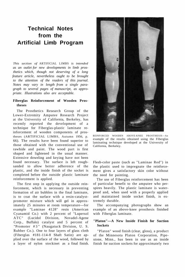
Technical Notes from the
Artificial Limb Program
This section of ARTIFICIAL LIMBS is intended
as an outlet for new developments in limb prosthetics which, though not deserving of a long feature article, nevertheless ought to be brought to the attention of the readers of this journal. Notes may vary in length from a single paragraph to several pages of manuscript, as appropriate. Illustrations also are acceptable.
Fiberglas Reinforcement of Wooden Prostheses
The Prosthetics Research Group of the Lower-Extremity Amputee Research Project at the University of California, Berkeley, has recently reported the development of a technique for Fiberglas-plastic laminate reinforcement of wooden components of prostheses (ARTIFICIAL LIMBS, Autumn 1956, p.
66). The results have been found superior to those obtained with the conventional use of rawhide and paint. The wood part is first shaped and lightened in the usual manner. Extensive doweling and keying have not been found necessary. The surface is left rough-sanded to allow better adherence of the plastic, and the inside finish of the socket is completed before the outside plastic laminate reinforcement is applied.
The first step in applying the outside reinforcement, which is necessary in preventing formation of air bubbles in the final laminate, is to coat the surface with a resin-catalyst-promoter mixture which will gel in approximately 25 minutes at room temperature—for example "Laminae 4128" resin (American Cyanamid Co.) with 2 percent of "Lupersol ATC" (Lucidol Division, Novadel-Agene Corp., Buffalo) catalyst and 5 percent of "Promoter # 3 " (Naugatuck Division, U. S. Rubber Co.). One to four layers of glass cloth (Fiberglas #181-114-8 Shaft Satin) are applied over the surface of the wood, followed by a layer of nylon stockinet as a final finish.
REINFORCED WOODEN ABOVE-KNEE PROSTHESIS—An
example of the results obtained using the Fiberglas laminating technique developed at the University of California, Berkeley.
Flesh-color paste (such as "Laminae Red") in the plastic used to impregnate the reinforcement gives a satisfactory skin color without the need for painting.
The use of Fiberglas reinforcement has been of particular benefit to the amputee who perspires heavily. The plastic laminate is waterproof and, when used with a properly applied and maintained inside socket finish, is extremely durable.
The accompanying photographs show an example of an above-knee prosthesis finished with Fiberglas laminate.
"Platon"—A New Inside Finish for Suction Sockets
"Platon" wood finish (clear, gloss), a product of the Minnesota Platon Corporation, Pipestone, Minn., has been in use as an inside finish for suction sockets for approximately two

years by the Prosthetics Research Group of the Lower-Extremity Amputee Research Project at the University of California, Berkeley. "Platon" is a phenolic-resin varnish requiring addition of an acid hardener before application. Five or six coats are necessary to build up a film thickness sufficient to provide a waterproof and durable finish. Approximately one hour between coats is required to evaporate solvents, and then there must be a 24-hour period of cure at room temperature. If the socket is used before the finish is completely cured, the finish will turn white.
A properly applied and cured finish is very durable. Many instances of two years' wear of
suction sockets without finish deterioration have been observed. There have been no reported cases of allergic reaction among amputees who have used sockets finished with "Platon."
Where socket modification is required after finishing, lacquers or cellulose acetate may be used as a temporary finish in modified areas, then removed and replaced by "Platon" when more time is available. Where time is available, and extensive socket modifications are not anticipated, the increased durability and hygienic advantages of a properly applied "Platon" socket finish are well worth the extra time required.

Abstracts of
Current Literature
This section of ARTIFICIAL LIMBS is intended to summarize the current literature of limb prosthetics, especially the less accessible reports literature arising from the several research groups participating in the Artificial Limb Program. Authors are hinted to submit, for review, copies of any such material, including papers published in scientific journals.
Training of the Lower Extremity Amputee, Donald Kerr and Signe Brunnstrom (with Introduction by T. Campbell Thompson), Charles C Thomas, Springfield, Ill., 1956. xx plus 272 pp., 153 illus. $6.50 (discounts for quantity lots). This volume—intended for use in re
habilitation centers, hospitals, and other places where amputees are being trained; as a textbook in schools of physical therapy, occupational therapy, and nursing; by members of the prosthetics profession; and, indeed, by amputees themselves—consists of 12 chapters. The first four are devoted, respectively, to adjustment to an amputation, to the principles of preprosthetic care, to a description of lower-extremity prostheses, and to practical suggestions with regard to stump socks and to dressing and undressing while wearing a leg prosthesis. The next seven chapters, each devoted to one or more problems (training, walking, daily activities) of a given level of lower-extremity amputation (unilateral and all possible combinations of bilateral leg amputations), give detailed instructions for the training of leg amputees and for walking with artificial legs. Chapter 12 gives information on the mastery of sports and social activities.
The techniques recommended for accomplishing the various requirements of daily living are depicted by both photograph and stick diagram. The illustrations in Chapter 12 show some of the special activities in which some leg amputees are able to engage.
Included at the back of this volume is a series of three checklists for leg amputees. The subject index runs to eight pages.
Development of Instrumentation for Measurement of Pressure in Stump Sockets and Prehension Force, Wallace E. Frank and Robert J. Gibson, Final Technical Report No. F-2400 [to the Veterans Administration], The Franklin Institute, Philadelphia, [covering the period] September 3, 1953, to June 30, 1956. 16 pp., illus. Free. For a number of years there has been in
progress at the Franklin Institute Laboratories for Research and Development a project aimed at the design and construction of thin, flexible, pressure-sensing elements which change capacitance with changing pressure. Supported first by the Quartermaster Research and Development Command of the U. S. Army, the work was later financed through a contract with the Veterans Administration, the objective being to develop the new instruments, known as "Filpips" (Franklin inst i tute Laboratories Pressure indicating Patch), for application to the measurement of prehension forces and of the pressures prevailing in the sockets of artificial legs.
At the beginning of the period covered by this report, the general feasibility of making the "Filpips" had been demonstrated, but application of the method to the study of the dynamic stump pressures in the socket of a prosthesis required further development. The work described covers efforts both to perfect the "Filpip" for use in prosthetics research and to develop instrumentation and techniques for transmitting the "Filpip" signal from a walking subject to the recording devices. Included are some recommendations for future work, it being suggested that development of the "Filpip" for the application intended, though promising, has only just begun.
Artificial Arms for Child Amputees—Fabrication and Fitting Developments to July 1, 1956, William H. Henderson, Department of Engineering and School of Medicine, University of California (Los Angeles), October 1, 1956. 70 pp., illus. Free. Until July 1, 1956, the Child Amputee
Prosthetics Project at the University of California at Los Angeles was a mutual undertaking of the Departments of Engineering and

of Pediatrics, Engineering having responsibility for technical matters relating to prosthetics while Pediatrics was responsible for developmental, clinical, and psychological factors. This report summarizes the experience of the Engineering part of the Project in the design, fabrication, fitting, and evaluation of upper-extremity prostheses and special devices for 82 child amputees ranging in age from 10 months to 18 years. The case load is analyzed by age, sex, and type and cause of amputation (congenital or traumatic); and there is included a tabulation of the prosthesis and harness types fitted and of the numbers of each component used.
Presented are 20 case summaries, with photographs, covering all the customary levels of amputation and a number of special problems such as quadrilateral amputations, phoco-melias, amelias, and various other anomalies. Performance with the prostheses, afteramputee training, is evaluated on a 12-point scale; and the test performances are compared by amputee type, prosthesis type, age of the patient, and cause of amputation (congenital or traumatic). An evaluation from the viewpoint of child prosthetics is given for each of the components used, including both commercial and experimental models. Indications for prescription are reported.
A total of 13 special developments in harnessing, modification of components, and adaptive equipment are described and illustrated. Sixty-five photographs, 13 graphs and charts, and four other drawings elucidate the discussion. Included are reports on 30 new components and developments.
Although many of the children studied had lower-extremity amputations and/or deformities, this report deals with the upper-extremity problems only. Artificial legs were fitted, as needed, by private prosthetists in the area.
Prosthesis Hinge for Very Short Below Elbow Amputees, Gerald E. Gwynne and Craig L. Taylor, Artificial Limbs Research, Department of Engineering, University of California (Los Angeles), Special Technical Report No. 24, September 1956. iv plus 46 pp., illus. Free. Although typically the amputee with a very
short below-elbow stump (stump length less
than about 35 percent of normal forearm length, epicondyle to styloid) is able to flex the free forearm stump through a full 135 deg., application of an arm socket to such a case usually limits forearm flexion to a practical maximum of about 90 deg. Yet for purposes of utility it is considered essential that the amputee be able to flex the prosthetic forearm through the full 135 deg. Accordingly, it has been the practice for a number of years to use on very short below-elbow stumps the so-called "split socket" (ARTIFICIAL LIMBS, September 1955, p. 18) with one or the other of two commercially available step-up elbow hinges (ARTIFICIAL LIMBS, September 1954, pp. 16 and 17), the Hosmer variable-ratio hinge (known as the "MA 200") or the Hosmer multiple-action hinge (known as the "MA 100").
For a variety of reasons, it is claimed, these two hinges, intended to amplify forearm motion with respect to stump motion while relying on the normal elbow for stabilization of the forearm, did not prove wholly satisfactory. It was therefore considered desirable to undertake a new study of a number of very short below-elbow cases, to review once again the biomechanics of forearm flexion, to test systematically the performance of existing step-up elbow hinges, and to attempt the experimental design of a step-up hinge that will meet all requirements of practicality and of functional regain.
This report, based on work conducted during 1955-56, analyzes the forearm-flexion characteristics of eight very short below-elbow stumps, reviews the kinematics of the existing step-up elbow hinges, and summarizes the results obtained with amputees wearing the existing designs. All considerations taken into account, it is concluded that essentially complete regain of forearm flexion can be afforded in the very short below-elbow case by application of an elbow hinge based on a planetary gear mechanism and having a fixed over-all ratio of flexion of the forearm shell to that of the short stump of 1.5:1. Experimental design studies of such a device (known as the "1.5 ratio hinge") are reported, together with kinematic analysis, the results of laboratory tests, and preliminary amputee evaluation.

Proceedings, Second Congress, World Confederation for Physical Therapy, American Physical Therapy Association, 1790 Broadway, New York 19, New York, 1957. x plus 208 pp., illus. Single copies, $3.00; ten or more copies ordered simultaneously, $2.50 each. During the period June 17 through 23, 1956,
there was conducted in New York City, under the sponsorship of the American Physical Therapy Association, the Second Conference of the World Confederation for Physical
Therapy. A part of that session (ARTIFICIAL LIMBS, Autumn 1956, p. 74) was devoted to a symposium entitled Prosthetics—Research, Developmental Aspects, and Training, in which a panel of recognized authorities combined to present the background of research in artificial limbs and to demonstrate the operation of the prosthetics clinic team in the management of amputees.
These Proceedings constitute all of the papers presented during the entire Congress, including the addresses and discussions offered during the symposium on prosthetics.

Digest of Major Activities of the
Artificial Limb Program
This section of ARTIFICIAL LIMBS is intended to present a summary of principal news events of interest in the Artificial Limb Program during the several months preceding issue. Stories of activities in the various laboratories and associated agencies, reports of meetings, photographs, and items about individuals all are acceptable.
Resignation of Col. Tyler
Col. Gerald R. Tyler, previously long with the Army's Corps of Engineers until his retirement in July 1955, resigned his position as Executive Director of the Prosthetics Research Board effective September 30, 1956. When, in December 1955, Col. Tyler accepted his position with the then newly constituted Board (ARTIFICIAL LIMBS, Spring 1956, p. 39), he pointed out that he would not be able to devote more than half time to the duties assigned him. By mid-1956, however, with the rapidly growing scope of the Artificial Limb Program through the support of additional agencies such as the Department of Health, Education, and Welfare, it became clear that eventually the position of Executive Director would necessarily have to be a full-time office, perhaps even with the assistance of other staff personnel. Since some change was indicated in any event, Col. Tyler suggested that he should step out promptly to make room for a full-time replacement. All concerned expressed regret that Col. Tyler's association with the Artificial Limb Program had to be so brief.
New West Coast Office
In view of the broadened activities already under way in the Artificial Limb Program during the current fiscal year, and considering indications of still further expansion in the years ahead, the Prosthetics Research Board has for some months had under consideration a general strengthening of staff functions both in
Washington and in the field. As a first step, a West Coast branch of the Office of the Executive Director was set in operation on October 1 at Suite 204, 8693 Wilshire Boulevard, Beverly Hills, Calif., under the management of Tonnes Dennison, formerly Field Engineer, who has been named Assistant Executive Director. Principal work of the new office will be concerned with coordinating the activities of the various research projects in the area and with assisting the Committee on Prosthetics Research and Development in the conduct of its duties.
New Staff Officers
Early last October, upon the recommendation of the Prosthetics Research Board, Dr. Detlev W. Bronk, President of the National Academy of Sciences— National Research Council, appointed Dr. Harold W. Glattly to the position of Executive Director, effective February 1, to fill the vacancy left by the resignation of Col. Tyler. Col. Robert E. Benjamin was appointed Executive Secretary, also effective February 1. Mr. A. Bennett Wilson, Jr., formerly Executive Secretary, was named to the new position of Staff Engineer.
Dr. Glattly, formerly Surgeon of the First Army at Governors Island, New York, with responsibility for medical and hospital services throughout New Jersey, New York, and the New England States, retired on January 31 as a Brigadier General, after more than 30 years' service in the Army's Medical Corps. A native of Minnesota, Dr. Glattly received his medical degree from Iowa State University in 1926 and entered the Service in August of that year. He has held many important assignments, one of which was Chief of the Personnel Division, Office of the Surgeon General. He served twice in the Philippines and during World War II was captured by the Japanese and held in prison camps for more than three years. In his retirement from active service, Dr. Glattly will devote his full time and energies to the further-
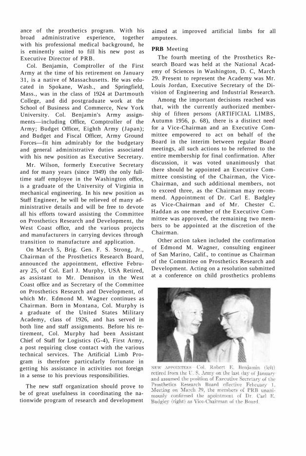
ance of the prosthetics program. With his broad administrative experience, together with his professional medical background, he is eminently suited to fill his new post as Executive Director of PRB.
Col. Benjamin, Comptroller of the First Army at the time of his retirement on January 31, is a native of Massachusetts. He was educated in Spokane, Wash., and Springfield, Mass., was in the class of 1924 at Dartmouth College, and did postgraduate work at the School of Business and Commerce, New York University. Col. Benjamin's Army assignments—including Office, Comptroller of the Army; Budget Officer, Eighth Army (Japan); and Budget and Fiscal Officer, Army Ground Forces—fit him admirably for the budgetary and general administrative duties associated with his new position as Executive Secretary.
Mr. Wilson, formerly Executive Secretary and for many years (since 1949) the only full-time staff employee in the Washington office, is a graduate of the University of Virginia in mechanical engineering. In his new position as Staff Engineer, he will be relieved of many administrative details and will be free to devote all his efforts toward assisting the Committee on Prosthetics Research and Development, the West Coast office, and the various projects and manufacturers in carrying devices through transition to manufacture and application.
On March 5, Brig. Gen. F. S. Strong, Jr., Chairman of the Prosthetics Research Board, announced the appointment, effective February 25, of Col. Earl J. Murphy, USA Retired, as assistant to Mr. Dennison in the West Coast office and as Secretary of the Committee on Prosthetics Research and Development, of which Mr. Edmond M. Wagner continues as Chairman. Born in Montana, Col. Murphy is a graduate of the United States Military Academy, class of 1926, and has served in both line and staff assignments. Before his retirement, Col. Murphy had been Assistant Chief of Staff for Logistics (G-4), First Army, a post requiring close contact with the various technical services. The Artificial Limb Program is therefore particularly fortunate in getting his assistance in activities not foreign in a sense to his previous responsibilities.
The new staff organization should prove to be of great usefulness in coordinating the nationwide program of research and development
aimed at improved artificial limbs for all amputees.
PRB Meeting
The fourth meeting of the Prosthetics Research Board was held at the National Academy of Sciences in Washington, D. C, March 29. Present to represent the Academy was Mr. Louis Jordan, Executive Secretary of the Division of Engineering and Industrial Research.
Among the important decisions reached was that, with the currently authorized membership of fifteen persons (ARTIFICIAL LIMBS, Autumn 1956, p. 68), there is a distinct need for a Vice-Chairman and an Executive Committee empowered to act on behalf of the Board in the interim between regular Board meetings, all such actions to be referred to the entire membership for final confirmation. After discussion, it was voted unanimously that there should be appointed an Executive Committee consisting of the Chairman, the Vice-Chairman, and such additional members, not to exceed three, as the Chairman may recommend. Appointment of Dr. Carl E. Badgley as Vice-Chairman and of Mr. Chester C. Haddan as one member of the Executive Committee was approved, the remaining two members to be appointed at the discretion of the Chairman.
Other action taken included the confirmation of Edmond M. Wagner, consulting engineer of San Marino, Calif., to continue as Chairman of the Committee on Prosthetics Research and Development. Acting on a resolution submitted at a conference on child prosthetics problems

held at the UCLA Medical Center October 26 (see below), the Board approved the appointment of the Committee on Child Prosthetics Problems, a permanent committee of the Board to consider problems peculiar to prosthetics for children.
The Board also extended its responsibilities to the field of braces and authorized the Chairman to take appropriate action in this regard.
After the meeting, the members gathered for luncheon at the Officers Club of Walter Reed Army Hospital as the guests of the Commanding General of Walter Reed Army Medical Center. After lunch, Col. Maurice J. Fletcher conducted the members on a tour of the Army Prosthetics Research Laboratory located in suburban Forest Glen, Md. Demonstrations by amputee subjects highlighted each major phase of APRL activity.
The next meeting of the Board is scheduled to be held in Los Angeles during the latter part of October.
Annual Fall Meeting, ALP
The University of California Medical Center, Los Angeles, was host to the committees and subcommittees of the Prosthetics Research Board for their annual fall meetings November 28 through December 1. All phases of the programs in research and development and in education were discussed by the appropriate subcommittees, who presented reports to their respective committees on the last day of the meetings.
After presentation and discussion of the reports, committee and subcommittee members present were invited by Brig. Gen. F. S. Strong, Jr., Chairman of the Prosthetics Research Board, to constitute a Committee of the Whole to discuss informally problems that had arisen as a result of expansion within the Artificial Limb Program during the previous year. Here it was pointed out that one of the major problems is the difficulty in locating well-qualified personnel for research projects. Those in attendance were requested to advise the Prosthetics Research Board of the availability of anyone interested in research and education in the field of limb prosthetics.
The next meetings of the committees of the Prosthetics Research Board are scheduled to be held at the National Academy of Sciences in Washington, D. C, May 21 through 25.
Conference on Child Prosthetics Problems
At the invitation of Dr. Stafford Warren, Dean of the Medical School, University of California at Los Angeles, there was held at that institution on October 26 a conference on child prosthetics problems. Led by Brig. Gen. F. S. Strong, Jr., Chairman of the Prosthetics Research Board, the conference was called to determine the feasibility of integrating the present research on the management of child amputees with that of the adult program.
The role of the Children's Bureau, Department of Health, Education, and Welfare, in both research and service was discussed by Dr. Arthur J. Lesser, Director of the Division of Health Services of that Bureau. The objectives of the state programs of New York, Michigan, and California for providing services for child amputees were set forth by Dr. Anne M. Bahlke, Dr. Carleton Dean, and Dr. Marcia Hays, respectively. Dr. Bahlke is Director of the Bureau of Medical Rehabilitation of the Department of Public Health of the State of New York, Dr. Dean is Director of the Michigan Crippled Children Commission, Grand Rapids, and Dr. Hays is Chief of the Bureau of Crippled Children Services of the Department of Public Health of the State of California.
Details of current research in child prosthetics were given in various papers by members of the research teams at the University of California at Los Angeles, the University of California at Berkeley, the Michigan Crippled Children Commission, the Army Prosthetics Research Laboratory, the Navy Prosthetics Research Laboratory, and New York University.
Dr. Charles H. Frantz, Chairman of the Interim Subcommittee on Child Prosthetics Problems (of the Committee on Prosthetics Research and Development), then outlined the activities of his group. In the round-table discussion that followed, the conference initiated a recommendation to the Prosthetics Research Board that the Interim Subcommittee be given permanent status as a committee of the Board to assist in coordinating the research to be conducted in the field of child prosthetics. The conference then proposed further that the Committee on Child Prosthetics Problems, if so established, consider holding during the spring of 1957 a

seminar involving individuals actively engaged in any phase of research in the field of management of the child amputee. [The Committee on Child Prosthetics Problems, Dr. Charles H. Frantz, Chairman, was approved by the Prosthetics Research Board on March 29. See page 109.]
Prosthetics Education Program
During the fall and winter, the Prosthetics Education Program for physicians, therapists, and prosthetistS was continued with additional courses at New York University and at the University of California at Los Angeles. At both institutions, students are afforded not only lectures but also laboratory sessions in which they are given an opportunity to do actual work with amputee subjects. During the last week of each course, the students are assigned to form clinic teams for practice in writing prescriptions and in checkout of prostheses.
For the 1956-57 academic year, NYU is offering, under the sponsorship of the Post-Graduate Medical School and in cooperation with the College of Engineering, six courses in above-knee prosthetics and one in upper-extremity prosthetics (ARTIFICIAL LIMBS, Autumn 1956, p. 72). Of these seven courses, four have been completed, with the following attendance:
The single course originally planned in upper-extremity prosthetics (Series A) was oversubscribed by therapists, so that the class had to be conducted in two sections (Series A, Series B).
Students of the several classes at NYU were as follows:
COURSE 741A (Above-Knee Prosthetics)
Physicians, October 1 through October 5—Dr. Arthur J. Heather, Wilmington, Del.; Dr. John J. Horwitz, Pittsburgh; Dr. Frederick W. Koenig, Pittsburgh; Dr. Harold H. Kuhn, Charleston, W. Va.; Dr. James G. McClure, Chapel Hill, N. C; Dr. Newton C. McCol-lough, Orlando, Fla.; Dr. Bernard J. Michela, Alexandria, Va.; Dr. Henry B. Moore, Winston-Salem, N. C; Dr. Frank N Potts, Buffalo; Dr. Victor M. Santana-Carlos, New York City; Dr. Charles D. Shields, Washington, D. C; Dr. Wayne 0. Southwick, Baltimore; Dr. Emery K. Stoner, Philadelphia; Dr. William W. Strange, Huntington, W. Va.; Dr. Thomas L. Waring, Memphis; Dr. Edward S. Whitesides, Gastonia, N. C; Dr. Milton J. Wilder, Baltimore; and Dr. George H. Zablocki, Huntington, W. Va.
COURSE 742A (Above-Knee Prosthetics)
Therapists, September 27 through October 5—John R-Brittle, Durham, N. C; Miriam B. Butzer, Ephrata, Pa.; Amy D. Campbell, Fayetteville, N. C; James D. Chan, Windham, N. Y.; Josephine Gardner, Maiden, W. Va.; Phyllis A. Koch, Washington, D. C; Dorothea J. Linn, Pittsburgh; R. M. Litaker, Chapel Hill, N. C; Mary Mason, Baltimore; William Neill, Ruxton, Md.; John J. Nostrant, Buffalo; Florence G. Rahrig, Kenova, W. Va.; Joseph B. Smolens, New York City; Emily Wellington, Nonquitt, Mass.; and Rose M. Zappala, Chester, Pa.
COURSE 743A (Above-Knee Prosthetics)
ProsthetistS, September 24 through October 5—Charles H. Dankmeyer, Baltimore; Harold Dobbs, Charlotte, N. C; Robert W. Hansbrough, Roanoke, Va.; Marion F. Kessler, Hyde Park, Mass.; Stanley J. Kordasz, Brooklyn, N. Y.; Ivan E. Letner, Washington, D. C; Martin D. Massey, Baltimore; Basil Peters, Glenolden, Pa.; Chester T. Shelton, Brentwood, Md.; Patrick E. Siress, Northlake, Ill.; Allen G. Talley, Raleigh, N. C; Bert R Titus, Durham, N. C; and Charles W. Weight, Springfield, Pa.
COURSE 741B (Above-Knee Prosthetics)
Physicians, November 5 through November 9—Dr-John L. Chandler, Augusta, Ga.; Dr. Harry L. Collins, Jr., Jacksonville, Fla.; Dr. Josh D. Davis, Montgomery, Ala.; Dr. Richard B. Donaldson, Chattanooga, Tenn.; Dr. William R. Fain, Jackson, Miss.; Dr. Charles L. Farrington, St. Petersburg, Fla.; Dr. William E. Gazeley, Schenectady, N. Y.; Dr. Harriet E. Gillette, Atlanta; Dr. William C. Hannon, Mobile, Ala.; Dr. Ralph J. Hobbs, Birmingham, Ala.; Dr. Richard E. King, Atlanta; Dr. Walter J. Lee, Knoxville, Tenn.; Dr. Irwin S. Leinbach, St. Petersburg, Fla.; Dr. Leilas R. Lonnergan, Gadsden, Ala.; Dr. Raymond J. Malzone, St. Petersburg, Fla.; Dr. Seymour Morse, Jacksonville, Fla.; Dr. Wesley L. Nolden, Little Rock, Ark.; Dr. Merritt B. Shobe, Kingsport, Tenn.; Dr. Dana M. Street, Memphis; Dr. John Q. U. Thompson, Jacksonville, Fla.; Dr. James C. Thoroughman, Atlanta; Dr. Edward G. White, Miami Beach, Fla.; Dr. Albert A. Wilson, Tampa, Fla.; Dr. William J. Wilson, Lake City, Fla.; and Dr. Chestley L. Yelton, Fairfield, Ala.

COURSE 742B (Above-Knee Prosthetics)
Therapists, November 1 through November 9—Leo W. Betzelberger, Memphis; Evan V. Davies, Knoxville, Tenn.; Mar-jorie G. Davis, Atlanta; Inez M.Fawbush, Little Rock, Ark ; Martin S. Garfinkel, Jacksonville, Fla.; Mable R. Gunter, Mobile, Ala.; John M. Hawk, Jackson, Miss.; Lois A. Hodges, Atlanta; Henrietta Huxford, Skancateles, N. Y.; Mildred I. Lineberger, Grand Rapids, Mich.; Margaret J. Neal, Birmingham, Ala.; Andrew A. Orsini, Madeira Beach, Fla.; William G. Redden, Jr., Lake City, Fla.; Marcus E. Spivey, Jr., Kingsport, Tenn.; Gerald Stern, Brooklyn, N. Y; Mary E, Stout, Atlanta; Ida M. Summers, Eden, Ala.; Ardis M. Traut, Orlando, Fla ; and Ar-lene R. Walker, Atlanta.
COURSE 743B (Above-Knee Prosthetics)
Prosthetists, October 29 through Novem ber 9—James M. Bonds, Knoxville, Tenn.; Jack L. Caldwell, Tampa, Fla.; William J. Ferris, Jr., Baltimore; Carlton E. Fillauer, Chattanooga, Tenn.; Edmund J. Gernannt, New York City; Herbert B. Hanger, Summit, N. J.; Richard M. Locke, Birmingham; Thomas L. Maples, Owens Cross Roads, Ala.; William C. McCall, St. Petersburg, Fla.; James A. McKown, Jacksonville, Fla ; Richard G. Moffatt, Richmond Hill, N. Y.; John Orzulak, Buffalo; Pierce A. Peacock, Morrow, Ga.; Roy R. Rice, Atlanta; Floyd D. Simmons, Memphis; and Moody L. Smitherman, Birmingham.
PROSTHETICS EDUCATION AT NYU—William A. Tosberg, instructor in the courses in above-knee prosthetics offered by New York University during the 1956-57 academic year, demonstrates to a group of students the rough fitting of a quadrilateral above-knee socket.
PROSTHETICS EDUCATION AT NYU—A clinic team examines a patient and writes the prescription during the course in upper-extremity prosthetics offered by New York University, January 28 through March 1.
COURSE 741C (Above-Knee Prosthetics'/ Physicians, December 10 through 14—Dr. Peter L.
Carnesale, Wood, Wis.; Dr. Kerwin A. Fischer, Louisville; Dr. Freeman P. Fountain, Louisville; Dr. Julio Garavito, Bogota, Colombia; Dr. James W. Gibson, New York City; Dr. Donald D Goldberg, Richmond Hill, N. Y.; Dr. William R. Harris, Toronto; Dr. Richard D. Hoover, Emory University, Ga.; Dr. Richard H. Kiene, Kansas City, Mo.; Dr. Frank H.
Lindeman, Tampa, Fla.; Dr. Heinz Lipp-mann, West Englewood, N J.; Dr. Wood Lovell, Atlanta; Dr Robert R. McKnight, Augusta, Ga.; Dr. Wrilliam K. Massie, Lexington, Ky.; Dr. Richard J. Miller, Tampa, Fla.; Dr. Ernest O. Ronneburger, Milwaukee; Dr. Norman P. Rotenberg, Grand Rapids, Mich.; Dr. John J. Untereker, Queens Village, N. Y ; and Dr. Robert M Yanchus, New Kensington, Pa.
COURSE 742C (Above-Knee Prosthetics)
Therapists, December 6 through 14— Joseph A. Corn, Tampa, Fla.; Arthur M. Grisa, Wauwatosa, Wis.; Sondra Lamp!, New York City; Annette C. Larson, St. Paul, Minn ; Mary E. McDonnell, Louisville; Robert D. Mears. East Orange, N. J.; Mary P. Murray, Milwaukee; Elmer E. Phelps, Jr , Webster Groves, Mo.; Marian H. Pratt, Albany, N Y ; and Robert O Wood, Wauwatosa, Wis.

COURSE 743C (Above-Knee Prosthetics)
Prosthelists, December 3 through 14—Herman P. Barghausen, Venetia, Pa.; Wesley G. Clemens, Grove City, Ohio; Asa L. Godbey, Miami; Henry T. Hertsch, New York City; John Kormylo, Hopkins, Minn ; James H. Luckett, Louisville; Chester C. Nelson, Minneapolis; Paul R. Rockett, Bronx, N. Y.; Addison H. Starkey, Wethersfield, Conn.; Robert K. Umstead, St. Petersburg, Fla.; Bun H. Vowell, St. Louis; James T. Vowell, St. Louis; and James R. Watkins, Lexington, Ky.
COURSE 744A (Upper-Extremity Prosthetics)
Physicians, February 25 through March 1—Dr. John H. Allan, Charlottesville, Va ; Dr. John F. Bell, Burlington, Vt.; Dr. Samuel W. Bridgham, Rumford, R. L: Dr. Nila K. Covalt, Winter Park, Fla.; Dr. K. A. Fischer, Louisville; Dr. James D. Fisher, Springfield, Mass.; Dr. Julio Garavito, Glen Oaks, N. Y.; Dr. William E. Gazeley, Schenectady, N. Y.; Dr. Maurice Gershman, Far Rockaway, N. Y.; Dr. Otto G. Goldkamp, West Hartford, Conn.; Dr. John E Gottsch, Tampa, Fla.; Dr. William C. Hannon, Mobile, Ala.; Dr. William R. Harris, Toronto; Dr. Robert C. Hartson, Holden, Mass.; Dr. Hla Pe, Rangoon, Burma; Dr. Robert Hoffman, Albany, N. Y'.; Dr. Ernest W. Johnson, Columbus, Ohio; Dr. Richard E. King, Atlanta; Dr. Richard W. Leong, Houston; Dr. Sung J. Liao, Boston; Dr. Heinz I. Lippmann, West Englewood, N. J.; Dr; John J. Lorentz, Methuen, Mass.; Dr. Edwin L. Lytle, Bronx, N. Y.; Dr. Joseph Rizzo, New York City; Dr. Herman L. Rudolph, Reading, Pa.; Dr. Thomas L. Waring, Memphis; and Dr. William F Enneking, Jackson, Miss.
COURSE 745A (Upper-Extremity Prosthetics)
Therapists, February 18 Ihrougli March 1—LieslBader, New York City; Florence Bailey, Cincinnati; Sara Bedini, Springfield, Mass.: Elizabeth Carey, Hudson Falls, N. Y.; Joan Crosby, Toronto; Cecilia Gil, New York City; Ellen Grover. Washington, D. C; Mable R. Gunter, Mobile, Ala.; Barbara Hanson, Canton Mass.; Louise Hayward, Albany, N .Y. William Hensley, Johnson City, Tenn. Hans M. Hillander, Schenectady, N. Y. M. Hope Keeney, Houston; Joseph Kramer, Ridgefield, N J.; Adelbert MacCaughey, Chicago; Anne Miller. Wilmington, Del.; Sonia Paniagua, New-York City; Frederic Sammons, La-Grange, 111.; Lyla Spelbring, Ann Arbor, Mich.; Mary Stout, Atlanta; Arlene Walker, Atlanta; and Dorothy White, Chicago.
COURSE 746A (Upper-Extremity Prosthetics)
Prosthelists, January 28 through March 1—Roland H. Daniel, Buffalo; Gunter H. Gehl, Chicago; Herbert E. Kramer, Little Neck, N. Y.; William J. Mcllmurray, Brooklyn, N. Y.; Robert J. Mitchell, Yonkers, N. Y'.; Armand L. Roy, Burlington, Vt.; Charles S. Schaeffer, Dearborn, Mich.; Michael Stone, Denver; and Reinhard Wurth, Schenectady, N. Y.
COURSE 745B (Upper-Extremity Prosthetics)
Therapists, March 4 through 15—Beverley Bates, Richmond, Va.; Catharine Bingaman, Boston; Jean Elson, New York City; Anna B. Fielding, Conimicut, R. I ; Mary Hannah, Philadelphia; Jean M. Lending, New York City; Erica M. Jokl, Lexington, Ky.; Jo Ann Logan, St. Louis; Marion McLenahan, Rochester,
PROSTHETICS EDUCATION AT NYU—Students of the courses in above-knee prosthetics offered by New York University during the 1956-57 academic year engage in shop practice in the construction of the above-knee prosthesis

N. Y.; Frances T. Miller, West Hartford, Conn.; Lucie W. Nagy, Wharton, N. J.; Cecilia Sattely, Flushing, N. Y.; Virginia Strandvold, Pittsburgh; Ann Teska, Jamaica, N. Y.; and Ella Westerman, New York City.
To complete the program at NYU for the
present academic year, a course in above-knee
prosthetics is scheduled for April 29 through
May 10 and another for June 3 through 14. It
is anticipated that a number of additional
courses both in upper-extremity and in above-
knee prosthetics will be scheduled during the
1957-58 academic year.
The Prosthetics Education Project at the
University of California at Los Angeles pre
sented during the fall semester of 1956 three
courses in Clinical Prosthetics: Above-Knee Am
putations. As in the case of the first course in
this series, given in May and June of 1956
(ARTIFICIAL LIMBS, Autumn 1956, p. 69), the
three courses in the fall (Sections I, I I , and III)
were given in the University of California
Medical Center (Los Angeles), where P E P has
classrooms, offices, and a teaching laboratory.
Students in attendance at the UCLA courses
were as follows:
SECTION I
Prosthetists, September 3 through 14—William E. Brownfield, Boise, Idaho; Lenart C. Ceder, Tacoma, Wash.; Clarence T. Cole, Portland, Ore.; John Galdik, San Francisco; Robert V. Home, Walla Walla, Wash.; Paul D. McCullough, Springfield, Mo.; Charles R. Newton, Portland, Ore.; Willard L. Parlette, Portland, Ore.; August W. Pruhsmeier, Portland, Ore.; Alpha O. Rogers, Anchorage, Alaska; and Marvin R. Sturtz, Detroit.
Therapists, September 10 through 14—Robert R. Gallucci, Tacoma, Wash.; Thomas B. Irwin, Seattle; Mildred G. Jett, Tigard, Ore.; Herbert C. Johnson, Oakland, Calif.; James G. Julius, Boise, Idaho; Bernard F. Knapik, Mercer Island, Wash.; Margaret V. Magee, Palo Alto, Calif.; Eugene F. Northrop, College Place, Wash.; Mildred A. Ramsey, Lanikai, Hawaii; Marten Schouten, Seattle; and Mercedes Weiss, Portland, Ore.
Physicians, September 10 through 14—Dr. Max J. Allen, Spokane, Wash.; Dr. T. Allen Casey, Portland, Ore.; Dr. Paul A. Grigorieff, Eureka, Calif.; Dr. Robert Kochsiek, Los Angeles; Dr. Bernard E. McConville, Seattle; Dr. Matthew Mendelsohn, Berkeley, Calif.; Dr. Jean Michaels, Los Angeles; Dr. Clyde D. Platner, Walla Walla, Wash.; Dr. Howard W. Rickett, Seattle; Dr. Manley B. Shaw, Boise, Idaho; Dr. Faulkner A. Short, Portland, Ore.; Dr. John E. Stewart, Seattle; and Dr. Lewis G. Wilson, Pasadena, Calif.
SECTION II
Prosthetists, October 29 through November 9—Michael M. Amrich, Chicago; George H. Botko, Minneapolis; Barnie R. Buddin, Oklahoma City; Eugene Coleman, Seattle; John G. Cranford, Richmond, Va.; Carl E. Gustavson, Seattle; Richard L. Kleiber, Denver; Flavel L. Lake, Oklahoma City; Frank Moos, San Jose, Calif.; Alvin Norell, Salt Lake City; Bruce A. Scott, Denver; and John S. Snyder, Tulsa, Okla.
Therapists, November 5 through 9—Eleanor B. Bar-haug, Lakewood, Colo.; Margaret E. Bryce, Madison, Wis.; Clyde R. Holway, Downey, Calif.; James C. Hufsey, Bethany, Okla.; Helen G. Loveless, Wichita; Frederick R. Murko, Fishersville, Va.; Lorraine G. Paulson, Denver; Alvin G. Russell, Jr., Salt Lake City; Troy T. Scholl, Eufaula, Okla.; Ozella L. Scruggs, Oklahoma City; Naomi Wesson, Wichita; and Don-namae Winters, Sausalito, Calif.
Physicians, November 5 through 9—Dr. Donald R. Adams, Muskogee, Okla.; Dr. Ernest M. Burgess, Seattle; Dr. Edward T. Evans, Minneapolis; Dr. Calvin E. Gantenbein, Portland, Ore.; Dr. Ella M. George, Oklahoma City; Dr. Thomas M. Hamilton, Charlottesville, Va.; Dr. Cline D. Hensley, Jr., Wichita; Dr. Carl C. Hoffman, Denver; Dr. Roy M. Hoover, Roanoke, Va.; Dr. Edward T. Kelley, Jr., Malibu, Calif.; Dr. Robert H. Lamb, Salt Lake City; Dr. Harold Rosenberg, Salt Lake City; Dr. Edwin R. Schottstaedt, San Francisco; Dr. Atha Thomas, Denver; Dr. George C. Twombly, Jr., Englewood, Calif.; and Dr. David K. Webster, Staunton, Va.
SECTION III
Prosthetists, January 7 through 18—George W. Berryman, New Orleans; Robert W. Brown, Edmonton, Alberta; Anthony Filippis, Detroit; Alexander Finlay, Milwaukee; Gustav R. F. Hinrichs, San Carlos, Calif.; Herman C. Hittenberger, Burlingame, Calif.; Satoru Kato, Honolulu, Hawaii; John L. Kolman, Los Angeles; George H. Lambert, Baton Rouge, La.; Lester J. Sabolich, Oklahoma City; George E. Snell, Little Rock, Ark.; and David C. McGraw, Shreveport, La.
Therapists, January 14 through 18—Aline F. Bletcher, Memphis; Jane E. Dunnigan, Santa Monica, Calif.; Dorothy J. Dutro, Williamsburg, Va.; Norman E. Hurl, Edmonton, Alberta; Johnnie M. Jackson, Coushatta, La.; Elizabeth Kirkpatrick, Santa Monica, Calif.; Eva M. LeBlanc, Seattle; Marilyn M. Shangraw, Ann Arbor, Mich.; Ethel A. Stagni, Baton Rouge, La.; and Julie Werner, Los Angeles.
Physicians, January 14 through 18—Dr. Norbert J. Bublis, Temple, Texas; Dr. Aeneas P. Cash, Lubbock, Texas; Dr. E. E. Chambers, Enid, Okla.; Dr. Joe B Davis, Portland, Ore.; Dr. John R. Fowler, Edmonton, Alberta; Dr. Edward T. Haslam, New Orleans; Dr. George H. Koepke, Ann Arbor, Mich.; Dr. James McC. Mitchner, Houston; and Dr. Donald H. Sitler, West Covina, Calif.
Similar courses scheduled for the remainder
of fiscal year 1956-57 are: Section IV, February
25 through March 8; Section V, March 18
through 29; and Section VI, April 22 through

PROSTHETICS EDUCATION AT UCLA—Continuation of the series. Pictured are the students and instructors in the courses in Clinical Prosthetics: Above-Knee Amputations presented by the Prosthetics Education Project at the University of California Medical Center, Los Angeles, during the fall semester, 1956. Top, Section I, September 3 through 14; middle, Section II, October 29 through November 9; bottom, Section III, January 7 through 18.

May 3. Section VII, which will deal with upper extremities, is scheduled to be held June 3 through 28.
other local facilities, such as Rancho Los Amigos (Los Angeles County Respirator Center) and certain private shops.
In-Service Training at VAPC
Early in March the Research and Development Division of the Prosthetic and Sensory Aids Service inaugurated an in-service training program for personnel of the Veterans Administration Prosthetics Center, New York City. Although originally the plan was designed specifically to meet the needs of some five or six people in the Limb and Brace Section who are preparing for future certification as ortho-tists, other staff members are permitted to participate as students. The course of instruction is that first recommended by the Advisory Committee on Educational Standards of the Orthopedic Appliance and Limb Manufacturers Association and later refined by the Educational Committee of OALMA.
Evening Classes at UCLA
The Prosthetics Education Project at the University of California at Los Angeles expanded its program of evening classes during the fall semester of 1956 to four nights a week. Previously, evening classes had been held once a week only. The new schedule provided for two-hour classes in Basic Principles of Orthotics on Monday and Wednesday evenings and two-hour classes in Basic Principles of Above-Knee Prosthetics on Tuesday and Thursday evenings. Enrollment for the fall semester was 11 for the classes in orthotics, 7 for the classes in prosthetics.
Approximately 60 percent of the class time in orthotics was used for actual laboratory practice, as compared with approximately 50 percent in the course in prosthetics. Lecture and laboratory practice sessions were held in the classrooms and teaching laboratory of the Prosthetics Education Project in the UCLA Medical Center. Included were a few trips to
OALMA National Assembly
The 1956 National Assembly of the Limb and Brace Profession, sponsored by the Orthopedic Appliance and Limb Manufacturers Association, convened in the Sheraton-Palace Hotel in San Francisco October 21 through 24. Charles A. Hennessy, President of the Peerless Artificial Limb Company, of Los Angeles, and a member of the faculty of the prosthetics school at UCLA, was named President of OALMA for the year 1956-57, succeeding W. Frank Harmon, of Atlanta. John A. McCann, of Burlington, N. J., was elected First Vice-President, and Karl W. Buschenfeldt, of Stoughton, Mass., was voted into the office of Second Vice-President. Re-elected as Secretary-Treasurer was M. P. Cestaro, of Washington, D. C.
Seminars and instructional courses offered during the Assembly included Appliances Used in Deformities and Functional Disorders of the Foot, by Dr. Paul W. Meyer and Ted R. Rey-
EVENING CLASSES AT UCLA—Kenneth L. Dodd, certified orthotist of Santa Monica, Calif., and Regional Director of Region IX of the Orthopedic Appliance and Limb Manufacturers Association, explains an important point in cast-taking to students of the evening course in Basic Principles of Orthotics presented by the Prosthetics Education Project, University of California at Los Angeles, during the fall semester of 1956.

nolds, both of Kansas City; Anatomy for the Limb and Brace Technician, by Dr. Charles G. Hutter, of Los Angeles; Cerebral Palsy Bracing, by C. O. Denison, of Baltimore, and Drs. Peter Cohen and Donald B. Lucas, both of San Francisco; Hand Splints, by Dr. Sterling Bunnell and Henry Weniger, both of San Francisco; and Harnessing, by Woodrow T. Yamaka, of Los Angeles, and Jerry Leavy, of San Jose, Calif.
Recent interest in the needs of the juvenile amputee was reflected in two additional features of the Assembly program. Problems Involved in Fitting the Child from One to Ten was the subject of a presentation by Drs. Charles H. Frantz and George T. Aitken, of the Michigan Crippled Children Commission, Grand Rapids. Drs. Robert Mazet, Jr., Craig L. Taylor, and Milo B. Brooks presented The Child Amputee Prosthetics Project, a report on the first year's findings in the research under way at the University of California at Los Angeles
Other technical papers and demonstrations included Functional Arm Bracing, by Dr. Edwin R. Schottstaedt, of San Francisco, and George B. Robinson, of Vallejo, Calif.; Cosmetic Appliances, by C. 0. Anderson, of San Francisco, and Carl Xielson, of Washington, D. C; and The Lower-Extremity Amputee-—A Clinical Picture as Revealed by Studies at the University of California, by Drs. Verne T. Inman, S. William Levy, Henry E. Loon, Malcolm R. Miller, Henry F. Albronda, and Henry J. Ralston, all of San Francisco.
Before adjourning, the delegates unanimously approved the selection of Washington, D. C, as the site of the 1957 National Assembly of the Limb and Brace Profession to be held September 29 through October 2. Headquarters will be in the Statler Hotel.
Robert C. Gruman, of Minneapolis, was named Program Chairman. Ralph Storrs, of Kankakee, Ill., was appointed Vice-Chairman of the Program Committee and will also serve as Chairman of the Exhibits Committee.
Shimmon, San Francisco
OALMA PAST-PRESIDENTS—Three Pas t -Pres idents of O A L M A welcome a new member to their " c lub" during the 195(5 Nat ional Assembly in San Francisco October 21 through 24. Left to right are Lee J. Fawver , of Kansas City, Mo., M c C a r t h y Hanger , Jr., of St. Louis, W. F rank Harmon, of Atlanta , and Clyde A Aunger, of Phoenix, Ariz.

Annual Meeting of ABC
At its annual meeting and luncheon at the Sheraton-Palace Hotel in San Francisco, October 22, the American Board for Certification of the Prosthetic and Orthopedic Appliance Industry, Inc., paid tribute to Brig. Gen. F. S. Strong, Jr., Chairman of the Prosthetics Research Board, National Research Council, for his leadership in research in limb prosthetics. Gen. Strong was presented with a bound volume of letters contributed by the managers of certified facilities, noted leaders in prosthetics and in orthopedic surgery, and others in a position to know about the contributions he has made to prosthetics research. Bound as the first letter in the volume was a special message from President Eisenhower.
Gen. and Mrs. Strong were guests of honor at the Board's luncheon meeting. Presentation of the volume was made by Mrs. Chester C. Haddan and Glenn E. Jackson, Executive Director of OALMA, Mrs. Haddan acting for her husband, who had been in charge of arrangements for assembling the volume. Unfortunately, Mr. Haddan was ill and therefore unable to be present.
The annual business session of the Certification Board was held at the conclusion of the presentation ceremony. The Board presented certificates of appreciation to its two retiring members, Dr. Robert Mazet, Jr., of Los Angeles, and Karl W. Buschenfeldt, certified prosthetist and ortholist of Boston. Dr. Mazet had served as President during the past year and as Vice-President in 1955.
Col. August W. Spittler, Chief of Orthopedic Surgery at Fitzsimons Army Hospital, Denver, and W. Frank Harmon, certified orthotist of Atlanta, were elected to the Board to fill these vacancies. Col. Spittler was nominated for membership by the American Academy of Orthopaedic Surgeons. Under the by-laws of the Board, three members are nominated by the Academy and four by the Orthopedic Appliance and Limb Manufac
turers Association. Mr. Harmon comes to the Certification Board after completing his term as President of OALMA (ARTIFICIAL LIMBS,
Spring 1956, p. 48). Now President of the Atlanta Brace Shop, he was formerly on the staff of the Warm Springs Foundation.
In the course of its meeting, the Board elected the following officers for the year 1956-57: President, Carlton E. Fillauer, certified prosthetist and orthotist of Chattanooga; Vice-President, Dr. Edward C. Holscher, of St. Louis; and Secretary-Treasurer, M. P. Cestaro, President of the J. E. Hanger Company, Washington, D. C. President Fillauer completed the Board's organization by appointing the four committees which carry on the Board's work. They are:
I. Committee on Credentials—To review and act upon all applications from persons desiring certification. To advise the national office as to any further information desired on any candidate The decisions of this committee determine who shall be eligible to take the examinations. Membership: W. Frank Harmon, Chairman, Dr. Roy M. Hoover, D. A. McKeever.
—Shimmon, San Francisco
HONORS FOR GENERAL STRONG—Brig. Gen. F. S. Strong, Jr., Chairman of the Prosthetics Research Board and formerly Executive Director of its predecessor, the Advisory Committee on Artificial Limbs, National Research Council, was guest of honor at the annual luncheon of the American Board for Certification at the Sheraton-Palace Hotel in San Francisco October 22. He was presented with a bound volume of letters attesting his contributions to the field of limb prosthetics. Left to right are Dr. Robert Mazet, Jr., President of ABC, Gen. Strong, and Dr John B. deC M. Saunders, Dean of the School of Medicine of the University of California, San Francisco, who delivered the annual Certification address.

— Shimmon, San Francisco NEW MEMBER or ABC—Col, August W. Spittler, Chief of Orthopedic Surgery at Fitzsimons Army Hospital, Denver, was elected during the 1956 OALMA National Assembly to serve on the American Board for Certification. Shown here, left to right, are, seated, Board members Dr. Roy M. Hoover, of Roanoke, Va., and Col. Spittler; standing, Board member Edward W. Snygg, of San Francisco, Treasurer M. P. Cestaro, of Washington, D.C., and Vice-President Karl W. Buschen-feldt, of Boston.
II. Committee on Facilities—To review and make decisions upon applications for certification of facilities. To advise the national office as to any further information needed. The decisions of this committee as to approval or disapproval are final unless an appeal is requested for review by the entire Board. Membership: McCarthy Hanger, Jr., Chairman, Dr. Edward C. Holscher, Karl W. Buschenfeldt.
III. Committee on Examinations—To administer and conduct the examinations in such places and on such dates as the Board shall designate. Membership: Edward W. Snygg, Chairman, David E. Stolpe, Dr. Miles H. Anderson.
IV. Committee on Standards and Practices—A nevv committee which absorbs the functions of the Judiciary Committee. To recommend standards and ethics in the best intere_st of the patients, doctors, and agencies served by certified facilities and personnel. This com mittee will receive, consider, and determine the appropriate disciplinary action in all cases of alleged infraction of established standards. Membership: Chester C Haddan, Chairman, Col August \Y. Spittler.
Fulbright Award for Dr. Murphy
Under the terms of a Fulbright award, Dr. Eugene F. Murphy, Chief of the Research and Development Division of the Prosthetic and Sensory Aids Service, Veterans Administration,
and long a well-known principal in, the Government-sponsored Artificial Limb Program, will leave for Denmark in July to begin a six-month assignment as lecturer and consultant at Copenhagen's renowned Home and Society for Cripples. The grant covering Dr. Murphy's special appointment was made by the Department of State through the International Educational Exchange Program of the U. S. Government (Fulbright Act), of which the purpose is to increase international good will and understanding through the mutual exchange of students, teachers, research scholars, and the like.
Oldest organization of its kind anywhere, the Home and Society supports orthopedic hospitals and research and clinical facilities throughout Denmark and maintains a world-wide information center in limb prosthetics. As one of his first duties, Dr. Murphy will present a series of lectures in
an international course for doctors, limb-makers, therapists, and others concerned with the rehabilitation of amputees or of persons
suffering from other orthopedic impairment. Later he will participate in clinic-team activities at the various institutions operated by the Home and Society.
Dr. Murphy, who recovered from polio in his youth to become one of America's outstanding researchers in orthopedic engineering, has been with the Veterans Administration
since 1948. Serving in various official capacities, he has been responsible for guiding the VA's participation in research and development aimed at improved artificial limbs, braces, hearing aids, aids for the blind, and similar devices. Before joining the VA, Dr. Murphy was on the faculty of the University of California at Berkeley and served as Staff Engineer

to the Committee on Artificial Limbs, forerunner of the Prosthetics Research Board.
Member of a number of scientific, professional, and honorary s o c i e t i e s , Dr. Murphy is a frequent lecturer and is the author of numerous publications in the field of prosthetic and orthopedic devices and in the application of engineering principles to medicine.
American Congress, PM&R
The 34th annual meeting of the American Congress of Physical Medicine and Rehabilitation was held in the Ambassador Hotel in Atlantic City, September 9 through 14, in conjunction with the annual session of the American Academy of Physical Medicine and Rehabilitation. Included among the scientific and technical exhibits, which were a feature of the meeting, was one by the American Board for Certification devoted to its project on brace terminology and to other activities intended to serve the physiatrist.
Papers presented during the scientific program of the Congress included The Essentials of Lower Extremity Bracing: The Orthotist's Viewpoint, by Karl W. Buschenfeldt; The Physician's Viewpoint on Lower Extremity Bracing, by Dr. Odon F. von Werssowetz: Rehabilitation of the Upper Extremity Amputee. by Dr. Henry H. Kessler; and The Rehabilitation Program in Pennsylvania at the Pennsylvania Rehabilitation Center, by John R. Torquato.
The 1957 meetings of the Congress and of
—Central Studios, Atlantic City
ABC EXHIBIT AT PM&R CONGRESS—Shown here is part of the display of the American Board for Certification during the 1956 Congress of Physical Medicine and Rehabilitation held in the Ambassador Hotel in Atlantic City the week of September 9. Left to right, Dr. Henry E. Loon, of the University of California Medical Center, San Francisco; Dr. O. Machek, a physiatrist of Kirkwood, Mo., and a compiler of the forthcoming OALMA Textbook of Anatomy; James Foort, an engineer at the University of California, Berkeley: and Cosmo L. Invidiato, certified prosthetist and orthotist of Paterson, N.J.
the Academy will be held at the Hotel Statler in Los Angeles September 8 through 13.
Appreciation The plea in the Spring 1956 issue of ARTI
FICIAL LIMBS (page 51) for a copy of the now-
out-of-print Amputation Prosthesis by Thomas and Haddan (Lippincott, Philadelphia, 1945) was rewarded forthwith when Doctor Gabriel Rosenkranz, of the Veterans Administration, New York City, donated to the Prosthetics Research Board his only copy of this volume otherwise seemingly unavailable. Earlier attempts to obtain a copy from booksellers had been unsuccessful. PRB is pleased to acknowledge Doctor Rosenkranz' gift. The book will be of genuine value in reference work in the Office of the Executive Director.

NATIONAL ACADEMY OF SCIENCES—NATIONAL RESEARCH COUNCIL
The National Academy of Sciences—National Research Council is a private, nonprofit organization of scientists, dedicated to the furtherance of science and to its use for the general welfare.
The Academy itself was established in 1863 under a Congressional charter signed by President Lincoln. Empowered to provide for all activities appropriate to academies of science, it was also required by its charter to act as an adviser to the Federal Government in scientific matters. This provision accounts for the close ties that have always existed between the Academy and the Government, although the Academy is not a governmental agency.
The National Research Council was established by the Academy in 1916, at the request of President Wilson, to enable scientists generally to associate their efforts with those of the limited membership of the Academy in service to the nation, to society, and to science at home and abroad. Members of the National Research Council receive their appointments from the President of the Academy. They include representatives nominated by the major scientific and technical societies, representatives of the Federal Government, and a number of members-at-large. In addition, several thousand scientists and engineers take part in the activities of the Research Council through membership on its various boards and committees.
Receiving funds from both public and private sources, by contribution, grant, or contract, the Academy and its Research Council thus work to stimulate research and its applications, to survey the broad possibilities of science, to promote effective utilization of the scientific and technical resources of the country, to serve the Government, and to further the general interests of science.