Embed Size (px)
Citation preview

Dividing Precursor Cells of the Embryonic Cortical Ventricular ZoneHave Morphological and Molecular Characteristics of Radial Glia
Stephen C. Noctor,1* Alexander C. Flint,1* Tamily A. Weissman,3 Winston S. Wong,1 Brian K. Clinton,3 andArnold R. Kriegstein1,2,3
Departments of 1Neurology, and 2Pathology, and 3Center for Neurobiology and Behavior, Columbia College of Physiciansand Surgeons, New York, New York 10032
The embryonic ventricular zone (VZ) of the cerebral cortexcontains migrating neurons, radial glial cells, and a large pop-ulation of cycling progenitor cells that generate newborn neu-rons. The latter two cell classes have been assumed for sometime to be distinct in both function and anatomy, but the cellularanatomy of the progenitor cell type has remained poorly de-fined. Several recent reports have raised doubts about thedistinction between radial glial and precursor cells by demon-strating that radial glial cells are themselves neuronal progenitorcells (Malatesta et al., 2000; Hartfuss et al., 2001; Miyata et al.,2001; Noctor et al., 2001). This discovery raises the possibilitythat radial glia and the population of VZ progenitor cells may beone anatomical and functional cell class. Such a hypothesispredicts that throughout neurogenesis almost all mitoticallyactive VZ cells and a substantial percentage of VZ cells overall
are radial glia. We have therefore used various anatomical,immunohistochemical, and electrophysiological techniques totest these predictions. Our data demonstrate that the majorityof VZ cells, and nearly all mitotically active VZ cells duringneurogenesis, both have radial glial morphology and expressradial glial markers. In addition, intracellular dye filling of elec-trophysiologically characterized progenitor cells in the VZ dem-onstrates that these cells have the morphology of radial glia.Because the vast majority cycling cells in the cortical VZ havecharacteristics of radial glia, the radial glial precursor cell maybe responsible for both the production of newborn neurons andthe guidance of daughter neurons to their destinations in thedeveloping cortex.
Key words: radial glia; ventricular zone; neuronal precursor;cortex; development; neurogenesis; cell cycle; electrophysiology
The ventricular zone (VZ) of the embryonic cerebral cortex is apseudostratified neuroepithelium that contains the precursor cellsfor most excitatory neurons contributing to the adult neocortex.The initial anatomical descriptions of the cells of the VZ werepublished over 100 years ago by several pioneering neuroanato-mists, including Kolliker, His, Golgi, Magini, and Ramon y Cajal(for review, see Bentivoglio and Mazzarello, 1999). Both Kolliker(1896) and His (1889) showed the existence of a neuroepitheliumlining the ventricular system, and Kolliker (1896) also demon-strated, using Golgi impregnations, that cells of this layer pos-sessed long radial fibers that extended into the cortical mantle.Magini (1888b), also using the Golgi method, provided an explicitanatomical description of the neuroepithelial cells of the embry-onic cortex: “The cylindrical epithelial cells which cover theventricular cavity are rather thin (4–6 �m in general), and fromeach one there escapes a very fine filament which radiates towardthe surface of the brain—often I could follow it after it crossed allthe thickness (3 mm approximately) of the medullary and corticalsubstance” (translated from the French). Ramon y Cajal’s (1911)
illustrations of embryonic cortex clearly show radial processesextending from the cells of the periventricular neuroepitheliumand terminating in branches at the pial surface of the cortex.Ramon y Cajal (1911) also suggested that the reticulated appear-ance of the “marginal veil,” just beneath the pia, resulted from“the interweaving of branches derived from neuroepithelialcells.”
Several terms were used by different authors to refer to theradial cells of the periventricular epithelium, including “epithelialcells” (Golgi, 1886; Ramon y Cajal, 1911), “neuroepithelial cells”(Ramon y Cajal, 1911), “ependymal cells” (Retzius, 1894), “spon-gioblasts” (His, 1889), and “neuroglial cells” (Magini, 1888b).Magini (1888a) was the first to use the term “radial neuroglialcells.”
Magini (1888b) observed numerous “varicosities” containingnuclei apposed to the radial fibers of these cells and argued thatthey represented migrating neurons. Although Ramon y Cajal(1911) agreed that the neuroepithelial cells possessed long radialfibers, he dismissed the idea that newborn neurons migrate alongthese fibers and postulated instead that the radial fibers serve asgeneral structural support for the developing brain.
Ramon y Cajal (1911) also suggested that neuroepithelial cellswere a distinct population from neural progenitors. He supposedthat the latter were the round cells with mitotic figures usuallyobserved near the ventricular surface. This proposal agreed withearlier conclusions made by His (1889), who had conceived ofseparate spongioblast and germinal cell populations. Decadeslater, however, Sauer (1935) showed that neuroepithelial cells inthe CNS undergo a process of interkinetic nuclear migration inwhich their nuclei descend to the ventricular surface to divide.
Received Dec. 26, 2001; revised Dec. 26, 2001; accepted Jan. 29, 2002.We thank Dr. David Owens, Dr. Lidija Ivic, Xiaoyan Wang, and Vivek Unni for
their helpful comments on this manuscript; Dr. Nathaniel Heintz, Dr. JamesGoldman, and Dr. Kohichi Nagata for kindly providing antibodies and reagents; andDr. Fred Gage for kindly providing the retroviral packaging cell line. Dr. Wen-BiaoGan, Dr. Carol Mason, Dr. Anna Dunaevsky, Holly Engelman, Sudhindra Swamy,and Theresa Swayne provided technical assistance and advice. This work wassupported by National Institutes of Health Grant NS35710, the Robert Lee andClara Guthrie Patterson Trust, and the March of Dimes Birth Defects Foundation.
* S.C.N. and A.C.F. contributed equally to this work.Correspondence should be addressed to Dr. Arnold Kriegstein, Columbia Col-
lege of Physicians and Surgeons, 630 W. 168th Street, P&S Building, Room 4-408/Box 31, New York, NY 10032. E-mail: [email protected] © 2002 Society for Neuroscience 0270-6474/02/223161-13$15.00/0
The Journal of Neuroscience, April 15, 2002, 22(8):3161–3173

Therefore, the neuroepithelial cells and the mitotic figures at theventricular surface are not distinct cell populations but representdifferent cell cycle stages of the same population. Despite theacceptance of this idea, a conceptual distinction has remainedbetween the elongated radial glial cells of the VZ and the popu-lation of neural precursor cells.
This conceptual distinction was reinforced by the finding thatmany but not all mitotic cells in the primate VZ express the glialmarker GFAP (Levitt et al., 1981, 1983). The findings that radialcells of the VZ can express GFAP and possess glycogen granules,a glial histologic characteristic (Rakic, 1972; Levitt and Rakic,1980), supported the idea that these cells are a specialized form ofglial cell. Rakic therefore first applied the term “radial glia”(Rakic, 1971a,b), and this appellation is now universally ac-cepted. Radial glia are defined by their characteristic morphol-ogy: an endfoot at the ventricular surface, an oval-shaped nucleusfound in the VZ or subventricular zone (SVZ), a long radial fibercontaining 24 nm microtubules and 9 nm intermediate filamentsthat extends to the “limiting membrane” just below the pia, andabundant glycogen granules in the terminal endfeet near the pia(for review, see Rakic, 1995).
Magini’s (1888a,b) original suggestion that newborn neuronsmigrate along the radial fibers of embryonic epithelial cells wasproven correct by Rakic in the 1970s using Golgi impregnationand electron microscopy (Rakic, 1972). Rakic’s electron micro-scopic reconstructions showed that the long radial fibers of pri-mate radial glia have bipolar migrating neurons apposed to themat various levels, showing that radial glia serve as migrationalguides for newborn neurons. Subsequently, much work has beendone to elucidate the specific molecular interactions underlyingthe migration of newborn neurons along their radial glial guides(for review see Hatten, 1999).
The finding that the long radial cells of the embryonic cortexare glial cells specialized to subserve neuronal migration wasinitially interpreted as additional evidence that this population ofcells is distinct from the population of neuronal progenitors in theVZ. Glial cells have not been considered previously to be poten-tial neuronal progenitors, and the demonstrated role of radial gliain supporting neuronal migration is consistent with the knownrole of glia as support cells in the CNS. However, recent evidencefrom several laboratories showing that radial glia generate neu-rons has called into question the distinction between radial gliaand neuronal progenitors (Malatesta et al., 2000; Hartfuss et al.,2001; Miyata et al., 2001; Noctor et al., 2001; Tamamaki et al.,2001).
Radial glia isolated by several different methods have beenshown recently to generate neurons in vitro (Malatesta et al.,2000; Hartfuss et al., 2001). Using in vivo lineage analysis with agreen fluorescent protein (GFP)-expressing retrovirus, we haveshown recently that radial glial cells are neuronal precursor cellsthat generate clones of neurons that migrate along parental radialglial fibers (Noctor et al., 2001). In this study, cells labeled in theVZ soon after infection were nearly all radial glia, suggesting thatmost dividing cells in the VZ are radial glia. Experiments pub-lished recently using a GFP-expressing adenovirus and 1,1�-dioctadecyl-3,3,3�,3�-tetramethylindocarbocyanine perchlorate(DiI) labeling have confirmed that cycling radial glia in the VZproduce neurons (Miyata et al., 2001; Tamamaki et al., 2001),showing that radial glial infection is not a selective property of theretrovirus used in the above study. Taken together, these datashow that radial glia, in addition to being scaffolds for neuronalmigration, also serve as neuronal progenitors.
It is unclear whether the production of neurons by radial glialcells represents the rule or exception in neocortical development.If the conceptual distinction between radial glia and precursorcells is incorrect, then most cortical VZ precursor cells may beradial glia. To explore this question we performed a quantitativeanalysis of the number of radial glial cells located in the corticalVZ. We first used multiple anatomical approaches to determinewhat proportion of VZ cells has the defining morphologic fea-tures of radial glia. Next, we randomly obtained whole-cell re-cordings with single-cell dye labeling to determine what percent-age of electrophysiologically identified VZ precursor cells hasradial glial morphology. Finally, we determined the percentage ofdividing VZ cells that express the radial glial markers vimentin(Alvarez-Buylla et al., 1987), 4A4 (Kamei et al., 1998), or RC2(Misson et al., 1988b) and have fibers reaching the pia. Through-out cortical neurogenesis, our results show that most VZ cellshave the morphology of radial glia, that all cells with the charac-teristic membrane properties of VZ precursor cells appear to beradial glia, and that nearly all dividing VZ cells express radialglial markers and also have radial glial morphology.
MATERIALS AND METHODSImmunohistochemistry. Animals used in these studies were maintainedaccording to protocols approved by the Institutional Animal Care andUse Committee at Columbia University. Timed pregnant Sprague Daw-ley rats (Taconic, NY) at embryonic day (E) 12, E15, and E18 weredeeply anesthetized using ketamine and xylazine, and embryos wereremoved. Embryos at E15 and E18 were perfused transcardially withice-cold PBS followed by 4% paraformaldehyde in PBS, and brains wereremoved, post-fixed overnight in paraformaldehyde, and then stored inPBS at 4°C. E12 embryos were fixed overnight by immersion, and thenbrains were removed. Cortical slabs were prepared by dissecting awaydorsolateral cortex, opening the lateral ventricle, and removing thehippocampal anlage to expose the ventricular surface. Cryostat sections(10–30 �m) were prepared after cryoprotection in 20% sucrose in PBSand embedding in Tissue-Tek (Sakura, Torrance, CA) before freezing.Immunohistochemistry was performed by rehydration in PBS for 30 min,blocking for 1 hr (10% normal goat or horse serum as appropriate, 0.1%Triton X-100, and 0.2% gelatin in PBS), washing for 10 min in PBS,incubation overnight at 4°C in primary antibody (dilutions as describedbelow in 2% normal goat or horse serum as appropriate, 0.1% TritonX-100, and 0.04% gelatin in PBS), washing three times for 10 min in PBS,and incubation in secondary antibody for 1–2 hr (dilutions as describedbelow in 2% normal goat or horse serum as appropriate, 0.1% TritonX-100, and 0.04% gelatin in PBS). For 5-bromo-2�-deoxyuridine (BrdU)immunohistochemistry, an intraperitoneal injection of BrdU (Sigma, St.Louis, MO) (160 mg/kg in 0.9% NaCl) was administered to pregnantrats 1–2 hr before they were killed, cryostat sections were pretreated with2N HCl in PBS for 30 min at 37°C before the blocking step, and afluorescent primary antibody (see below) was used without a secondaryantibody step. Primary antibodies were obtained from the followingsources and used at the indicated dilutions: fluorescein-conjugated ratmonoclonal anti-BrdU (Accurate Chemical; 1:10), mouse monoclonalanti-vimentin (Sigma; 1:40), mouse monoclonal 4A4 (kind gift of Dr. K.Nagata, Aichi Cancer Center Research Institute, Nagoya, Japan; 1:1000),mouse monoclonal RC2 (Developmental Studies Hybridoma Bank; 1:100),guinea pig polyclonal anti-L-glutamate/L-aspartate transporter (GLAST)(gift of Dr. J. Goldman, Columbia University, New York; 1:1000), andrabbit polyclonal anti-brain lipid binding protein (BLBP) (gift of Dr. N.Heintz, Rockefeller University, New York; 1:5000). Secondary antibodieswere obtained from the following sources and used at the indicated dilu-tions: Cy5-conjugated goat anti-mouse (Jackson ImmunoResearch, WestGrove, PA; 1:50), Texas Red-conjugated goat anti-mouse (Vector Labora-tories, Burlingame, CA; 1:100), Texas Red-conjugated goat anti-rabbit(Vector; 1:100), and rhodamine-conjugated donkey anti-guinea pig (gift ofDr. J. Goldman; 1:1000).
Fluorescent cell staining. Fluorescent labeling of chromatin withSyto-11 or bisbenzamide (Molecular Probes, Eugene, OR) was per-formed by incubation of sections with 5 �M Syto-11 and 0.1% DMSO inPBS or 0.001% bisbenzamide in PBS for 20 min followed by washingthree times for 10 min in PBS. Fluorescent labeling of cell membranes
3162 J. Neurosci., April 15, 2002, 22(8):3161–3173 Noctor et al. • VZ Precursors Have Radial Glial Characteristics

with conjugated concanavalin A (ConA) was performed by incubation ofsections in 0.02 mg/ml tetramethylrhodamine- or Alexa Fluor-488-conjugated ConA (Molecular Probes), 2 �M CaCl2, and 200 �M sodiumbicarbonate in PBS for 1 hr followed by washing three times for 10 minin PBS.
DiOlistic labeling of the VZ with DiI. E12, E15, and E18 brains preparedas above were transferred to PBS containing 0.4% EDTA for 24–48 hr at4°C and then removed to PBS containing 0.1% EDTA. EDTA was addedto all solutions for DiI (Molecular Probes) experiments to preventcell-to-cell transfer of the fluorophore (Hofmann and Bleckmann, 1999).For ventricular surface labeling, cortical slabs were dissected as describedabove. Slabs were placed pial side down in preparation for delivery ofDiI. For coronal slice labeling, E18 brains were embedded in low-meltingagarose (Fisher Biotech, Fair Lawn, NJ) and sliced coronally at 200 �mon a Vibratome (Ted Pella, St. Louis). Cells were labeled with DiI usingthe recently developed “DiOlistics” method (Gan et al., 2000). A 3%solution of DiI in methylene chloride (Sigma) was precipitated onto 0.7�m tungsten microcarriers (Bio-Rad, Hercules, CA). Tefzel tubing (Bio-Rad) was then sparsely coated along its inner wall with the preparedmicrocarriers and cut down to 8 mm for use as bullets. DiI-coatedmicrocarriers were delivered by gene gun using a 150–200 psi heliumpulse. A 3.0 �m Isopore membrane filter (Millipore, Bedford, MA) wasinserted between the gun barrel and the tissue to reduce delivery ofclumped particles. DiI was allowed to diffuse throughout the membraneof labeled cells for 24 hr in the dark at 25°C in 4% paraformaldehydewith 0.1% EDTA. Slabs were then embedded in low-melting agarose(Fisher Biotech), sliced coronally at 250 �m on a Vibratome (Ted Pella)for imaging, and stored at 4°C in PBS with 0.1% EDTA. For counting ofcells and fiber length analysis, sections were examined on an epifluores-cence microscope, and all distinct DiI-labeled cells were followed acrossfocal planes to trace fibers from individual cells. Cells were binned bythree independent counters according to radial glial or nonradial glialmorphology and fiber length by identifying the borders of the VZ,intermediate zone (IZ), cortical plate (CP), and marginal zone (MZ) ondifferential interference contrast (DIC) microscopy. Cells were includedin the fiber length analysis only if the cell body was located within the VZand if individual cells and their fibers could be unequivocally identified.Cells with fibers that were transected at the cut edge of the Vibratomesection were excluded from analysis. Fibers that became too dim toresolve as they passed deep into the tissue section were binned accordingto their furthest resolvable point. For the images shown in Figure 3,projections were constructed from confocal Z stacks (see below), and insome cases neighboring cells in other focal planes were masked forclarity.
Latex microsphere labeling. Cortical slabs were prepared from E18 ratsas described above. Fluospheres (Molecular Probes) were painted di-rectly onto the pial surfaces of the slabs, which were maintained for 9 hrat room temperature in artificial CSF (ACSF) that was bubbled contin-uously with 95% O2/5% CO2. BrdU, 20 �M, was added to the ACSF forthe final 2 hr. The 9 hr incubation period allowed sufficient time for thelatex microspheres to be absorbed and transported throughout those cellswith processes reaching the pia. Slabs were fixed overnight in 4% para-formaldehyde and embedded in 4% agar, and 100 �m coronal sectionswere cut on a Vibratome. Sections were immunostained with anti-BrdUantibodies, and the tissue was counterstained with fluorescently labeledConA as described above. Sections were mounted on slides and cover-slipped with Aquamount. Serial optical sections obtained at high magni-fication were taken on an FV300 laser scanning confocal microscope(Olympus). BrdU-labeled soma in the VZ were then assayed for thepresence of Fluospheres. Only those BrdU-labeled cells in which it waspossible to discern the entire soma with ConA membrane labeling wereincluded for analysis (see Fig. 5A, right panel ). The use of triple-labelingand confocal microscopy allowed us to confirm colocalization of BrdUand Fluospheres within individual cells.
Microscopy. Epifluorescence imaging was performed using either aZeiss Axioskop or an Olympus BX50WI upright microscope and appro-priate fluorescence filter sets for fluorescein or rhodamine. Confocalmicroscopy was performed on a Zeiss LSM410 or LSM510 confocal laserscanning microscope using the following filter sets with the indicatedwavelengths (in nm) for the excitation laser line and emission filters:fluorescein (green), 488/(510–540); rhodamine (red), 568/(590–610);and Cy5 (far red), 647/(670–810). For double- or triple-staining exper-iments, the confocal pinhole was set to reduce the depth of opticalsectioning as much as possible (always �3 �m), and the same pinholesetting was used across channels to ensure equal section thickness for the
different channels. For imaging of bisbenzamide, dual photon excitationwith a Ti:sapphire laser tuned to 770 nm was used. In some cases,maximum intensity projections were made from Z stacks (sets of stackedoptical sections), but projection data were never used for double- ortriple-labeling experiments.
Electrophysiology. Whole-cell recordings from living brain slices wereobtained as described previously (Blanton et al., 1989). Briefly, embryoswere removed at E17–E18, brains were dissected, and cortical slices werecut coronally at 350–400 �m on a Vibratome (Leica). The extracellularACSF solution contained (in mM): NaCl 125, KCl 5, NaH2PO4 1.25,MgSO4 1, CaCl2 2, NaHCO3 25, and glucose 20, pH 7.4, at 25°C, 310mOsm/l, and was bubbled with 95% O2/5% CO2. Voltage and current-clamp experiments were performed using an EPC-9 patch-clamp ampli-fier (HEKA Electronic) controlled by a Macintosh computer runningPulse v. 8.0 software (HEKA Electronic). Whole-cell recordings wereobtained “blinded” at room temperature from slices that were super-fused continuously with 95% O2/5% CO2-oxygenated ACSF. Electrodes(8–12 M�) were lowered onto the ventricular surface of a cortical sliceand slowly advanced until a resistance increase was detected (10–50M�), after which a suction pulse was immediately applied to form agigaohm seal. To establish whole-cell recording, additional suction wasapplied to rupture the underlying plasma membrane. Patch electrodeswere filled with (in mM): KCl 130, NaCl 5, CaCl2 0.4, MgCl2 1, HEPES10, pH 7.3, EGTA 1.1, to which 500 �M Alexa 594-conjugated biocytin(Molecular Probes) was added to identify recorded cells. Membraneresistance was calculated by measuring the steady-state current deflec-tion during 200 msec voltage steps (�10–30 mV) from a holding poten-tial of �60 mV. I–V relationship was determined by applying 20 msecvoltage steps from �80 to �60 mV. Leak-subtracted voltage-clamprecordings in response to voltage steps from �80 to �10 mV were usedto examine the presence of voltage-activated inward currents. Fluores-cently labeled cells were subsequently visualized by epifluorescence onan upright, fixed-stage microscope.
Retrovirus production. Replication-incompetent enhanced GFP-expressing retrovirus was produced from a stably transfected packagingcell line (293gp NIT-GFP; gift of Dr. Fred Gage, Salk Institute, La Jolla,CA). Cells were transiently transfected at �80% confluence withpVSV-G using the Calphos Mammalian Transfection kit (Clontech, PaloAlto, CA). Supernatant was harvested 48 hr after transfection, filteredthrough 0.45 �m low-protein binding filters (Fisher Scientific), andconcentrated 1.000-fold at 35,000 rpm, 4°C for 1.5 hr. Pellets wereresuspended in Opti-MEM (Invitrogen, Rockville, MD) and stored at�80°C. Final titer of retrovirus was 1 � 10 7 colony-forming units/ml � 1.
Retrovirus injection. Uterine horns of E17 timed pregnant SpragueDawley rats were exposed in a sterile biosafety level II hood. Retrovirus(0.5–1.0 �l) with Fast Green (2.5 mg/ml) (Sigma) was injected into thecerebral ventricles through a beveled calibrated glass micropipette(Drummond Scientific, Broomall, PA). After injection, the peritonealcavity was lavaged with 10 ml of 0.9% NaCl, the uterine horns werereplaced, and the wounds were closed.
Laminar analysis. Postnatal day 6 (P6) pups were anesthetized andtranscardially perfused as above. Brains were removed, post-fixed, andsectioned at 100 �m on a Vibratome (Ted Pella). Optical sections werecollected on a Zeiss 410 laser-scanning confocal microscope, and Z stackprojections were made to analyze the appearance and position of all theGFP-labeled cells. The laminar position of each GFP-positive cell wasdetermined by comparing its normalized distance from the pia with layerposition determined from a transmitted light image of the same sections.
RESULTSRetroviral infection of radial glia demonstratespostnatal labeling of pyramidal neuronsWe have recently demonstrated that radial glial cells generateradial clones of cortical neurons during embryonic stages ofdevelopment (Noctor et al., 2001). At 24 hr after infection, 96%of infected cells in the VZ are radial glia (Fig. 1A), and at latertime points (36, 48, and 72 hr), several clonally related cells,including neurons, are arrayed along the parental radial glial fiber(Noctor et al., 2001). To extend these studies, we examined thepostnatal distribution of GFP-labeled cells at stages when mostneurons have completed migration and taken up their final lam-inar position.
Noctor et al. • VZ Precursors Have Radial Glial Characteristics J. Neurosci., April 15, 2002, 22(8):3161–3173 3163

Figure 1. GFP-retroviral labeling at E17 demonstrates radial glia at 30 hr and upper layer cortical neurons at P6. A, Representative labeling of radialglia 30 hr after infection. B1, At P6, GFP-expressing neurons are distributed mainly in the upper layers of cortex. B2, Histogram of the laminardistribution of labeled cells confirms localization to upper cortical layers. C1, At P6, many pyramidal-like cortical cells are GFP positive, as are the apicalprocesses of transforming radial glia (arrowhead). C3, Optical sections through the GFP-expressing cortical cells shown in C2 demonstrate the presenceof the neuronal marker TuJ1. Layers I–VI are indicated. Scale bars: A, 10 �m; B1, 20 �m; C, 20 �m.
3164 J. Neurosci., April 15, 2002, 22(8):3161–3173 Noctor et al. • VZ Precursors Have Radial Glial Characteristics
When we analyzed the brains of P6 animals that had beeninfected at E17, we found a high density of GFP-positive cellsconcentrated in the upper cortical layers (Fig. 1B1), as shown bylaminar analysis of cell position (Fig. 1B2) (�2; p value 6.3 �10� 49). This result is consistent with the well characterizedinside-out pattern of cortical lamination in the postnatal cortex(Angevine and Sidman, 1961), because retroviral labeling at latestages of neurogenesis such as E17 should give rise to neuronalprogeny that primarily populate the upper layers of the postnatalcortex. In addition, GFP-labeled pial endfeet of radial glial pro-cesses were interspersed among labeled cortical neurons (Fig.1C1, arrowhead). As expected for P6, the radial glia appeared tobe transforming into astrocytes by losing their ventricular end-feet, developing stellate branches, and translocating away fromthe VZ (Fig. 1B1, arrow) (Voigt, 1989; Misson et al., 1991). TheGFP-positive cells in the upper cortical layers expressed theneuron-specific marker TuJ1 (Geisert and Frankfurter, 1989),confirming neuronal identity (Fig. 1C3), and the majority hadpyramidal neuron morphology (Fig. 1B1,C1). It has been sug-gested recently that pyramidal neurons derive from precursorcells located in the cortical VZ, whereas nonpyramidal neuronsare generated elsewhere (Anderson et al., 1997; Mione et al.,1997; Tan et al., 1998; for review see Parnavelas, 2001). Thereforethe GFP-positive pyramidal neurons that we observe postnatally(Fig. 1B,C) may derive from initially infected radial glial precur-sors observed in the cortical VZ at 24 hr (Fig. 1A). Although wecannot rule out the possibility that some of the GFP-positivecortical cells were generated by precursors in other brain regionsand migrated into the cortex, these data are consistent with theconcept that radial glia are neuronal precursors in the VZ (Mala-testa et al., 2000; Miyata et al., 2001; Noctor et al., 2001). How-ever, these studies do not address whether radial glia representthe predominant precursor cell type or one subset among a largerprecursor population during corticogenesis. We therefore soughtto determine what proportion of VZ cells are radial glia.
Simultaneous electrophysiological recording anddye-filling demonstrate that cells with precursor cellphysiology are radial gliaPrevious studies using whole-cell recording techniques have de-scribed the characteristic physiological properties of cycling pre-cursor cells in the embryonic cortical VZ (LoTurco and Krieg-stein, 1991; LoTurco et al., 1995; Bittman et al., 1997). Theseprecursor cells have a low membrane resistance caused by exten-sive gap-junction coupling and a relative absence of voltage-gatedinward conductances. Previous whole-cell recording studies usedgap junction-permeable dyes to confirm the extensive coupling ofthis cell population (LoTurco and Kriegstein, 1991; Bittman etal., 1997). To characterize the morphology of individual electro-physiologically identified VZ precursor cells, we labeled cellsintracellularly using electrodes filled with Alexa-594-conjugatedbiocytin, a fluorescent dye that does not readily pass through gapjunctions. Whole-cell recordings were randomly obtained fromVZ cells in coronal slices from E12 to E19, ages that span bothearly and late stages of neurogenesis in the rat cortex. Recordingswere made by advancing electrodes into the ventricular surfaceusing the blind technique (Blanton et al., 1989), an approach usedin previous studies to characterize VZ precursor cell physiology(LoTurco and Kriegstein, 1991; Bittman et al., 1997). Thismethod selects cells that have an endfoot contacting the ventric-ular surface, thus allowing for recordings from precursor cells inthe VZ but only rarely from postmitotic neurons.
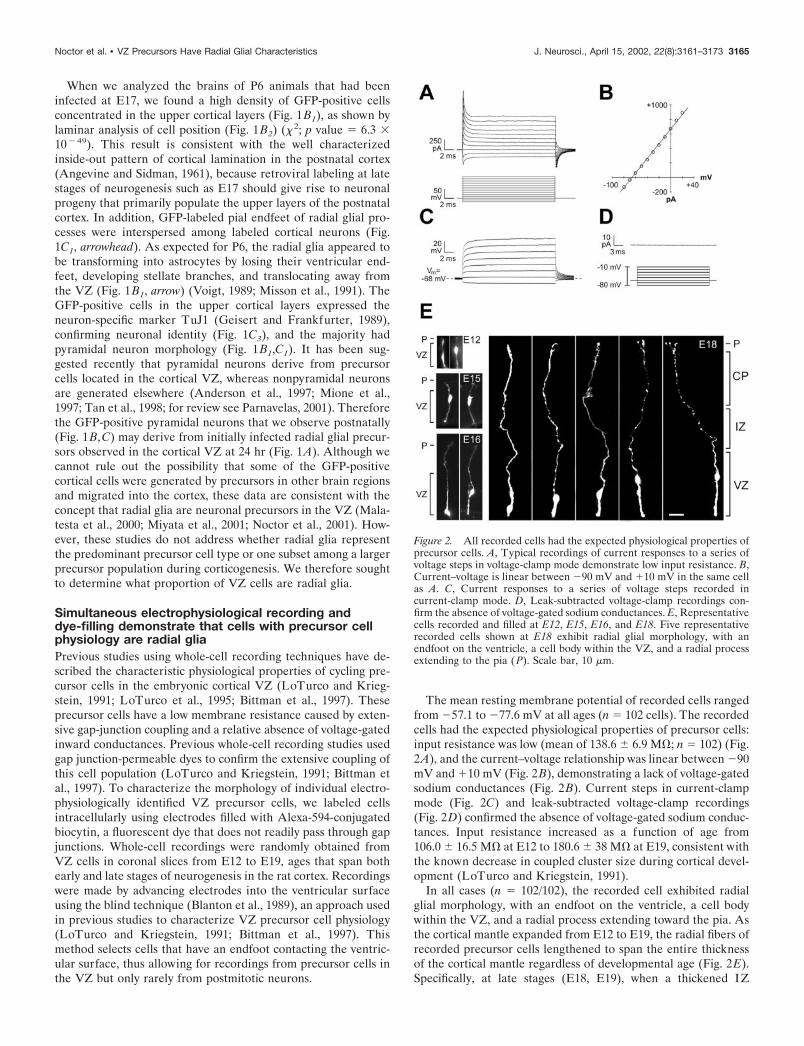
The mean resting membrane potential of recorded cells rangedfrom �57.1 to �77.6 mV at all ages (n 102 cells). The recordedcells had the expected physiological properties of precursor cells:input resistance was low (mean of 138.6 � 6.9 M�; n 102) (Fig.2A), and the current–voltage relationship was linear between �90mV and �10 mV (Fig. 2B), demonstrating a lack of voltage-gatedsodium conductances (Fig. 2B). Current steps in current-clampmode (Fig. 2C) and leak-subtracted voltage-clamp recordings(Fig. 2D) confirmed the absence of voltage-gated sodium conduc-tances. Input resistance increased as a function of age from106.0 � 16.5 M� at E12 to 180.6 � 38 M� at E19, consistent withthe known decrease in coupled cluster size during cortical devel-opment (LoTurco and Kriegstein, 1991).
In all cases (n 102/102), the recorded cell exhibited radialglial morphology, with an endfoot on the ventricle, a cell bodywithin the VZ, and a radial process extending toward the pia. Asthe cortical mantle expanded from E12 to E19, the radial fibers ofrecorded precursor cells lengthened to span the entire thicknessof the cortical mantle regardless of developmental age (Fig. 2E).Specifically, at late stages (E18, E19), when a thickened IZ
Figure 2. All recorded cells had the expected physiological properties ofprecursor cells. A, Typical recordings of current responses to a series ofvoltage steps in voltage-clamp mode demonstrate low input resistance. B,Current–voltage is linear between �90 mV and �10 mV in the same cellas A. C, Current responses to a series of voltage steps recorded incurrent-clamp mode. D, Leak-subtracted voltage-clamp recordings con-firm the absence of voltage-gated sodium conductances. E, Representativecells recorded and filled at E12, E15, E16, and E18. Five representativerecorded cells shown at E18 exhibit radial glial morphology, with anendfoot on the ventricle, a cell body within the VZ, and a radial processextending to the pia ( P). Scale bar, 10 �m.
Noctor et al. • VZ Precursors Have Radial Glial Characteristics J. Neurosci., April 15, 2002, 22(8):3161–3173 3165

separates the pia from the VZ and one can unambiguously iden-tify radial glial morphology, filled radial fibers extended to the pia(n 60/61) or CP (n 1/61) (Fig. 2E). These results show thatcells with the membrane properties of precursor cells have themorphology of radial glia.
Random DiOlistic labeling demonstrates anatomicallythat most of the VZ cells are radial gliaThe VZ is a pseudostratified neuroepithelium where all cells,with the exception of postmitotic neurons, contact the ventricularsurface (Sauer, 1935). To characterize the morphology of VZcells, we randomly labeled VZ cells contacting the ventricle withthe recently developed DiOlistics technique (Gan et al., 2000).Small DiI-coated tungsten beads were delivered via gene gun tothe ventricular surface of intact fixed cortical slabs, and the DiIwas allowed to diffuse throughout the membrane of individualcells (see Materials and Methods). This labeling technique al-lowed us to analyze individual VZ cell morphology and quantifythe number of cells with radial glial features.
DiOlistics experiments were performed at E12, E15, and E18.Previous observations have shown that radial glial fibers some-times terminate in the marginal zone, cortical plate, or upperintermediate zone (Gadisseux et al., 1992). Therefore, our crite-ria for radial glia included a cell body in the VZ, a ventricularendfoot, and a radial process extending out of the VZ and into theMZ, CP, or upper IZ. At E18 the cortical plate and intermediatezone are relatively thick, and these regions can be distinguishedeasily. Most of the cells labeled by this method at E18 (86.6 �4.5%; n 21 slices; 799 cells) (Fig. 3E) had distinct radial glialmorphology (Fig. 3A,B) with a radial process extending either tothe CP/MZ (60.4 � 4.0%) or the upper IZ (26.2 � 3.6%) (Fig.3B). In our tissue labeled from the ventricular surface, long fibersbecome progressively dimmer as they approach the pia. Wemeasured fiber length based on the furthest resolvable point andrarely observed abrupt fiber terminations in the IZ. Thereforeour counts most likely under-represent actual fiber length. Weobserved a smaller percentage of VZ cells (13.4 � 4.5%) that hadprocesses restricted to the VZ (Fig. 3C). This latter group of cellsmay include newborn neurons, as one would expect (Takahashi etal., 1996; O’Rourke et al., 1997), or other cells with shortprocesses.
To examine the morphology of VZ cells at earlier ages, we alsoperformed ventricular surface DiOlistic labeling at E12 and E15.At E15 the VZ is separated from the pia only by the marginalzone, because the cortical plate has yet to develop (BoulderCommittee, 1970). Most VZ cells labeled with DiOlistics at E15had a process that spanned nearly the entire thickness of thecortical mantle, to the upper marginal zone or pia (84.8 � 3.1%;n 28 slices; 788 cells) (Fig. 3D). The remainder (15.2 � 3.1%)had processes restricted to the VZ. At E12, because the neuro-epithelium spans the entire cortical width (Altman and Bayer,1990), virtually every cell labeled from the ventricular surfacecontacted the pial surface.
To control for potential bias that could be introduced in ven-tricular surface labeling if there is a nonrandom distribution ofendfoot size among cell classes, we also performed DiOlistics onthe cut surface of coronal sections of E18 cortex. In these exper-iments, 70.1 � 4.9% of cells with a soma in the VZ were radialglia, whereas 29.9 � 4.9% were VZ-restricted cells (n 6 slices;187 cells). The increased number of nonradial glial cells in the VZin these experiments was primarily accounted for by an increase
in cells lacking a contact on the ventricular surface that had themorphology expected of newborn neurons.
Taken together, the results of our electrophysiological andDiOlistics experiments show that most of the VZ cells identifiedeither physiologically or anatomically have radial glial morphol-ogy. The nonradial glial cells labeled by both approaches likelyincluded newborn neurons, but on the basis of these data wecannot rule out the possibility that some of these cells representa distinct population of cycling progenitor cells. We thereforeperformed a series of experiments to further characterize the VZby examining the characteristics of mitotically active VZ cells.
VZ cells in S-phase express the radial glial markersvimentin and RC2Neuronal precursors in the embryonic VZ undergo a process ofinterkinetic nuclear migration in which cells pass through S-phaseof the cell cycle with their nuclei in the outer half of the VZ andthen enter M-phase along the border of the lateral ventricle
Figure 3. Random labeling of VZ cells by DiOlistics suggests that mostof the VZ cells are radial glia. A, Shown are labeled cells at E18 withsomata (arrowheads) in the upper, middle, and lower zones of the VZ andlong fibers extending to the pia. B, Cells identified by DiOlistic labelinghave expected morphological features of radial glial cells, such asbranched pial endfeet of the long radial fiber (lef t) and small side branches(right). C, A small percentage of cells with processes restricted to the VZare also observed, with cell bodies (arrowheads) in the middle (lef t) andlower (right) zones of the VZ. D, The majority of VZ cells labeled byDiOlistics at E15 resembled these two examples with long radial fibers. E,Quantitative analysis of the percentage of cells with long pially directedfibers (Long) versus cells with fibers restricted to the VZ (Short) identifiedby random DiOlistic labeling of the VZ surface at E15 and E18. Scalebars: A, C, 10 �m; B, D, 5 �m. CP, Cortical plate; IZ, intermediate zone;P, pia; SVZ, subventricular zone.
3166 J. Neurosci., April 15, 2002, 22(8):3161–3173 Noctor et al. • VZ Precursors Have Radial Glial Characteristics

(Sauer, 1935). To assess what percentage of S-phase cells in theVZ are radial glia, we labeled S-phase cells in utero at three agesspanning the neuronogenetic interval (E12, E15, and E18) anddetermined what percentage of S-phase cells at these ages expressradial glial markers. Pregnant rats received a single injection ofBrdU and were killed 1–2 hr later. VZ cells were labeled with anantibody to the radial glial marker, vimentin, an intermediatefilament protein that has been shown to specifically label radialglial cells in the cortex at the ages used here (Dahl et al., 1981;Pixley and de Vellis, 1984; Kalman and Ajtai, 2001). Vimentinlabeling is localized to the cytoplasm, whereas the BrdU labelis limited to the nucleus. To ensure that individual double-labeled cells were identified correctly, we used a third marker,tetramethylrhodamine-conjugated ConA, to label cellularmembranes (Tarasova et al., 1997), thereby outlining individ-ual cells. We used confocal microscopy to visualize all fluo-rescent labels in the same optical section. The use of thistriple-labeling and imaging protocol allowed us to confirmcolocalization of BrdU and vimentin immunolabeling in indi-vidual cells (Fig. 4 A2).
As shown in Figure 4A1, vimentin is expressed in the vastmajority of BrdU-positive VZ cells. To limit counting of newbornneurons that might have exited the cell cycle between the injec-tion of BrdU and the time rats were killed, we counted onlyBrdU-positive cells in the outer half of the VZ, where S-phasenuclei reside. At E12, 99.3 � 0.3% of cells labeled with BrdU inthe S-phase zone were positive for vimentin (n 13 sections; 548cells). At E15, the percentage was 98.6 � 0.5% (n 7 sections;564 cells), and at E18, the percentage was 98.3 � 0.4% (n 9sections; 539 cells).
To confirm these data, we also performed a quantitative analysisof the colocalization of BrdU and vimentin labeling in dissociatedVZ cell suspensions to unequivocally identify individual cells. Inthese experiments, 94.5% (n 310 cells) of BrdU-labeled cellswere vimentin positive (Fig. 4B). These data demonstrate thatmost of the proliferative VZ cells in S-phase express the radial glialmarker vimentin throughout neocortical neurogenesis.
We repeated these experiments in mouse using the murineradial glial-specific marker RC2 (Misson et al., 1988b). S-phaseVZ cells were pulse labeled with BrdU in E15.5 mice (roughlyequivalent to E18 in the rat), and mice were killed 1–2 hr later.We then labeled radial glia with antibodies to RC2 and deter-mined the percentage of BrdU-labeled cells that were RC2 pos-itive. RC2 densely labeled the entire VZ (Fig. 4C1,C2) and ap-peared qualitatively identical to vimentin staining in the rat. As inthe vimentin double-labeling experiments described above, weused concanavalin-A to confirm colocalization of the markerswithin VZ cells. We found that in E15.5 mice, 96.1 � 0.8% (n 5 sections; 342 cells) of the BrdU-positive VZ cells were positivefor RC2. We also dissociated BrdU-labeled mouse cortical cellsand stained them with RC2 to unambiguously calculate the per-centage of BrdU cells that also expressed RC2. We found that97% (233/239) of dissociated BrdU-positive cells coexpressedRC2 (Fig. 4D). In addition we stained embryonic brain slicesusing antibodies against BLBP (Feng et al., 1994) and theastrocyte-specific glutamate transporter, GLAST (Shibata et al.,1997), both of which are proteins expressed in subsets of radialglial cells (Hartfuss et al., 2001). In cortical regions where thesemarkers are expressed, 93.0 � 2.7% (n 5 sections; 204 cells) ofBrdU-positive cells were positive for BLBP and 95.4 � 0.3% (n 4 section; 313 cells) of BrdU-positive cells were positive for
GLAST. These data indicate that the vast majority of S-phasecells in the embryonic VZ express a range of radial glial markers.
VZ cells in S-phase have fibers that extend to the piaIf S-phase cells are radial glia, they should have characteristicradial glial fibers that extend to the pial surface. To examine thismorphological feature of S-phase VZ cells, we applied fluores-cently labeled latex microspheres (beads) to the pial surface ofacute cortical slabs. The slabs were incubated for 9 hr to allowretrograde labeling of those cells in the VZ with processes con-tacting the pia (Katz et al., 1984), and BrdU was added during thefinal 2 hr to label S-phase cells. Slabs were then fixed, and coronalslices were examined to quantify the number of BrdU-positivecells in the VZ that contained fluorescent beads. All sections werecounterstained with ConA to label cell membranes and outlineindividual VZ cells. With this triple-staining protocol, and the useof serial confocal sectioning, we were able to unambiguouslyidentify VZ cell somata that contained beads (Fig. 5A).
After pial application of fluorescent beads, we found that thebeads retrogradely labeled VZ cells in regions underlying the pialpainting, whereas neighboring regions of VZ remained unlabeled(Fig. 5A, middle panel). When we examined serial optical sections,we found that 90.5 � 2.5% (n 11 sections; 658 cells) ofBrdU-positive VZ cells contained beads in their perinuclearcytoplasm (Fig. 5A, right panel). It is likely that this numberunder-represents the actual percentage of bead-labeled VZ cells,because in the optical sections we could only examine the BrdU-labeled cell body and not the endfeet or radial fibers of VZ cells.Therefore, we could not determine whether cells without perinu-clear beads contained beads in their processes. These data indi-cate that in addition to expressing radial glial markers, most VZcells in S-phase have radial processes extending to the pia. Toexamine cycling VZ cells at a different stage in the mitotic cycle,we next analyzed the expression of phosphorylated vimentin incells undergoing M-phase at the ventricular surface.
VZ cells in M-phase express 4A4, a specific marker fordividing radial gliaVimentin is phosphorylated by the cell cycle kinase cell divisioncycle 2 (cdc2) in radial glial cells during cytokinesis (Kamei et al.,1998). An antibody raised against the cdc2 phosphorylation siteon vimentin, termed 4A4, has been shown to specifically labelradial glia in M-phase (Kamei et al., 1998). We used 4A4 immu-nohistochemistry to determine what percentage of VZ cells inM-phase are radial glia. We stained coronal sections and intactcortical slabs with the 4A4 antibody and then labeled all cellnuclei using the fluorophore Syto-11. Using this approach, allM-phase cells at the ventricular surface can be clearly identifiedmorphologically by their condensed chromatin and arrangementof chromatin into mitotic figures. As shown in Figure 5B, confocalimaging of intact neocortex from the ventricular surface revealedSyto-11-labeled cells in various stages of M-phase (arrows). All ofthese Syto-11-labeled mitotic figures (Fig. 5B, green) are sur-rounded with positive immunolabeling for 4A4 (Fig. 5B, red).Other cells in the field without condensed chromatin (Fig. 5B,arrowheads) are negative for 4A4. Cells in specific stages ofM-phase—metaphase, anaphase, and telophase—could be iden-tified in both coronal sections and intact cortical slabs, as shownin Figure 5C. As shown previously for 4A4 immunostaining(Kamei et al., 1998), positive staining is found almost exclusivelyin cells with nuclei along the ventricular border (Fig. 5D), whereVZ precursor cells undergo M-phase. To determine what per-
Noctor et al. • VZ Precursors Have Radial Glial Characteristics J. Neurosci., April 15, 2002, 22(8):3161–3173 3167

centage of M-phase cells express 4A4, we performed quantitativeanalysis in coronal sections. To conservatively identify M-phasecells for quantification, we counted only cells in anaphase andtelophase, stages that could be easily and unambiguously identi-
fied (Fig. 5C). Results were virtually identical at the ages studied.At E12, 98.7 � 0.1% of anaphase and telophase cells lining theVZ surface were positive for 4A4 (n 3 brains; 551 cells). AtE15, this percentage was 98.3 � 0.3% (n 3 brains; 555 cells),
Figure 4. S-phase cells in the VZ ex-press the radial glial markers vimentinand RC2. A1, BrdU labeling (BrdU,green) in E18 rat tissue fixed 1 hr afterBrdU injection to identify S-phase cells.Vimentin labeling (vim, red) labels mostcells in the VZ. Overlay of the two chan-nels is shown in the right panel (merge).A2, Colocalization of BrdU and vimentinlabeling in brain slices was confirmedusing a third marker, concanavalin A(ConA, blue) to label cellular mem-branes. Overlay of the BrdU, ConA, andvimentin channels is shown on the right(merge). B, BrdU-positive dissociatedsingle cells are also vimentin positive.Some cells (arrows in DIC panel ) do notexpress either marker, and somevimentin-positive cells do not expressBrdU (arrowheads). C1, In E15 mousebrain slices, BrdU ( green) colocalizeswith the murine radial glial marker, RC2(red). C2, Colocalization of BrdU andRC2 was determined at higher magnifi-cation than in C1, as shown here. D,BrdU-positive dissociated single cells arealso RC2 positive. Scale bars: A, C, 15�m; B, 5 �m; D, 20 �m.
3168 J. Neurosci., April 15, 2002, 22(8):3161–3173 Noctor et al. • VZ Precursors Have Radial Glial Characteristics

and at E18, this percentage was 98.8 � 0.1% (n 3 brains; 597cells). Therefore, consistent with our data for vimentin expres-sion in S-phase cells, virtually all M-phase cells in the VZ expressthe radial glial-specific marker 4A4 throughout development.
Because low levels of 4A4 staining were also present within thecytoplasm of labeled cells, we were able to examine the morphol-ogy of M-phase VZ cells to determine whether they resembledradial glia. It has been shown recently that dividing radial glialcells retain their radial fibers even through M-phase of the cellcycle (Miyata et al., 2001); thus we are able to ask whether mitoticVZ cells have radial processes characteristic of radial glial cells.We found that dividing cells often had 4A4-positive radial pro-cesses extending from the VZ to the marginal zone (Fig. 5D2).
To examine whether 4A4 is specific for dividing radial glialcells and not ubiquitously expressed in dividing cells in the brain,we also examined 4A4 staining in sections of P8 cerebellum. Atthis stage of cerebellar development, significant numbers of gran-ule cells are dividing in the external granule cell layer (EGL)where radial glia are not found. In contrast, a population ofcerebellar radial glial cells, the Bergmann glia, have their nucleiat the top of the inner granule cell layer (Hatten and Heintz,1995). In P8 cerebellum, specific 4A4 immunoreactivity wasfound in some Bergmann glial cells, but not in mitotic cells in theEGL (Fig. 5E), consistent with previous data that 4A4 is specificfor dividing cells of the radial glial lineage (Kamei et al., 1998).
DISCUSSIONPrecursor cells in the VZ are radial gliaThe cortex develops from a proliferative neuroepithelium thatinitially spans the entire cortical mantle and thickens progres-sively during neurogenesis. During this process, radial glia con-tact the ventricle and elongate their apical fibers far beyond theupper border of the VZ. A distinction has traditionally been madebetween radial glia that extend processes to the pia and neuralprecursor cells that are believed to be restricted to the VZ.However, these two cell populations actually share a number ofcommon features, including interkinetic nuclear migration duringthe cell cycle (Misson et al., 1988a), expression of the neuralprecursor marker nestin (Hockfield and McKay, 1985; Lendahl etal., 1990), and a characteristic radial morphology with endfeetcontacting the ventricular surface (Sauer, 1935). These commonfeatures, together with evidence that cortical radial glia generateneurons (Malatesta et al., 2000; Noctor et al., Miyata et al., 2001;2001), raise the possibility that neuronal precursor cells and radialglia may represent overlapping populations of VZ cells. Further-more, the population of neuronal precursors that are presumed tobe distinct from radial glia has not been clearly characterized.
We find here that most of the cycling VZ cells have morpho-logical, molecular, and physiological characteristics of radial glia.We first specifically examined VZ cells that had characteristicmembrane properties of precursor cells and found that they haveradial glial morphology. We next randomly labeled VZ cells usingthe DiOlistics technique and found that most exhibit radial glialmorphology, including ventricular endfeet, lamellate projectionsthat frequently contact blood vessels, and radial processes withbranched endings on the pia. Because postmitotic neurons aregenerated by cell divisions at the ages we studied, we nextexamined mitotic VZ cells at specific stages in the cell cycle todetermine whether they have radial glial characteristics. We la-beled S-phase cells using BrdU and found that most of theproliferating cells express the specific radial glial markers vimen-tin, RC2, BLBP, and GLAST. Colabeling of M-phase cells with
Syto-11 and 4A4 confirmed that dividing VZ cells also express thephosphorylated form of the radial glial-specific marker vimentin.Moreover, visualization of phosphorylated vimentin within thelong radial processes of M-phase cells directly demonstrated thatdividing VZ cells have radial glial morphology. Finally, pialapplication of fluorescent microspheres led to labeling of 90% ofVZ cells in S-phase, indicating that most cycling VZ cells haveprocesses that reach the pia. Therefore, most VZ cells overallhave radial glial morphology and virtually all proliferating VZcells both express specific radial glial markers and have fibers thatspan the cortex. Throughout the period of neuronal production,we do not find evidence for a nonradial glial population ofdividing VZ precursor cells. Therefore, we propose that mostprecursor cells in the embryonic ventricular zone are radial glialcells.
Radial glia have traditionally been classed as specialized glialcells with a unique developmental role in guiding neuronal mi-gration (Rakic, 1988). Recent data, however, indicate that radialglia serve dual roles as both neuronal precursors and migrationalguides (Alvarez-Buylla et al., 1990; Malatesta et al., 2000; Noctoret al., 2001), suggesting that there may be no appropriate distinc-tion between radial glia and neuronal progenitors in the VZ(Alvarez-Buylla et al., 2001; Parnavelas and Nadarajah, 2001). Weshow here that nearly all mitotically active VZ cells appear to beradial glia. Taken together with recent evidence that radial glialcells generate neurons, the data presented here raise the possi-bility that radial glia may be the predominant neuronal precursorcells in the embryonic neocortical VZ.
Are radial glia, glia?If radial glial cells are to be considered neuronal precursors, is itappropriate to term them “glia”? In the late 19th century, it wasthought that most, if not all, neuroepithelial cells in the cortexextend a long radial process across the cortical mantle (Magini,1888b; Kolliker, 1896). In today’s nomenclature, these cells wouldbe called radial glial cells. It has been presumed that these cellsare differentiated support cells committed to the glial lineage, inpart because they were shown to express the astrocyte markerGFAP and possess glycogen granules (Rakic, 1972; Levitt andRakic, 1980). Glial cells specialized to guide neuronal migration(Rakic, 1978; Schmechel and Rakic, 1979a; Levitt and Rakic,1980) were not believed to be capable of producing neurons aswell. However, it is now clear that these cells are multipotential.Therefore, it may now be timely to reconsider the general natureof radial glial cells. Radial glia have many features in commonwith astrocytes beyond common expression of molecular markerssuch as glial filaments. For example, both cell types make spe-cialized contacts with blood vessels (Chanas-Sacre et al., 2000),contain glycogen storage granules (Rakic, 1972; Bruckner andBiesold, 1981), are coupled together with gap junction channels(Massa and Mugnaini, 1982; LoTurco and Kriegstein, 1991), andsustain intracellular calcium waves (T. A. Weissman and A. R.Kriegstein, unpublished observations). In addition, radial glialcells are known to transform into astrocytes. Thus it is seemsappropriate to consider radial glia a form of glial cell. However,because astrocytes and radial glia can generate neurons (Doetschet al., 1999; Malatesta et al., 2000; Noctor et al., 2001; Seri et al.,2001), the radial glial cell can be considered less a specialized glialcell and more a precursor cell with glial characteristics and thepotential to generate cells of both neuronal and glial lineages.This formulation recalls the concept of the “radial neuroglialcells” described by Magini in the 19th century.
Noctor et al. • VZ Precursors Have Radial Glial Characteristics J. Neurosci., April 15, 2002, 22(8):3161–3173 3169
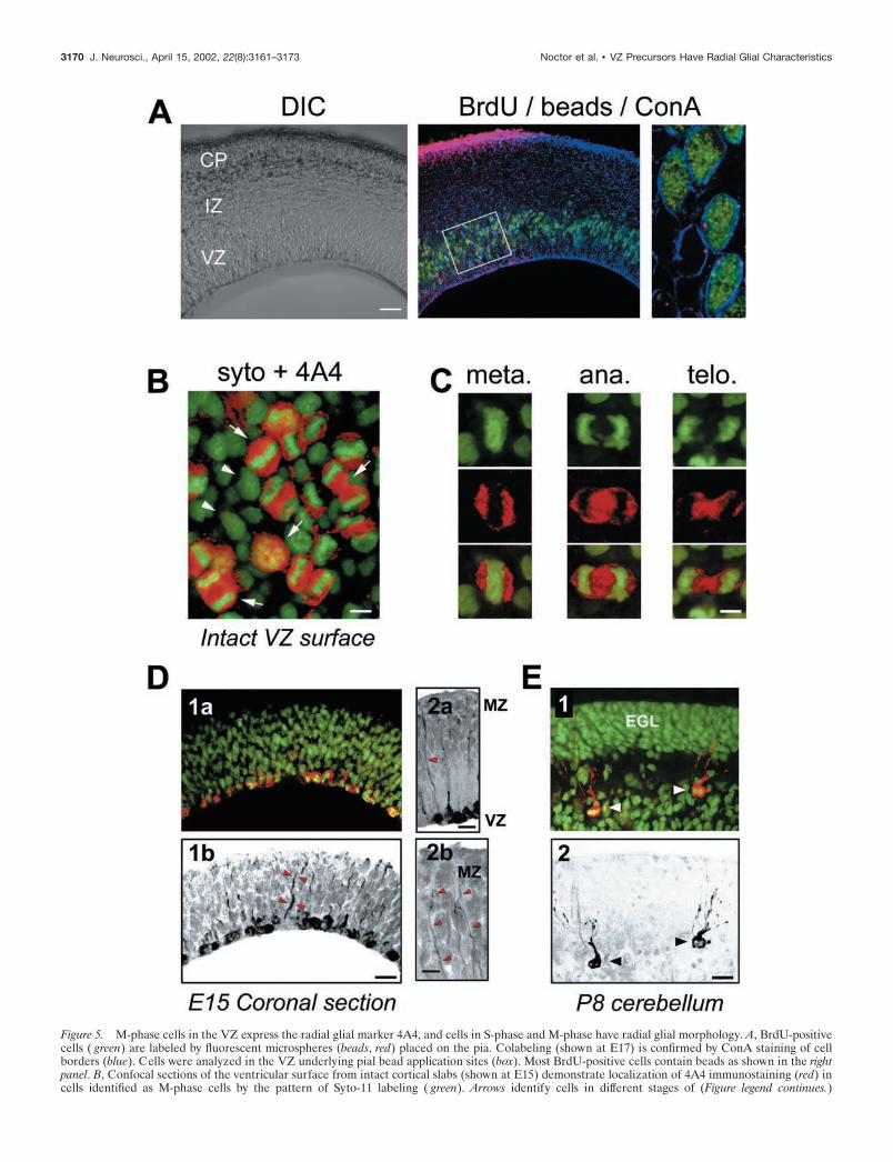
Figure 5. M-phase cells in the VZ express the radial glial marker 4A4, and cells in S-phase and M-phase have radial glial morphology. A, BrdU-positivecells ( green) are labeled by fluorescent microspheres (beads, red) placed on the pia. Colabeling (shown at E17) is confirmed by ConA staining of cellborders (blue). Cells were analyzed in the VZ underlying pial bead application sites (box). Most BrdU-positive cells contain beads as shown in the rightpanel. B, Confocal sections of the ventricular surface from intact cortical slabs (shown at E15) demonstrate localization of 4A4 immunostaining (red) incells identified as M-phase cells by the pattern of Syto-11 labeling ( green). Arrows identify cells in different stages of (Figure legend continues.)
3170 J. Neurosci., April 15, 2002, 22(8):3161–3173 Noctor et al. • VZ Precursors Have Radial Glial Characteristics
Because radial glia self-renew (Levitt et al., 1981; Misson et al.,1988a), generate neurons (Malatesta et al., 2000; Noctor et al.,2001), and later transform into astrocytes (Schmechel and Rakic,1979a; Misson et al., 1991), it may be most accurate to refer tothem as bipotential or multipotential precursor cells. There maybe restrictions on the diversity of neuronal and glial cell typesgenerated by cortical radial glia. For example, neurons generatedby radial glia in the cortical VZ may include only principal cells(pyramidal and projection neurons) rather than interneurons(Mione et al., 1997; Tan et al., 1998; Anderson et al., 1999). Theglial cells generated by cortical radial glia may be restricted to theastrocytes into which they are thought to differentiate (Schmecheland Rakic, 1979a; Misson et al., 1991). However, it has beensuggested that in mammals, some radial glial cells might also bethe precursors for the major classes of adult glial cells, includingoligodendrocytes as well as astrocytes (Choi and Kim, 1985;Hirano and Goldman, 1988; Rakic, 1995). It is also possible thatthere are subclasses of radial glial precursor cells (Schmechel andRakic, 1979b; Qian et al., 1998; Hartfuss et al., 2001) with moreor less restricted potential to act as neuronal or astrocytic pro-genitors. For example, the transcription factor genes Emx2 andPax6 are expressed within precursor cells of the cortical VZ alongtwo complementary gradients (Gulisano et al., 1996; Bishop et al.,2000; Muzio et al., 2002). These patterns may reflect expression ofdifferent transcription factors by radial glial cells in differentproliferative regions. Such selective expression has been demon-strated for the transcription factor Pax6, which is expressed inradial glia of the cortex but not radial glia of the basal telenceph-alon (Gotz et al., 1998),
Radial glial cells and neuronal migration in the cortexRecently, lineage analysis experiments using chimeric embryos aswell as retroviral labeling indicated that radially migrating corti-cal neurons are primarily projection neurons, whereas tangen-tially migrating neurons are primarily interneurons (Mione et al.,1997; Tan et al., 1998). Moreover, most of the tangentially mi-grating neurons appear to be inhibitory interneurons generated inthe proliferative zone of the adjacent ganglionic eminence(Anderson et al., 1997; Wichterle et al., 1999), whereas theexcitatory projection neurons are thought to migrate radially fromthe cortical VZ (Anderson et al., 1997; Mione et al., 1997; Tan etal., 1998). Thus there appear to be different proliferative zonesfor the generation of distinct classes of cortical neurons. Weobserve that after infection of radial glia in the VZ with aGFP-expressing retrovirus, many of the postnatal GFP-expressing cells in the cortex appear to be pyramidal neurons.This observation is consistent with the hypothesis that corticalradial glia may generate clones consisting primarily of pyramidalneurons.
Our anatomical labeling shows that most of the VZ cells haveradial glial morphology, with both an endfoot on the ventricle anda long radial fiber extending toward the pia (Magini, 1888b; His,
1889; Kolliker, 1896; Schmechel and Rakic, 1979a; for review, seeBentivoglio and Mazzarello, 1999). Until recently it was thoughtthat only radial glial cells have this morphology. It has beensuggested, however, that at early stages of neurogenesis somemigrating neurons may resemble radial glia (Brittis et al., 1995;Nadarajah et al., 2001). A new time lapse study from Miyata et al.(2001) suggests that newborn neurons may span the width of thecortex by inheriting the morphology of their parent radial glialcell. This study indicates that production of neurons by radial glialcells is quite common, which supports our conclusion that mostVZ precursor cells have radial glial characteristics. In addition,Miyata et al. (2001) suggest that radial glial cells have short radialprocesses transiently that re-extend after mitosis. These radialglia with processes that do not reach the pia may correspond tothe “freely arborizing spongioblasts” described by Stensaas(1967). Other studies have also indicated heterogeneous radialglial fiber length during periods of neurogenesis (Gadisseux et al.,1992). One possible explanation is that some radial glial cells inG1-phase may have shorter regrowing processes after mitosis,whereas at subsequent cell cycle stages, S-, G2- and M-phase,radial glial fibers are full length. This interpretation is consistentwith our observations of long fibers in some dividing cells. Al-though some newly generated postmitotic neurons may have beenincluded with our radial glial counts in the DiOlistics experi-ments, we find no evidence that a separate population of mitoti-cally active nonradial glial precursor cells exists in the VZ.
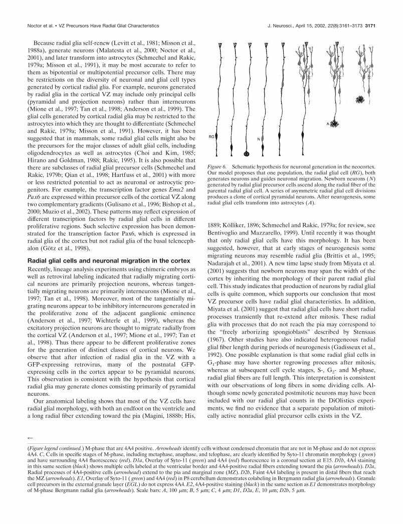
Figure 6. Schematic hypothesis for neuronal generation in the neocortex.Our model proposes that one population, the radial glial cell (RG), bothgenerates neurons and guides neuronal migration. Newborn neurons (N)generated by radial glial precursor cells ascend along the radial fiber of theparental radial glial cell. A series of asymmetric radial glial cell divisionsproduces a clone of cortical pyramidal neurons. After neurogenesis, someradial glial cells transform into astrocytes (A).
4
(Figure legend continued.) M-phase that are 4A4 positive. Arrowheads identify cells without condensed chromatin that are not in M-phase and do not express4A4. C, Cells in specific stages of M-phase, including metaphase, anaphase, and telophase, are clearly identified by Syto-11 chromatin morphology ( green)and have surrounding 4A4 fluorescence (red). D1a, Overlay of Syto-11 ( green) and 4A4 (red) fluorescence in a coronal section at E15. D1b, 4A4 stainingin this same section (black) shows multiple cells labeled at the ventricular border and 4A4-positive radial fibers extending toward the pia (arrowheads). D2a,Radial processes of 4A4-positive cells (arrowhead) extend to the pia and marginal zone (MZ). D2b, Faint 4A4 labeling is present in distal fibers that reachthe MZ (arrowheads). E1, Overlay of Syto-11 ( green) and 4A4 (red) in P8 cerebellum demonstrates colabeling in Bergmann radial glia (arrowheads). Granulecell precursors in the external granule layer (EGL) do not express 4A4. E2, 4A4-positive staining (black) in the same section as E1 demonstrates morphologyof M-phase Bergmann radial glia (arrowheads). Scale bars: A, 100 �m; B, 5 �m; C, 4 �m; D1, D2a, E, 10 �m; D2b, 5 �m.
Noctor et al. • VZ Precursors Have Radial Glial Characteristics J. Neurosci., April 15, 2002, 22(8):3161–3173 3171

Radial glial cells as neuronal precursor cells inadulthood and throughout the CNSRadial glial cells may play a role in neurogenesis in multiple CNSregions, in both development and adulthood. In the vertebrateretina, radial glial cells known as Muller glia have been suspectedto be retinal precursor cells both during early development andduring regeneration (Reh and Levine, 1998; Fischer and Reh,2001). In the chick optic tectum, radial glia appear to generateradial clones of neurons during development (Gray and Sanes,1992). At postnatal stages of cortical development, some radialglial cells transform into subventricular zone astrocytes(Schmechel and Rakic, 1979a), which in the adult have beenshown to generate neurons that subsequently migrate into theolfactory bulb (Doetsch et al., 1999). In the dentate gyrus of thehippocampus, radial glia persist into adulthood in specific regionswhere adult neurogenesis occurs (Gould et al., 1997), and re-cently a link between hippocampal radial glia and adult neuro-genesis has been suggested (Seri et al., 2001). In the adult avianventricular zone, “hotspots” of dividing radial glia correspond tolocal regions of neurogenesis (Alvarez-Buylla et al., 1990). Thusradial glia may generate neurons in the adult nervous system aswell as produce neurons during embryonic development.
Developmental insults can affect radial glia, migration, andneurogenesis. In utero radiation exposure during periods ofpeak neurogenesis disrupts neurogenesis and causes severedisruption of radial glia (Roper et al., 1997). Similarly, expo-sure to an antimitotic agent disrupts radial glial cells andimpairs normal cortical layering (Noctor et al., 1999). In hu-mans, developmental causes of cortical malformations such asheterotopias, lissencephaly, polymicrogyria, and related con-ditions have been interpreted to represent varying forms ofneuronal migration disorders. However, in many of these con-ditions, there is evidence for greatly decreased neuronal num-bers in addition to the failure of neurons to migrate to theirappropriate destinations. Our finding that radial glia subserveboth proliferation and migration may explain the apparentcombination of proliferative and migrational defects in pa-tients affected by these disorders. It remains to be seen, how-ever, whether radial glia play the same roles in primate devel-opment that we have observed in rodent development.
ConclusionOur findings expand the role of radial glia in neocortical devel-opment. The prevailing model for cortical neurogenesis and neu-ronal migration holds that a distinct population of precursorsrestricted to the VZ give rise to neurons that then migrate out ofthe VZ along nearby radial glial cells. The present data support amodified model in which radial glial neuronal precursor cells firstgenerate neurons in the VZ and then guide the migration of theirneuronal progeny into the cortex (Fig. 6). The centralization ofneurogenesis and migrational guidance in one cell type expandsthe role of the cortical VZ precursor cell and provides an efficientmechanism for establishing the laminar and columnar organiza-tion of the developing neocortex. The sequential migration ofclonally related neurons along the parent radial glia fiber enablesrelated cells to take up positions in different cortical layers duringthe inside-out formation of the cortex as predicted by the radialunit hypothesis (Rakic, 1988). Mutual contact with the parentalradial glial fiber (Noctor et al., 2001) may also allow for theformation of synaptic connections among neurons within theclone to form a local micronetwork across the cortical layers.Therefore the clonal relationship between radial glia and their
neuronal daughter cells may underlie the establishment of localcortical circuitry to form functional radial units.
REFERENCESAltman J, Bayer SA (1990) Vertical compartmentation and cellular
transformations in the germinal matrices of the embryonic rat cerebralcortex. Exp Neurol 107:23–35.
Alvarez-Buylla A, Buskirk DR, Nottebohm F (1987) Monoclonal anti-body reveals radial glia in adult avian brain. J Comp Neurol264:159–170.
Alvarez-Buylla A, Theelen M, Nottebohm F (1990) Proliferation “hotspots” in adult avian ventricular zone reveal radial cell division. Neuron5:101–109.
Alvarez-Buylla A, Garcia-Verdugo JM, Tramontin AD (2001) A unifiedhypothesis on the lineage of neural stem cells. Nat Rev Neurosci2:287–293.
Anderson S, Mione M, Yun K, Rubenstein JL (1999) Differential originsof neocortical projection and local circuit neurons: role of Dlx genes inneocortical interneuronogenesis. Cereb Cortex 9:646–654.
Anderson SA, Eisenstat DD, Shi L, Rubenstein J (1997) Interneuronmigration from basal forebrain to neocortex: dependence on dlx genes.Science 278:474–476.
Angevine JBJ, Sidman RL (1961) Autoradiographic study of cell migra-tion during histogenesis of cerebral cortex in the mouse. Nature192:766–768.
Bentivoglio M, Mazzarello P (1999) The history of radial glia. Brain ResBull 49:305–315.
Bishop KM, Goudreau G, O’Leary DD (2000) Regulation of area iden-tity in the mammalian neocortex by Emx2 and Pax6. Science288:344–349.
Bittman K, Owens DF, Kriegstein AR, LoTurco JJ (1997) Cell couplingand uncoupling in the ventricular zone of developing neocortex. J Neu-rosci 17:7037–7044.
Blanton MG, LoTurco JJ, Kriegstein AR (1989) Whole cell recordingfrom neurons in slices of reptilian and mammalian cerebral cortex.J Neurosci Methods 30:203–210.
Boulder Committee (1970) Embryonic vertebrate central nervous sys-tem: revised terminology. Anat Rec 166:257–261.
Brittis PA, Meiri K, Dent E, Silver J (1995) The earliest patterns ofneuronal differentiation and migration in the mammalian central ner-vous system. Exp Neurol 134:1–12.
Bruckner G, Biesold D (1981) Histochemistry of glycogen deposition inperinatal rat brain: importance of radial glial cells. J Neurocytol10:749–757.
Chanas-Sacre G, Rogister B, Moonen G, Leprince P (2000) Radial gliaphenotype: origin, regulation, and transdifferentiation. J Neurosci Res61:357–363.
Choi BH, Kim RC (1985) Expression of glial fibrillary acidic protein byimmature oligodendroglia and its implications. J Neuroimmunol8:215–235.
Dahl D, Rueger DC, Bignami A, Weber K, Osborn M (1981) Vimentin,the 57,000 molecular weight protein of fibroblast filaments, is the majorcytoskeletal component in immature glia. Eur J Cell Biol 24:191–196.
Doetsch F, Caille I, Lim DA, Garcia-Verdugo JM, Alvarez-Buylla A(1999) Subventricular zone astrocytes are neural stem cells in the adultmammalian brain. Cell 97:703–716.
Feng L, Hatten ME, Heintz N (1994) Brain lipid-binding protein(BLBP): a novel signaling system in the developing mammalian CNS.Neuron 12:895–908.
Fischer AJ, Reh TA (2001) Muller glia are a potential source of neuralregeneration in the postnatal chicken retina. Nat Neurosci 4:247–252.
Gadisseux JF, Evrard P, Mission JP, Caviness Jr VS (1992) Dynamicchanges in the density of radial glial fibers of the developing murinecerebral wall: a quantitative immunohistological analysis. J Comp Neu-rol 322:246–254.
Gan WB, Grutzendler J, Wong WT, Wong RO, Lichtman JW (2000)Multicolor “DiOlistic” labeling of the nervous system using lipophilicdye combinations. Neuron 27:219–225.
Geisert Jr EE, Frankfurter A (1989) The neuronal response to injury asvisualized by immunostaining of class III beta-tubulin in the rat. Neu-rosci Lett 102:137–141.
Golgi C (1886) Sulla fina anatomia degli organi centrali del sistemanervoso. Milano: Hoepli.
Gotz M, Stoykova A, Gruss P (1998) Pax6 controls radial glia differen-tiation in the cerebral cortex. Neuron 21:1031–1044.
Gould E, McEwen BS, Tanapat P, Galea LA, Fuchs E (1997) Neuro-genesis in the dentate gyrus of the adult tree shrew is regulated bypsychosocial stress and NMDA receptor activation. J Neurosci17:2492–2498.
Gray GE, Sanes JR (1992) Lineage of radial glia in the chicken optictectum. Development 114:271–283.
Gulisano M, Broccoli V, Pardini C, Boncinelli E (1996) Emx1 and Emx2show different patterns of expression during proliferation and differen-
3172 J. Neurosci., April 15, 2002, 22(8):3161–3173 Noctor et al. • VZ Precursors Have Radial Glial Characteristics

tiation of the developing cerebral cortex in the mouse. Eur J Neurosci8:1037–1050.
Hartfuss E, Galli R, Heins N, Gotz M (2001) Characterization of CNSprecursor subtypes and radial glia. Dev Biol 229:15–30.
Hatten ME (1999) Central nervous system neuronal migration. AnnuRev Neurosci 22:511–539.
Hatten ME, Heintz N (1995) Mechanisms of neural patterning andspecification in the developing cerebellum. Annu Rev Neurosci18:385–408.
Hirano M, Goldman JE (1988) Gliogenesis in rat spinal cord: evidencefor origin of astrocytes and oligodendrocytes from radial precursors.J Neurosci Res 21:155–167.
His W (1889) Die Neuroblasten und deren Entstehung im embryonalenMark. Abh Kgl sachs Ges Wissensch math phys Kl 15:311–372.
Hockfield S, McKay RD (1985) Identification of major cell classes in thedeveloping mammalian nervous system. J Neurosci 5:3310–3328.
Hofmann MH, Bleckmann H (1999) Effect of temperature and calciumon transneuronal diffusion of DiI in fixed brain preparations. J NeurosciMethods 88:27–31.
Kalman M, Ajtai BM (2001) A comparison of intermediate filamentmarkers for presumptive astroglia in the developing rat neocortex:immunostaining against nestin reveals more detail than GFAP orvimentin. Int J Dev Neurosci 19:101–108.
Kamei Y, Inagaki N, Nishizawa M, Tsutsumi O, Taketani Y, Inagaki M(1998) Visualization of mitotic radial glial lineage cells in the devel-oping rat brain by Cdc2 kinase-phosphorylated vimentin. Glia23:191–199.
Katz LC, Burkhalter A, Dreyer WJ (1984) Fluorescent latex micro-spheres as a retrograde neuronal marker for in vivo and in vitro studiesof visual cortex. Nature 310:498–500.
Kolliker A (1896) Handbuch der Gewebelehre des Menschen, Ed 6.Leipzig: W. Engleman.
Lendahl U, Zimmerman LB, McKay RD (1990) CNS stem cells expressa new class of intermediate filament protein. Cell 60:585–595.
Levitt P, Rakic P (1980) Immunoperoxidase localization of glial fibril-lary acidic protein in radial glial cells and astrocytes of the developingrhesus monkey brain. J Comp Neurol 193:815–840.
Levitt P, Cooper ML, Rakic P (1981) Coexistence of neuronal and glialprecursor cells in the cerebral ventricular zone of the fetal monkey: anultrastructural immunoperoxidase analysis. J Neurosci 1:27–39.
Levitt P, Cooper ML, Rakic P (1983) Early divergence and changingproportions of neuronal and glial precursor cells in the primate cerebralventricular zone. Dev Biol 96:472–484.
LoTurco JJ, Kriegstein AR (1991) Clusters of coupled neuroblasts inembryonic neocortex. Science 252:563–566.
LoTurco JJ, Owens DF, Heath MJS, Davis MBE, Kriegstein AR (1995)GABA and glutamate depolarize cortical progenitor cells and inhibitDNA synthesis. Neuron 15:1287–1298.
Magini G (1888a) Sur la neuroglie et les cellules nerveuses cerebraleschez les foetus. Arch Ital Biol 9:59–60.
Magini J (1888b) Nouvelles recherches histologiques sur le cerveau dufoetus. Arch Ital Biol 10:384–387.
Malatesta P, Hartfuss E, Gotz M (2000) Isolation of radial glial cells byfluorescent-activated cell sorting reveals a neuronal lineage. Develop-ment 127:5253–5263.
Massa PT, Mugnaini E (1982) Cell junctions and intramembrane parti-cles of astrocytes and oligodendrocytes: a freeze-fracture study. Neu-roscience 7:523–538.
Mione MC, Cavanagh JFR, Harris B, Parnavelas JG (1997) Cell fatespecification and symmetrical /asymmetrical divisions in the developingcerebral cortex. J Neurosci 17:2018–2029.
Misson JP, Edwards MA, Yamamoto M, Caviness Jr VS (1988a) Mitoticcycling of radial glial cells of the fetal murine cerebral wall: a combinedautoradiographic and immunohistochemical study. Brain Res466:183–190.
Misson JP, Edwards MA, Yamamoto M, Caviness Jr VS (1988b) Iden-tification of radial glial cells within the developing murine centralnervous system: studies based upon a new immunohistochemicalmarker. Brain Res Dev Brain Res 44:95–108.
Misson JP, Takahashi T, Caviness VS Jr (1991) Ontogeny of radial andother astroglial cells in murine cerebral cortex. Glia 4:138–148.
Miyata T, Kawaguchi A, Okano H, Ogawa M (2001) Asymmetric inher-itance of radial glial fibers by cortical neurons. Neuron 31:727–741.
Muzio L, DiBenedetto B, Stoykova A, Boncinelli E, Gruss P, MallamaciA (2002) Emx2 and Pax6 control regionalization of the pre-neuronogenic cortical primordium. Cereb Cortex 12:129–139.
Nadarajah B, Brunstrom JE, Grutzendler J, Wong RO, Pearlman AL(2001) Two modes of radial migration in early development of thecerebral cortex. Nat Neurosci 4:143–150.
Noctor SC, Palmer SL, Hasling T, Juliano SL (1999) Interference withthe development of early generated neocortex results in disruption ofradial glia and abnormal formation of neocortical layers. Cereb Cortex9:121–136.
Noctor SC, Flint AC, Weissman TA, Dammerman RS, Kriegstein AR(2001) Neurons derived from radial glial cells establish radial units inneocortex. Nature 409:714–720.
O’Rourke NA, Chenn A, McConnell SK (1997) Postmitotic neuronsmigrate tangentially in the cortical ventricular zone. Development124:997–1005.
Parnavelas JG, Nadarajah B (2001) Radial glial cells. Are they reallyglia? Neuron 31:881–884.
Pixley SK, de Vellis J (1984) Transition between immature radial gliaand mature astrocytes studied with a monoclonal antibody to vimentin.Brain Res 317:201–209.
Qian X, Goderie SK, Shen Q, Stern JH, Temple S (1998) Intrinsicprograms of patterned cell lineages in isolated vertebrate CNS ventric-ular zone cells. Development 125:3143–3152.
Rakic P (1971a) Guidance of neurons migrating to the fetal monkeyneocortex. Brain Res 33:471–476.
Rakic P (1971b) Neuron-glia relationship during granule cell migrationin developing cerebellar cortex. A Golgi and electron microscopic studyin Macacus Rhesus. J Comp Neurol 141:283–312.
Rakic P (1972) Mode of cell migration to the superficial layers of fetalmonkey neocortex. J Comp Neurol 145:61–83.
Rakic P (1978) Neuronal migration and contact guidance in the primatetelencephalon. Postgraduate Medical J 1:25–40.
Rakic P (1988) Specification of cerebral cortical areas. Science241:170–176.
Rakic P (1995) Radial gial cells: scaffolding for brain construction. In:Neuroglia (Kettenmann H, Ransom BR, eds), pp 746–762. Oxford:Oxford UP.
Ramon y Cajal S (1911) Histologie du systeme nerveux de l’hommes etdes vertebres. Paris: Maloine.
Ramon y Cajal S (1995) Histology of the nervous system. Oxford: Ox-ford UP.
Reh TA, Levine EM (1998) Multipotential stem cells and progenitors inthe vertebrate retina. J Neurobiol 36:206–220.
Retzius G (1894) Die Neuroglia des Gehirns beim Menschen und beiSaugetieren. Biologische Untersuchungen 6:1–24.
Roper SN, Abraham LA, Streit WJ (1997) Exposure to in utero irradi-ation produces disruption of radial glia in rats. Dev Neurosci19:521–528.
Sauer FC (1935) Mitosis in the neural tube. J Comp Neurol 62:377–405.Schmechel DE, Rakic P (1979a) A Golgi study of radial glial cells in
developing monkey telencephalon: morphogenesis and transformationinto astrocytes. Anat Embryol 156:115–152.
Schmechel DE, Rakic P (1979b) Arrested proliferation of radial glialcells during midgestation in rhesus monkey. Nature 277:303–305.
Seri B, Garcia-Verdugo JM, McEwen BS, Alvarez-Buylla A (2001) As-trocytes give rise to new neurons in the adult mammalian hippocampus.J Neurosci 21:7153–7160.
Shibata T, Yamada K, Watanabe M, Ikenaka K, Wada K, Tanaka K,Inoue Y (1997) Glutamate transporter GLAST is expressed in theradial glia-astrocyte lineage of developing mouse spinal cord. J Neuro-sci 17:9212–9219.
Stensaas LJ (1967) The development of hippocampal and dorsolateralpallial regions of the cerebral hemisphere in fetal rabbits. 3. Fifteenmillimeter stage, spongioblast morphology. J Comp Neurol 129:59–70.
Takahashi T, Nowakowski RS, Caviness Jr VS (1996) Interkinetic andmigratory behavior of a cohort of neocortical neurons arising in theearly embryonic murine cerebral wall. J Neurosci 16:5762–5776.
Tamamaki N, Nakamura K, Okamoto K, Kaneko T (2001) Radial glia isa progenitor of neocortical neurons in the developing cerebral cortex.Neurosci Res 41:51–60.
Tan SS, Kalloniatis M, Sturm K, Tam PP, Reese BE, Faulkner-Jones B(1998) Separate progenitors for radial and tangential cell dispersionduring development of the cerebral neocortex. Neuron 21:295–304.
Tarasova NI, Stauber RH, Choi JK, Hudson EA, Czerwinski G, MillerJL, Pavlakis GN, Michejda CJ, Wank SA (1997) Visualization of Gprotein-coupled receptor trafficking with the aid of the green fluores-cent protein. Endocytosis and recycling of cholecystokinin receptortype A. J Biol Chem 272:14817–14824.
Voigt T (1989) Development of glial cells in the cerebral wall of ferrets:direct tracing of their transformation from radial glia into astrocytes.J Comp Neurol 289:74–88.
Wichterle H, Garcia-Verdugo JM, Herrera DG, Alvarez-Buylla A (1999)Young neurons from medial ganglionic eminence disperse in adult andembryonic brain. Nat Neurosci 2:461–466.
Noctor et al. • VZ Precursors Have Radial Glial Characteristics J. Neurosci., April 15, 2002, 22(8):3161–3173 3173


![Combining gene and stem cell therapy for peripheral nerve ... Cells... · 112 precursor cells [39], induced pluripotent stem cells [40] and embryonic stem cells [41]. Therapeutic](https://img.dokumen.tips/doc/110x75/5f041a0d7e708231d40c53c8/combining-gene-and-stem-cell-therapy-for-peripheral-nerve-cells-112-precursor.jpg)