Embed Size (px)
Citation preview

Joy Hills 2013 | RN, BSN, MSN (Cancer), SpecCertCR (Onc)
Peripheral IV
Cannulation
DISTRIBUTED SIMULATION PROJECT

Objectives Having completed the IV cannulation workshop you will be able to:
• Describe the basic anatomy and physiology of the superficial veins of the arms and
hands
• Locate and assemble required equipment for IV cannulation or venepuncture
• Perform a successful IV cannulation on the training arm
• Demonstrate awareness of Infection Control guidelines and O H & S practices as
per organisation policy
• Be aware of documentation requirements in relation to cannulation
• Demonstrate critical thinking relevant to managing the risks and complications of IV
cannulation
• Identify patient education requirements

Anatomy and physiology
• Approximately 2/3 of total blood volume is in the veins which
transport deoxygenated blood to the heart from the tissues
• Veins are thin-walled, fibrous, have a large diameter and low
pressure
• Veins contract to propel the blood through the vein towards the
heart
• Some veins contain valves to regulate the one way flow to the
heart (usually lower limbs)
• The skeletal muscle pump influences venous return
• Superficial and deep veins

Anatomy and physiology cont
Blood vessel walls have three layers
• Tunic Intima
innermost, epithelial lining
• Tunic Media
Elastic and Smooth muscle fibres and nerve supply
• Tunic Externa Outer coat

Anatomy and physiology cont
Major veins of the arm
• Dorsal
• Cephalic
• Basilic
• Cubital Fossa

Anatomy and
physiology cont

Anatomy and physiology cont

Cannulae
• 14G
• 16G
• 18G
• 20G
• 22G
• 24G
• Large volume replacement
• Rapid transfusion of whole blood or blood components
• IV maintenance, NBM patients
• IV analgesia
• Paediatrics, elderly, chemotherapy patients
• Paediatrics, neonates

Equipment • Dressing/IV trolley with sharps container and waste bag
• Dressing pack
• Gloves (sterile)
• Alcoholic chlorhexidine
• Transparent semi permeable dressing
• Cannula (size depending on need)
• Giving and Extension set (and prescribed IV fluids)
• Water proof protective cover
• Syringe 10ml with 0.9% Normal saline
• Tourniquet
• Fluid balance sheet

Assessing & preparing the patient
• Check patient for baseline vital signs, diagnosis
and allergies to medications, cleansing fluids &
dressings
• Provide a clear explanation of the procedure
including potential adverse and side effects
• A relaxed patient is generally easier to cannulate
• Assess the dominant/non-dominant side and
check the veins for status and suitability

Preparing equipment
• Equipment should be gathered on trolley in
treatment room with sharps container
• IV fluids should be prepared by priming the giving
set
• The equipment should not be opened until in the
patient’s room and patient education, assessment of
vein and appropriate positioning has been attended

Positioning the patient
• If possible use the non dominant arm
• Raise bed prior to procedure
• Place the arm in a supported comfortable
position
• Use a tourniquet to find vein but release it while
you are getting equipment ready
• Position patient with pillows or towels
• Have IV trolley close by

Preparing vein
Warm veins by
• Rubbing
• Washing client’s hands under warm water
• Apply warmed towel
• If limb is warm ask the patient to gently clench and
unclench their hand
• Or gently rub up and down the vein

Before inserting cannula
• The tourniquet is applied above the IV insertion
site and should not be left on for more than 2-3
minutes
• Don gloves and clean site with appropriate
solution using a circular outward movement
• Allow site to air dry or dry with sterile swab

Inserting the cannula
• Hold cannula and rotate the barrel 360 degrees
• Apply skin traction to immobilise the vein
• Ensure cannula has bevel side UP and insert at
approximately 30 degree angle
• You will see a flashback of blood in the chamber once
you have pierced the vein
• Then advance the cannula a few more millimetres and
then flatten the cannula, stabilise the device and advance
the cannula until at skin level
• Remove the stylet and apply pressure just beyond the
catheter tip
Inserting the cannula cont
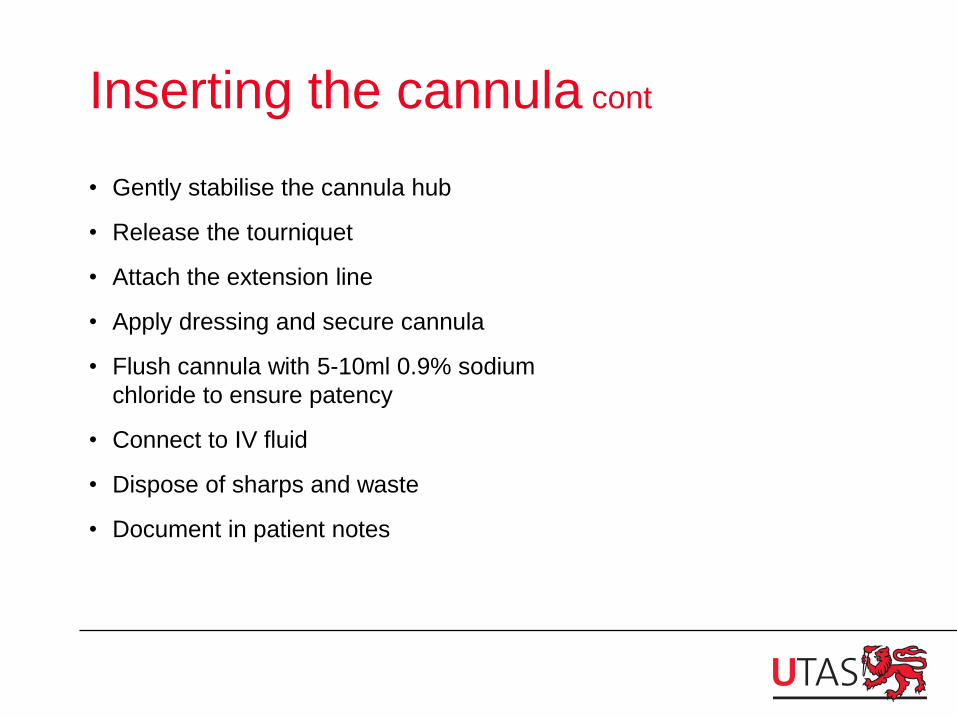
• Gently stabilise the cannula hub
• Release the tourniquet
• Attach the extension line
• Apply dressing and secure cannula
• Flush cannula with 5-10ml 0.9% sodium
chloride to ensure patency
• Connect to IV fluid
• Dispose of sharps and waste
• Document in patient notes

Dressing
• A transparent sterile occlusive dressing is the
optimal dressing to use
• Before applying dressing, ensure site is clean of
blood and moisture
• Check with patient re allergies to dressings

Documentation
• Site of insertion-vein and arm/hand
• Type and gauge of cannula
• Date and time of insertion
• Type and amount of IV solution
• Reason for IV therapy
“Because of the invasive nature of I.V. therapy, patients may
experience unexpected problems, even with diligent nursing
care. Your careful documentation helps prove that the care
you provided met the standard of care…” (Rosenthal, 2005,
p.28)

Potential complications • Extravasation
• Haematoma
• Phlebitis
• Venous Spasm
• Occlusion
• Thrombophlebitis
• Infection
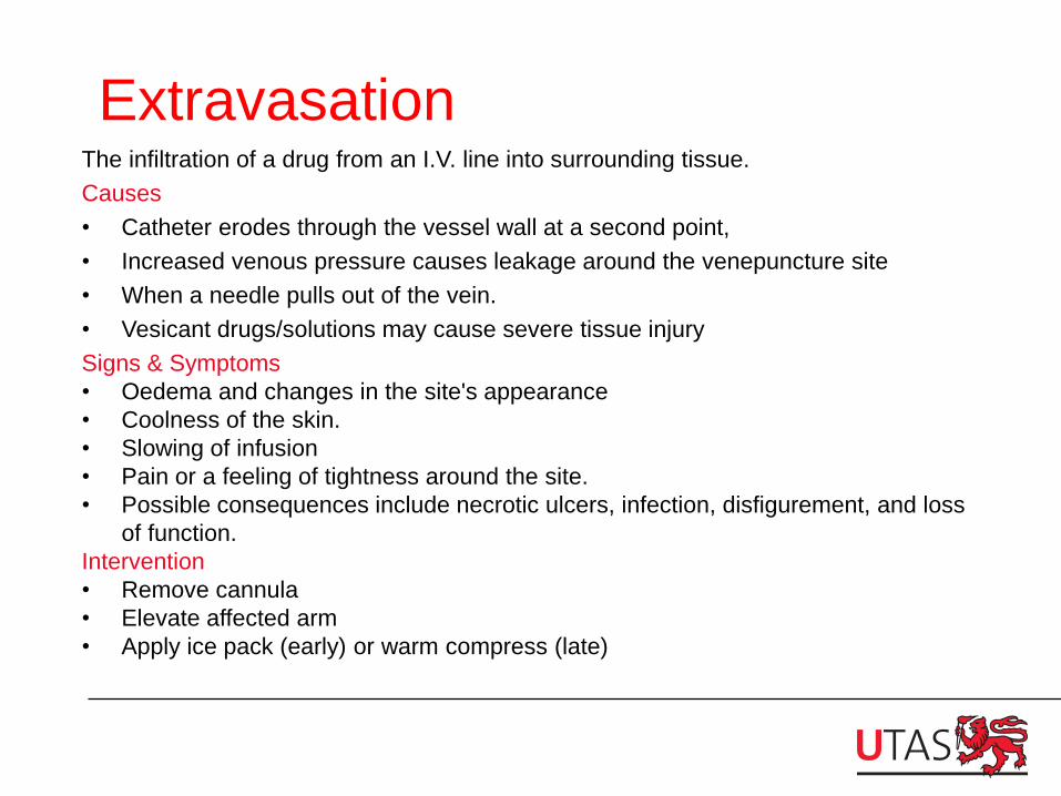
Extravasation The infiltration of a drug from an I.V. line into surrounding tissue.
Causes
• Catheter erodes through the vessel wall at a second point,
• Increased venous pressure causes leakage around the venepuncture site
• When a needle pulls out of the vein.
• Vesicant drugs/solutions may cause severe tissue injury
Signs & Symptoms
• Oedema and changes in the site's appearance
• Coolness of the skin.
• Slowing of infusion
• Pain or a feeling of tightness around the site.
• Possible consequences include necrotic ulcers, infection, disfigurement, and loss
of function.
Intervention
• Remove cannula
• Elevate affected arm
• Apply ice pack (early) or warm compress (late)

Haematoma
Localised collection of extravasated blood, usually clotted, in an organ or tissue.
Cause
• Blood leaking out of the vein into the tissue due to puncture or trauma
Signs & Symptoms
• Swelling, tenderness and discolouration
Prevention
• Proper device insertion
• Pressure over site on removal of cannula
Intervention
• Apply appropriate pressure bandage, monitor the site

Phlebitis
Inflammation of the vein
Cause
• Poor aseptic technique
• High osmolarity I.V. infusions or
drugs
• Trauma to the vein during
insertion/incorrect cannula gauge
• Prolonged use of the same site
Signs & Symptoms
• Tenderness, redness, heat and
oedema
• Advanced-induration, palpable
venous cord
Intervention
• Remove cannula
• Apply warm compress
• Observe for signs of
infection
• If phlebitis is advanced
antibiotics may be required
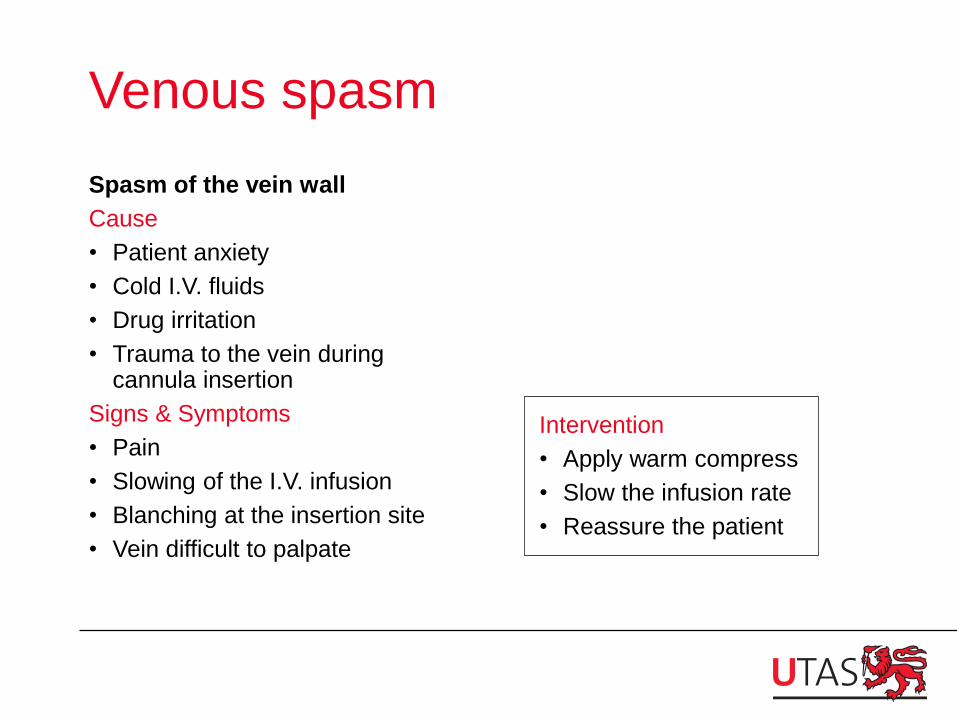
Venous spasm
Spasm of the vein wall
Cause
• Patient anxiety
• Cold I.V. fluids
• Drug irritation
• Trauma to the vein during cannula insertion
Signs & Symptoms
• Pain
• Slowing of the I.V. infusion
• Blanching at the insertion site
• Vein difficult to palpate
Intervention
• Apply warm compress
• Slow the infusion rate
• Reassure the patient

Occlusion
Slowing or cessation of fluid infusion due to:
• Fibrin formation in or around the tip of the cannula
• Mechanical occlusion (kink) of the cannula
Cause
• Cannula not flushed
• Kinking of the cannula
• Back flow or interrupted flow
Signs & Symptoms
• I.V. not running
• Blood in the line
• Discomfort
Intervention
• Check for kinks in cannula
• Raise IV higher
• Remove cannula

Thrombophlebitis
Formation of a thrombus and inflammation in the vein, usually occurs after phlebitis.
Cause
• Injury to the vein
• Infection
• Chemical irritation
• Prolonged use of the same vein
Signs & Symptoms
• Tenderness/redness
• Heat/oedema
• Cordlike appearance of the vein
• Slowing of the IV infusion
Intervention
• Remove cannula
• Observe for signs of infection
• Change cannula frequently (48-72hrs)

Infection
Pathogen in the surrounding tissue of the I.V. site.
Cause
• Lack of asepsis
• Prolonged use of the same site
Signs & Symptoms
• Tenderness and swelling
• Erythema/purulent drainage
Intervention
• Remove cannula
• Antibiotics may be required
• Documentation

STOP! CHECK. FAIL!

Preventing complications
• Know your organisation’s policy on IV therapy
• Check and inspect the IV site regularly as per
your facility policy
• Ask the patient how the IV site feels
• Document
• Troubleshoot at first sign
• Report any problems

Hints
Successful peripheral intravenous cannulation
increases with
•Meticulous attention to proper technique
•The use of proper equipment
•Familiarity with anatomy
•Knowledge of a variety of approaches to
accessing peripheral veins
•Knowledge of the latest evidence in the area
(this should drive your organisation’s policies
around cannulation and IV management)

IV access decision tree Edwards, A., Muir, Y., Grieves, L., & Willis, K 2010.

Infusion therapy process model Edwards, A., Muir, Y., Grieves, L., & Willis, K 2010.

References • Ahlqvist, M., Bogren, A., Hagran, S., Nazar, I., Nilssen, K. Nordin, K. et al., 2006. Handling of
peripheral intravenous cannulae: effects of evidence-based clinical guidelines. Journal of
Clinical Nursing, 15, 1354–1361
• Hindley, G. 2004. Infection control in peripheral cannulae. Nursing Standard. 18:(27) p. 37-39.
• Institute of Health and Nursing Australia, IV cannulation video.
http://www.youtube.com/watch?v=WvA6LR_g8yQ
• Joanna Briggs Institute. 2008. Management of Peripheral Intravenous Devices. Best Practice.
12:(5) pp.1-4.
• Lavery, I. 2003. Peripheral intravenous cannulation and patient consent. Nursing Standard.
17:(28) p.40-42.
• Ortega, R. Sekhar, P., Song, M., Hansen, C.J., and Peterson, L. 2008. Peripheral Intravenous
Cannulation. The New England Journal of Medicine. 359:(21) p. e26-29.
• Probert, S. 2010. Peripheral Intravenous Cannulation. Accessed via the intranet of the
Department of Health and Human Services, Tasmania.
• Rosenthal, K. 2005. Documenting peripheral I.V. therapy. Nursing 2005. 35:(7) p. 28.
• Edwards, A., Muir, Y., Grieves, L. & Willis, K 2010. Cannulation Resource Package. Accessed
via the intranet of the Department of Health and Human Services (NW), Tasmania.